ISSN: 3006-6514 (Online)
3006-6506 (Print)
Department of Medicine, Columbia Center for Human Development, Columbia University Medical Center, New York, NY, USA
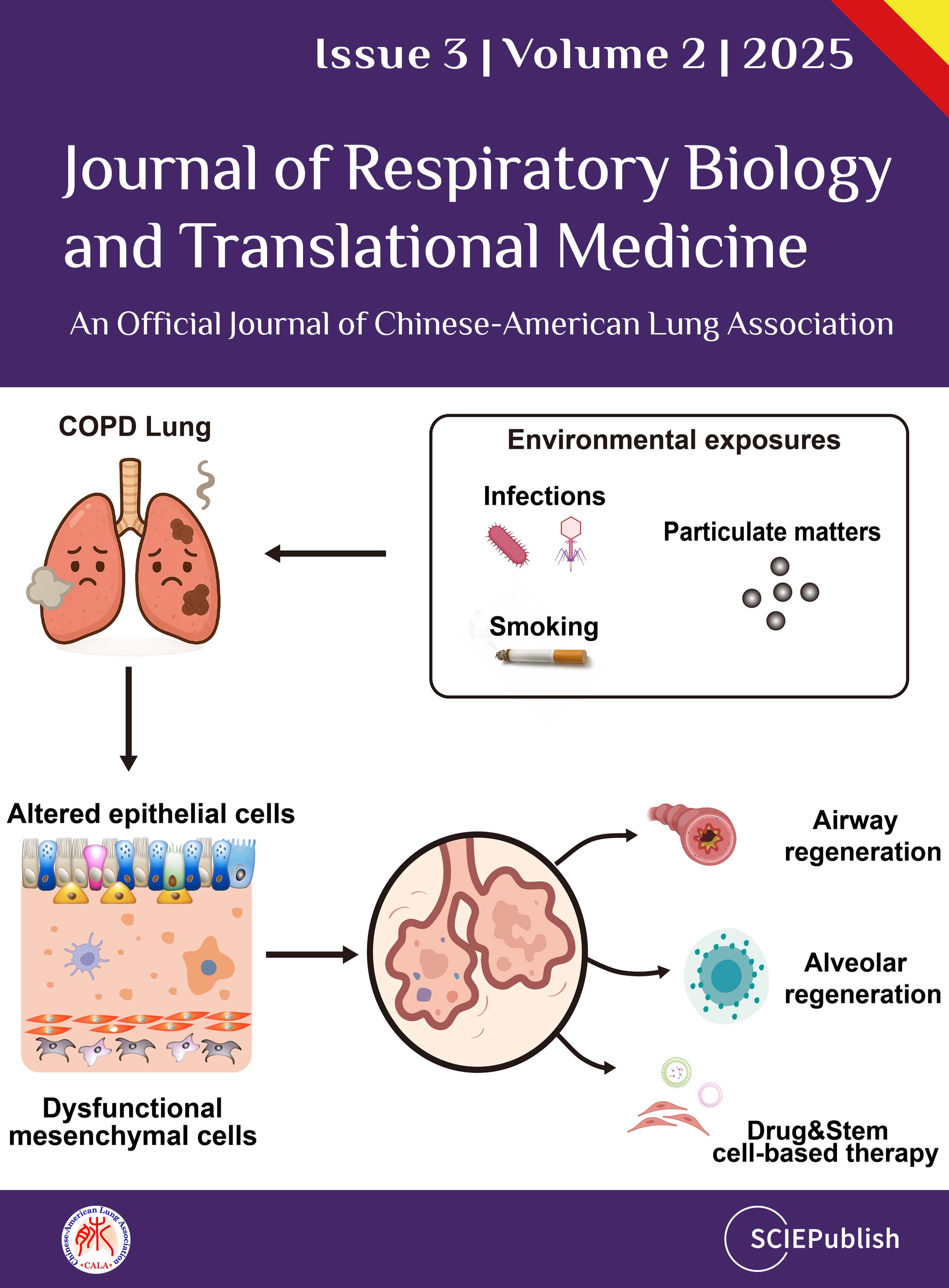
Chronic obstructive pulmonary disease (COPD) is a leading cause of global morbidity and mortality, characterized by progressive 
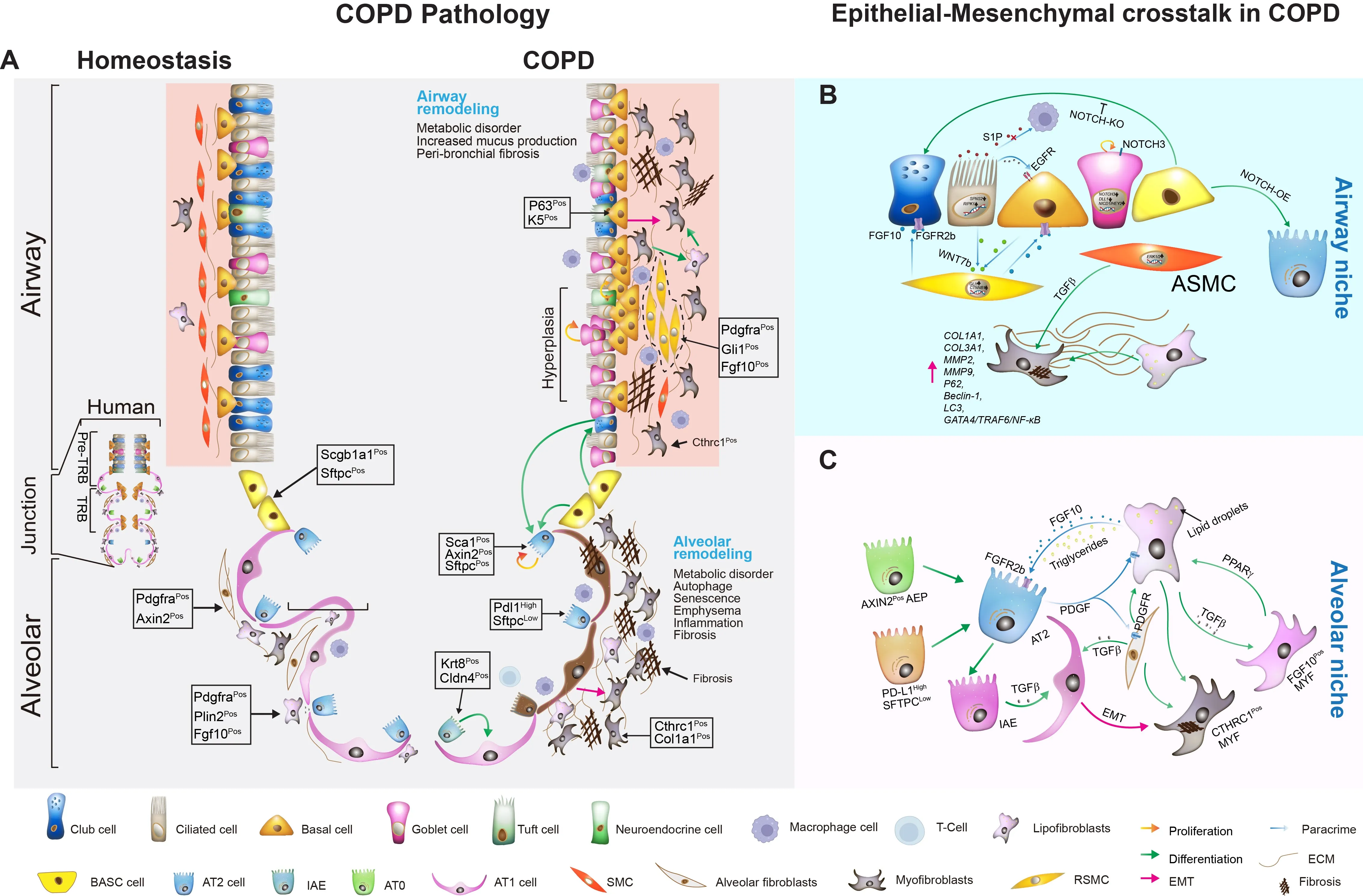
airway and alveolar remodeling. The disease pathogenesis is commonly driven by chronic environmental insults, leading to airway obstruction, emphysema, and chronic bronchitis. This review synthesizes emerging evidence that altered epithelial cell behavior and dysfunctional epithelial-mesenchymal interactions serve as pivotal drivers of COPD pathogenesis, orchestrating failed repair and structural degeneration. We detail how altered responses of 
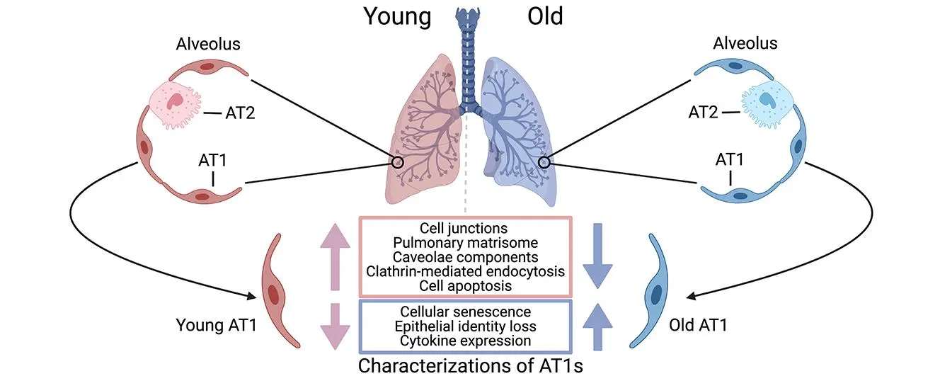
airway (ciliated, club, basal, goblet) and alveolar (AT1 and AT2) epithelial cells lead to cellular senescence, metaplasia, defective regeneration, and barrier disruption, acting as primary instigators of pathogenesis. We also summarize current knowledge on the mechanisms of activation and pathogenic role of mesenchymal cells, which drive peribronchiolar fibrosis, alveolar destruction, and metabolic reprogramming, alongside the compromised reparative function of mesenchymal stem cells (MSCs). We emphasize how distinct mesenchymal niches (e.g., PDGFRαPos MANCs, FGF10Pos lipofibroblasts, SFRP1Pos fibroblasts) and distinct epithelial stem/progenitor subpopulations critically contribute to pathogenesis. Key signaling pathways—including FGF10/FGFR2b, WNT, Hippo, NOTCH, and TGF-β—mediate epithelial-mesenchymal transition (EMT), stem cell niche function, and structural remodeling. By dissecting how epithelial injury responses and mesenchymal niche failure collaboratively drive COPD progression, we identify actionable targets to disrupt pathogenesis and restore endogenous repair. We propose targeting EMT, including inhibiting EMT/fibrosis, promoting alveolar regeneration, MSC-based therapies, exosome-delivered biomolecules, and precision cell transplantation strategies, as promising future therapeutic strategies.
Congenital Diaphragmatic Hernia (CDH) is a rare neonatal disorder causing diaphragmatic defects and cardiopulmonary hypoplasia, traditionally attributed to mechanical compression from organ herniation. However, emerging evidence suggests genetic mutations may independently impair lung development, prompting debate over CDH etiology. Here, we investigated the requirement of mitochondrial function guarded by LON peptidase 1 (Lonp1), a CDH risk gene, in either diaphragm or lung development. Lonp1 loss in skeletal muscles of the diaphragm led to its thinning and membranization, recapitulating the pathology of sac-type CDH. On the other hand, lung-specific inactivation caused severe hypoplasia with defective branching morphogenesis, independent of diaphragm anomalies. Molecularly, Lonp1 disruption dysregulated key transcription factors and signaling pathways known to be critical for early lung development. Our findings here revealed that mitochondrial defects contribute to the pathogenesis of CDH in an organ and cell type specific manner, opening new avenues for drug and therapeutic development.
The activation of IL-17 signaling has been linked to the pathogenesis of many chronic, inflammatory lung diseases including cystic fibrosis. Through unbiased single-cell RNAseq screening, we found that IL-17+ T cells highly express Srm and Smox, which encode two key enzymes involved in spermidine synthesis, spermidine synthase and spermine oxidase respectively. Spermidine has been shown to reduce inflammation by regulating macrophage activation and balancing Th17/Treg differentiation; however its direct effects on Th17 cytokine production have not been carefully investigated. Here, using already differentiated Th17 cells from cultured mouse splenocytes, we found that exogenous spermidine directly inhibits IL-1β/IL-23-induced IL-17 production. Blockade of endogenous spermidine synthesis enhanced IL-17 production above native levels, further supporting the notion that spermidine is a direct regulator of cytokine secretion independent of differentiation. In vivo, spermidine alleviates lung inflammation in both Pseudomonas aeruginosa (PA) and LPS induced acute lung injury models. Further RNA-seq analysis suggests that spermidine suppression of Th17 cytokine production is mediated through its PRDX1-dependent antioxidant activity. Our data suggests that spermidine is a direct regulator of Type-17 T cell cytokine production and has potent anti-inflammatory effects against lung inflammation.
Idiopathic pulmonary fibrosis (IPF) is a fatal disease with limited therapeutic options. Lung transplantation is the only curative treatment, but it is rarely available due to a lack of suitable donors. In a recent publication in Science Immunology, Farhat et al. demonstrated that bone marrow transplantation from young donors alleviates fibrosis by restoring immune resolution in aged hosts in animal models. Aged hematopoietic cells exacerbate fibrosis through the persistence of inflammatory macrophages and impaired Treg-derived IL-10, highlighting bone marrow rejuvenation as a potential treatment strategy for IPF.
Spatial transcriptomics technologies have emerged as powerful tools for understanding cellular identity and function within the natural spatial context of tissues. Traditional transcriptomics techniques, such as bulk and single-cell RNA sequencing, lose this spatial information, which is critical for addressing many biological questions. Here, we present a protocol for high-resolution spatial transcriptomics using fixed frozen mouse lung sections mounted on 10X Genomics Xenium slides. This method integrates multiplexed fluorescent in situ hybridization (FISH) with high-throughput imaging to reveal the spatial distribution of mRNA molecules in lung tissue sections, allowing detailed analysis of gene expression changes in a mouse model of pulmonary hypertension (PH). We compared two tissue preparation methods, fixed frozen and fresh frozen, for compatibility with the Xenium platform. Our fixed frozen approach, utilizing a free-floating technique to mount thin lung sections onto Xenium slides at room temperature, preserved tissue integrity and maximized the imaging area, resulting in high-fidelity spatial transcriptomics data. Using a predesigned 379-gene mouse panel, we identified 40 major lung cell types. We detected key cellular changes in PH, including an increase in arterial endothelial cells (AECs) and fibroblasts, alongside a reduction in capillary endothelial cells (CAP1 and CAP2). Through differential gene expression analysis, we observed markers of endothelial-to-mesenchymal transition and fibroblast activation in PH lungs. High-resolution spatial mapping further confirmed increased arterialization in the distal microvasculature. These findings underscore the utility of spatial transcriptomics in preserving the native tissue architecture and enhancing our understanding of cellular heterogeneity in disease. Our protocol provides a reliable method for integrating spatial and transcriptomic data using fixed frozen lung tissues, offering significant potential for future studies in complex diseases such as PH.
Pulmonary arterial hypertension (PAH) is a progressive, lethal, and incurable disease of the pulmonary vasculature. A previous genome-wide association study (GWAS) with Affymetrix microarray analysis data exhibited elevated histidine triad nucleotide-binding protein 3 (HINT3) in the lung samples of PAH compared to control subjects (failed donors, FD) and the positive correlations of HINT3 with deubiquitinase USP11 and B-cell lymphoma 2 (BCL2). In this study, we aim to investigate the roles and interplay of USP11 and HINT3 in the apoptosis resistance of PAH. The levels of USP11 and HINT3 were increased in the lungs of idiopathic PAH (IPAH) patients and Hypoxia/Sugen-treated mice. USP11 and HINT3 interacted physically, as shown by co-immunoprecipitation (co-IP) assay in human pulmonary arterial endothelial cells (HPAECs). HINT3 was degraded by polyubiquitination, which was reversed by USP11. Furthermore, HINT3 interacted with the anti-apoptotic mediator, BCL2. Overexpression of USP11 increased BCL2 content, congruent to elevated lung tissue levels seen in IPAH patients and Hypoxia/Sugen-treated mice. Conversely, the knockdown of HINT3 function led to a depletion of BCL2. Thus, we conclude that USP11 stabilizes HINT3 activation, which contributes to endothelial apoptosis-resistance of pulmonary arterial endothelial cells in PAH. This can potentially be a novel therapeutic target for ubiquitination modulators for PAH.
Idiopathic pulmonary fibrosis (IPF) is a progressive, irreversible, and fatal disease with an increasing incidence and limited therapeutic options. It is characterized by the formation and deposition of excess extracellular matrix proteins resulting in the gradual replacement of normal lung architecture by fibrous tissue. The cellular and molecular mechanism of IPF has not been fully understood. A hallmark in IPF is pulmonary fibroblast to myofibroblast transformation (FMT). During excessive lung repair upon exposure to harmful stimuli, lung fibroblasts transform into myofibroblasts under stimulation of cytokines, chemokines, and vesicles from various cells. These mediators interact with lung fibroblasts, initiating multiple signaling cascades, such as TGFβ1, MAPK, Wnt/β-catenin, NF-κB, AMPK, endoplasmic reticulum stress, and autophagy, contributing to lung FMT. Furthermore, single-cell transcriptomic analysis has revealed significant heterogeneity among lung myofibroblasts, which arise from various cell types and are adapted to the altered microenvironment during pathological lung repair. This review provides an overview of recent research on the origins of lung myofibroblasts and the molecular pathways driving their formation, with a focus on the interactions between lung fibroblasts and epithelial cells, endothelial cells, and macrophages in the context of lung fibrosis. Based on these molecular insights, targeting the lung FMT could offer promising avenues for the treatment of IPF.
Fibrosis is a progressive pathological process that severely impairs normal organ function. Current treatments for fibrosis are extremely limited, with no curative approaches available. In a recent article published in Cell, Zhang and colleagues employed drug screening using ACTA2 reporter iPSC-derived cardiac fibroblasts and identified artesunate as a potent antifibrotic drug by targeting MD2/TLR4 signaling. This study provides new insights into strategies for exploiting existing drugs to treat fibrosis.
Tissue lymphatic vessels network plays critical roles in immune surveillance and tissue homeostasis in response to pathogen invasion, but how lymphatic system per se is remolded during infection is less understood. Here, we observed that influenza infection induces a significant increase of lymphatic vessel numbers in the lung, accompanied with extensive proliferation of lymphatic endothelial cells (LECs). Single-cell RNA sequencing illustrated the heterogeneity of LECs, identifying a novel PD-L1+ subpopulation that is present during viral infection but not at steady state. Specific deletion of Pd-l1 in LECs elevated the expansion of lymphatic vessel numbers during viral infection. Together these findings elucidate a dramatic expansion of lung lymphatic network in response to viral infection, and reveal a PD-L1+ LEC subpopulation that potentially modulates lymphatic vessel remolding.
Alveolar macrophages (AMs) are critical for normal lung homeostasis, surfactant metabolism, and host defense against various respiratory pathogens. Despite being terminally differentiated cells, AMs are able to proliferate and self-renew to maintain their compartment without the input of the hematopoietic system in the adulthood during homeostasis. However, the molecular and metabolic mechanisms modulating AM proliferative responses are still incompletely understood. Here we have investigated the metabolic regulation of AM proliferation and self-renewal. Inhibition of glucose uptake or fatty acid oxidation did not significantly impact AM proliferation. Rather, inhibition of the glutamine uptake and/or glutaminase activity impaired AM mitochondrial respiration and cellular proliferation in vitro and in vivo in response to growth factor stimulation. Furthermore, mice with a genetic deletion of glutaminase in macrophages showed decreased proliferation. Our data indicate that glutamine is a critical substrate for fueling mitochondrial metabolism that is required for AM proliferation. Overall, our study is expected to shed light on the AM maintenance and repopulation by glutamine during homeostasis and following acute respiratory viral infection.
The Asthma Risk Gene, GSDMB, Promotes Mitochondrial DNA-induced ISGs Expression
Released mitochondrial DNA (mtDNA) in cells activates cGAS-STING pathway, which induces expression of interferon-stimulated genes (ISGs) and thereby promotes inflammation, as frequently seen in asthmatic airways. However, whether the genetic determinant, Gasdermin B (GSDMB), the most replicated asthma risk gene, regulates this pathway remains unknown. We set out to determine whether and how GSDMB regulates mtDNA-activated cGAS-STING pathway and subsequent ISGs induction in human airway epithelial cells. Using qPCR, ELISA, native polyacrylamide gel electrophoresis, co-immunoprecipitation and immunofluorescence assays, we evaluated the regulation of GSDMB on cGAS-STING pathway in both BEAS-2B cells and primary normal human bronchial epithelial cells (nHBEs). mtDNA was extracted in plasma samples from human asthmatics and the correlation between mtDNA levels and eosinophil counts was analyzed. GSDMB is significantly associated with RANTES expression in asthmatic nasal epithelial brushing samples from the Genes-environments and Admixture in Latino Americans (GALA) II study. Over-expression of GSDMB promotes DNA-induced IFN and ISGs expression in bronchial epithelial BEAS-2B cells and nHBEs. Conversely, knockout of GSDMB led to weakened induction of interferon (IFNs) and ISGs in BEAS-2B cells. Mechanistically, GSDMB interacts with the C-terminus of STING, promoting the translocalization of STING to Golgi, leading to the phosphorylation of IRF3 and induction of IFNs and ISGs. mtDNA copy number in serum from asthmatics was significantly correlated with blood eosinophil counts especially in male subjects. GSDMB promotes the activation of mtDNA and poly (dA:dT)-induced activation of cGAS-STING pathway in airway epithelial cells, leading to enhanced induction of ISGs.
Thioredoxin-interacting protein (TXNIP) plays a critical role in regulation of cellular redox reactions and inflammatory responses by interacting with thioredoxin (TRX) or the inflammasome. The role of TXNIP in lung fibrosis and molecular regulation of its stability have not been well studied. Therefore, here we investigated the molecular regulation of TXNIP stability and its role in TGF-β1-mediated signaling in lung fibroblasts. TXNIP protein levels were significantly decreased in lung tissues from bleomycin-challenged mice. Overexpression of TXNIP attenuated transforming growth factor-β1 (TGF-β1)-induced phosphorylation of Smad2/3 and fibronectin expression in lung fibroblasts, suggesting that decrease in TXNIP may contribute to the pathogenesis of lung fibrosis. Further, we observed that TGF-β1 lowered TXNIP protein levels, while TXNIP mRNA levels were unaltered by TGF-β1 exposure. TGF-β1 induced TXNIP degradation via the ubiquitin-proteasome system. A serine residue mutant (TNXIP-S308A) was resistant to TGF-β1-induced degradation. Furthermore, downregulation of ubiquitin-specific protease-13 (USP13) promoted the TGF-β1-induced TXNIP ubiquitination and degradation. Mechanistic studies revealed that USP13 targeted and deubiquitinated TXNIP. The results of this study revealed that the decrease of TXNIP in lungs apparently contributes to the pathogenesis of pulmonary fibrosis and that USP13 can target TXNP for deubiquitination and regulate its stability.
The molecular mechanisms that regulate progressive pulmonary fibrosis remain poorly understood. Type 2 alveolar epithelial cells (AEC2s) function as adult stem cells in the lung. We previously showed that there is a loss of AEC2s and a failure of AEC2 renewal in the lungs of idiopathic pulmonary fibrosis (IPF) patients. We also reported that beta-arrestins are the key regulators of fibroblast invasion, and beta-arrestin 1 and 2 deficient mice exhibit decreased mortality, decreased matrix deposition, and increased lung function in bleomycin-induced lung fibrosis. However, the role of beta-arrestins in AEC2 regeneration is unclear. In this study, we investigated the role and mechanism of Arrestin beta 1 (ARRB1) in AEC2 renewal and in lung fibrosis. We used conventional deletion as well as cell type-specific deletion of ARRB1 in mice and found that Arrb1 deficiency in fibroblasts protects mice from lung fibrosis, and the knockout mice exhibit enhanced AEC2 regeneration in vivo, suggesting a role of fibroblast-derived ARRB1 in AEC2 renewal. We further found that Arrb1-deficient fibroblasts promotes AEC2 renewal in 3D organoid assays. Mechanistically, we found that CCL7 is among the top downregulated cytokines in Arrb1 deficient fibroblasts and CCL7 inhibits AEC2 regeneration in 3D organoid experiments. Therefore, fibroblast ARRB1 mediates AEC2 renewal, possibly by releasing chemokine CCL7, leading to fibrosis in the lung.
Fibrosis is a progressive pathological process that severely impairs normal organ function. Current treatments for fibrosis are extremely limited, with no curative approaches available. In a recent article published in Cell, Zhang and colleagues employed drug screening using ACTA2 reporter iPSC-derived cardiac fibroblasts and identified artesunate as a potent antifibrotic drug by targeting MD2/TLR4 signaling. This study provides new insights into strategies for exploiting existing drugs to treat fibrosis.
Respiratory diseases pose a major public health challenge globally, necessitating collaborative efforts between basic researchers and clinicians for effective solutions. China, which is heavily impacted by a broad spectrum of respiratory disorders, has made notable strides in both research and clinical management of these diseases. The International Respiratory Medicine (IRM) meeting was organized with the primary goal of facilitating the exchange of recent research developments and promoting collaboration between Chinese and American scientists in both basic and clinical research fields. This article summarizes key insights from IRM2024, held in Shanghai, where a wide range of topics were discussed, including lung tissue development, disease mechanisms, and innovative therapeutic strategies. By integrating perspectives from basic, translational, and clinical research, IRM2024 highlighted recent advancements, addressed persistent challenges, and explored future directions in respiratory science and clinical practice. The insights gained from IRM2024 are poised to be pivotal in shaping future research and therapeutic approaches, further reinforcing the global commitment to enhancing respiratory health and improving patient outcomes.
Pulmonary arterial hypertension (PAH) is a devastating disease characterized by high blood pressure in the pulmonary arteries, which can potentially lead to heart failure over time. Previously, our lab found that endothelia-specific knockout of Egln1, encoding prolyl 4-hydroxylase-2 (PHD2), induced spontaneous pulmonary hypertension (PH). Recently, we elucidated that Tmem100 is a lung-specific endothelial gene using Tmem100-CreERT2 mice. We hypothesize that lung endothelial-specific deletion of Egln1 could lead to the development of PH without affecting Egln1 gene expression in other organs. Tmem100-CreERT2 mice were crossed with Egln1flox/flox mice to generate Egln1f/f;Tmem100-CreERT2 (LiCKO) mice. Western blot and immunofluorescent staining were performed to verify the knockout efficacy of Egln1 in multiple organs of LiCKO mice. PH phenotypes, including hemodynamics, right heart size and function, pulmonary vascular remodeling, were evaluated by right heart catheterization and echocardiography measurements. Tamoxifen treatment induced Egln1 deletion in the lung endothelial cells (ECs) but not in other organs of adult LiCKO mice. LiCKO mice exhibited an increase in right ventricular systolic pressure (RVSP, ~35 mmHg) and right heart hypertrophy. Echocardiography measurements showed right heart hypertrophy, as well as cardiac and pulmonary arterial dysfunction. Pulmonary vascular remodeling, including increased pulmonary wall thickness and muscularization of distal pulmonary arterials, was enhanced in LiCKO mice compared to wild-type mice. Tmem100 promoter-mediated lung endothelial knockout of Egln1 in mice leads to development of spontaneous PH. LiCKO mice could serve as a novel mouse model for PH to study lung and other organ crosstalk.
As alveolar epithelial stem cells, alveolar type II (AT2) cells play a pivotal role in sustaining alveolar homeostasis and facilitating repair processes. However, the sources of AT2 cell regeneration have remained contentious due to the non-specific labeling limitations of traditional single recombinase-based lineage tracing techniques. To address this issue, we employed dual recombination systems to develop more precise lineage tracing methodologies, effectively bypassing the shortcomings of conventional approaches and enabling specific labeling of lung epithelial cells. Our findings demonstrate that, following lung injury, regenerated AT2 cells do not originate from alveolar type I (AT1) cells, but instead derive from bronchiolar club cells and bronchioalveolar stem cells (BASCs), alongside the self-renewal of resident AT2 cells. Furthermore, we discovered that the transition of club cells and BASCs into AT2 cells is distinctly modulated by the Notch signaling pathway. This study not only provides novel insights into lung regeneration, but the innovative lineage tracing technology developed herein also holds promise as a technical support for research in diverse fields.
Deficiency of surfactant protein-C (SPC) increases susceptibility to lung infections and injury, and suppressed expression of SPC has been associated with the severity of acute respiratory distress syndrome (ARDS). Alveolar type 2 epithelial cells (AT2) are critical for maintenance and repair of the lung. However, the role of the SPC in the regulation of AT2 cell lineage and the underlying mechanisms are not completely understood. This study aimed to investigate the mechanisms by which SPC regulates AT2 lineages. Sftpc−/− mice were used to model the SPC deficiency in ARDS patients. We utilized three-dimensional (3D) organoids to compare AT2 lineage characteristics between wild type (WT) and Sftpc−/− mice by analyzing AT2 proliferation, alveolar type 1 cells (AT1) differentiation and CD74 expression, using colony-formation assay, immunofluorescence, flow cytometry, and immunoblots. The results showed that Sftpc−/− mice demonstrated a reduced AT2 cell population. Influenza A virus subtype H1N1 (H1N1) infected Sftpc−/− mice demonstrated reduced AT2 proliferation and AT1 differentiation. Western blot indicated elevated levels of CD74 protein in AT2 cells of Sftpc−/− mice. Colony-forming efficiency was significantly attenuated in AT2 cells isolated from Sftpc−/− mice compared to the WT controls. Podoplanin (PDPN, a marker of AT1 cells) expression and transient cell count significantly increased in Sftpc−/− organoids. Moreover, siRNA-mediated gene silencing of CD74 in AT2 cells significantly increased AT2 proliferation and AT1 differentiation in Sftpc−/− organoids. This study suggests that SPC regulates AT2 lineage in vitro and in vivo. The SPC might influence AT2 lineage during the lung epithelium repair by activating signaling mechanism involving CD74 receptor.
The Asthma Risk Gene, GSDMB, Promotes Mitochondrial DNA-induced ISGs Expression
Released mitochondrial DNA (mtDNA) in cells activates cGAS-STING pathway, which induces expression of interferon-stimulated genes (ISGs) and thereby promotes inflammation, as frequently seen in asthmatic airways. However, whether the genetic determinant, Gasdermin B (GSDMB), the most replicated asthma risk gene, regulates this pathway remains unknown. We set out to determine whether and how GSDMB regulates mtDNA-activated cGAS-STING pathway and subsequent ISGs induction in human airway epithelial cells. Using qPCR, ELISA, native polyacrylamide gel electrophoresis, co-immunoprecipitation and immunofluorescence assays, we evaluated the regulation of GSDMB on cGAS-STING pathway in both BEAS-2B cells and primary normal human bronchial epithelial cells (nHBEs). mtDNA was extracted in plasma samples from human asthmatics and the correlation between mtDNA levels and eosinophil counts was analyzed. GSDMB is significantly associated with RANTES expression in asthmatic nasal epithelial brushing samples from the Genes-environments and Admixture in Latino Americans (GALA) II study. Over-expression of GSDMB promotes DNA-induced IFN and ISGs expression in bronchial epithelial BEAS-2B cells and nHBEs. Conversely, knockout of GSDMB led to weakened induction of interferon (IFNs) and ISGs in BEAS-2B cells. Mechanistically, GSDMB interacts with the C-terminus of STING, promoting the translocalization of STING to Golgi, leading to the phosphorylation of IRF3 and induction of IFNs and ISGs. mtDNA copy number in serum from asthmatics was significantly correlated with blood eosinophil counts especially in male subjects. GSDMB promotes the activation of mtDNA and poly (dA:dT)-induced activation of cGAS-STING pathway in airway epithelial cells, leading to enhanced induction of ISGs.utf-8
Heart failure (HF) is a common clinical syndrome marked by reduced cardiac output, elevated intracardiac pressures, and heart dysfunction. Chronic HF (CHF) is a syndrome characterized by a lack of blood flow and impaired pumping ability to the heart over time, while acute HF (AHF) arises suddenly due to incidents like myocardial infarction or cardiac arrest. HF has a significant impact on pulmonary health and function, leading to conditions such as pulmonary edema and restrictive lung patterns. Clinical evidence highlights the bidirectional relationship between HF and lung dysfunction. Declining lung function serves as a predictor for HF progression and severity, while HF contributes to worsening lung health. Animal models that induce HF through surgical methods further demonstrate the connection between heart and lung pathology. The main mechanisms linking HF and lung dysfunction are pressure overload and chronic systemic inflammation, with changes in the extracellular matrix (ECM) also playing a role. Additionally, environmental factors like air pollution exacerbate lung inflammation, increasing the risk of both HF and chronic obstructive pulmonary disease (COPD) incidence. Combined treatment approaches involving pharmaceutical drugs such as statins, Angiotensin-converting enzyme (ACE) inhibitors, and Angiotensin receptor blockers (ARBs) may benefit by reducing inflammation. This review will explore the complex interplay between HF and lung function, emphasizing their interconnected pathophysiology and potential integrated treatment strategies.utf-8
Respiratory diseases pose a major public health challenge globally, necessitating collaborative efforts between basic researchers and clinicians for effective solutions. China, which is heavily impacted by a broad spectrum of respiratory disorders, has made notable strides in both research and clinical management of these diseases. The International Respiratory Medicine (IRM) meeting was organized with the primary goal of facilitating the exchange of recent research developments and promoting collaboration between Chinese and American scientists in both basic and clinical research fields. This article summarizes key insights from IRM2024, held in Shanghai, where a wide range of topics were discussed, including lung tissue development, disease mechanisms, and innovative therapeutic strategies. By integrating perspectives from basic, translational, and clinical research, IRM2024 highlighted recent advancements, addressed persistent challenges, and explored future directions in respiratory science and clinical practice. The insights gained from IRM2024 are poised to be pivotal in shaping future research and therapeutic approaches, further reinforcing the global commitment to enhancing respiratory health and improving patient outcomes.utf-8
Thioredoxin-interacting protein (TXNIP) plays a critical role in regulation of cellular redox reactions and inflammatory responses by interacting with thioredoxin (TRX) or the inflammasome. The role of TXNIP in lung fibrosis and molecular regulation of its stability have not been well studied. Therefore, here we investigated the molecular regulation of TXNIP stability and its role in TGF-β1-mediated signaling in lung fibroblasts. TXNIP protein levels were significantly decreased in lung tissues from bleomycin-challenged mice. Overexpression of TXNIP attenuated transforming growth factor-β1 (TGF-β1)-induced phosphorylation of Smad2/3 and fibronectin expression in lung fibroblasts, suggesting that decrease in TXNIP may contribute to the pathogenesis of lung fibrosis. Further, we observed that TGF-β1 lowered TXNIP protein levels, while TXNIP mRNA levels were unaltered by TGF-β1 exposure. TGF-β1 induced TXNIP degradation via the ubiquitin-proteasome system. A serine residue mutant (TNXIP-S308A) was resistant to TGF-β1-induced degradation. Furthermore, downregulation of ubiquitin-specific protease-13 (USP13) promoted the TGF-β1-induced TXNIP ubiquitination and degradation. Mechanistic studies revealed that USP13 targeted and deubiquitinated TXNIP. The results of this study revealed that the decrease of TXNIP in lungs apparently contributes to the pathogenesis of pulmonary fibrosis and that USP13 can target TXNP for deubiquitination and regulate its stability.utf-8
Tissue lymphatic vessels network plays critical roles in immune surveillance and tissue homeostasis in response to pathogen invasion, but how lymphatic system per se is remolded during infection is less understood. Here, we observed that influenza infection induces a significant increase of lymphatic vessel numbers in the lung, accompanied with extensive proliferation of lymphatic endothelial cells (LECs). Single-cell RNA sequencing illustrated the heterogeneity of LECs, identifying a novel PD-L1+ subpopulation that is present during viral infection but not at steady state. Specific deletion of Pd-l1 in LECs elevated the expansion of lymphatic vessel numbers during viral infection. Together these findings elucidate a dramatic expansion of lung lymphatic network in response to viral infection, and reveal a PD-L1+ LEC subpopulation that potentially modulates lymphatic vessel remolding.utf-8
Alveolar macrophages (AMs) are critical for normal lung homeostasis, surfactant metabolism, and host defense against various respiratory pathogens. Despite being terminally differentiated cells, AMs are able to proliferate and self-renew to maintain their compartment without the input of the hematopoietic system in the adulthood during homeostasis. However, the molecular and metabolic mechanisms modulating AM proliferative responses are still incompletely understood. Here we have investigated the metabolic regulation of AM proliferation and self-renewal. Inhibition of glucose uptake or fatty acid oxidation did not significantly impact AM proliferation. Rather, inhibition of the glutamine uptake and/or glutaminase activity impaired AM mitochondrial respiration and cellular proliferation in vitro and in vivo in response to growth factor stimulation. Furthermore, mice with a genetic deletion of glutaminase in macrophages showed decreased proliferation. Our data indicate that glutamine is a critical substrate for fueling mitochondrial metabolism that is required for AM proliferation. Overall, our study is expected to shed light on the AM maintenance and repopulation by glutamine during homeostasis and following acute respiratory viral infection.utf-8
It is well established that Nrf2 plays a crucial role in anti-oxidant and anti-inflammatory functions. However, its antiviral capabilities remain less explored. Despite this, several Nrf2 activators have demonstrated anti-SARS-CoV-2 properties, though the mechanisms behind these effects are not fully understood. In this study, using two mouse models of SARS-CoV-2 infection, we observed that the absence of Nrf2 significantly increased viral load and altered inflammatory responses. Additionally, we evaluated five Nrf2 modulators. Notably, epigallocatechin gallate (EGCG), sulforaphane (SFN), and dimethyl fumarate (DMF) exhibited significant antiviral effects, with SFN being the most effective. SFN did not impact viral entry but appeared to inhibit the main protease (MPro) of SARS-CoV-2, encoded by the Nsp5 gene, as indicated by two protease inhibition assays. Moreover, using two Nrf2 knockout cell lines, we confirmed that SFN's antiviral activity occurs independently of Nrf2 activation in vitro. Paradoxically, in vivo tests using the MA30 model showed that SFN's antiviral function was completely lost in Nrf2 knockout mice. Thus, although SFN and potentially other Nrf2 modulators can inhibit SARS-CoV-2 independently of Nrf2 activation in cell models, their Nrf2-dependent activities might be crucial for antiviral defense under physiological conditions. utf-8
Chronic obstructive pulmonary disease (COPD) and lung cancer are closely linked, with individuals suffering from COPD at a significantly higher risk of developing lung cancer. The mechanisms driving this increased risk are multifaceted, involving genomic instability, immune dysregulation, and alterations in the lung environment. Neutrophils, the most abundant myeloid cells in human blood, have emerged as critical regulators of inflammation in both COPD and lung cancer. Despite their short lifespan, neutrophils contribute to disease progression through various forms of programmed cell death, including apoptosis, necroptosis, ferroptosis, pyroptosis, and NETosis, a form of neutrophil death with neutrophil extracellular traps (NETs) formation. These distinct death pathways affect inflammatory responses, tissue remodeling, and disease progression in COPD and lung cancer. This review provides an in-depth exploration of the mechanisms regulating neutrophil death, the interplay between various cell death pathways, and their influence on disease progression. Additionally, we highlight emerging therapeutic approaches aimed at targeting neutrophil death pathways, presenting promising new interventions to enhance treatment outcomes in COPD and lung cancer.utf-8
Asthma is a prevalent respiratory condition with multifaceted pathomechanisms, presenting challenges for therapeutic development. The SLC (Solute Carrier) gene family, encompassing diverse membrane transport proteins, plays pivotal roles in various human diseases by facilitating solute movement across biological membranes. These solutes include ions, sugars, amino acids, neurotransmitters, and drugs. Mutations in these ion channels have been associated with numerous disorders, underscoring the significance of SLC gene families in physiological processes. Among these, the SLC26A4 gene encodes pendrin, an anion exchange protein involved in transmembrane transport of chloride, iodide, and bicarbonate. Mutations in SLC26A4 are associated with Pendred syndrome. Elevated SLC26A4 expression has been linked to airway inflammation, hyperreactivity, and mucus production in asthma. Here, we review novel insights from SLC gene family members into the mechanisms of substrate transport and disease associations, with specific emphasis on SLC26A4. We explore triggers inducing SLC26A4 expression and its contributions to the pathogenesis of pulmonary diseases, particularly asthma. We summarize the inhibitors of SLC26A4 that have shown promise in the treatment of different phenotypes of diseases. While SLC26A4 inhibitors present potential treatments for asthma, further research is imperative to delineate their precise role in asthma pathogenesis and develop efficacious therapeutic strategies targeting this protein.utf-8
Spatial transcriptomics technologies have emerged as powerful tools for understanding cellular identity and function within the natural spatial context of tissues. Traditional transcriptomics techniques, such as bulk and single-cell RNA sequencing, lose this spatial information, which is critical for addressing many biological questions. Here, we present a protocol for high-resolution spatial transcriptomics using fixed frozen mouse lung sections mounted on 10X Genomics Xenium slides. This method integrates multiplexed fluorescent in situ hybridization (FISH) with high-throughput imaging to reveal the spatial distribution of mRNA molecules in lung tissue sections, allowing detailed analysis of gene expression changes in a mouse model of pulmonary hypertension (PH). We compared two tissue preparation methods, fixed frozen and fresh frozen, for compatibility with the Xenium platform. Our fixed frozen approach, utilizing a free-floating technique to mount thin lung sections onto Xenium slides at room temperature, preserved tissue integrity and maximized the imaging area, resulting in high-fidelity spatial transcriptomics data. Using a predesigned 379-gene mouse panel, we identified 40 major lung cell types. We detected key cellular changes in PH, including an increase in arterial endothelial cells (AECs) and fibroblasts, alongside a reduction in capillary endothelial cells (CAP1 and CAP2). Through differential gene expression analysis, we observed markers of endothelial-to-mesenchymal transition and fibroblast activation in PH lungs. High-resolution spatial mapping further confirmed increased arterialization in the distal microvasculature. These findings underscore the utility of spatial transcriptomics in preserving the native tissue architecture and enhancing our understanding of cellular heterogeneity in disease. Our protocol provides a reliable method for integrating spatial and transcriptomic data using fixed frozen lung tissues, offering significant potential for future studies in complex diseases such as PH.utf-8