Issue 2, Volume 2 – 3 articles
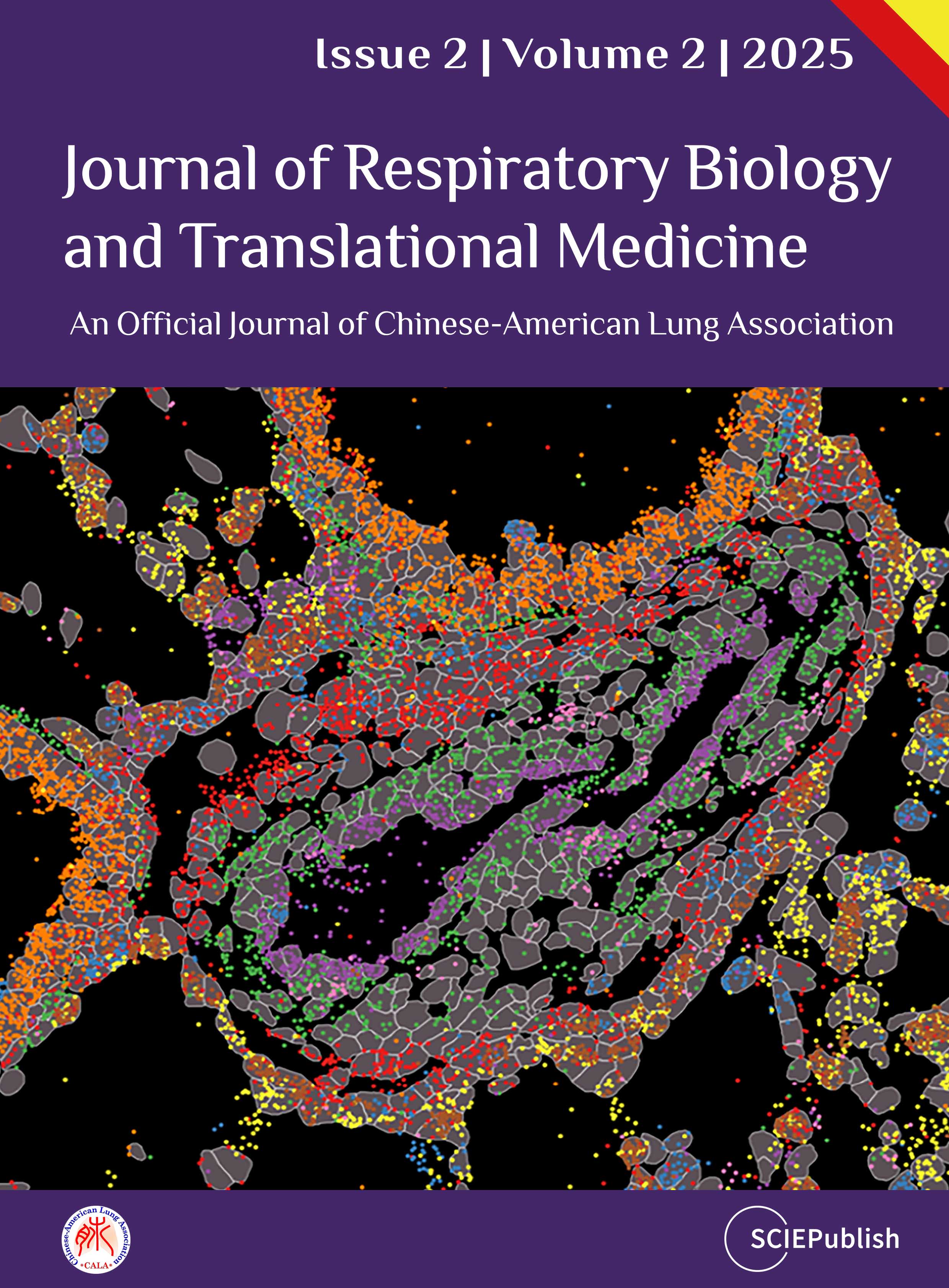
Cover Story (View full-size image):
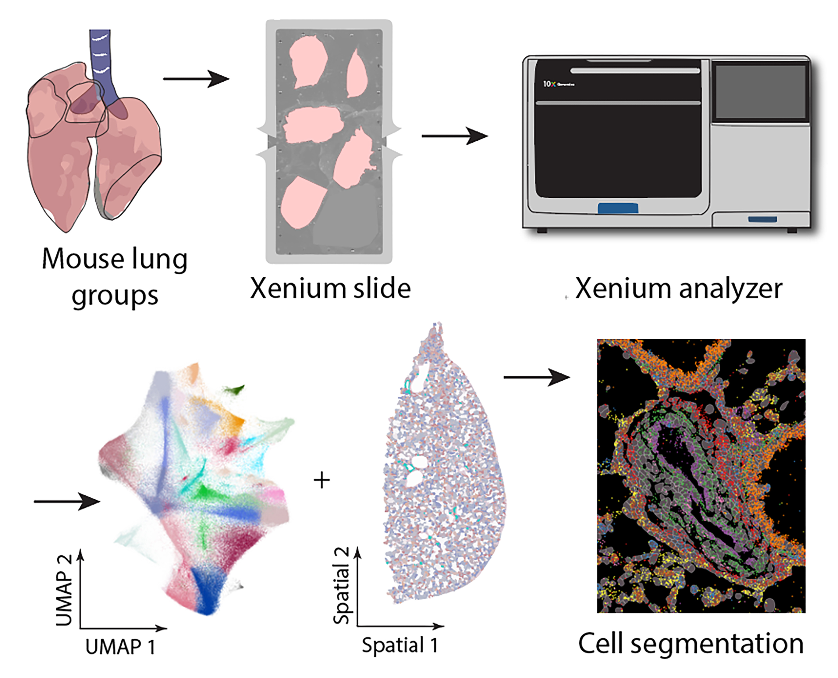
Obliterative vascular remodeling is a defining characteristic of pulmonary arterial hypertension (PAH). Unfortunately, none of the current therapies can reverse this process. Understanding the molecular alterations in the obliterative lesions is crucial for comprehending the disease’s pathogenesis and identifying novel therapeutic approaches. To achieve this, this study developed an enhanced high-resolution spatial transcriptomic technique to profile mouse lung tissue with severe PAH. Spatial imaging revealed the heterogeneity in cell populations within the obliterative arterial lesions of PAH mouse lungs. Each dot represents a molecular RNA, while each color represents a distinct cell population.