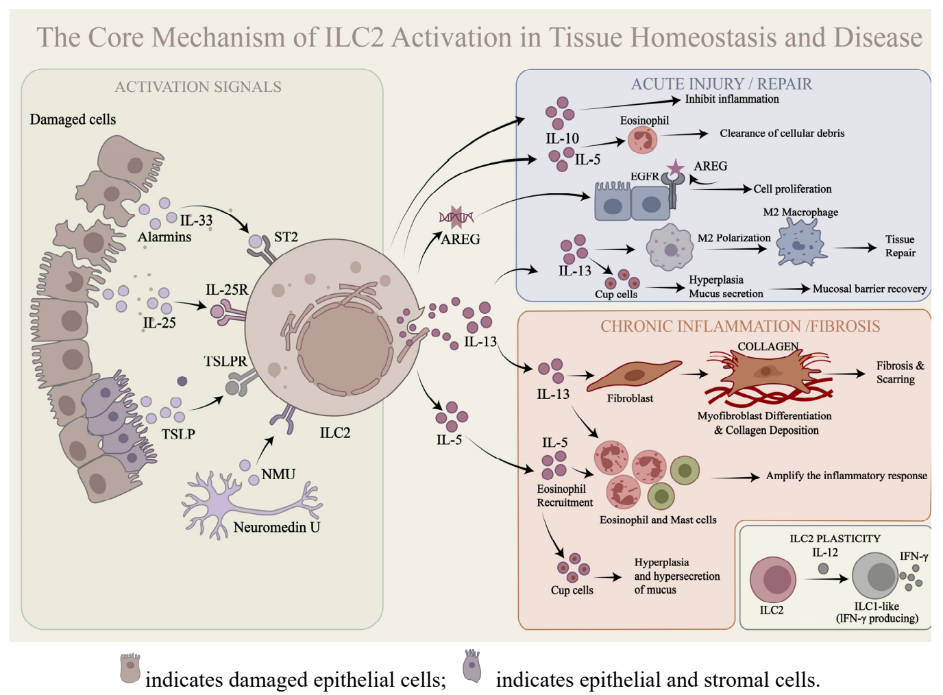
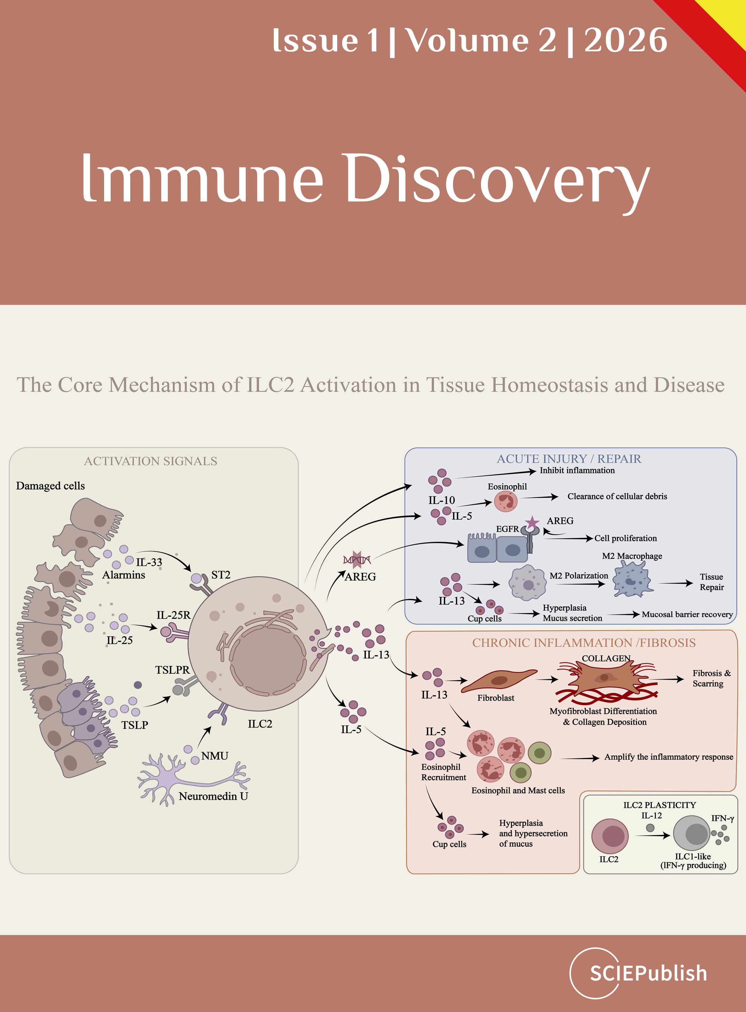
ISSN: 3007-6730 (Online)
3007-6722 (Print)
1. Institute of Transfusion Medicine and Immunology, Mannheim Institute of Innate Immunosciences (MI3), Medical Faculty Mannheim, Heidelberg University, Mannheim, Germany
2. Laboratory for Translational Cellular and Molecular Biomedicine, Tomsk State University, Tomsk, Russia
Henan Key Laboratory of Immunology and Targeted Drug, Henan Collaborative Innovation Center of Molecular Diagnosis and Laboratory Medicine, School of Medical Technology, Henan Medical University, Xinxiang, China
Autoimmune encephalitis has reshaped the understanding of neuropsychiatric disorders by highlighting the role of autoantibodies in psychosis symptoms, which often mimic primary psychosis conditions. This review synthesizes recent research on autoimmune encephalitis-related psychosis, broadening the focus from humoral immunity to T cell autoimmunity and the communication between the peripheral and central nervous systems. We discuss the identification of neuronal antigen targets, particularly the N-methyl-D-aspartate receptor (NMDAR), and their involvement in disease pathogenesis. Current treatments, such as plasma exchange and intravenous immunoglobulin, primarily target the pathogenicity of autoantibodies. However, emerging evidence suggests a crucial role for T cells, glia cell, and B cell in the immunopathogenesis of autoimmune encephalitis-related psychosis diseases. Autoimmune factors, including T and B cells, can either infiltrate the brain from the periphery or propagate via interacting with other cells, like glia, within the brain itself. This review advocates for a comprehensive approach to studying and treating these conditions, integrating both humoral and cellular mechanisms.
Chronic inflammation is widely considered a risk factor for T2DM by inducing insulin resistance, but all attempts to translate the concept into clinical therapies have failed in the past 30 years. Anti-inflammatory medicines, including anti-TNF-α antibody (Etanercept), anti-IL1 antibody (Anakinra), anti-IL6 (Ziltivekimab), and NLRP3 inflammasome inhibitor (Colchicine) have excellent activities in the control of inflammation in arthritis. They reduced inflammation in T2DM patients in the clinical trials, but none improved insulin sensitivity. Some of them exhibited a mild and transient activity in the control of blood glucose, but the activities were related to the improvement of insulin secretion by β-cells. The failure may be related to followings: over-interpretation of TNF-α activity; ignoring the role of anti-inflammatory cytokines; differences between mice and humans. However, the species difference cannot fully explain the failure as these therapies did not work in the animal models as well. Moreover, genome-wide association studies (GWAS) show that T2DM is not associated with proinflammatory cytokine genes, including TNF-α, IL-1β, IL-6, and CCL2(MCP1). More studies suggest that inflammation has beneficial activities in the mobilization of energy stores and promotion of energy expenditure to prevent energy surplus, a risk factor of obesity-associated T2DM. Inflammatory cytokines induce lipolysis, thermogenesis, and satiety. In this regard, the inflammatory response is a compensatory event to obesity-associated stress with beneficial effects on energy metabolism. It is time to reconsider inflammation activity in obesity for protective activities.
Chimeric Antigen Receptor T (CAR-T) cell therapy represents a groundbreaking advancement in cancer treatment, demonstrating remarkable antitumor efficacy in hematological malignancies. However, despite the substantial clinical progress achieved with CAR-T cell therapy, its application in the treatment of solid tumors remains limited by various factors, including the intricate tumor microenvironment (TME), CAR-T cell exhaustion, CAR-T cell infiltration into tumor tissue, and antigen heterogeneity. Scientists are relentlessly pursuing research in this domain, driven by the aim of offering newfound hope to cancer patients. In this comprehensive review, we delineate the fundamental principles underlying CAR-T cell therapy, delve into the challenges it encounters, and provide an insightful exploration of the advancements and progress made in the application of CAR-T cell therapy for solid tumors.
Cytotoxic CD8 T cells play a crucial role in controlling tumor progression. However, T cells infiltrating tumor tissues upregulate inhibitory receptors, reduce cytokine secretion, and lose their killing function, a state known as exhaustion. Thus, preventing or reversing T cell exhaustion is essential for sustaining a successful antitumor immune response. Recent studies have shown that T cell immunity not only requires the three primary signals—antigen receptor signaling, costimulation, and cytokines—but is largely shaped by endogenous and ambient metabolites as a fourth regulatory signal. Therefore, metabolic changes in the tumor microenvironment, caused by tumor cell proliferation and tissue remodeling, have a significant impact on the function of tumor-infiltrating T cells. This paper will review mechanisms by which three major types of metabolites—carbohydrates, lipids, and amino acids—influence T cell exhaustion in the tumor microenvironment, providing insights and directions for exploring metabolic targets in antitumor immunity.
As mankind breaks the boundaries of potential years to live, the process of aging imposes various cellular challenges, from less capacity of cell repair and damage to impaired protein formation, causing chronic low-level inflammation on tissues including the brain. Persistent chronic neuroinflammation can harm neurons, contributing to the development of neurodegeneration, a pathological process that affects cognitive function and is often reflected by dementia. This opinion article tries to recapitulate the influence that major histocompatibility class I (MHC-I) molecules have on brain homeostasis and how abnormalities in their expression can lead to cognitive deterioration. Studies carried out during recent years not only demonstrated that neurons and other central nervous system (CNS) cells express MHC-I molecules, but also that these molecules play essential roles in the establishment, function, and modeling of synapses in the CNS during the embryonic period, at birth and during adulthood, namely during inflammatory conditions. The accumulated body of evidence suggests that MHC-I molecules and the signaling pathways they regulate could provide clues on some of the molecular and cellular mechanisms regulating brain homeostasis and neuroregeneration in health and disease, thus becoming potential biomarkers of cognitive decline and targets for innovative immunotherapies.
The deubiquitinating enzyme cylindromatosis (CYLD) plays a fundamental role in regulating T cell development and activation. Previous studies have shown that CYLD is associated with autophagy, while AMP activated protein kinase (AMPK) pathway regulates the development of autophagy and affects cell metabolism. However, the mechanism by which CYLD affects autophagy and whether it affects the downstream metabolism of AMPKα remains unclear. In this study, we used the CYLD gene knockout model in Jurkat cells to investigate the mechanism of CYLD and autophagy and its relationship with cellular metabolism. The results show that CYLD deletion promotes autophagy through AMPKα/mTOR/ULK1 signaling pathway, promotes mitochondrial autophagy to improve mitochondrial function and attenuates cell lipid metabolism in Jurkat cells.
In Drosophila melanogaster, the siRNA-directed RNAi pathway provides crucial antiviral defenses. Cell-autonomously, Dicer-2 (Dcr-2) recognizes and cleaves viral dsRNA into siRNAs, which are incorporated into the RNA-induced silencing complex (RISC). Argonaute 2 (Ago2) then targets and cleaves viral RNA, preventing replication. Non-cell-autonomously, infected hemocytes secrete exosomes containing viral siRNAs, spreading antiviral signals to other cells. Additionally, tunneling nanotubes can transfer RNAi components between neighboring cells, further enhancing systemic immunity. These findings highlight the sophisticated antiviral strategies in Drosophila, offering insights for broader antiviral research.
Cytotoxic CD8 T cells play a crucial role in controlling tumor progression. However, T cells infiltrating tumor tissues upregulate inhibitory receptors, reduce cytokine secretion, and lose their killing function, a state known as exhaustion. Thus, preventing or reversing T cell exhaustion is essential for sustaining a successful antitumor immune response. Recent studies have shown that T cell immunity not only requires the three primary signals—antigen receptor signaling, costimulation, and cytokines—but is largely shaped by endogenous and ambient metabolites as a fourth regulatory signal. Therefore, metabolic changes in the tumor microenvironment, caused by tumor cell proliferation and tissue remodeling, have a significant impact on the function of tumor-infiltrating T cells. This paper will review mechanisms by which three major types of metabolites—carbohydrates, lipids, and amino acids—influence T cell exhaustion in the tumor microenvironment, providing insights and directions for exploring metabolic targets in antitumor immunity.
In Drosophila melanogaster, the siRNA-directed RNAi pathway provides crucial antiviral defenses. Cell-autonomously, Dicer-2 (Dcr-2) recognizes and cleaves viral dsRNA into siRNAs, which are incorporated into the RNA-induced silencing complex (RISC). Argonaute 2 (Ago2) then targets and cleaves viral RNA, preventing replication. Non-cell-autonomously, infected hemocytes secrete exosomes containing viral siRNAs, spreading antiviral signals to other cells. Additionally, tunneling nanotubes can transfer RNAi components between neighboring cells, further enhancing systemic immunity. These findings highlight the sophisticated antiviral strategies in Drosophila, offering insights for broader antiviral research.
The deubiquitinating enzyme cylindromatosis (CYLD) plays a fundamental role in regulating T cell development and activation. Previous studies have shown that CYLD is associated with autophagy, while AMP activated protein kinase (AMPK) pathway regulates the development of autophagy and affects cell metabolism. However, the mechanism by which CYLD affects autophagy and whether it affects the downstream metabolism of AMPKα remains unclear. In this study, we used the CYLD gene knockout model in Jurkat cells to investigate the mechanism of CYLD and autophagy and its relationship with cellular metabolism. The results show that CYLD deletion promotes autophagy through AMPKα/mTOR/ULK1 signaling pathway, promotes mitochondrial autophagy to improve mitochondrial function and attenuates cell lipid metabolism in Jurkat cells.
Chimeric Antigen Receptor T (CAR-T) cell therapy represents a groundbreaking advancement in cancer treatment, demonstrating remarkable antitumor efficacy in hematological malignancies. However, despite the substantial clinical progress achieved with CAR-T cell therapy, its application in the treatment of solid tumors remains limited by various factors, including the intricate tumor microenvironment (TME), CAR-T cell exhaustion, CAR-T cell infiltration into tumor tissue, and antigen heterogeneity. Scientists are relentlessly pursuing research in this domain, driven by the aim of offering newfound hope to cancer patients. In this comprehensive review, we delineate the fundamental principles underlying CAR-T cell therapy, delve into the challenges it encounters, and provide an insightful exploration of the advancements and progress made in the application of CAR-T cell therapy for solid tumors.
As mankind breaks the boundaries of potential years to live, the process of aging imposes various cellular challenges, from less capacity of cell repair and damage to impaired protein formation, causing chronic low-level inflammation on tissues including the brain. Persistent chronic neuroinflammation can harm neurons, contributing to the development of neurodegeneration, a pathological process that affects cognitive function and is often reflected by dementia. This opinion article tries to recapitulate the influence that major histocompatibility class I (MHC-I) molecules have on brain homeostasis and how abnormalities in their expression can lead to cognitive deterioration. Studies carried out during recent years not only demonstrated that neurons and other central nervous system (CNS) cells express MHC-I molecules, but also that these molecules play essential roles in the establishment, function, and modeling of synapses in the CNS during the embryonic period, at birth and during adulthood, namely during inflammatory conditions. The accumulated body of evidence suggests that MHC-I molecules and the signaling pathways they regulate could provide clues on some of the molecular and cellular mechanisms regulating brain homeostasis and neuroregeneration in health and disease, thus becoming potential biomarkers of cognitive decline and targets for innovative immunotherapies.
Chronic inflammation is widely considered a risk factor for T2DM by inducing insulin resistance, but all attempts to translate the concept into clinical therapies have failed in the past 30 years. Anti-inflammatory medicines, including anti-TNF-α antibody (Etanercept), anti-IL1 antibody (Anakinra), anti-IL6 (Ziltivekimab), and NLRP3 inflammasome inhibitor (Colchicine) have excellent activities in the control of inflammation in arthritis. They reduced inflammation in T2DM patients in the clinical trials, but none improved insulin sensitivity. Some of them exhibited a mild and transient activity in the control of blood glucose, but the activities were related to the improvement of insulin secretion by β-cells. The failure may be related to followings: over-interpretation of TNF-α activity; ignoring the role of anti-inflammatory cytokines; differences between mice and humans. However, the species difference cannot fully explain the failure as these therapies did not work in the animal models as well. Moreover, genome-wide association studies (GWAS) show that T2DM is not associated with proinflammatory cytokine genes, including TNF-α, IL-1β, IL-6, and CCL2(MCP1). More studies suggest that inflammation has beneficial activities in the mobilization of energy stores and promotion of energy expenditure to prevent energy surplus, a risk factor of obesity-associated T2DM. Inflammatory cytokines induce lipolysis, thermogenesis, and satiety. In this regard, the inflammatory response is a compensatory event to obesity-associated stress with beneficial effects on energy metabolism. It is time to reconsider inflammation activity in obesity for protective activities.
Autoimmune encephalitis has reshaped the understanding of neuropsychiatric disorders by highlighting the role of autoantibodies in psychosis symptoms, which often mimic primary psychosis conditions. This review synthesizes recent research on autoimmune encephalitis-related psychosis, broadening the focus from humoral immunity to T cell autoimmunity and the communication between the peripheral and central nervous systems. We discuss the identification of neuronal antigen targets, particularly the N-methyl-D-aspartate receptor (NMDAR), and their involvement in disease pathogenesis. Current treatments, such as plasma exchange and intravenous immunoglobulin, primarily target the pathogenicity of autoantibodies. However, emerging evidence suggests a crucial role for T cells, glia cell, and B cell in the immunopathogenesis of autoimmune encephalitis-related psychosis diseases. Autoimmune factors, including T and B cells, can either infiltrate the brain from the periphery or propagate via interacting with other cells, like glia, within the brain itself. This review advocates for a comprehensive approach to studying and treating these conditions, integrating both humoral and cellular mechanisms.
Cytotoxic CD8 T cells play a crucial role in controlling tumor progression. However, T cells infiltrating tumor tissues upregulate inhibitory receptors, reduce cytokine secretion, and lose their killing function, a state known as exhaustion. Thus, preventing or reversing T cell exhaustion is essential for sustaining a successful antitumor immune response. Recent studies have shown that T cell immunity not only requires the three primary signals—antigen receptor signaling, costimulation, and cytokines—but is largely shaped by endogenous and ambient metabolites as a fourth regulatory signal. Therefore, metabolic changes in the tumor microenvironment, caused by tumor cell proliferation and tissue remodeling, have a significant impact on the function of tumor-infiltrating T cells. This paper will review mechanisms by which three major types of metabolites—carbohydrates, lipids, and amino acids—influence T cell exhaustion in the tumor microenvironment, providing insights and directions for exploring metabolic targets in antitumor immunity.utf-8
Chronic inflammation is widely considered a risk factor for T2DM by inducing insulin resistance, but all attempts to translate the concept into clinical therapies have failed in the past 30 years. Anti-inflammatory medicines, including anti-TNF-α antibody (Etanercept), anti-IL1 antibody (Anakinra), anti-IL6 (Ziltivekimab), and NLRP3 inflammasome inhibitor (Colchicine) have excellent activities in the control of inflammation in arthritis. They reduced inflammation in T2DM patients in the clinical trials, but none improved insulin sensitivity. Some of them exhibited a mild and transient activity in the control of blood glucose, but the activities were related to the improvement of insulin secretion by β-cells. The failure may be related to followings: over-interpretation of TNF-α activity; ignoring the role of anti-inflammatory cytokines; differences between mice and humans. However, the species difference cannot fully explain the failure as these therapies did not work in the animal models as well. Moreover, genome-wide association studies (GWAS) show that T2DM is not associated with proinflammatory cytokine genes, including TNF-α, IL-1β, IL-6, and CCL2(MCP1). More studies suggest that inflammation has beneficial activities in the mobilization of energy stores and promotion of energy expenditure to prevent energy surplus, a risk factor of obesity-associated T2DM. Inflammatory cytokines induce lipolysis, thermogenesis, and satiety. In this regard, the inflammatory response is a compensatory event to obesity-associated stress with beneficial effects on energy metabolism. It is time to reconsider inflammation activity in obesity for protective activities.utf-8
Autoimmune encephalitis has reshaped the understanding of neuropsychiatric disorders by highlighting the role of autoantibodies in psychosis symptoms, which often mimic primary psychosis conditions. This review synthesizes recent research on autoimmune encephalitis-related psychosis, broadening the focus from humoral immunity to T cell autoimmunity and the communication between the peripheral and central nervous systems. We discuss the identification of neuronal antigen targets, particularly the N-methyl-D-aspartate receptor (NMDAR), and their involvement in disease pathogenesis. Current treatments, such as plasma exchange and intravenous immunoglobulin, primarily target the pathogenicity of autoantibodies. However, emerging evidence suggests a crucial role for T cells, glia cell, and B cell in the immunopathogenesis of autoimmune encephalitis-related psychosis diseases. Autoimmune factors, including T and B cells, can either infiltrate the brain from the periphery or propagate via interacting with other cells, like glia, within the brain itself. This review advocates for a comprehensive approach to studying and treating these conditions, integrating both humoral and cellular mechanisms.utf-8
In Drosophila melanogaster, the siRNA-directed RNAi pathway provides crucial antiviral defenses. Cell-autonomously, Dicer-2 (Dcr-2) recognizes and cleaves viral dsRNA into siRNAs, which are incorporated into the RNA-induced silencing complex (RISC). Argonaute 2 (Ago2) then targets and cleaves viral RNA, preventing replication. Non-cell-autonomously, infected hemocytes secrete exosomes containing viral siRNAs, spreading antiviral signals to other cells. Additionally, tunneling nanotubes can transfer RNAi components between neighboring cells, further enhancing systemic immunity. These findings highlight the sophisticated antiviral strategies in Drosophila, offering insights for broader antiviral research.utf-8
The deubiquitinating enzyme cylindromatosis (CYLD) plays a fundamental role in regulating T cell development and activation. Previous studies have shown that CYLD is associated with autophagy, while AMP activated protein kinase (AMPK) pathway regulates the development of autophagy and affects cell metabolism. However, the mechanism by which CYLD affects autophagy and whether it affects the downstream metabolism of AMPKα remains unclear. In this study, we used the CYLD gene knockout model in Jurkat cells to investigate the mechanism of CYLD and autophagy and its relationship with cellular metabolism. The results show that CYLD deletion promotes autophagy through AMPKα/mTOR/ULK1 signaling pathway, promotes mitochondrial autophagy to improve mitochondrial function and attenuates cell lipid metabolism in Jurkat cells.utf-8
As mankind breaks the boundaries of potential years to live, the process of aging imposes various cellular challenges, from less capacity of cell repair and damage to impaired protein formation, causing chronic low-level inflammation on tissues including the brain. Persistent chronic neuroinflammation can harm neurons, contributing to the development of neurodegeneration, a pathological process that affects cognitive function and is often reflected by dementia. This opinion article tries to recapitulate the influence that major histocompatibility class I (MHC-I) molecules have on brain homeostasis and how abnormalities in their expression can lead to cognitive deterioration. Studies carried out during recent years not only demonstrated that neurons and other central nervous system (CNS) cells express MHC-I molecules, but also that these molecules play essential roles in the establishment, function, and modeling of synapses in the CNS during the embryonic period, at birth and during adulthood, namely during inflammatory conditions. The accumulated body of evidence suggests that MHC-I molecules and the signaling pathways they regulate could provide clues on some of the molecular and cellular mechanisms regulating brain homeostasis and neuroregeneration in health and disease, thus becoming potential biomarkers of cognitive decline and targets for innovative immunotherapies.utf-8
Chimeric Antigen Receptor T (CAR-T) cell therapy represents a groundbreaking advancement in cancer treatment, demonstrating remarkable antitumor efficacy in hematological malignancies. However, despite the substantial clinical progress achieved with CAR-T cell therapy, its application in the treatment of solid tumors remains limited by various factors, including the intricate tumor microenvironment (TME), CAR-T cell exhaustion, CAR-T cell infiltration into tumor tissue, and antigen heterogeneity. Scientists are relentlessly pursuing research in this domain, driven by the aim of offering newfound hope to cancer patients. In this comprehensive review, we delineate the fundamental principles underlying CAR-T cell therapy, delve into the challenges it encounters, and provide an insightful exploration of the advancements and progress made in the application of CAR-T cell therapy for solid tumors.utf-8