On the Utility of Nailfold Capillaroscopy in Detecting the Effects of Fibrinaloid Microclots in Diseases Involving Blood Stasis

Review Open Access

On the Utility of Nailfold Capillaroscopy in Detecting the Effects of Fibrinaloid Microclots in Diseases Involving Blood Stasis

Author Information
1
Department of Biochemistry, Cell and Systems Biology, Institute of Systems, Molecular and Integrative Biology, Faculty of Health and Life Sciences, University of Liverpool, Crown St, Liverpool L69 7ZB, UK
2
The Novo Nordisk Foundation Center for Biosustainability, Building 220, Søltofts Plads, Technical University of Denmark, 2800 Kongens Lyngby, Denmark
3
Department of Physiological Sciences, Faculty of Science, Stellenbosch University, Stellenbosch 7602, Private Bag X1 Matieland, South Africa
*
Authors to whom correspondence should be addressed.

Received: 31 May 2025 Accepted: 15 August 2025 Published: 20 August 2025

Creative Commons

© 2025 The authors. This is an open access article under the Creative Commons Attribution 4.0 International License (https://creativecommons.org/licenses/by/4.0/).

Views:49
Downloads:7
Immune Discov. 2025, 1(3), 10011; DOI: 10.70322/immune.2025.10011
ABSTRACT: A variety of chronic, inflammatory vascular and autoimmune diseases are accompanied by fibrinaloid microclots. Such diseases reflect endothelial dysfunction and may be detected using a ‘structural’ assay in the form of the fluorescence microscopic or flow ‘clotometry’ analysis of suitably stained platelet-poor plasma. Their amyloid nature and the presence of anti-fibrinolytic molecules therein make the fibrinaloid microclots comparatively resistant to the normal processes of clot degradation. By inhibiting the free flow of blood, the many effects of fibrinaloid microclots include those causing hypoxia, oxidative stress, and ‘blood stasis’ in the microcirculation. Nailfold capillaroscopy is an established observational technique (with both ‘structural’ and ‘functional’ elements) for assessing the microcirculation, and it is thus of interest to establish whether it too demonstrates changes when these syndromes are diagnosed. All diseases in which both methods have been applied show both the presence of fibrinaloid microclots and changes in capillary properties, indicating the complementary value of the structural and functional assays. This also suggests the potential value of nailfold capillaroscopy in a variety of other diseases involving coagulopathies or a deficient microcirculation, which has been little studied to date.
Keywords: Nailfold capillaroscopy; Nailfold videocapillaroscopy; Fibrinaloid microclots; Microcirculation; Inflammation; Coagulopathies; Blood stasis; Vascular and immunological disorders

1. Introduction

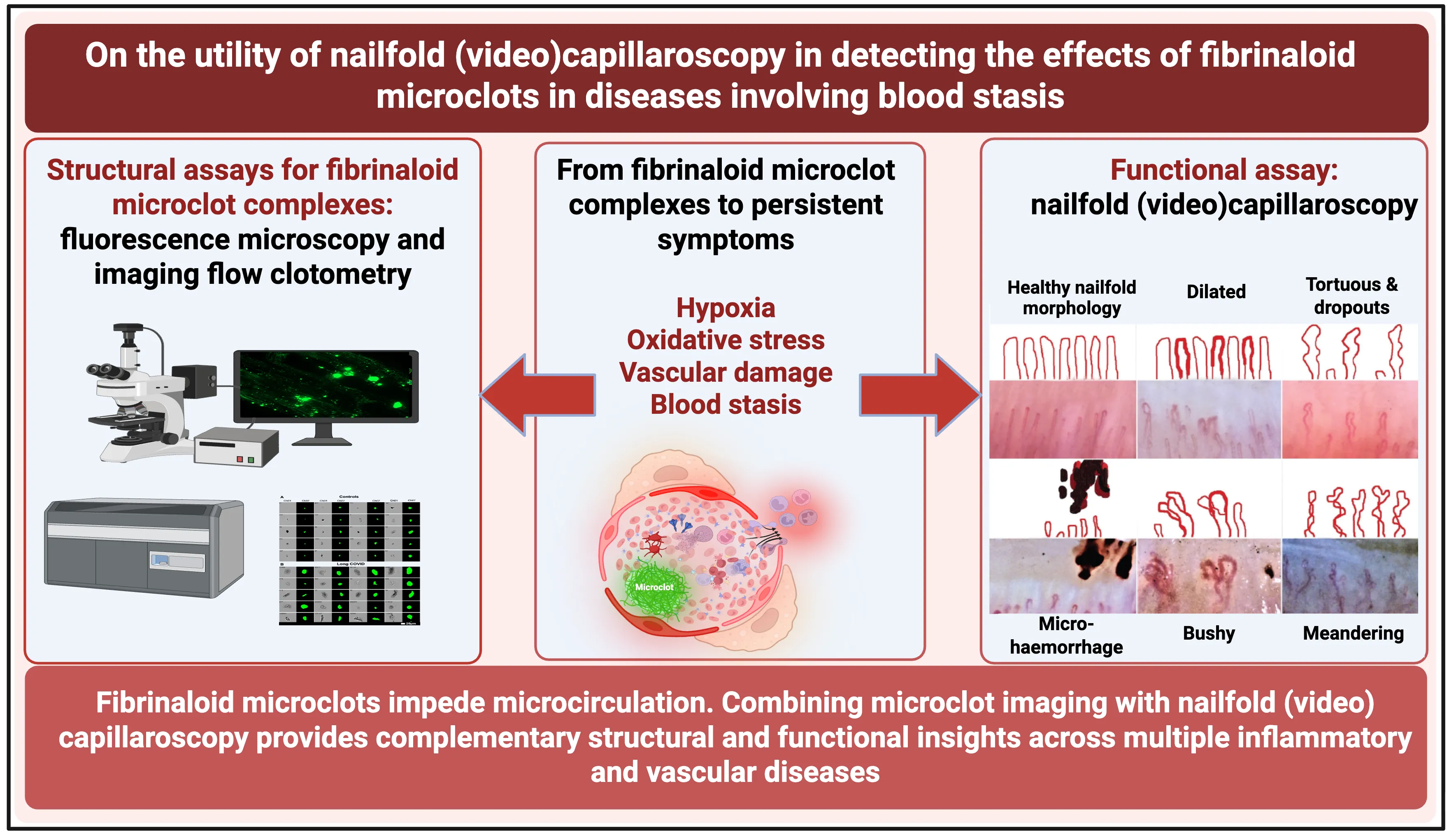
More than a decade ago it was discovered, via scanning electron microscopy, that blood can clot into a highly anomalous form, which in contrast to the normal thrombi with individual fibres that appear like nicely cooked spaghetti, adopted a morphology, referred to as ‘dense matted deposits’, that instead resembled parboiled spaghetti that had congealed into an amorphous mass (e.g., [1,2,3]). It was subsequently recognised (e.g., [4,5,6]) that the ‘anomalous form’ was amyloid in nature, as it could be stained with the well-established amyloid stain thioflavin T [7,8,9,10]. Such clots were commonly in the range 2–200 μm diameter, were found in a variety of chronic, inflammatory diseases [11] (including Alzheimer’s [12,13,14], Parkinson’s [15,16], type 2 diabetes [12,17,18] and rheumatoid arthritis [19]) and—as all amyloids—were significantly resistant to the normal routes of fibrinolysis via plasmin(ogen) [20]. Because of their amyloid nature, we have come to refer to them as fibrinaloid microclots [21,22,23,24,25]. Figure 1 shows a typical example of the microclots as stained with three different amyloid-selective stains.
Figure 1
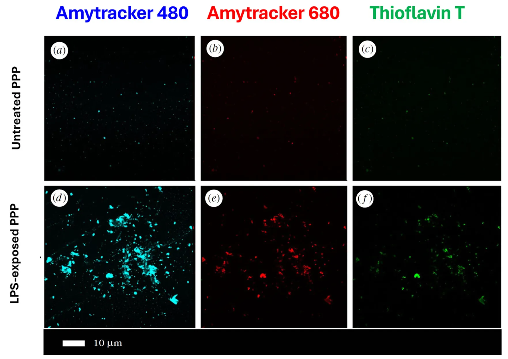
. A typical example of fibrinaloid microclots. Platelet-poor plasma was treated with thrombin and stained either with one of two oligothiophene [<a href="#B26" class="html-bibr">26</a>,<a href="#B27" class="html-bibr">27</a>,<a href="#B28" class="html-bibr">28</a>] Amytracker™ dyes (<strong>a</strong>,<strong>b</strong>,<strong>d</strong>,<strong>e</strong>) or with thioflavin T (<strong>c</strong>,<strong>f</strong>) . In one case (<strong>d</strong>–<strong>f</strong>), a small amount of bacterial lipopolysaccharide was added prior to the thrombin. Modified from the CC-BY 4.0 publication [<a href="#B6" class="html-bibr">6</a>].
More recently, fibrinaloid microclots have been recognised as a major feature accompanying both acute [18,29] and Long COVID [24,30,31,32,33,34,35,36], as well as in the related Myalgic encephalopathy/chronic fatigue syndrome (ME/CFS) [23,34,37] and sepsis [38]. As with any inert matter of this type and size (including microplastics [39,40,41]), fibrinaloid microclots can become trapped in microcapillaries, thereby restricting blood flow and oxygen transport to tissues. This provides ready explanations for a variety of phenomena, especially those associated with Long COVID, such as fatigue [21], post-exertional symptom exacerbation [42], postural orthostatic tachycardia syndrome (POTS) [43], and fibromyalgia [44]. Proteomic studies [30,38,45] indicate [46] a high prevalence of amyloidogenic proteins in fibrinaloid microclots that, in contrast to normal clots [47], do not reflect those in the typical normal plasma proteome [46], and this has predictive power [48,49]. In addition, both the microclots [50,51,52] and the thrombectomised macroclots observed [53,54] following an ischaemic stroke are amyloid in nature. Amyloidogenic cross-seeding can significantly change the conformation of other proteins, and this can also explain the generation of autoantibodies since such proteins would then be seen as not-self [22]. A chief consequence of the blockage of microcapillaries by fibrinaloid microclots is a slowing down of the blood flow, sometimes referred to (including by Virchow in his ‘triad’ [55,56,57]) as Blood Stasis. ‘Blood stasis’ is also a highly important concept in Traditional Asian Medicines (see e.g., [58,59,60,61]), and seems strongly correlated with the presence of fibrinaloid microclots [62]. Ischaemia or hypoxia are inevitable consequences, and indeed this property, leading to oxidative stress, is consequently common to all the chronic, inflammatory syndromes being considered here (e.g., [11,63,64,65,66,67,68,69,70,71,72,73]). To date, the most frequent types of measurements of fibrinaloid microclots have mainly been ‘structural’ using thioflavin T staining and observation by fluorescence microscopy (e.g., [4,6,12,31,33,38,74]) or flow cytometry [32] (‘flow clotometry’ [34]). A more ‘functional’ assay would be highly desirable to complement this. Although many of the phenomena may occur deep inside the body, our chief focus here is on the human skin microcirculation [75] and its attendant disorders (e.g., [76,77,78,79]). Capillaroscopy refers to a general technique that can potentially determine blood flow rates in the microcirculation in a simple and non-invasive manner, and, given the above, might potentially serve as an excellent functional assay for blood stasis and microclot presence. Sublingual microscopy [80], retinal [81], and nailfold capillaroscopy (NFC) are in common use and—while other methods such as laser speckle contrast imaging [82,83,84,85,86,87,88] and thermography [89,90] may also be used—we focus here on NFC. To this end, the chief purpose of the present review is to develop the idea that NFC, including in particular its derivative nailfold videocapillaroscopy (NVC), will be of value in understanding syndromes in which fibrinaloid microclots are involved, and to provide the evidence-based reasoning for this. A preprint has been lodged [91].

2. Nailfold Capillaroscopy

2.1. History and Modern Implementations Capillaroscopy is a technique for looking at the microcirculation [75]. The idea that the study of capillaries might have disease-diagnostic value goes back to the 17th Century, when Johan Christophorous Kolhaus used a primitive microscope to observe the small blood vessels surrounding the nails, and later Giovanni Rasori, using a magnifying glass, related the observed properties to conjunctival inflammation [92]. A list of recent reviews is given in Table 1. Modern microscopes, commonly using 200× magnification, coupled to well engineered illumination and focusing optics, are all that is necessary to acquire static (capillaroscopy) and dynamic (videocapillaroscopy) images, with a second element being how these images are interpreted—manually or computationally—to provide useful diagnostic information (Figure 2). Because our focus lies on the relationship with fibrinaloid microclots, we do not provide detailed analyses of the precise procedures involved (e.g., [93,94,95]). Many commercial systems are available, although we neither seek to discriminate among them nor recommend particular models. They provide interfaces with laptops and even cellphones. Since interpretation by the human eye alone is neither straightforward nor objective [96,97], the more refined instrumentation usually comes with software to assist or even provide interpretation. It typically costs a few hundred pounds for entry-level models. Parameters (strictly, variables) that are measured or calculated include capillary density [98,99] and percentage of abnormal and giant capillaries, tortuosities, and haemorrhages [94,97,100,101,102,103]. EULAR (The European League Against Rheumatism) has agreed on a consented standardised capillaroscopic description protocol [93,104,105,106,107].
Figure 2. Cartoon illustrating the basic steps in nailfold capillaroscopy. Image includes elements of the Open Access (CC-BY 4.0) materials at [94,100,108]. The three fingers adjacent to the thumb may be profiled, with 4 regions on each [100]. The normal or healthy pattern (as illustrated) displays a regular architecture with uniform distribution and diameter and a hairpin shape [109].
Figure 2 also illustrates the kinds of capillary structure seen in healthy controls (and see e.g., [75,94,110]). Figure 3 
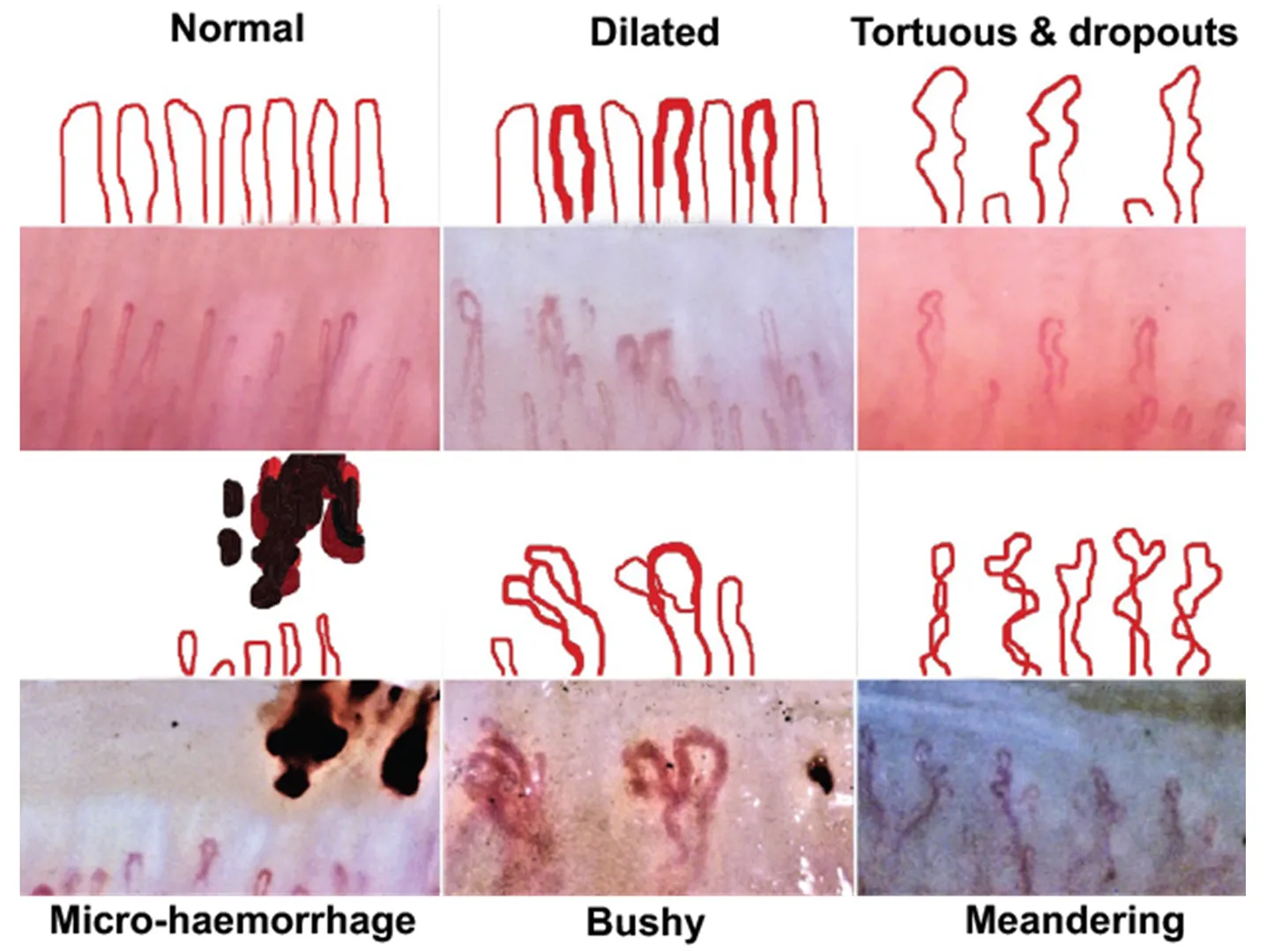
provides a feel for the kinds of images that are obtainable under different conditions.
Figure 3. Some images and explanatory cartoons of various nailfold capillary morphologies. Taken from the CC-BY 4.0 Open Access paper [94].
Clearly, the strategy is generic, and while possibly the majority of studies have focused on Raynaud’s disease, rheumatology, and systemic sclerosis (scleroderma), the range is very wide. This is partly because many are chronic inflammatory diseases with common vascular causes and/or symptoms [11], including endothelial dysfunction. Systemic sclerosis is recognised as a vascular disease involving endothelial dysfunction [111,112,113,114,115,116,117], thus bearing significant similarities to COVID-19 [118] that most certainly does [36,119,120,121,122,123,124,125], so it is not surprising that such commonalities exist. In a sense, this survey thus adds weight to the view that there are common co-occurrences of various symptoms. The advantage of nailfold capillaroscopy over, e.g., cytokine-based assessment of inflammation is that capillaroscopy is non-invasive, quick, and cheap, providing useful information for diagnosis and treatment. To illustrate its broad general utility, a sample of such studies in which it has been used is included in Table 1. While these are seen mostly as immunological in character, we recognise that the symptoms of many other syndromes can be reflected in the microcirculation. Specifically, Table 1 has three purposes. These are to show readers (i) that every disease where microclots have been measured shows abnormalities in the microcirculation as judged by NFC/NVC, (ii) for the microclot community to see how widespread and valuable the usage of NFC/NVC is and thus to encourage them to try it, and (iii) to point the NFC/NVC community at other diseases where (based on microclot measurements) we suggest their methods would be of value.

Table 1. Some disorders involving the microcirculation in which (mainly) nailfold (video) capillaroscopy has been found to have diagnostic utility or where fibrinaloid microclots have been demonstrated. Disorders in which fibrinaloid microclots have been demonstrated are rendered in bold face; note that every disorder in which microclots have been demonstrated has anomalies as assessed using nailfold capillaroscopy.

Disease or Syndrome Comments Selected Nailfold Capillaroscopy References Selected Fibrinaloid Microclot References
(Where Tested)
Acute COVID-19 Capillary tortuosity, meandering, haemosiderin deposition, and microhaemorrhage increased; capillary density and length, and blood flow decreased [126,127,128,129,130,131,132,133,134,135,136,137,138] [18,29,139,140,141,142]
Alzheimer’s dementia Greater nailfold capillary tortuosity in individuals with Alzheimer’s dementia [143,144] [12,13,14,145,146]
Anderson-Fabry disease Also known as Fabry disease. Promising but underexplored. Subclasses detected depend on the α-galactosidase A variant. [147,148,149,150]
Anorexia nervosa Related to connective tissue disorders [151,152]
Antineutrophil Cytoplasmic Antibody-Associated (ANCA) Vasculitis Neoangiogenesis, capillary
loss, microhaemorrhages, and bushy and enlarged capillaries
[153,154,155,156]
Atopic dermatitis Many more lesions, e.g., pitting, capillary density, tortuosity [157]
Behçet’s disease Increases in capillary dilatation, tortuous and branched capillaries, and microhaemorrhages [158,159]
Biliary cirrhosis Multiple abnormalities [160]
Cancer Not yet widely deployed, but some suggestive data imply it could have diagnostic or prognostic value. [161]
Chronic obstructive pulmonary disorder (COPD) Review [162] (but cf. [163])
Chronic smokers Abnormalities are more common in chronic smokers [164]
Chronic urticaria Lower capillary density, more capillary malformations, and more irregular capillary dilations [165]
Connective tissue disorders Various [166,167,168]
Coronary heart disease Assessment of capillary blood flow and relation to erythrocyte aggregation [169]
Dermatomyositis Diminished capillary density and abnormal capillary morphology (including enlargement) in patients. It can be related to antibody levels. Haemosiderin deposits can occur. [104,170,171,172,173,174,175,176,177,178,179]
Diabetes mellitus, type 1 General [180,181]
Use of deep learning [182]
Diabetes mellitus,
type 2
Capillary dilatation, avascular zones, and tortuous capillaries [183,184,185] [12,17,18,186,187]
Peak capillary blood flow velocity (CBFV) post-occlusion is much lower [188]
Use of deep learning [182]
Changes closely related to the quality of glucose control [189,190]
Capillaries are larger but less dense [191]
Various differences [192,193]
Diabetic complications Listed separately below
Diabetic foot Significant difference in pulp of the big toe [194]
Diabetic nephropathy Significant correlation between lowered glomerular filtration rate and e.g., reduced fundus transparency and visibility of the sub-venous plexus [195,196]
Diabetic neuropathy Improved by α-lipoic acid [197]
Diabetic retinopathy Decreased capillary length, width, number, and turbidity, crossing capillaries, and other abnormalities [183,184,198,199,200,201]
Digital ulcers A common accompaniment to systemic sclerosis and Raynaud’s disease [202,203,204]
Endothelial dysfunction Reviews [117,128] [125]
In livedoid vasculopathy [205]
Association with rheumatoid arthritis [206]
Fibromyalgia Raynaud’s disease and impaired microvascular function are common in patients with fibromyalgia, where nailfold abnormalities are common. [207,208,209,210]
Fewer capillaries, apical limb width, and capillary width decreased [78,211] See [44], and for amyloid deposition in skeletal muscle [212]
Mean capillary loop diameter, micro-aneurysm number, avascular areas, and neoangiogenic
capillaries significantly higher
[213]
Gaucher disease Various subtypes. Microangiopathy is the most obvious NFC finding [214]
General reviews [77,94,100,215,216,217]
Non-rheumatic diseases [218]
Vascular disease [219]
Glaucoma Huge decrease in capillary blood flow [220]
Heart failure Greater abnormalities with preserved ejection fraction [221]
Hepatitis, viral Changes in the avascular area, capillary dilatation, capillary tortuosity, and capillary enlargement were observed [222]
Hypertension (general) Many differences, including lowered capillary density and other morphological changes [223]
Idiopathic inflammatory myopathy Significant differences, including between subtypes [224,225,226,227,228]
Idiopathic macular telangiectasia type 2 Increased capillary tortuosity, ‘bizarre’ capillaries, and microhaemorrhage in the patient group compared to the controls [229]
Leprosy Capillary dropouts are the most frequent, followed by tortuous, receding, and dilated capillaries [230]
Long COVID Compares Long COVID patients without and with systemic sclerosis. Long COVID patients show more microvascular alterations than recovered COVID patients. [231] [21,24,30,31,32,33,34,35,43,45,125,232]
Lupus (systemic lupus erythematosus) [105,173,233,234,235,236]
Migraine Seemingly little studied recently by nailfold capillaroscopy, but major differences observed vs controls, especially when cold-induced [237,238] [239]
Neutropenia Indirect assessment from videocapillaroscopy [240,241]
Obstructive sleep apnoea All capillaroscopy findings were significantly higher in the patient group [242]
Parkinson’s disease Seemingly not yet studied by nailfold capillaroscopy in any detail. [12,16,243]
Polycythemia vera Increase in the diameter of vessels [244]
Polymyositis Scleroderma pattern predominated and improved after treatment. [104,170,179,245,246]
Pre-eclampsia Change in capillary length and density with pregnancy-induced hypertension, whether pre-eclamptic or not. Some relationship to maternal blood pressure [247]. [248]
Decreased capillary density in pre-eclamptics [249]
Review of capillaroscopy in pregnancy [250]
Psoriasis [251,252,253,254,255,256,257]
Pulmonary
arterial hypertension
(Can accompany systemic sclerosis.) Lowered capillary density [223,258,259]
Raynaud’s disease or Raynaud’s phenomenon Common accompaniment of systemic sclerosis. NFC especially used in discriminating primary Raynaud’s from secondary Raynaud’s due to systemic sclerosis [89,204,260,261,262,263,264,265,266,267,268,269,270,271]
Standardisation [106]
Rheumatology, especially rheumatoid arthritis Reviews [92,251,272,273,274,275,276,277,278,279,280] [19,281,282]
Sarcoidosis Early but promising [283,284,285]
Sarcopenia Common in rheumatoid arthritis and systemic sclerosis [286,287]
Sepsis and septic shock See Section 2.3 [288] [38] and see [289]
Decreased capillary density sublingually [290]
Sickle cell disease Lower capillary density and more dilated capillaries [291]
Sjögren’s syndrome [292,293]
Systemic sclerosis It is increasingly seen as a vasculopathy [114]. Reviews on the role of NFC are in the adjacent column. Arguably, the commonest area of study. [107,172,294,295,296,297,298,299,300,301,302,303,304,305,306,307,308,309]
Use of the NEMO score (the number of microhaemorrhages
plus micro-thromboses)
[310,311,312]
Patients with primary biliary cholangitis [313]
Differences when pulmonary arterial hypertension is also present [314,315]
The widespread occurrence of alterations in the microcirculation, as judged by nailfold capillaroscopy, shows how extensive this is in multiple syndromes that plausibly share common causes. The important point here is that, where tested, all examples in which fibrinaloid microclots have been measured in plasma also show microcirculation disorders, as one would expect. One reviewer questioned whether these techniques, that are well accepted for their ability to measure the morphological properties of microcapillaries, are also suitable for measuring blood flow; clearly individual static images can not do that directly, although it is pretty obvious (given that these methods are routinely used for assessing the microcirculation) that changes in capillary diameter and tortuosity necessarily have such effects [107,143,144,217,273,315,316]. Equally clearly, video methods that image capillaries at intervals and with a suitable magnification can precisely detect the flow of blood, as the following illustrative references that demonstrate it explicitly amply show (e.g., [100,117,143,168,188,217,317,318,319,320,321,322,323,324,325,326]). This does not seem remotely controversial. 2.2. Long COVID as an Example Following infection with the SARS-CoV-2 virus, a significant fraction of individuals fail to return fully to health, and after three months are said to suffer from post-acute sequelae of COVID-19 (PASC), commonly known as Long COVID [327]. Long COVID is estimated to affect or have affected over 400 million individuals worldwide, contributing to an annual economic burden of $1 trillion or ~1% of the global economy [328,329]. Symptoms vary widely [330,331,332,333,334,335], with fatigue being the most common [336,337]. It is a disease in which many studies have shown the presence of fibrinaloid microclots [21,24,30,31,32,33,34,35,43,45,125,232], which provide ready explanations for the various symptoms [21,22,42,43]. Importantly, Cutolo and colleagues carried out a valuable study [231] using NFC in patients with Long COVID and matched controls. Specifically, nailfold videocapillaroscopy (NVC) showed significant microvascular damage in long covid (LC) patients compared with matched healthy controls. Dilated capillaries, microhaemorrhages, abnormal shapes, and reduced capillary density were detectable in LC patients even 12 months after acute SARS-CoV-2 infection. Importantly, NVC demonstrated that these observables were normalised in patients who had recovered. It would seem that NVC is a valuable functional diagnostic for individuals with Long COVID, and a potentially valuable complement to the measurement of fibrinaloid microclots. 2.3. Sepsis and Septic Shock Sepsis and septic shock are of special interest here for a number of reasons. First, they can be highly dangeous. Secondly, they are accompanied by microthrombi [338] that we suggested are our fibrinaloid microclots [289], and thirdly, the fibrinaloid microclot burden, as now so measured, is highly predictive of survival in the ICU (odds ratio > 5) [38]. (The microclots are even more predictive of disseminated intravascular coagulation [
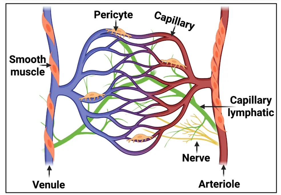
38].) Finally, the microcirculation (Figure 4) is intimately involved in sepsis and septic shock.
Figure 4. The anatomy of the microcirculation is the largest part of the vascular system and consists of the smallest vessels, referred to as arterioles, capillaries, and venules. The lymphatic capillaries carry the extravascular fluid into the venous system, while the arterioles are surrounded by vascular smooth muscle cells responsible for regulating arteriole tone. Taken and slightly adapted from the CC-BY 4.0 publication [339].
Specifically, disorders of the endothelium and microcirculation leading to hypoxia are recognised as an intimate part of sepsis and septic shock (e.g., [288,340,341,342,343,344,345,346,347,348,349,350,351,352,353,354,355,356,357,358,359,360,361,362,363]). However, NFC is said to be not much used as the nailfold vascular bed is sensitive to peripheral vasoconstriction, vasopressor agents, and temperature changes. None of these seems like insurmountable issues [364], and it would seem that the recognition of the importance of both the microcirculation and fibrinaloid microclots in sepsis mortality warrants a far more detailed analysis, to include a comparison between microclot burden, nailfold capillaroscopic findings, any effects of clotbusters or other treatments, and disease outcomes. Indeed, the generally less common sublingual capillaroscopy is known to be very useful [365] and even predictive of mortality [366]. 2.4. ‘Blood Stasis’ and Treatments to Improve It As mentioned above, ‘Blood stasis’, a term used as part of Virchow’s triad [55,56,57,367,368], is an extremely important concept in Traditional Chinese Medicine [58,369,370,371,372,373,374,375] (known there as Xue Yu (血瘀) [376]). Exactly equivalent ideas exist in Japanese Kampo medicine (where it is termed Oketsu [377,378,379,380]) and in Traditional Korean Medicine (where blood stasis is known as ‘Ouhyul’ or ‘Eohyul’) [61,381]). We recently summarised the extensive evidence to the effect that ‘blood stasis’ is precisely a manifestation of the presence and effects of fibrinaloid microclots [62], in that all syndromes known to display fibrinaloid microclots are considered to be diseases of blood stasis. While such traditional medical formalisms also recognise that one should treat the patient as an individual, standard herbal formulas for diseases of blood stasis do exist, in the form of XueFu ZhuYu [62,382,383,384,385,386,387] and Keishibukuryogan [377,388,389], and are known to improve the microcirculation. Consequently, it would be of considerable interest to assess this via nailfold capillaroscopy. 2.5. Future Directions Although many of the commercial systems are served by reasonably sophisticated software to assist interpretation [390], much of the downstream analysis remains in the hands and minds of skilled clinicians, and, especially depending on the image quality [391], is necessarily subjective [323,392,393,394]. This said, where it has been studied inter-rater reliability has been found to be good (e.g., [92,96,392,395,396]). An easy prediction is that these kinds of tasks will soon be taken over by intelligent systems trained using modern data-driven generative methods of machine learning, commonly referred to as artificial intelligence or ‘AI’. This particular revolution is, of course, already underway [390,393,394,397,398,399,400,401,402,403,404,405]. To date, the lack of availability of labelled datasets has confined most efforts to transfer learning or ‘fine tuning’ of other models [406]. However, much as with equivalent developments in general image processing [407,408], chemistry [409], and protein science [410,411,412], progress will be greatly assisted [275] by the availability of a database of tagged images, videos and appropriate metadata taken directly from nailfold capillaroscopy, as well as the ability [413] to explain the bases (in terms of features extracted) for any such disease classifications. A further point is that while many diseases have been studied using nailfold capillaroscopy (Table 1), there are quite a number more, some with high prevalence, that have not. These would seem to offer considerable opportunities for devising analyses with prognostic value. For instance, myalgic encephalopathy/chronic fatigue syndrome (ME/CFS) shares many of the hallmarks of the other diseases (including Long COVID) listed in Table 1 [414,415,416], including endothelial dysfunction [37,417,418,419,420], fibrinaloid microclots [23,37,421], and a disrupted microcirculation [422] (as in Figure 5), but we know of no attempt to assess its severity using nailfold capillaroscopy, which would provide further evidence beyond that existing (see [232]) for a deranged microcirculation in this syndrome.
Figure 5
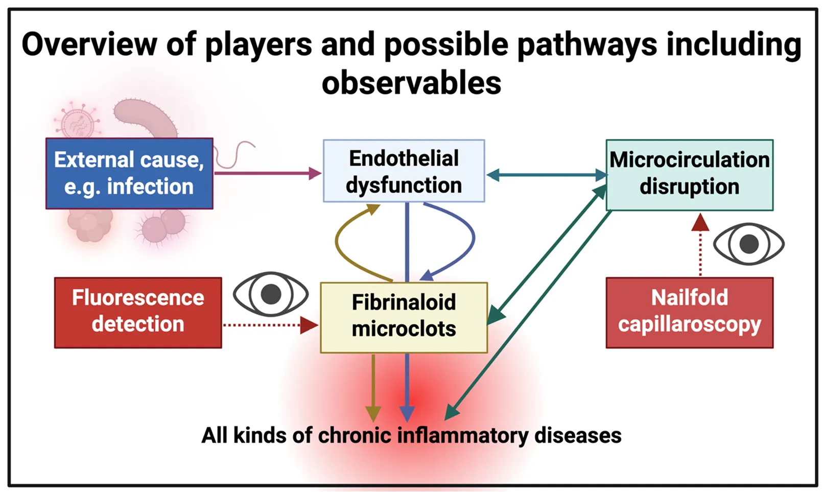
. Cartoon illustrating the systems aspects of the main features of this prospective review. Endothelial dysfunction, the result of a deterministic ‘external’ cause such as (a new or reactivated) infection, trauma, or stress, leads to the disruption of the microcirculation and to fibrinaloid microclots, each of which can potentially exacerbate the others. Our focus here is on nailfold capillaroscopy, though we have rehearsed the methods for microclot detection. Endothelial (dys)function <i>in vivo</i> is commonly assessed via flow-mediated dilatation [117,423] or various biomarkers (e.g., [424,425,426,427]). We anticipate that the combination of all three will be especially powerful in understanding the nature and severity of these kinds of disease, thereby assessing mechanisms and candidate treatments.
This review has purposely confined itself to nailfold capillaroscopy since relatively inexpensive equipment to implement the method is widely available. This said, we recognise that more sophisticated methods will likely provide much more power. 3D optoacoustic imaging (raster scanning optoacoustic mesoscopy) [428,429,430] is one such approach, mirroring (in a certain sense) the developments [431,432,433,434,435] in optoacoutstic infrared and Raman spectroscopy and imaging. Another trend towards using smart phones is taking photographs directly to assess digital lesions [436,437,438].

3. Conclusions

The novelty of this synthetic review (see [439]) lies in bringing together two fields that have until now pursued entirely separate paths, namely the fields of fibrinaloid microclot measurements and nailfold (video) capillaroscopy. Nailfold capillaroscopy and nailfold videocapillaroscopy enjoy widespread usage in a large number of syndromes involving vascular issues and/or autoimmunity where they manifest in the microcirculation (Table 1). However, until now, there has been no recognition of the possibility that changes in the static and especially the video images thereby observed might be related to the well-established presence of fibrinaloid microclots in many of these diseases. We believe that the arguments deployed here now make a comparison of the prevalence of the ‘structural’ microclots in images seen in microscopy or imaging flow cytometry with the structural and importantly more ‘functional’ images observable by capillaroscopic assays a very worthwhile endeavour.

Author Contributions

Conceptualization, D.B.K. & E.P.; Formal Analysis, D.B.K. & E.P.; Resources, D.B.K. & E.P.; Writing—Original Draft Preparation, D.B.K.; Writing—Review & Editing, D.B.K. & E.P.; Visualization, D.B.K. & E.P.; Funding Acquisition, D.B.K. & E.P.

Funding

DBK thanks the Balvi Foundation (grant 18) and the Novo Nordisk Foundation for funding (grant NNF20CC0035580). EP thanks PolyBio Research Foundation and Kanro Foundation for funding. The content and findings reported and illustrated are the sole deduction, view and responsibility of the researchers and do not reflect the official position and sentiments of the funders. The funders had no role in study design, data collection and analysis, decision to publish, or preparation of the manuscript.

Declaration of Competing Interest

EP is a named inventor on a patent disclosing the use of fluorescence microscopy in Long COVID.

References

1.
Pretorius E, Oberholzer HM, van der Spuy WJ, Meiring JH. The changed ultrastructure of fibrin networks during use of oral contraception and hormone replacement. J. Thromb. Thrombolysis 2010, 30, 502–506. doi:10.1007/s11239-010-0502-4. [Google Scholar]
2.
Pretorius E, Swanepoel AC, Oberholzer HM, van der Spuy WJ, Duim W, Wessels PF. A descriptive investigation of the ultrastructure of fibrin networks in thrombo-embolic ischemic stroke. J. Thromb. Thrombolysis 2011, 31, 507–513. doi:10.1007/s11239-010-0538-5. [Google Scholar]
3.
Pretorius E, Vermeulen N, Bester J, Lipinski B, Kell DB. A novel method for assessing the role of iron and its functional chelation in fibrin fibril formation: the use of scanning electron microscopy. Toxicol. Mech. Methods 2013, 23, 352–359. doi:10.3109/15376516.2012.762082. [Google Scholar]
4.
Pretorius E, Mbotwe S, Bester J, Robinson CJ, Kell DB. Acute induction of anomalous and amyloidogenic blood clotting by molecular amplification of highly substoichiometric levels of bacterial lipopolysaccharide. J. R. Soc. Interface 2016, 123, 20160539. doi:10.1098/rsif.2016.0539. [Google Scholar]
5.
Kell DB, Pretorius E. Proteins behaving badly. Substoichiometric molecular control and amplification of the initiation and nature of amyloid fibril formation: lessons from and for blood clotting. Progr. Biophys. Mol. Biol. 2017, 123, 16–41. doi:10.1016/j.pbiomolbio.2016.08.006. [Google Scholar]
6.
Pretorius E, Page MJ, Hendricks L, Nkosi NB, Benson SR, Kell DB. Both lipopolysaccharide and lipoteichoic acids potently induce anomalous fibrin amyloid formation: assessment with novel Amytracker™ stains. J. R. Soc. Interface 2018, 15, 20170941. doi:10.1098/rsif.2017.0941. [Google Scholar]
7.
Biancalana M, Makabe K, Koide A, Koide S. Molecular mechanism of thioflavin-T binding to the surface of beta-rich peptide self-assemblies. J. Mol. Biol. 2009, 385, 1052–1063. doi:10.1016/j.jmb.2008.11.006. [Google Scholar]
8.
Biancalana M, Koide S. Molecular mechanism of Thioflavin-T binding to amyloid fibrils. Biochim. Biophys. Acta 2010, 1804, 1405–1412. doi:10.1016/j.bbapap.2010.04.001. [Google Scholar]
9.
Amdursky N, Erez Y, Huppert D. Molecular rotors: what lies behind the high sensitivity of the thioflavin-T fluorescent marker. Acc. Chem. Res. 2012, 45, 1548–1557. doi:10.1021/ar300053p. [Google Scholar]
10.
Gade Malmos K, Blancas-Mejia LM, Weber B, Buchner J, Ramirez-Alvarado M, Naiki H, et al. ThT 101: a primer on the use of thioflavin T to investigate amyloid formation. Amyloid 2017, 24, 1–16. doi:10.1080/13506129.2017.1304905. [Google Scholar]
11.
Kell DB, Pretorius E. No effects without causes. The Iron Dysregulation and Dormant Microbes hypothesis for chronic, inflammatory diseases. Biol. Rev. 2018, 93, 1518–1557. doi:10.1111/brv.12407. [Google Scholar]
12.
de Waal GM, Engelbrecht L, Davis T, de Villiers WJS, Kell DB, Pretorius E. Correlative Light-Electron Microscopy detects lipopolysaccharide and its association with fibrin fibres in Parkinson's Disease, Alzheimer's Disease and Type 2 Diabetes Mellitus. Sci. Rep. 2018, 8, 16798. doi:10.1038/s41598-018-35009-y. [Google Scholar]
13.
Pretorius E, Bester J, Kell DB. A bacterial component to Alzheimer-type dementia seen via a systems biology approach that links iron dysregulation and inflammagen shedding to disease. J. Alzheimers. Dis. 2016, 53, 1237–1256. doi:10.3233/JAD-160318. [Google Scholar]
14.
Pretorius E, Bester J, Page MJ, Kell DB. The potential of LPS-binding protein to reverse amyloid formation in plasma fibrin of individuals with Alzheimer-type dementia. Front. Aging Neurosci. 2018, 10, 257. doi:10.3389/fnagi.2018.00257. [Google Scholar]
15.
Adams B, Nunes JM, Page MJ, Roberts T, Carr J, Nell TA, et al. Parkinson’s disease: a systemic inflammatory disease accompanied by bacterial inflammagens. Front. Aging Neurosci. 2019, 11, 210. doi:10.3389/fnagi.2019.00210. [Google Scholar]
16.
Pretorius E, Page MJ, Mbotwe S, Kell DB. Lipopolysaccharide-binding protein (LBP) can reverse the amyloid state of fibrin seen or induced in Parkinson’s disease. PLoS ONE 2018, 13, e0192121. doi:10.1371/journal.pone.0192121. [Google Scholar]
17.
Pretorius E, Page MJ, Engelbrecht L, Ellis GC, Kell DB. Substantial fibrin amyloidogenesis in type 2 diabetes assessed using amyloid-selective fluorescent stains. Cardiovasc. Diabetol. 2017, 16, 141. doi:10.1186/s12933-017-0624-5. [Google Scholar]
18.
Pretorius E, Venter C, Laubscher GJ, Lourens PJ, Steenkamp J, Kell DB. Prevalence of readily detected amyloid blood clots in ‘unclotted’ Type 2 Diabetes Mellitus and COVID-19 plasma: A preliminary report. Cardiovasc. Diabetol. 2020, 19, 193. doi:10.1186/s12933-020-01165-7. [Google Scholar]
19.
Pretorius E, Akeredolu O-O, Soma P, Kell DB. Major involvement of bacterial components in rheumatoid arthritis and its accompanying oxidative stress, systemic inflammation and hypercoagulability. Exp. Biol. Med. 2017, 242, 355–373. doi:10.1177/1535370216681549. [Google Scholar]
20.
Kell DB, Pretorius E. The simultaneous occurrence of both hypercoagulability and hypofibrinolysis in blood and serum during systemic inflammation, and the roles of iron and fibrin(ogen). Integr. Biol. 2015, 7, 24–52. doi:10.1039/c4ib00173g. [Google Scholar]
21.
Kell DB, Laubscher GJ, Pretorius E. A central role for amyloid fibrin microclots in long COVID/PASC: origins and therapeutic implications. Biochem. J. 2022, 479, 537–559. doi:10.1042/BCJ20220016. [Google Scholar]
22.
Kell DB, Pretorius E. Are fibrinaloid microclots a cause of autoimmunity in Long Covid and other post-infection diseases? Biochem. J. 2023, 480, 1217–1240. doi:10.1042/BCJ20230241. [Google Scholar]
23.
Nunes JM, Kruger A, Proal A, Kell DB, Pretorius E. The occurrence of hyperactivated platelets and fibrinaloid microclots in Myalgic Encephalomyelitis/Chronic Fatigue Syndrome (ME/CFS). Pharmaceuticals 2022, 15, 931. doi:10.3390/ph15080931. [Google Scholar]
24.
Turner S, Khan MA, Putrino D, Woodcock A, Kell DB, Pretorius E. Long COVID: pathophysiological factors and abnormal coagulation. Trends Endocrinol. Metab. 2023, 34, 321–344. doi:10.1016/j.tem.2023.03.002. [Google Scholar]
25.
Kell DB, Lip GYH, Pretorius E. Fibrinaloid Microclots and Atrial Fibrillation. Biomedicines 2024, 12, 891. doi:10.3390/biomedicines12040891. [Google Scholar]
26.
Klingstedt T, Shirani H, Åslund KOA, Cairns NJ, Sigurdson CJ, Goedert M, et al. The structural basis for optimal performance of oligothiophene-based fluorescent amyloid ligands: conformational flexibility is essential for spectral assignment of a diversity of protein aggregates. Chemistry 2013, 19, 10179–10192. doi:10.1002/chem.201301463. [Google Scholar]
27.
Nilsson KP, Lindgren M, Hammarström P. A pentameric luminescent-conjugated oligothiophene for optical imaging of in vitro-formed amyloid fibrils and protein aggregates in tissue sections. Methods Mol. Biol. 2012, 849, 425–434. doi:10.1007/978-1-61779-551-0_29. [Google Scholar]
28.
Stepanchuk A, Tahir W, Nilsson KPR, Schatzl HM, Stys PK. Early detection of prion protein aggregation with a fluorescent pentameric oligothiophene probe using spectral confocal microscopy. J. Neurochem. 2021, 156, 1033–1048. doi:10.1111/jnc.15148. [Google Scholar]
29.
Laubscher GJ, Lourens PJ, Venter C, Kell DB, Pretorius E. TEG®, Microclot and Platelet Mapping for Guiding Early Management of Severe COVID-19 Coagulopathy. J. Clin. Med. 2021, 10, 5381. doi:10.3390/jcm10225381. [Google Scholar]
30.
Pretorius E, Vlok M, Venter C, Bezuidenhout JA, Laubscher GJ, Steenkamp J, et al. Persistent clotting protein pathology in Long COVID/ Post-Acute Sequelae of COVID-19 (PASC) is accompanied by increased levels of antiplasmin. Cardiovasc. Diabetol. 2021, 20, 172. doi:10.1186/s12933-021-01359-7. [Google Scholar]
31.
Pretorius E, Venter C, Laubscher GJ, Kotze MJ, Oladejo S, Watson LR, et al. Prevalence of symptoms, comorbidities, fibrin amyloid microclots and platelet pathology in individuals with Long COVID/ Post-Acute Sequelae of COVID-19 (PASC). Cardiovasc. Diabetol. 2022, 21, 148. doi:10.1186/s12933-022-01579-5. [Google Scholar]
32.
Turner S, Laubscher GJ, Khan MA, Kell DB, Pretorius E. Accelerating discovery: A novel flow cytometric method for detecting fibrin(ogen) amyloid microclots using long COVID as a model. Heliyon 2023, 9, e19605. doi:10.1016/j.heliyon.2023.e19605. [Google Scholar]
33.
Dalton CF, de Oliveira MIR, Stafford P, Peake N, Kane B, Higham A, et al. Increased fibrinaloid microclot counts in platelet-poor plasma are associated with Long COVID. medRxiv 2024, 2024-04. doi:10.1101/2024.04.04.24305318.
34.
Pretorius E, Nunes M, Pretorius J, Kell DB. Flow Clotometry: Measuring Amyloid Microclots in ME/CFS, Long COVID, and Healthy Samples with Imaging Flow Cytometry. Research Square. 2024. Available online: https://www.researchsquare.com/article/rs-4507472/v4507471 (accessed on 18 July 2025).
35.
Pretorius E, Thierry A, Sanchez C, Ha T, Pastor B, Mirandola A, et al. Circulating Microclots Are Structurally Associated with Neutrophil Extracellular Traps and Their Amounts Are Strongly Elevated in Long COVID Patients. Research Square. 2024. Available online: https://www.researchsquare.com/article/rs-4666650/v4666651 (accessed on 18 July 2025).
36.
Turner S, Naidoo CA, Usher TJ, Kruger A, Venter C, Laubscher GJ, et al. Increased levels of inflammatory and endothelial biomarkers in blood of long COVID patients point to thrombotic endothelialitis. Semin. Thromb. Hemost. 2024, 50, 288–294. doi:10.1055/s-0043-1769014. [Google Scholar]
37.
Nunes JM, Kell DB, Pretorius E. Cardiovascular and haematological pathology in Myalgic Encephalomyelitis/Chronic Fatigue Syndrome (ME/CFS): a role for Viruses. Blood Rev. 2023, 60, 101075. doi:10.1016/j.blre.2023.101075. [Google Scholar]
38.
Schofield J, Abrams ST, Jenkins R, Lane S, Wang G, Toh CH. Microclots, as defined by amyloid-fibrinogen aggregates, predict risks of disseminated intravascular coagulation and mortality. Blood Adv. 2024, 8, 2499–2508. doi:10.1182/bloodadvances.2023012473. [Google Scholar]
39.
Marfella R, Prattichizzo F, Sardu C, Fulgenzi G, Graciotti L, Spadoni T, et al. Microplastics and nanoplastics in atheromas and cardiovascular events. N. Engl. J. Med. 2024, 390, 900–910. doi:10.1056/NEJMoa2309822. [Google Scholar]
40.
Wang S, Lu W, Cao Q, Tu C, Zhong C, Qiu L, et al. Microplastics in the Lung Tissues Associated with Blood Test Index. Toxics 2023, 11, 759. doi:10.3390/toxics11090759. [Google Scholar]
41.
Zhao B, Rehati P, Yang Z, Cai Z, Guo C, Li Y. The potential toxicity of microplastics on human health. Sci. Total Environ. 2024, 912, 168946. doi:10.1016/j.scitotenv.2023.168946. [Google Scholar]
42.
Kell DB, Pretorius E. The potential role of ischaemia-reperfusion injury in chronic, relapsing diseases such as rheumatoid arthritis, long COVID and ME/CFS: evidence, mechanisms, and therapeutic implications. Biochem. J. 2022, 479, 1653–1708. doi:10.1042/BCJ20220154. [Google Scholar]
43.
Kell DB, Khan MA, Kane B, Lip GYH, Pretorius E. Possible role of fibrinaloid microclots in Postural Orthostatic Tachycardia Syndrome (POTS): focus on Long COVID. J. Pers. Med. 2024, 14, 170. doi:10.3390/jpm14020170. [Google Scholar]
44.
Kell DB, Pretorius E. Potential Roles of Fibrinaloid Microclots in Fibromyalgia Syndrome. OSF Preprint. 2024. Available online: https://osf.io/9e2y5/ (accessed on 18 July 2025).
45.
Kruger A, Vlok M, Turner S, Venter C, Laubscher GJ, Kell DB, et al. Proteomics of fibrin amyloid microclots in Long COVID/ Post-Acute Sequelae of COVID-19 (PASC) shows many entrapped pro-inflammatory molecules that may also contribute to a failed fibrinolytic system. Cardiovasc. Diabetol. 2022, 21, 190. doi:10.1186/s12933-022-01623-4. [Google Scholar]
46.
Kell DB, Pretorius E. Proteomic evidence for amyloidogenic cross-seeding in fibrinaloid microclots. Int. J. Mol. Sci. 2024, 25, 10809. doi:10.3390/ijms251910809. [Google Scholar]
47.
Ząbczyk M, Stachowicz A, Natorska J, Olszanecki R, Wiśniewski JR, Undas A. Plasma fibrin clot proteomics in healthy subjects: relation to clot permeability and lysis time. J. Proteom. 2019, 208, 103487. doi:10.1016/j.jprot.2019.103487. [Google Scholar]
48.
Kell DB, Pretorius E. The proteome content of blood clots observed under different conditions: Successful role in predicting clot amyloid(ogenicity). Molecules 2025, 30, 668. doi:10.3390/molecules30030668. [Google Scholar]
49.
Kell DB, Doyle KM, Salcedo-Sora E, Sekhar A, Walker M, Pretorius E. AmyloGram reveals amyloidogenic potential in stroke thrombus proteomes. bioRxiv 2025, 2025-07. doi:10.1101/2025.07.07.663482.
50.
Pretorius E, Windberger UB, Oberholzer HM, Auer RE. Comparative ultrastructure of fibrin networks of a dog after thrombotic ischaemic stroke. Onderstepoort J. Vet. Res. 2010, 77, E1–E4. doi:10.4102/ojvr.v77i1.4. [Google Scholar]
51.
Pretorius E, Steyn H, Engelbrecht M, Swanepoel AC, Oberholzer HM. Differences in fibrin fiber diameters in healthy individuals and thromboembolic ischemic stroke patients. Blood Coagul. Fibrinolysis 2011, 22, 696–700. doi:10.1097/MBC.0b013e32834bdb32. [Google Scholar]
52.
Pretorius E. The use of a desktop scanning electron microscope as a diagnostic tool in studying fibrin networks of thrombo-embolic ischemic stroke. Ultrastruct. Pathol. 2011, 35, 245–250. doi:10.3109/01913123.2011.606659. [Google Scholar]
53.
Grixti JM, Chandran A, Pretorius JH, Walker M, Sekhar A, Pretorius E, et al. The clots removed from ischaemic stroke patients by mechanical thrombectomy are amyloid in nature. medRxiv 2024, 2024-11. doi:10.1101/2024.11.01.24316555.
54.
Grixti JM, Chandran A, Pretorius JH, Walker M, Sekhar A, Pretorius E, et al. Amyloid presence in acute ischemic stroke thrombi: observational evidence for fibrinolytic resistance. Stroke 2025, 56, e165–e167. doi:10.1161/STROKEAHA.124.050033. [Google Scholar]
55.
Bagot CN, Arya R. Virchow and his triad: a question of attribution. Br. J. Haematol. 2008, 143, 180–190. doi:10.1111/j.1365-2141.2008.07323.x. [Google Scholar]
56.
Gonzalez-Gonzalez FJ, Ziccardi MR, McCauley MD. Virchow's Triad and the Role of Thrombosis in COVID-Related Stroke. Front. Physiol. 2021, 12, 769254. doi:10.3389/fphys.2021.769254. [Google Scholar]
57.
Mehta JL, Calcaterra G, Bassareo PP. COVID-19, thromboembolic risk, and Virchow's triad: Lesson from the past. Clin. Cardiol. 2020, 43, 1362–1367. doi:10.1002/clc.23460. [Google Scholar]
58.
Choi TY, Jun JH, Park B, Lee JA, You SS, Jung JY, et al. Concept of blood stasis in Chinese medical textbooks: A systematic review. Eur. J. Integr. Med. 2016, 8, 158–164. doi:10.1016/j.eujim.2015.09.137. [Google Scholar]
59.
Huang H, Pan J, Han Y, Zeng L, Liang G, Yang W, et al. Chinese Herbal Medicines for Promoting Blood Circulation and Removing Blood Stasis for Preventing Deep Venous Thrombosis after Total Hip Arthroplasty: A Systematic Review and Meta-Analysis. Comb. Chem. High. Throughput Screen. 2021, 24, 893–907. doi:10.2174/1386207323666200901103732. [Google Scholar]
60.
Li HQ, Wei JJ, Xia W, Li JH, Liu AJ, Yin SB, et al. Promoting blood circulation for removing blood stasis therapy for acute intracerebral hemorrhage: a systematic review and meta-analysis. Acta Pharmacol. Sin. 2015, 36, 659–675. doi:10.1038/aps.2014.139. [Google Scholar]
61.
Park MS, Kim J, Kim KH, Yoo HR, Chae I, Lee J, et al. Modern concepts and biomarkers of blood stasis in cardio- and cerebrovascular diseases from the perspectives of Eastern and Western medicine: A scoping review protocol. JBI Evid. Synth. 2023, 21, 214–222. doi:10.11124/JBIES-22-00020. [Google Scholar]
62.
Kell DB, Pretorius E, Zhao H. A direct relationship between ‘blood stasis’ and fibrinaloid microclots in chronic, inflammatory and vascular diseases, and some traditional natural products approaches to treatment. Pharmaceuticals 2025, 18, 712. doi:10.3390/ph18050712. [Google Scholar]
63.
Jomova K, Raptova R, Alomar SY, Alwasel SH, Nepovimova E, Kuca K, et al. Reactive oxygen species, toxicity, oxidative stress, and antioxidants: chronic diseases and aging. Arch. Toxicol. 2023, 97, 2499–2574. doi:10.1007/s00204-023-03562-9. [Google Scholar]
64.
Biswas SK. Does the Interdependence between Oxidative Stress and Inflammation Explain the Antioxidant Paradox? Oxid. Med. Cell Longev. 2016, 2016, 5698931. doi:10.1155/2016/5698931. [Google Scholar]
65.
Fischer R, Maier O. Interrelation of oxidative stress and inflammation in neurodegenerative disease: role of TNF. Oxid. Med. Cell Longev. 2015, 2015, 610813. doi:10.1155/2015/610813. [Google Scholar]
66.
Gambini J, Stromsnes K. Oxidative Stress and Inflammation: From Mechanisms to Therapeutic Approaches. Biomedicines 2022, 10, 753. doi:10.3390/biomedicines10040753. [Google Scholar]
67.
Hussain T, Tan B, Yin Y, Blachier F, Tossou MCB, Rahu N. Oxidative Stress and Inflammation: What Polyphenols Can Do for Us? Oxid. Med. Cell Longev. 2016, 2016, 7432797. doi:10.1155/2016/7432797. [Google Scholar]
68.
Kell DB. Iron behaving badly: inappropriate iron chelation as a major contributor to the aetiology of vascular and other progressive inflammatory and degenerative diseases. BMC Med. Genom. 2009, 2, 2. doi:10.1186/1755-8794-2-2. [Google Scholar]
69.
Liguori I, Russo G, Curcio F, Bulli G, Aran L, Della-Morte D, et al. Oxidative stress, aging, and diseases. Clin. Interv. Aging 2018, 13, 757–772. doi:10.2147/CIA.S158513. [Google Scholar]
70.
McGarry T, Biniecka M, Veale DJ, Fearon U. Hypoxia, oxidative stress and inflammation. Free Radic. Biol. Med. 2018, 125, 15–24. doi:10.1016/j.freeradbiomed.2018.03.042. [Google Scholar]
71.
Reuter S, Gupta SC, Chaturvedi MM, Aggarwal BB. Oxidative stress, inflammation, and cancer: How are they linked? Free Radic. Biol. Med. 2010, 49, 1603–1616. doi:10.1016/j.freeradbiomed.2010.09.006. [Google Scholar]
72.
Siti HN, Kamisah Y, Kamsiah J. The role of oxidative stress, antioxidants and vascular inflammation in cardiovascular disease (a review). Vascul Pharmacol. 2015, 71, 40–56. doi:10.1016/j.vph.2015.03.005. [Google Scholar]
73.
Zhazykbayeva S, Pabel S, Mugge A, Sossalla S, Hamdani N. The molecular mechanisms associated with the physiological responses to inflammation and oxidative stress in cardiovascular diseases. Biophys. Rev. 2020, 12, 947–968. doi:10.1007/s12551-020-00742-0. [Google Scholar]
74.
Grixti JM, Theron CW, Salcedo-Sora JE, Pretorius E, Kell DB. Automated microscopic measurement of fibrinaloid microclots and their degradation by nattokinase, the main natto protease. J. Exp. Clin. Appl. Chin. Med. 2024, 5, 30–55. doi:10.62767/jecacm504.6557. [Google Scholar]
75.
Cracowski JL, Roustit M. Human Skin Microcirculation. Compr. Physiol. 2020, 10, 1105–1154. doi:10.1002/cphy.c190008. [Google Scholar]
76.
Jung F, Pindur G, Ohlmann P, Spitzer G, Sternitzky R, Franke RP, et al. Microcirculation in hypertensive patients. Biorheology 2013, 50, 241–255. doi:10.3233/BIR-130645. [Google Scholar]
77.
Jung C, Kelm M. Evaluation of the microcirculation in critically ill patients. Clin. Hemorheol. Microcirc. 2015, 61, 213–224. doi:10.3233/CH-151994. [Google Scholar]
78.
Morf S, Amann-Vesti B, Forster A, Franzeck UK, Koppensteiner R, Uebelhart D, et al. Microcirculation abnormalities in patients with fibromyalgia—measured by capillary microscopy and laser fluxmetry. Arthritis Res. Ther. 2005, 7, R209–R216. doi:10.1186/ar1459. [Google Scholar]
79.
Lutze S, Westphal T, Jünger M, Arnold A. Microcirculation disorders of the skin. J. Dtsch. Dermatol. Ges. 2024, 22, 236–264. doi:10.1111/ddg.15242. [Google Scholar]
80.
Radic M, Thomas J, McMillan S, Frech T. Does sublingual microscopy correlate with nailfold videocapillaroscopy in systemic sclerosis? Clin. Rheumatol. 2021, 40, 2263–2266. doi:10.1007/s10067-020-05495-5. [Google Scholar]
81.
Jakhar D, Grover C, Singal A, Das GK. Nailfold Capillaroscopy and Retinal Findings in Patients with Systemic Sclerosis: Is There An Association? Indian. Dermatol. Online J. 2020, 11, 382–386. doi:10.4103/idoj.IDOJ_264_19. [Google Scholar]
82.
Briers D, Duncan DD, Hirst E, Kirkpatrick SJ, Larsson M, Steenbergen W, et al. Laser speckle contrast imaging: theoretical and practical limitations. J. Biomed. Opt. 2013, 18, 066018. doi:10.1117/1.JBO.18.6.066018. [Google Scholar]
83.
Couturier A, Bouvet R, Cracowski JL, Roustit M. Reproducibility of high-resolution laser speckle contrast imaging to assess cutaneous microcirculation for wound healing monitoring in mice. Microvasc. Res. 2022, 141, 104319. doi:10.1016/j.mvr.2022.104319. [Google Scholar]
84.
Hellmann M, Kalinowski L, Cracowski JL. Laser speckle contrast imaging to assess microcirculation. Cardiol. J. 2022, 29, 1028–1030. doi:10.5603/CJ.a2022.0097. [Google Scholar]
85.
Lazaridis A, Triantafyllou A, Mastrogiannis K, Malliora A, Doumas M, Gkaliagkousi E. Assessing skin microcirculation in patients at cardiovascular risk by using laser speckle contrast imaging. A narrative review. Clin. Physiol. Funct. Imaging 2023, 43, 211–222. doi:10.1111/cpf.12819. [Google Scholar]
86.
Linkous C, Pagan AD, Shope C, Andrews L, Snyder A, Ye T, et al. Applications of Laser Speckle Contrast Imaging Technology in Dermatology. JID Innov. 2023, 3, 100187. doi:10.1016/j.xjidi.2023.100187. [Google Scholar]
87.
Senarathna J, Rege A, Li N, Thakor NV. Laser Speckle Contrast Imaging: theory, instrumentation and applications. IEEE Rev. Biomed. Eng. 2013, 6, 99–110. doi:10.1109/RBME.2013.2243140. [Google Scholar]
88.
Kell DB, Zhao H, Pretorius E. Assessment of the impacts of fibrinaloid microclots on the microcirculation and endothelial function, using laser speckle and laser Doppler imaging. Preprints 2025, 2025062239. doi:10.20944/preprints202506.2239.v1. [Google Scholar]
89.
Herrick AL, Murray A. The role of capillaroscopy and thermography in the assessment and management of Raynaud's phenomenon. Autoimmun. Rev. 2018, 17, 465–472. doi:10.1016/j.autrev.2017.11.036. [Google Scholar]
90.
Wilkinson JD, Leggett SA, Marjanovic EJ, Moore TL, Allen J, Anderson ME, et al. A Multicenter Study of the Validity and Reliability of Responses to Hand Cold Challenge as Measured by Laser Speckle Contrast Imaging and Thermography: Outcome Measures for Systemic Sclerosis-Related Raynaud’s Phenomenon. Arthritis Rheumatol. 2018, 70, 903–911. doi:10.1002/art.40457. [Google Scholar]
91.
Kell DB, Pretorius E. On the utility of nailfold capillaroscopy in detecting the effects of fibrinaloid microclots in diseases involving blood stasis. Preprints 2025, 202505.202356/v202501. doi:10.20944/preprints202505.2356.v1. [Google Scholar]
92.
Cutolo M, Smith V. State of the art on nailfold capillaroscopy: a reliable diagnostic tool and putative biomarker in rheumatology? Rheumatology 2013, 52, 1933–1940. doi:10.1093/rheumatology/ket153. [Google Scholar]
93.
El Miedany Y, Ismail S, Wadie M, Hassan M. Nailfold capillaroscopy: tips and challenges. Clin. Rheumatol. 2022, 41, 3629–3640. doi:10.1007/s10067-022-06354-1. [Google Scholar]
94.
Grover C, Jakhar D, Mishra A, Singal A. Nail-fold capillaroscopy for the dermatologists. Indian. J. Dermatol. Venereol. Leprol. 2022, 88, 300–312. doi:10.25259/IJDVL_514_20. [Google Scholar]
95.
Karbalaie A, Emrani Z, Fatemi A, Etehadtavakol M, Erlandsson BE. Practical issues in assessing nailfold capillaroscopic images: A summary. Clin. Rheumatol. 2019, 38, 2343–2354. doi:10.1007/s10067-019-04644-9. [Google Scholar]
96.
Rodriguez-Reyna TS, Bertolazzi C, Vargas-Guerrero A, Gutiérrez M, Hernández-Molina G, Audisio M, et al. Can nailfold videocapillaroscopy images be interpreted reliably by different observers? Results of an inter-reader and intra-reader exercise among rheumatologists with different experience in this field. Clin. Rheumatol. 2019, 38, 205–210. doi:10.1007/s10067-018-4041-2. [Google Scholar]
97.
Gracia Tello BDC, SáezComet L, Lledó G, Freire Dapena M, Mesa MA, Martín-Cascón M, et al. Capi-score: a quantitative algorithm for identifying disease patterns in nailfold videocapillaroscopy. Rheumatology 2024, 63, 3315–3321. doi:10.1093/rheumatology/keae197. [Google Scholar]
98.
Emrani Z, Karbalaie A, Fatemi A, Etehadtavakol M, Erlandsson BE. Capillary density: An important parameter in nailfold capillaroscopy. Microvasc. Res. 2017, 109, 7–18. doi:10.1016/j.mvr.2016.09.001. [Google Scholar]
99.
Karbalaie A, Abtahi F, Fatemi A, Etehadtavakol M, Emrani Z, Erlandsson BE. Elliptical broken line method for calculating capillary density in nailfold capillaroscopy: Proposal and evaluation. Microvasc. Res. 2017, 113, 1–8. doi:10.1016/j.mvr.2017.04.002. [Google Scholar]
100.
Kintrup S, Listkiewicz L, Arnemann PH, Wagner NM. Nailfold videocapillaroscopy—A novel method for the assessment of hemodynamic incoherence on the ICU. Crit. Care 2024, 28, 400. doi:10.1186/s13054-024-05194-6. [Google Scholar]
101.
El Miedany Y, Ismail S, Wadie Fawzy M, Muller-Ladner U, Giacomelli R, Liakouli V, et al. Towards a consensus on the clinical applications and interpretations of the nailfold capillaroscopy standards in clinical practice: An initiative by the Egyptian Society of Microcirculation. Arch. Rheumatol. 2023, 38, 451–460. doi:10.46497/ArchRheumatol.2023.9875. [Google Scholar]
102.
El Miedany Y, Ismail S, Wadie M, Muller-Ladneru U, Giacomelli R, Liakouli V, et al. Development of a core domain set for nailfold capillaroscopy reporting. Reumatol. Clin. 2024, 20, 345–352. doi:10.1016/j.reumae.2024.07.003. [Google Scholar]
103.
Etehad Tavakol M, Fatemi A, Karbalaie A, Emrani Z, Erlandsson BE. Nailfold Capillaroscopy in Rheumatic Diseases: Which Parameters Should Be Evaluated? Biomed. Res. Int. 2015, 2015, 974530. doi:10.1155/2015/974530. [Google Scholar]
104.
Bertolazzi C, Cutolo M, Smith V, Gutierrez M. State of the art on nailfold capillaroscopy in dermatomyositis and polymyositis. Semin. Arthritis Rheum. 2017, 47, 432–444. doi:10.1016/j.semarthrit.2017.06.001. [Google Scholar]
105.
Cutolo M, Melsens K, Wijnant S, Ingegnoli F, Thevissen K, De Keyser F, et al. Nailfold capillaroscopy in systemic lupus erythematosus: A systematic review and critical appraisal. Autoimmun. Rev. 2018, 17, 344–352. doi:10.1016/j.autrev.2017.11.025. [Google Scholar]
106.
Smith V, Herrick AL, Ingegnoli F, Damjanov N, De Angelis R, Denton CP, et al. Standardisation of nailfold capillaroscopy for the assessment of patients with Raynaud's phenomenon and systemic sclerosis. Autoimmun. Rev. 2020, 19, 102458. doi:10.1016/j.autrev.2020.102458. [Google Scholar]
107.
Smith V, Ickinger C, Hysa E, Snow M, Frech T, Sulli A, et al. Nailfold capillaroscopy. Best. Pract. Res. Clin. Rheumatol. 2023, 37, 101849. doi:10.1016/j.berh.2023.101849. [Google Scholar]
108.
Patil A, Sood I. Nailfold Capillaroscopy in Rheumatic Diseases. Intech Open 2020, 72602. doi:10.5772/intechopen.92786. [Google Scholar]
109.
Ocampo-Garza SS, Villarreal-Alarcon MA, Villarreal-Trevino AV, Ocampo-Candiani J. Capillaroscopy: A Valuable Diagnostic Tool. Actas. Dermosifiliogr. 2019, 110, 347–352. doi:10.1016/j.ad.2018.10.018. [Google Scholar]
110.
Dundar HA, Adrovic A, Demir S, Demir F, Cakmak F, Ayaz NA, et al. Description of the characteristics of the nailfold capillary structure in healthy children: a multi-centric study. Rheumatology 2024, 63, SI152–SI159. doi:10.1093/rheumatology/keae296. [Google Scholar]
111.
Altorok N, Wang Y, Kahaleh B. Endothelial dysfunction in systemic sclerosis. Curr. Opin. Rheumatol. 2014, 26, 615–620. doi:10.1097/BOR.0000000000000112. [Google Scholar]
112.
Matucci-Cerinic M, Kahaleh B, Wigley FM. Review: evidence that systemic sclerosis is a vascular disease. Arthritis Rheum. 2013, 65, 1953–1962. doi:10.1002/art.37988. [Google Scholar]
113.
Moschetti L, Piantoni S, Vizzardi E, Sciatti E, Riccardi M, Franceschini F, et al. Endothelial Dysfunction in Systemic Lupus Erythematosus and Systemic Sclerosis: A Common Trigger for Different Microvascular Diseases. Front. Med. 2022, 9, 849086. doi:10.3389/fmed.2022.849086. [Google Scholar]
114.
Mostmans Y, Cutolo M, Giddelo C, Decuman S, Melsens K, Declercq H, et al. The role of endothelial cells in the vasculopathy of systemic sclerosis: A systematic review. Autoimmun. Rev. 2017, 16, 774–786. doi:10.1016/j.autrev.2017.05.024. [Google Scholar]
115.
Ota Y, Kuwana M. Endothelial cells and endothelial progenitor cells in the pathogenesis of systemic sclerosis. Eur. J. Rheumatol. 2020, 7, S139–S146. doi:10.5152/eurjrheum.2019.19158. [Google Scholar]
116.
Patnaik E, Lyons M, Tran K, Pattanaik D. Endothelial Dysfunction in Systemic Sclerosis. Int. J. Mol. Sci. 2023, 24, 14385. doi:10.3390/ijms241814385. [Google Scholar]
117.
Silva I, Teixeira A, Oliveira J, Almeida I, Almeida R, Aguas A, et al. Endothelial dysfunction and nailfold videocapillaroscopy pattern as predictors of digital ulcers in systemic sclerosis: a cohort study and review of the literature. Clin. Rev. Allergy Immunol. 2015, 49, 240–252. doi:10.1007/s12016-015-8500-0. [Google Scholar]
118.
Matucci-Cerinic M, Hughes M, Taliani G, Kahaleh B. Similarities between COVID-19 and systemic sclerosis early vasculopathy: A “viral” challenge for future research in scleroderma. Autoimmun. Rev. 2021, 20, 102899. doi:10.1016/j.autrev.2021.102899. [Google Scholar]
119.
Kuchler T, Gunthner R, Ribeiro A, Hausinger R, Streese L, Wohnl A, et al. Persistent endothelial dysfunction in post-COVID-19 syndrome and its associations with symptom severity and chronic inflammation. Angiogenesis 2023, 26, 547–563. doi:10.1007/s10456-023-09885-6. [Google Scholar]
120.
Santoro L, Zaccone V, Falsetti L, Ruggieri V, Danese M, Miro C, et al. Role of Endothelium in Cardiovascular Sequelae of Long COVID. Biomedicines 2023, 11, 2239. doi:10.3390/biomedicines11082239. [Google Scholar]
121.
Xu SW, Ilyas I, Weng JP. Endothelial dysfunction in COVID-19: An overview of evidence, biomarkers, mechanisms and potential therapies. Acta Pharmacol. Sin. 2023, 44, 695–709. doi:10.1038/s41401-022-00998-0. [Google Scholar]
122.
Aljadah M, Khan N, Beyer AM, Chen Y, Blanker A, Widlansky ME. Clinical Implications of COVID-19-Related Endothelial Dysfunction. JACC Adv. 2024, 3, 101070. doi:10.1016/j.jacadv.2024.101070. [Google Scholar]
123.
Perico L, Benigni A, Remuzzi G. SARS-CoV-2 and the spike protein in endotheliopathy. Trends Microbiol. 2024, 32, 53–67. doi:10.1016/j.tim.2023.06.004. [Google Scholar]
124.
Wu X, Xiang M, Jing H, Wang C, Novakovic VA, Shi J. Damage to endothelial barriers and its contribution to long COVID. Angiogenesis 2024, 27, 5–22. doi:10.1007/s10456-023-09878-5. [Google Scholar]
125.
Kruger A, Joffe D, Lloyd-Jones G, Khan MA, Šalamon Š, Laubscher GJ, et al. Vascular pathogenesis in acute and long covid: current insights and therapeutic outlook. Semin. Throm. Hemost. 2025, 51, 256–271. doi:10.1055/s-0044-1790603. [Google Scholar]
126.
Çakmak F, Demirbuga A, Demirkol D, Gümüs S, Torun SH, Kayaalp GK, et al. Nailfold capillaroscopy: A sensitive method for evaluating microvascular involvement in children with SARS-CoV-2 infection. Microvasc. Res. 2021, 138, 104196. doi:10.1016/j.mvr.2021.104196. [Google Scholar]
127.
Jud P, Gressenberger P, Muster V, Avian A, Meinitzer A, Strohmaier H, et al. Evaluation of Endothelial Dysfunction and Inflammatory Vasculopathy After SARS-CoV-2 Infection-A Cross-Sectional Study. Front. Cardiovasc. Med. 2021, 8, 750887. doi:10.3389/fcvm.2021.750887. [Google Scholar]
128.
Natalello G, De Luca G, Gigante L, Campochiaro C, De Lorenzis E, Verardi L, et al. Nailfold capillaroscopy findings in patients with coronavirus disease 2019: Broadening the spectrum of COVID-19 microvascular involvement. Microvasc. Res. 2021, 133, 104071. doi:10.1016/j.mvr.2020.104071. [Google Scholar]
129.
Wollina U, Kanitakis J, Baran R. Nails and COVID-19—A comprehensive review of clinical findings and treatment. Dermatol. Ther. 2021, 34, e15100. doi:10.1111/dth.15100. [Google Scholar]
130.
Armağan B, Özdemir B, Aypak A, Akıncı E, Karakaş Ö, Güven SC, et al. Evaluation of Coronavirus Disease-2019 Patients with Nailfold Capillaroscopy. Namik Kemal Med. J. 2022, 10, 80–86. doi:10.4274/nkmj.galenos.2021.97269. [Google Scholar]
131.
Mostmans Y, Smith V, Cutolo M, Melsens K, Battist S, Benslimane A, et al. Nailfold videocapillaroscopy and serum vascular endothelial growth factor in probable COVID-19-induced chilblains: a cross-sectional study to assess microvascular impairment. Br. J. Dermatol. 2022, 187, 1017–1019. doi:10.1111/bjd.21785. [Google Scholar]
132.
Rosei CA, Gaggero A, Fama F, Malerba P, Chiarini G, Nardin M, et al. Skin capillary alterations in patients with acute SarsCoV2 infection. J. Hypertens. 2022, 40, 2385–2393. doi:10.1097/HJH.0000000000003271. [Google Scholar]
133.
Sulli A, Gotelli E, Bica PF, Schiavetti I, Pizzorni C, Aloe T, et al. Detailed videocapillaroscopic microvascular changes detectable in adult COVID-19 survivors. Microvasc. Res. 2022, 142, 104361. doi:10.1016/j.mvr.2022.104361. [Google Scholar]
134.
Cutolo M, Sulli A, Smith V, Gotelli E. Emerging nailfold capillaroscopic patterns in COVID-19: From acute patients to survivors. Reumatismo 2023, 74, 139–143. doi:10.4081/reumatismo.2022.1555. [Google Scholar]
135.
Mondini L, Confalonieri P, Pozzan R, Ruggero L, Trotta L, Lerda S, et al. Microvascular Alteration in COVID-19 Documented by Nailfold Capillaroscopy. Diagnostics 2023, 13, 1905. doi:10.3390/diagnostics13111905. [Google Scholar]
136.
Kaplan H, Cengiz G, Şaş S, Kara H. Comparison of nailfold capillaroscopy findings in COVID-19 survivors with and without rheumatic disease: a case-control study. Cucurova Med. J. 2024, 49, 71–80. doi:10.17826/cumj.1382804. [Google Scholar]
137.
Kastarli Bakay OS, Cetin N, Bakay U, Cinar G, Goksin S. A Window into the Vascular Endothelium in COVID-19: Nails. Dermatol. Pract. Concept. 2025, 15, 4927. doi:10.5826/dpc.1501a4927. [Google Scholar]
138.
Wilkinson S, Wilkinson J, Grace A, Lyon D, Mellor M, Yunus T, et al. Imaging the microvasculature using nailfold capillaroscopy in patients with coronavirus disease-2019; A cross-sectional study. Microvasc. Res. 2025, 159, 104796. doi:10.1016/j.mvr.2025.104796. [Google Scholar]
139.
Bunch CM, Moore EE, Moore HB, Neal MD, Thomas AV, Zackariya N, et al. Immuno-thrombotic Complications of COVID-19: Implications for Timing of Surgery and Anticoagulation. Front. Surg. 2022, 9, 889999. doi:10.3389/fsurg.2022.889999. [Google Scholar]
140.
Grobbelaar LM, Venter C, Vlok M, Ngoepe M, Laubscher GJ, Lourens PJ, et al. SARS-CoV-2 spike protein S1 induces fibrin(ogen) resistant to fibrinolysis: implications for microclot formation in COVID-19. Biosci. Rep. 2021, 41BSR20210611. doi:10.1042/BSR20210611. [Google Scholar]
141.
Grobbelaar LM, Kruger A, Venter C, Burger EM, Laubscher GJ, Maponga TG, et al. Relative hypercoagulopathy of the SARS-CoV-2 Beta and Delta variants when compared to the less severe Omicron variants is related to TEG parameters, the extent of fibrin amyloid microclots, and the severity of clinical illness. Semin. Thromb. Haemost. 2022, 48, 858–868. doi:10.1055/s-0042-1756306. [Google Scholar]
142.
Grobler C, Maphumulo SC, Grobbelaar LM, Bredenkamp’ J, Laubscher J, Lourens PJ, et al. COVID-19: The Rollercoaster of Fibrin(ogen), D-dimer, von Willebrand Factor, P-selectin and Their Interactions with Endothelial Cells, Platelets and Erythrocytes. Int. J. Mol. Sci. 2020, 21, 5168. doi:10.3390/ijms21145168. [Google Scholar]
143.
Cousins CC, Alosco ML, Cousins HC, Chua A, Steinberg EG, Chapman KR, et al. Nailfold Capillary Morphology in Alzheimer's Disease Dementia. J. Alzheimers. Dis. 2018, 66, 601–611. doi:10.3233/JAD-180658. [Google Scholar]
144.
Ciaffi J, Ajasllari N, Mancarella L, Brusi V, Meliconi R, Ursini F. Nailfold capillaroscopy in common non-rheumatic conditions: A systematic review and applications for clinical practice. Microvasc. Res. 2020, 131, 104036. doi:10.1016/j.mvr.2020.104036. [Google Scholar]
145.
Grobler C, van Tongeren M, Gettemans J, Kell D, Pretorius E. Alzheimer-type dementia: a systems view Provides a unifying explanation of its development. J. Alzheimer’s Dis. 2023, 91, 43–70. doi:10.3233/JAD-220720. [Google Scholar]
146.
Pretorius L, Kell DB, Pretorius E. Iron Dysregulation and Dormant Microbes as Causative Agents for Impaired Blood Rheology and Pathological Clotting in Alzheimer's Type Dementia. Front. Neurosci. 2018, 12, 851. doi:10.3389/fnins.2018.00851. [Google Scholar]
147.
Deshayes S, Auboire L, Jaussaud R, Lidove O, Parienti JJ, Triclin N, et al. Prevalence of Raynaud phenomenon and nailfold capillaroscopic abnormalities in Fabry disease: A cross-sectional study. Medicine 2015, 94, e780. doi:10.1097/MD.0000000000000780. [Google Scholar]
148.
Faro DC, Di Pino FL, Monte IP. Inflammation, Oxidative Stress, and Endothelial Dysfunction in the Pathogenesis of Vascular Damage: Unraveling Novel Cardiovascular Risk Factors in Fabry Disease. Int. J. Mol. Sci. 2024, 25, 8273. doi:10.3390/ijms25158273. [Google Scholar]
149.
Faro DC, Di Pino FL, Rodolico MS, Costanzo L, Losi V, Di Pino L, et al. Relationship between Capillaroscopic Architectural Patterns and Different Variant Subgroups in Fabry Disease: Analysis of Cases from a Multidisciplinary Center. Genes 2024, 15, 1101. doi:10.3390/genes15081101. [Google Scholar]
150.
Wasik JS, Simon RW, Meier T, Steinmann B, Amann-Vesti BR. Nailfold capillaroscopy: Specific features in Fabry disease. Clin. Hemorheol. Microcirc. 2009, 42, 99–106. doi:10.3233/CH-2009-1158. [Google Scholar]
151.
De Martinis M, Sirufo MM, Ginaldi L. Raynaud’s phenomenon and nailfold capillaroscopic findings in anorexia nervosa. Curr. Med. Res. Opin. 2018, 34, 547–550. doi:10.1080/03007995.2017.1417828. [Google Scholar]
152.
Sirufo MM, Ginaldi L, De Martinis M. Peripheral Vascular Abnormalities in Anorexia Nervosa: A Psycho-Neuro-Immune-Metabolic Connection. Int. J. Mol. Sci. 2021, 22, 5043. doi:10.3390/ijms22095043. [Google Scholar]
153.
Matsuda S, Kotani T, Wakura R, Suzuka T, Kuwabara H, Kiboshi T, et al. Examination of nailfold videocapillaroscopy findings in ANCA-associated vasculitis. Rheumatology 2023, 62, 747–757. doi:10.1093/rheumatology/keac402. [Google Scholar]
154.
Triggianese P, D’Antonio A, Nesi C, Kroegler B, Di Marino M, Conigliaro P, et al. Subclinical microvascular changes in ANCA-vasculitides: The role of optical coherence tomography angiography and nailfold capillaroscopy in the detection of disease-related damage. Orphanet J. Rare Dis. 2023, 18, 184. doi:10.1186/s13023-023-02782-7. [Google Scholar]
155.
Screm G, Mondini L, Confalonieri P, Salton F, Trotta L, Barbieri M, et al. Nailfold Capillaroscopy Analysis Can Add a New Perspective to Biomarker Research in Antineutrophil Cytoplasmic Antibody-Associated Vasculitis. Diagnostics 2024, 14, 254. doi:10.3390/diagnostics14030254. [Google Scholar]
156.
Sullivan MM, Abril A, Aslam N, Ball CT, Berianu F. Nailfold videocapillaroscopy in antineutrophil cytoplasmic antibody-associated vasculitis. Arthritis Res. Ther. 2024, 26, 4. doi:10.1186/s13075-023-03227-z. [Google Scholar]
157.
Arslan Uku S, Demir B, Cicek D, Inan Yuksel E. Assessment of nail findings in children with atopic dermatitis. Clin. Exp. Dermatol. 2021, 46, 1511–1517. doi:10.1111/ced.14783. [Google Scholar]
158.
Aytekin S, Yuksel EP, Aydin F, Senturk N, Ozden MG, Canturk T, et al. Nailfold capillaroscopy in Behçet disease, performed using videodermoscopy. Clin. Exp. Dermatol. 2014, 39, 443–447. doi:10.1111/ced.12343. [Google Scholar]
159.
Mercadé-Torras JM, Guillén-Del-Castillo A, Buján S, Solans-Laque R. Nailfold videocapillaroscopy abnormalities and vascular manifestations in Behçet’s syndrome. Clin. Exp. Rheumatol. 2024, 42, 2065–2070. doi:10.55563/clinexprheumatol/v5mz8d. [Google Scholar]
160.
Monoe K, Takahashi A, Abe K, Kanno Y, Watanabe H, Ohira H. Evaluation of nail fold capillaroscopy findings in patients with primary biliary cirrhosis. Hepatol. Res. 2014, 44, E129–E136. doi:10.1111/hepr.12255. [Google Scholar]
161.
Kim M. Nail fold capillaroscopy as a potential tool to evaluate breast tumor. J. Anal. Sci. Technol. 2024, 15, 35. doi:10.1186/s40543-024-00449-x. [Google Scholar]
162.
Screm G, Mondini L, Salton F, Confalonieri P, Trotta L, Barbieri M, et al. Vascular Endothelial Damage in COPD: Where Are We Now, Where Will We Go? Diagnostics 2024, 14, 950. doi:10.3390/diagnostics14090950. [Google Scholar]
163.
Corrado A, Carpagnano GE, Gaudio A, Foschino-Barbaro MP, Cantatore FP. Nailfold capillaroscopic findings in systemic sclerosis related lung fibrosis and in idiopathic lung fibrosis. Jt. Bone Spine 2010, 77, 570–574. doi:10.1016/j.jbspin.2010.02.019. [Google Scholar]
164.
Yuksel EP, Yuksel S, Soylu K, Aydin F. Microvascular abnormalities in asymptomatic chronic smokers: A videocapillaroscopic study. Microvasc. Res. 2019, 124, 51–53. doi:10.1016/j.mvr.2019.03.004. [Google Scholar]
165.
Mostmans Y, Maurer M, Richert B, Smith V, Melsens K, De Maertelaer V, et al. Chronic spontaneous urticaria: Evidence of systemic microcirculatory changes. Clin. Transl. Allergy 2024, 14, e12335. doi:10.1002/clt2.12335. [Google Scholar]
166.
Bernardino V, Rodrigues A, Lladó A, Panarra A. Nailfold capillaroscopy and autoimmune connective tissue diseases in patients from a Portuguese nailfold capillaroscopy clinic. Rheumatol. Int. 2020, 40, 295–301. doi:10.1007/s00296-019-04427-0. [Google Scholar]
167.
Munteanu A, Kundnani NR, Caraba A. Nailfold capillaroscopy abnormalities and pulmonary hypertension in mixed connective tissue disease and systemic sclerosis patients. Eur. Rev. Med. Pharmacol. Sci. 2024, 28, 1314–1326. doi:10.26355/eurrev_202402_35453. [Google Scholar]
168.
Tang Z, Yang F, Wu H, Zhao Y, Shen J, Hong H, et al. Alterations in nailfold videocapillaroscopy among patients with connective tissue diseases combined with pulmonary arterial hypertension: A cross-sectional study. Sci. Rep. 2025, 15, 8647. doi:10.1038/s41598-025-92093-7. [Google Scholar]
169.
Maslianitsyna A, Ermolinskiy P, Lugovtsov A, Pigurenko A, Sasonko M, Gurfinkel Y, et al. Multimodal Diagnostics of Microrheologic Alterations in Blood of Coronary Heart Disease and Diabetic Patients. Diagnostics 2021, 11, 76. doi:10.3390/diagnostics11010076. [Google Scholar]
170.
Manfredi A, Sebastiani M, Cassone G, Pipitone N, Giuggioli D, Colaci M, et al. Nailfold capillaroscopic changes in dermatomyositis and polymyositis. Clin. Rheumatol. 2015, 34, 279–284. doi:10.1007/s10067-014-2795-8. [Google Scholar]
171.
McBride JD, Sontheimer RD. Proximal nailfold microhemorrhage events are manifested as distal cuticular (eponychial) hemosiderin-containing deposits (CEHD) (syn. Maricq sign) and can aid in the diagnosis of dermatomyositis and systemic sclerosis. Dermatol. Online J. 2016, 22, 13030. [Google Scholar]
172.
Cutolo M, Smith V. Detection of microvascular changes in systemic sclerosis and other rheumatic diseases. Nat. Rev. Rheumatol. 2021, 17, 665–677. doi:10.1038/s41584-021-00685-0. [Google Scholar]
173.
Monfort JB, Chasset F, Barbaud A, Frances C, Senet P. Nailfold capillaroscopy findings in cutaneous lupus erythematosus patients with or without digital lesions and comparison with dermatomyositis patients: A prospective study. Lupus 2021, 30, 1207–1213. doi:10.1177/09612033211010329. [Google Scholar]
174.
Pachman LM, Morgan G, Klein-Gitelman MS, Ahsan N, Khojah A. Nailfold capillary density in 140 untreated children with juvenile dermatomyositis: an indicator of disease activity. Pediatr. Rheumatol. Online J. 2023, 21, 118. doi:10.1186/s12969-023-00903-x. [Google Scholar]
175.
Flatley EM, Collins D, Lukowiak TM, Miller JH. Nailfold microscopy in adult-onset dermatomyositis in association with myositis antibodies. Arch. Dermatol. Res. 2024, 317, 34. doi:10.1007/s00403-024-03521-z. [Google Scholar]
176.
Trevisan G, Bonin S, Tucci S, Bilancini S. Dermatomyositis: Nailfold capillaroscopy patterns and a general survey. Acta Dermatovenerol. Alp. Pannonica Adriat. 2024, 33, 69–79. [Google Scholar]
177.
Xu H, Qian J. The role of nailfold video-capillaroscopy in the assessment of dermatomyositis. Rheumatology 2025, 64, 2987–2994. doi:10.1093/rheumatology/keae677. [Google Scholar]
178.
Yılmaz Tuğan B, Sönmez HE, Güngör M, Yüksel N, Karabaş L. Preclinical ocular microvascular changes in juvenile dermatomyositis: A pilot optical coherence tomography angiography study. Microvasc. Res. 2022, 143, 104382. doi:10.1016/j.mvr.2022.104382. [Google Scholar]
179.
Piette Y, Reynaert V, Vanhaecke A, Bonroy C, Gutermuth J, Sulli A, et al. Standardised interpretation of capillaroscopy in autoimmune idiopathic inflammatory myopathies: A structured review on behalf of the EULAR study group on microcirculation in Rheumatic Diseases. Autoimmun. Rev. 2022, 21, 103087. doi:10.1016/j.autrev.2022.103087. [Google Scholar]
180.
Abdelmaksoud AA, Daifallah SM, Salah NY, Saber AS. Nail fold microangiopathy in adolescents with type 1 diabetes: Relation to diabetic vascular complications. Microcirculation 2022, 29, e12771. doi:10.1111/micc.12771. [Google Scholar]
181.
Kaminska-Winciorek G, Deja G, Polańska J, Jarosz-Chobot P. Diabetic microangiopathy in capillaroscopic examination of juveniles with diabetes type 1. Adv. Hyg. Exp. Med./Postepy Higieny i Medycyny Doswiadczalnej 2012, 66, 51–59. [Google Scholar]
182.
Shah R, Petch J, Nelson W, Roth K, Noseworthy MD, Ghassemi M, et al. Nailfold capillaroscopy and deep learning in diabetes. J. Diabetes 2023, 15, 145–151. doi:10.1111/1753-0407.13354. [Google Scholar]
183.
Abd El-Khalik DM, Hafez EA, Hassan HE, Mahmoud AE, Ashour DM, Morshedy NA. Nail Folds Capillaries Abnormalities Associated With Type 2 Diabetes Mellitus Progression and Correlation with Diabetic Retinopathy. Clin. Med. Insights Endocrinol. Diabetes 2022, 15, 11795514221122828. doi:10.1177/11795514221122828. [Google Scholar]
184.
Ahmad S, Pai VV, Sharath A, Ghodge R, Shukla P. Qualitative analysis of nailfold capillaries in diabetes and diabetic retinopathy using dermatoscope in patients with coloured skin. Indian J. Dermatol. Venereol Leprol 2024, 90, 139–149. doi:10.25259/IJDVL_710_2022. [Google Scholar]
185.
Maldonado G, Guerrero R, Paredes C, Rios C. Nailfold capillaroscopy in diabetes mellitus. Microvasc. Res. 2017, 112, 41–46. doi:10.1016/j.mvr.2017.03.001. [Google Scholar]
186.
Pretorius E, Oberholzer HM, van der Spuy WJ, Swanepoel AC, Soma P. Qualitative scanning electron microscopy analysis of fibrin networks and platelet abnormalities in diabetes. Blood Coagul. Fibrinol 2011, 22, 463–467. doi:10.1097/MBC.0b013e3283468a0d. [Google Scholar]
187.
Pretorius E, Bester J, Vermeulen N, Alummoottil S, Soma P, Buys AV, et al. Poorly controlled type 2 diabetes is accompanied by significant morphological and ultrastructural changes in both erythrocytes and in thrombin-generated fibrin: implications for diagnostics. Cardiovasc. Diabetol. 2015, 134, 30. doi:10.1186/s12933-015-0192-5. [Google Scholar]
188.
Pazos-Moura CC, Moura EG, Bouskela E, Torres Filho IP, Breitenbach MM. Nailfold capillaroscopy in non-insulin dependent diabetes mellitus: blood flow velocity during rest and post-occlusive reactive hyperaemia. Clin. Physiol. 1990, 10, 451–461. doi:10.1111/j.1475-097x.1990.tb00825.x. [Google Scholar]
189.
Maldonado G, Chacko A, Lichtenberg R, Ionescu M, Rios C. Nailfold capillaroscopy in diabetes mellitus: A case of neo-angiogenesis after achieving normoglycemia. Oxf. Med. Case Rep. 2022, 2022omac088. doi:10.1093/omcr/omac088. [Google Scholar]
190.
Lisco G, Triggiani V. Computerized nailfold video-capillaroscopy in type 2 diabetes: A cross-sectional study on 102 outpatients. J. Diabetes 2023, 15, 890–899. doi:10.1111/1753-0407.13442. [Google Scholar]
191.
Elumalai S, Krishnamoorthi N, Periyasamy N, Farazullah M, Raj K, Mahadevan S. Analysis of microvascular pattern in diabetes mellitus condition using the nailfold capillaroscopy images. Proc. Inst. Mech. Eng. H 2024, 238, 340–347. doi:10.1177/09544119231224510. [Google Scholar]
192.
Bakirci S, Celik E, Acikgoz SB, Erturk Z, Tocoglu AG, Imga NN, et al. The evaluation of nailfold videocapillaroscopy findings in patients with type 2 diabetes with and without diabetic retinopathy. North. Clin. Istanb. 2019, 6, 146–150. doi:10.14744/nci.2018.02222. [Google Scholar]
193.
Uyar S, Balkarlı A, Erol MK, Yeşil B, Tokuç A, Durmaz D, et al. Assessment of the Relationship between Diabetic Retinopathy and Nailfold Capillaries in Type 2 Diabetics with a Noninvasive Method: Nailfold Videocapillaroscopy. J. Diabetes Res. 2016, 2016, 7592402. doi:10.1155/2016/7592402. [Google Scholar]
194.
Chao CYL, Zheng YP, Cheing GLY. The association between skin blood flow and edema on epidermal thickness in the diabetic foot. Diabetes Technol. Ther. 2012, 14, 602–609. doi:10.1089/dia.2011.0301. [Google Scholar]
195.
Yilmaz U, Ayan A, Uyar S, Inci A, Ozer H, Yilmaz FT, et al. Capillaroscopic appearance of nailfold vasculature of diabetic nephropathy patients. Arch. Endocrinol. Metab. 2022, 66, 295–302. doi:10.20945/2359-3997000000475. [Google Scholar]
196.
Hsu PC, Liao PY, Huang SW, Chang HH, Chiang JY, Lo LC. Nailfold capillary abnormalities as indicators of diabetic nephropathy progression: a cross-sectional study in type 2 diabetes. Ann. Med. 2025, 57, 2458766. doi:10.1080/07853890.2025.2458766. [Google Scholar]
197.
Haak ES, Usadel KH, Kohleisen M, Yilmaz A, Kusterer K, Haak T. The effect of alpha-lipoic acid on the neurovascular reflex arc in patients with diabetic neuropathy assessed by capillary microscopy. Microvasc. Res. 1999, 58, 28–34. doi:10.1006/mvre.1999.2151. [Google Scholar]
198.
Gou H, Liu J. Non-ocular biomarkers for early diagnosis of diabetic retinopathy by non-invasive methods. Front. Endocrinol. 2025, 16, 1496851. doi:10.3389/fendo.2025.1496851. [Google Scholar]
199.
Mahajan M, Kaur T, Singh K, Mahajan BB. Evaluation of nail fold capillaroscopy changes in patients with diabetic retinopathy and healthy controls, and its correlation with disease duration, HbA1c levels and severity of diabetic retinopathy: An observational study. Indian. J. Dermatol. Venereol. Leprol. 2024, 90, 782–788. doi:10.25259/IJDVL_232_2023. [Google Scholar]
200.
Okabe T, Kunikata H, Yasuda M, Kodama S, Maeda Y, Nakano J, et al. Relationship between nailfold capillaroscopy parameters and the severity of diabetic retinopathy. Graefes Arch. Clin. Exp. Ophthalmol. 2024, 262, 759–768. doi:10.1007/s00417-023-06220-z. [Google Scholar]
201.
Shikama M, Sonoda N, Morimoto A, Suga S, Tajima T, Kozawa J, et al. Association of crossing capillaries in the finger nailfold with diabetic retinopathy in type 2 diabetes mellitus. J. Diabetes Investig. 2021, 12, 1007–1014. doi:10.1111/jdi.13444. [Google Scholar]
202.
Hughes M, Herrick AL. Digital ulcers in systemic sclerosis. Rheumatology 2017, 56, 14–25. doi:10.1093/rheumatology/kew047. [Google Scholar]
203.
Hughes M, Allanore Y, Chung L, Pauling JD, Denton CP, Matucci-Cerinic M. Raynaud phenomenon and digital ulcers in systemic sclerosis. Nat. Rev. Rheumatol. 2020, 16, 208–221. doi:10.1038/s41584-020-0386-4. [Google Scholar]
204.
Herrick AL. Raynaud’s phenomenon and digital ulcers: advances in evaluation and management. Curr. Opin. Rheumatol. 2021, 33, 453–462. doi:10.1097/BOR.0000000000000826. [Google Scholar]
205.
Apti Sengun O, Ergun T, Guctekin T, Alibaz Oner F. Endothelial dysfunction, thrombophilia, and nailfold capillaroscopic features in livedoid vasculopathy. Microvasc. Res. 2023, 150, 104591. doi:10.1016/j.mvr.2023.104591. [Google Scholar]
206.
Angeloudi E, Bekiari E, Pagkopoulou E, Anyfanti P, Doumas M, Garyfallos A, et al. Study of Peripheral Microcirculation Assessed by Nailfold Video-Capillaroscopy and Association with Markers of Endothelial Dysfunction and Inflammation in Rheumatoid Arthritis. Mediterr. J. Rheumatol. 2022, 33, 375–379. doi:10.31138/mjr.33.3.375. [Google Scholar]
207.
Frödin T, Bengtsson A, Skogh M. Nail fold capillaroscopy findings in patients with primary fibromyalgia. Clin. Rheumatol. 1988, 7, 384–388. doi:10.1007/BF02239197. [Google Scholar]
208.
Bennett RM, Clark SR, Campbell SM, Ingram SB, Burckhardt CS, Nelson DL, et al. Symptoms of Raynaud's syndrome in patients with fibromyalgia. A study utilizing the Nielsen test, digital photoplethysmography, and measurements of platelet alpha 2-adrenergic receptors. Arthritis Rheum. 1991, 34, 264–269. doi:10.1002/art.1780340303. [Google Scholar]
209.
Scolnik M, Vasta B, Hart DJ, Shipley JA, McHugh NJ, Pauling JD. Symptoms of Raynaud’s phenomenon (RP) in fibromyalgia syndrome are similar to those reported in primary RP despite differences in objective assessment of digital microvascular function and morphology. Rheumatol. Int. 2016, 36, 1371–1377. doi:10.1007/s00296-016-3483-6. [Google Scholar]
210.
Esen E, Çetin A. Microvascular functions in patients with fibromyalgia syndrome: effects of physical exercise. Turk. J. Phys. Med. Rehabil. 2017, 63, 215–223. doi:10.5606/tftrd.2017.351. [Google Scholar]
211.
Choi DH, Kim HS. Quantitative analysis of nailfold capillary morphology in patients with fibromyalgia. Korean J. Intern. Med. 2015, 30, 531–537. doi:10.3904/kjim.2015.30.4.531. [Google Scholar]
212.
Appelman B, Charlton BT, Goulding RP, Kerkhoff TJ, Breedveld EA, Noort W, et al. Muscle abnormalities worsen after post-exertional malaise in long COVID. Nat. Commun. 2024, 15, 17. doi:10.1038/s41467-023-44432-3. [Google Scholar]
213.
Coşkun Benlidayı I, Kayacan Erdoğan E, Sarıyıldız A. The evaluation of nailfold capillaroscopy pattern in patients with fibromyalgia. Arch. Rheumatol. 2021, 36, 341–348. doi:10.46497/ArchRheumatol.2021.8359. [Google Scholar]
214.
Salah NY. Vascular endothelial growth factor (VEGF), tissue inhibitors of metalloproteinase-1 (TIMP-1) and nail fold capillaroscopy changes in children and adolescents with Gaucher disease; relation to residual disease severity. Cytokine 2020, 133, 155120. doi:10.1016/j.cyto.2020.155120. [Google Scholar]
215.
Dima A, Berza I, Popescu DN, Parvu MI. Nailfold capillaroscopy in systemic diseases: short overview for internal medicine. Rom. J. Intern. Med. 2021, 59, 201–217. doi:10.2478/rjim-2021-0007. [Google Scholar]
216.
Herrick AL, Berks M, Taylor CJ. Quantitative nailfold capillaroscopy-update and possible next steps. Rheumatology 2021, 60, 2054–2065. doi:10.1093/rheumatology/keab006. [Google Scholar]
217.
Komai M, Takeno D, Fujii C, Nakano J, Ohsaki Y, Shirakawa H. Nailfold capillaroscopy: A comprehensive review on its usefulness in both clinical diagnosis and improving unhealthy dietary lifestyles. Nutrients 2024, 16, 1914. doi:10.3390/nu16121914. [Google Scholar]
218.
Mansueto N, Rotondo C, Corrado A, Cantatore FP. Nailfold capillaroscopy : a comprehensive review on common findings and clinical usefulness in non-rheumatic disease. J. Med. Investig. 2021, 68, 6–14. doi:10.2152/jmi.68.6. [Google Scholar]
219.
Abularrage CJ, Sidawy AN, Aidinian G, Singh N, Weiswasser JM, Arora S. Evaluation of the microcirculation in vascular disease. J. Vasc. Surg. 2005, 42, 574–581. doi:10.1016/j.jvs.2005.05.019. [Google Scholar]
220.
Cousins CC, Chou JC, Greenstein SH, Brauner SC, Shen LQ, Turalba AV, et al. Resting nailfold capillary blood flow in primary open-angle glaucoma. Br. J. Ophthalmol. 2019, 103, 203–207. doi:10.1136/bjophthalmol-2018-311846. [Google Scholar]
221.
Yüksel S, Yüksel EP, Meriç M. Abnormal nailfold videocapillaroscopic findings in heart failure patients with preserved ejection fraction. Clin. Hemorheol. Microcirc. 2021, 77, 115–121. doi:10.3233/CH-200968. [Google Scholar]
222.
Pancar GS, Kaynar T. Nailfold capillaroscopic changes in patients with chronic viral hepatitis. Microvasc. Res. 2020, 129, 103970. doi:10.1016/j.mvr.2019.103970. [Google Scholar]
223.
Mishra A, Grover C, Singal A, Narang S, Das GK. Nailfold capillary changes in newly diagnosed hypertensive patients: An observational analytical study. Microvasc. Res. 2021, 136, 104173. doi:10.1016/j.mvr.2021.104173. [Google Scholar]
224.
Kubo S, Todoroki Y, Nakayamada S, Nakano K, Satoh M, Nawata A, et al. Significance of nailfold videocapillaroscopy in patients with idiopathic inflammatory myopathies. Rheumatology 2019, 58, 120–130. doi:10.1093/rheumatology/key257. [Google Scholar]
225.
Sambataro D, Sambataro G, Libra A, Vignigni G, Pino F, Fagone E, et al. Nailfold Videocapillaroscopy is a Useful Tool to Recognize Definite Forms of Systemic Sclerosis and Idiopathic Inflammatory Myositis in Interstitial Lung Disease Patients. Diagnostics 2020, 10, 253. doi:10.3390/diagnostics10050253. [Google Scholar]
226.
Mugii N, Hamaguchi Y, Horii M, Fushida N, Ikeda T, Oishi K, et al. Longitudinal changes in nailfold videocapillaroscopy findings differ by myositis-specific autoantibody in idiopathic inflammatory myopathy. Rheumatology 2023, 62, 1326–1334. doi:10.1093/rheumatology/keac401. [Google Scholar]
227.
Bogojevic M, Markovic Vlaisavljevic M, Medjedovic R, Strujic E, Pravilovic Lutovac D, Pavlov-Dolijanovic S. Nailfold Capillaroscopy Changes in Patients with Idiopathic Inflammatory Myopathies. J. Clin. Med. 2024, 13, 5550. doi:10.3390/jcm13185550. [Google Scholar]
228.
Sieiro Santos C, Tandaipan JL, Castillo D, Codes H, Martínez-Martínez L, Magallares B, et al. Nailfold videocapillaroscopy findings correlate with lung outcomes in idiopathic inflammatory myopathies-related interstitial lung disease. Rheumatology 2024, 63, keae669. doi:10.1093/rheumatology/keae669.  [Google Scholar]
229.
Gedik B, Erol MK, Bulut M, Dogan B, Bozdogan YC, Ekinci R, et al. Proximal nailfold videocapillaroscopy findings of patients with idiopathic macular telangiectasia type 2. Indian J. Ophthalmol. 2024, 72, S148–S152. doi:10.4103/IJO.IJO_1731_23. [Google Scholar]
230.
Aggarwal B, Gandhi V, Singal A, Aggarwal A, Saha S. Nail fold capillaroscopy in leprosy: Unveiling the microvascular changes. Microvasc. Res. 2024, 155, 104712. doi:10.1016/j.mvr.2024.104712. [Google Scholar]
231.
Gotelli E, Campitiello R, Pizzorni C, Sammori S, Aitella E, Ginaldi L, et al. Multicentre retrospective detection of nailfold videocapillaroscopy abnormalities in long covid patients. RMD Open 2025, 11. doi:10.1136/rmdopen-2025-005469. [Google Scholar]
232.
Kell DB, Khan MA, Pretorius E. Fibrinaloid microclots in Long COVID: Assessing the actual evidence properly. Res. Pract. Thromb. Haemost. 2024, 8, 102566. doi:10.1016/j.rpth.2024.102566. [Google Scholar]
233.
Zhao T, Lin FA, Chen HP. Pattern of Nailfold Capillaroscopy in Patients With Systemic Lupus Erythematosus. Arch. Rheumatol. 2020, 35, 568–574. doi:10.46497/ArchRheumatol.2020.7763. [Google Scholar]
234.
Bergkamp SC, Schonenberg-Meinema D, Nassar-Sheikh Rashid A, Melsens K, Vanhaecke A, Boumans MJH, et al. Reliable detection of subtypes of nailfold capillary haemorrhages in childhood-onset systemic lupus erythematosus. Clin. Exp. Rheumatol. 2021, 39, 1126–1131. doi:10.55563/clinexprheumatol/n4gkg1. [Google Scholar]
235.
Schonenberg-Meinema D, Bergkamp SC, Nassar-Sheikh Rashid A, Gruppen MP, Middelkamp-Hup MA, Armbrust W, et al. Nailfold capillary scleroderma pattern may be associated with disease damage in childhood-onset systemic lupus erythematosus: important lessons from longitudinal follow-up. Lupus Sci. Med. 2022, 9, e000572. doi:10.1136/lupus-2021-000572. [Google Scholar]
236.
Makarem YS, Selim ZI, Ismail S, Imam Mekkawy A, Galal H, El Nouby FH. Nailfold capillaroscopy changes in systemic lupus erythematosus patients: Correlation with disease activity and anti-uridin1-ribonucleoprotein antibodies. Reumatol. Clin. 2025, 21, 501840. doi:10.1016/j.reumae.2025.501840. [Google Scholar]
237.
Gasser P, Meienberg O. Finger microcirculation in classical migraine. A video-microscopic study of nailfold capillaries. Eur. Neurol. 1991, 31, 168–171. doi:10.1159/000116670. [Google Scholar]
238.
Hegyalijai T, Meienberg O, Dubler B, Gasser P. Cold-induced acral vasospasm in migraine as assessed by nailfold video-microscopy: Prevalence and response to migraine prophylaxis. Angiology 1997, 48, 345–349. doi:10.1177/000331979704800407. [Google Scholar]
239.
de Villiers S, Bester J, Kell DB, Pretorius E. Erythrocyte health and the possible role of amyloidogenic blood clotting in the evolving haemodynamics of female migraine-with-aura pathophysiology: Results from a pilot study. Front. Neurol. 2019, 10, 1262. doi:10.3389/fneur.2019.01262. [Google Scholar]
240.
Bourquard A, Pablo-Trinidad A, Butterworth I, Sánchez-Ferro Á, Cerrato C, Humala K, et al. Non-invasive detection of severe neutropenia in chemotherapy patients by optical imaging of nailfold microcirculation. Sci. Rep. 2018, 8, 5301. doi:10.1038/s41598-018-23591-0. [Google Scholar]
241.
McKay GN, Mohan N, Butterworth I, Bourquard A, Sánchez-Ferro Á, Castro-González C, et al. Visualization of blood cell contrast in nailfold capillaries with high-speed reverse lens mobile phone microscopy. Biomed. Opt. Express 2020, 11, 2268–2276. doi:10.1364/BOE.382376. [Google Scholar]
242.
Arslan NG, Pancar GS. Nailfold capillaroscopic changes of sleep apnea patients. Microvasc. Res. 2021, 137, 104177. doi:10.1016/j.mvr.2021.104177. [Google Scholar]
243.
van Vuuren MJ, Nell TA, Carr JA, Kell DB, Pretorius E. Iron dysregulation and inflammagens related to oral and gut health are central to the development of Parkinson’s disease. Biomolecules 2021, 11, 30. doi:10.3390/biom11010030. [Google Scholar]
244.
Ersozlu ED, Bakirci S, Sunu C, Erturk Z, Acikgoz SB, Tamer A. Use of nailfold video capillaroscopy in polycythemia vera. Arch. Rheumatol. 2022, 37, 404–410. doi:10.46497/ArchRheumatol.2022.9271. [Google Scholar]
245.
Pinal-Fernandez I, Fonollosa-Pla V, Selva-O’Callaghan A. Improvement of the nailfold capillaroscopy after immunosuppressive treatment in polymyositis. QJM 2016, 109, 205–206. doi:10.1093/qjmed/hcv196. [Google Scholar]
246.
Shenavandeh S, Rashidi F. Nailfold capillaroscopy changes with disease activity in patients with inflammatory myositis including overlap myositis, pure dermatomyositis, and pure polymyositis. Reumatologia 2022, 60, 42–52. doi:10.5114/reum.2022.114109. [Google Scholar]
247.
Pacini G, Schenone C, Pogna A, Ferraiolo A, Ferrero S, Gustavino C, et al. Full longitudinal nailfold videocapillaroscopy analysis of microvascular changes during normal pregnancy. Microvasc. Res. 2022, 141, 104343. doi:10.1016/j.mvr.2022.104343. [Google Scholar]
248.
Rusavy Z, Pitrova B, Korecko V, Kalis V. Changes in capillary diameters in pregnancy-induced hypertension. Hypertens. Pregnancy 2015, 34, 307–313. doi:10.3109/10641955.2015.1033925. [Google Scholar]
249.
Thevissen K, Demir M, Cornette J, Gyselaers W. Nailfold Video Capillaroscopy in Pregnant Women With and Without Cardiovascular Risk Factors. Front. Med. 2022, 9, 904373. doi:10.3389/fmed.2022.904373. [Google Scholar]
250.
Thevissen K, Gyselaers W. Capillaroscopy in pregnancy. Expert. Rev. Med. Devices 2017, 14, 961–967. doi:10.1080/17434440.2017.1409113. [Google Scholar]
251.
Graceffa D, Amorosi B, Maiani E, Bonifati C, Chimenti MS, Perricone R, et al. Capillaroscopy in psoriatic and rheumatoid arthritis: a useful tool for differential diagnosis. Arthritis 2013, 2013, 957480. doi:10.1155/2013/957480. [Google Scholar]
252.
Fink C, Kilian S, Bertlich I, Hoxha E, Bardehle F, Enk A, et al. Evaluation of capillary pathologies by nailfold capillaroscopy in patients with psoriasis vulgaris: study protocol for a prospective, controlled exploratory study. BMJ Open 2018, 8, e021595. doi:10.1136/bmjopen-2018-021595. [Google Scholar]
253.
Bardehle F, Sies K, Enk A, Rosenberger A, Fink C, Haenssle H. Nailfold videocapillaroscopy identifies microvascular pathologies in psoriasis vulgaris: Results of a prospective controlled study. J. Dtsch. Dermatol. Ges. 2021, 19, 1736–1744. doi:10.1111/ddg.14606. [Google Scholar]
254.
Lazar LT, Guldberg-Moller J, Lazar BT, Mogensen M. Nailfold capillaroscopy as diagnostic test in patients with psoriasis and psoriatic arthritis: A systematic review. Microvasc. Res. 2023, 147, 104476. doi:10.1016/j.mvr.2023.104476. [Google Scholar]
255.
Cafaro G, Bursi R, Valentini V, Hansel K, Perricone C, Venerito V, et al. Combined semiquantitative nail-enthesis complex ultrasonography and capillaroscopy in psoriasis and psoriatic arthritis. Front. Immunol. 2024, 15, 1505322. doi:10.3389/fimmu.2024.1505322. [Google Scholar]
256.
Santhosh P, Riyaz N, Bagde P, Binitha MP, Sasidharanpillai S. A Cross-Sectional Study of Nailfold Capillary Changes in Psoriasis. Indian Dermatol. Online J. 2021, 12, 873–878. doi:10.4103/idoj.IDOJ_793_20. [Google Scholar]
257.
Sivasankari M, Arora S, Vasdev V, Mary EM. Nailfold capillaroscopy in psoriasis. Med. J. Armed Forces India 2021, 77, 75–81. doi:10.1016/j.mjafi.2020.01.013. [Google Scholar]
258.
Smits AJ, Isebia K, Combee-Duffy C, van der Wal S, Nossent EJ, Boonstra A, et al. Low nailfold capillary density in patients with pulmonary arterial hypertension and chronic thromboembolic pulmonary hypertension: biomarker of clinical outcome? Sci. Rep. 2024, 14, 19467. doi:10.1038/s41598-024-69017-y. [Google Scholar]
259.
Sugimoto T, Dohi Y, Yoshida Y, Mokuda S, Hirata S. Ameliorated nailfold capillary morphology of patients with pulmonary arterial hypertension in systemic sclerosis, treated with riociguat. Rheumatol. Adv. Pract. 2023, 7, rkad011. doi:10.1093/rap/rkad011. [Google Scholar]
260.
Brunner-Ziegler S, Dassler E, Muller M, Pratscher M, Forstner NFM, Koppensteiner R, et al. Capillaroscopic differences between primary Raynaud phenomenon and healthy controls indicate potential microangiopathic involvement in benign vasospasms. Vasc. Med. 2024, 29, 200–207. doi:10.1177/1358863X231223523. [Google Scholar]
261.
Kapten K, Orczyk K, Smolewska E. The effect of vitamin D3 and thyroid hormones on the capillaroscopy-confirmed microangiopathy in pediatric patients with a suspicion of systemic connective tissue disease-a single-center experience with Raynaud phenomenon. Rheumatol. Int. 2021, 41, 1485–1493. doi:10.1007/s00296-021-04919-y. [Google Scholar]
262.
Koenig M, Joyal F, Fritzler MJ, Roussin A, Abrahamowicz M, Boire G, et al. Autoantibodies and microvascular damage are independent predictive factors for the progression of Raynaud’s phenomenon to systemic sclerosis: A twenty-year prospective study of 586 patients, with validation of proposed criteria for early systemic sclerosis. Arthritis Rheum. 2008, 58, 3902–3912. doi:10.1002/art.24038. [Google Scholar]
263.
Smith V, Vanhaecke A, Herrick AL, Distler O, Guerra MG, Denton CP, et al. Fast track algorithm: How to differentiate a “scleroderma pattern” from a “non-scleroderma pattern”. Autoimmun. Rev. 2019, 18, 102394. doi:10.1016/j.autrev.2019.102394. [Google Scholar]
264.
Nawaz I, Nawaz Y, Nawaz E, Manan MR, Mahmood A. Raynaud’s Phenomenon: Reviewing the Pathophysiology and Management Strategies. Cureus 2022, 14, e21681. doi:10.7759/cureus.21681. [Google Scholar]
265.
Roberts-Thomson PJ, Patterson KA, Walker JG. Clinical utility of nailfold capillaroscopy. Intern. Med. J. 2023, 53, 671–679. doi:10.1111/imj.15966. [Google Scholar]
266.
Amaral MC, Paula FS, Caetano J, Ames PR, Alves JD. Re-evaluation of nailfold capillaroscopy in discriminating primary from secondary Raynaud's phenomenon and in predicting systemic sclerosis: A randomised observational prospective cohort study. Expert. Rev. Clin. Immunol. 2024, 20, 665–672. doi:10.1080/1744666X.2024.2313642. [Google Scholar]
267.
Wu PC, Huang MN, Kuo YM, Hsieh SC, Yu CL. Clinical applicability of quantitative nailfold capillaroscopy in differential diagnosis of connective tissue diseases with Raynaud's phenomenon. J. Formos. Med. Assoc. 2013, 112, 482–488. doi:10.1016/j.jfma.2012.02.029. [Google Scholar]
268.
Herrick AL, Wigley FM. Raynaud’s phenomenon. Best. Pract. Res. Clin. Rheumatol. 2020, 34, 101474. doi:10.1016/j.berh.2019.101474. [Google Scholar]
269.
Herrick AL, Dinsdale G, Murray A. New perspectives in the imaging of Raynaud's phenomenon. Eur. J. Rheumatol. 2020, 7, S212–S221. doi:10.5152/eurjrheum.2020.19124. [Google Scholar]
270.
van Roon AM, Smit AJ, van Roon AM, Bootsma H, Mulder DJ. Digital ischaemia during cooling is independently related to nailfold capillaroscopic pattern in patients with Raynaud's phenomenon. Rheumatology 2016, 55, 1083–1090. doi:10.1093/rheumatology/kew028. [Google Scholar]
271.
Screm G, Mondini L, Salton F, Confalonieri P, Bozzi C, Torregiani C, et al. Assessment of Treatment Effects of Aminaphtone by Capillaroscopy in a Patient with Raynaud's Phenomenon. Pharmaceuticals 2025, 18, 203. doi:10.3390/ph18020203. [Google Scholar]
272.
do Rosário e Souza EJ, Kayser C. Nailfold capillaroscopy: relevance to the practice of rheumatology. Rev. Bras. Reumatol. 2015, 55, 264–271. doi:10.1016/j.rbr.2014.09.003. [Google Scholar]
273.
Chojnowski MM, Felis-Giemza A, Olesińska M. Capillaroscopy—a role in modern rheumatology. Reumatologia 2016, 54, 67–72. doi:10.5114/reum.2016.60215. [Google Scholar]
274.
Anyfanti P, Angeloudi E, Dara A, Arvanitaki A, Bekiari E, Kitas GD, et al. Nailfold Videocapillaroscopy for the Evaluation of Peripheral Microangiopathy in Rheumatoid Arthritis. Life 2022, 12, 1167. doi:10.3390/life12081167. [Google Scholar]
275.
Eden M, Wilkinson S, Murray A, Bharathi PG, Vail A, Taylor CJ, et al. Nailfold capillaroscopy: A survey of current UK practice and 'next steps' to increase uptake among rheumatologists. Rheumatology 2022, 62, 335–340. doi:10.1093/rheumatology/keac320. [Google Scholar]
276.
Ingegnoli F, Cornalba M, De Angelis R, Guiducci S, Giuggioli D, Pizzorni C, et al. Nailfold capillaroscopy in the rheumatological current clinical practice in Italy: results of a national survey. Reumatismo 2022, 74, 97–102. doi:10.4081/reumatismo.2022.1508. [Google Scholar]
277.
Angeloudi E, Anyfanti P, Dara A, Pagkopoulou E, Bekiari E, Sgouropoulou V, et al. Peripheral nailfold capillary microscopic abnormalities in rheumatoid arthritis are associated with arterial stiffness: Results from a cross-sectional study. Microvasc. Res. 2023, 150, 104576. doi:10.1016/j.mvr.2023.104576. [Google Scholar]
278.
Anghel D, Sirbu CA, Petrache OG, Opris-Belinski D, Negru MM, Bojinca VC, et al. Nailfold Videocapillaroscopy in Patients with Rheumatoid Arthritis and Psoriatic Arthropathy on ANTI-TNF-ALPHA Therapy. Diagnostics 2023, 13, 2079. doi:10.3390/diagnostics13122079. [Google Scholar]
279.
Schonenberg-Meinema D, Cutolo M, Smith V. Capillaroscopy in the daily clinic of the pediatric rheumatologist. Best. Pract. Res. Clin. Rheumatol. 2024, 38, 101978. doi:10.1016/j.berh.2024.101978. [Google Scholar]
280.
Anghel D, Prioteasă OG, Nicolau IN, Bucurică S, Belinski DO, Popescu GG, et al. The Role of Nailfold Videocapillaroscopy in the Diagnosis and Monitoring of Interstitial Lung Disease Associated with Rheumatic Autoimmune Diseases. Diagnostics 2025, 15, 362. doi:10.3390/diagnostics15030362. [Google Scholar]
281.
Bezuidenhout J, Venter C, Roberts T, Tarr G, Kell D, Pretorius E. The Atypical Fibrin Fibre Network in Rheumatoid Arthritis and its Relation to Autoimmunity, Inflammation and Thrombosis. bioRxiv 2020, 2020-05. doi:10.1101/2020.05.28.121301.
282.
Pretorius E, Oberholzer HM, van der Spuy WJ, Swanepoel AC, Soma P. Scanning electron microscopy of fibrin networks in rheumatoid arthritis: A qualitative analysis. Rheumatol. Int. 2012, 32, 1611–1615. doi:10.1007/s00296-011-1805-2. [Google Scholar]
283.
Acemoğlu ŞŞZ, Türk I, Aşık MA, Bircan AÖ, Deniz PP, Arslan D, et al. Microvascular damage evaluation based on nailfold videocapillarosopy in sarcoidosis. Clin. Rheumatol. 2023, 42, 1951–1957. doi:10.1007/s10067-023-06582-z. [Google Scholar]
284.
Cattelan F, Hysa E, Gotelli E, Pizzorni C, Bica PF, Grosso M, et al. Microvascular capillaroscopic abnormalities and occurrence of antinuclear autoantibodies in patients with sarcoidosis. Rheumatol. Int. 2022, 42, 2199–2210. doi:10.1007/s00296-022-05190-5. [Google Scholar]
285.
Chianese M, Screm G, Confalonieri P, Salton F, Trotta L, Da Re B, et al. Nailfold Video-Capillaroscopy in Sarcoidosis: New Perspectives and Challenges. Tomography 2024, 10, 1547–1563. doi:10.3390/tomography10100114. [Google Scholar]
286.
Paolino S, Goegan F, Cimmino MA, Casabella A, Pizzorni C, Patane M, et al. Advanced microvascular damage associated with occurence of sarcopenia in systemic sclerosis patients: results from a retrospective cohort study. Clin. Exp. Rheumatol. 2020, 38 (Suppl 125), 65–72. [Google Scholar]
287.
Zhang L, Mao D, Zhang Q. Correlation between sarcopenia and nailfold microcirculation, serum 25-hydroxycholecalciferol (vitamin D3) and IL-17 levels in female patients with rheumatoid arthritis. Biomed. Pap. Med. Fac. Univ. Palacky. Olomouc. Czech Repub. 2021, 165, 264–269. doi:10.5507/bp.2020.036. [Google Scholar]
288.
Miranda M, Balarini M, Caixeta D, Bouskela E. Microcirculatory dysfunction in sepsis: Pathophysiology, clinical monitoring, and potential therapies. Am. J. Physiol. Heart Circ. Physiol. 2016, 311, H24–H35. doi:10.1152/ajpheart.00034.2016. [Google Scholar]
289.
Kell DB, Pretorius E. To what extent are the terminal stages of sepsis, septic shock, SIRS, and multiple organ dysfunction syndrome actually driven by a toxic prion/amyloid form of fibrin? Semin. Thromb. Hemost. 2018, 44, 224–238. doi:10.1055/s-0037-1604108. [Google Scholar]
290.
De Backer D, Creteur J, Preiser JC, Dubois MJ, Vincent JL. Microvascular blood flow is altered in patients with sepsis. Am. J. Respir. Crit. Care Med. 2002, 166, 98–104. doi:10.1164/rccm.200109-016oc. [Google Scholar]
291.
Sapozhnikov M, Rehman M, Johnson C, Daich J, Salciccioli L, Gillette P, et al. Characterization of microvascular disease in patients with sickle cell disease using nailfold capillaroscopy. Microvasc. Res. 2019, 125, 103877. doi:10.1016/j.mvr.2019.04.007. [Google Scholar]
292.
Melsens K, Leone MC, Paolino S, Elewaut D, Gerli R, Vanhaecke A, et al. Nailfold capillaroscopy in Sjögren's syndrome: a systematic literature review and standardised interpretation. Clin. Exp. Rheumatol. 2020, 38 (Suppl 126), 150–157. [Google Scholar]
293.
Lercara A, Malattia C, Hysa E, Gattorno M, Cere A, Lavarello C, et al. Microvascular status in juvenile Sjögren's disease: the first nailfold videocapillaroscopy investigation. Clin. Rheumatol. 2024, 43, 733–741. doi:10.1007/s10067-023-06857-5. [Google Scholar]
294.
Soulaidopoulos S, Triantafyllidou E, Garyfallos A, Kitas GD, Dimitroulas T. The role of nailfold capillaroscopy in the assessment of internal organ involvement in systemic sclerosis: A critical review. Autoimmun. Rev. 2017, 16, 787–795. doi:10.1016/j.autrev.2017.05.019. [Google Scholar]
295.
Paxton D, Pauling JD. Does nailfold capillaroscopy help predict future outcomes in systemic sclerosis? A systematic literature review. Semin. Arthritis Rheum. 2018, 48, 482–494. doi:10.1016/j.semarthrit.2018.02.005. [Google Scholar]
296.
Nikolova Lambova S, Müller-Ladner U. Nailfold capillaroscopy in systemic sclerosis—State of the art: The evolving knowledge about capillaroscopic abnormalities in systemic sclerosis. J. Scleroderma Relat. Disord. 2019, 4, 200–211. doi:10.1177/2397198319833486. [Google Scholar]
297.
Smith V, Vanhaecke A, Vandecasteele E, Guerra M, Paolino S, Melsens K, et al. Nailfold Videocapillaroscopy in Systemic Sclerosis-related Pulmonary Arterial Hypertension: A Systematic Literature Review. J. Rheumatol. 2020, 47, 888–895. doi:10.3899/jrheum.190296. [Google Scholar]
298.
Paolino S, Gotelli E, Goegan F, Casabella A, Ferrari G, Patane M, et al. Body composition and bone status in relation to microvascular damage in systemic sclerosis patients. J. Endocrinol. Investig. 2021, 44, 255–264. doi:10.1007/s40618-020-01234-4. [Google Scholar]
299.
van Leeuwen NM, Ciaffi J, Schoones JW, Huizinga TWJ, de Vries-Bouwstra JK. Contribution of Sex and Autoantibodies to Microangiopathy Assessed by Nailfold Videocapillaroscopy in Systemic Sclerosis: A Systematic Review of the Literature. Arthritis Care Res. 2021, 73, 722–731. doi:10.1002/acr.24149. [Google Scholar]
300.
Minopoulou I, Theodorakopoulou M, Boutou A, Arvanitaki A, Pitsiou G, Doumas M, et al. Nailfold Capillaroscopy in Systemic Sclerosis Patients with and without Pulmonary Arterial Hypertension: A Systematic Review and Meta-Analysis. J. Clin. Med. 2021, 10, 1528. doi:10.3390/jcm10071528. [Google Scholar]
301.
Mandujano A, Golubov M. Animal Models of Systemic Sclerosis: Using Nailfold Capillaroscopy as a Potential Tool to Evaluate Microcirculation and Microangiopathy: A Narrative Review. Life 2022, 12, 703. doi:10.3390/life12050703. [Google Scholar]
302.
Hysa E, Campitiello R, Sammori S, Gotelli E, Cere A, Pesce G, et al. Specific Autoantibodies and Microvascular Damage Progression Assessed by Nailfold Videocapillaroscopy in Systemic Sclerosis: Are There Peculiar Associations? An Update. Antibodies 2023, 12, 3. doi:10.3390/antib12010003. [Google Scholar]
303.
Ma Z, Mulder DJ, Gniadecki R, Cohen Tervaert JW, Osman M. Methods of Assessing Nailfold Capillaroscopy Compared to Video Capillaroscopy in Patients with Systemic Sclerosis-A Critical Review of the Literature. Diagnostics 2023, 13, 2204. doi:10.3390/diagnostics13132204. [Google Scholar]
304.
De Angelis R, Riccieri V, Cipolletta E, Del Papa N, Ingegnoli F, Bosello S, et al. Significant nailfold capillary loss and late capillaroscopic pattern are associated with pulmonary arterial hypertension in systemic sclerosis. Rheumatology 2024, 63, 1616–1623. doi:10.1093/rheumatology/kead445. [Google Scholar]
305.
Zanatta E, Famoso G, Boscain F, Montisci R, Pigatto E, Polito P, et al. Nailfold avascular score and coronary microvascular dysfunction in systemic sclerosis: A newsworthy association. Autoimmun. Rev. 2019, 18, 177–183. doi:10.1016/j.autrev.2018.09.002. [Google Scholar]
306.
Elsayed SA, Mounir A, Mostafa EM, Saif DS, Mounir O. The correlation between retinal microvascular changes by optical coherence tomography angiography and nailfold capillaroscopic findings in patients with systemic sclerosis. J Rheum Dis. 2025, 32, 198-210. doi.org/10.4078/jrd.2024.0124. [Google Scholar]
307.
Hammoda RM, El-Gharbawy NH, Khalifa AA, Moharram AA, Elziaty RA. Neutrophil-to-lymphocyte ratio: association with microcirculatory changes detected by nailfold capillaroscopy in scleroderma patients and its relation to disease severity. Eqyptian Rheumatol. Rehab. 2024, 52, 4. doi:10.1186/s43166-024-00299-w. [Google Scholar]
308.
Vanhaecke A, Cutolo M, Distler O, Riccieri V, Allanore Y, Denton CP, et al. Nailfold capillaroscopy in SSc: innocent bystander or promising biomarker for novel severe organ involvement/progression? Rheumatology 2022, 61, 4384–4396. doi:10.1093/rheumatology/keac079. [Google Scholar]
309.
Smith V, Vanhaecke A, Guerra MG, Melsens K, Vandecasteele E, Paolino S, et al. May capillaroscopy be a candidate tool in future algorithms for SSC-ILD: Are we looking for the holy grail? A systematic review. Autoimmun. Rev. 2020, 19, 102619. doi:10.1016/j.autrev.2020.102619. [Google Scholar]
310.
Andracco R, Irace R, Zaccara E, Vettori S, Maglione W, Riccardi A, et al. The cumulative number of micro-haemorrhages and micro-thromboses in nailfold videocapillaroscopy is a good indicator of disease activity in systemic sclerosis: A validation study of the NEMO score. Arthritis Res. Ther. 2017, 19, 133. doi:10.1186/s13075-017-1354-5. [Google Scholar]
311.
Pignataro F, Maglione W, Minniti A, Sambataro D, Sambataro G, Campanaro F, et al. NEMO score in nailfold videocapillaroscopy is a good tool to assess both steady state levels and overtime changes of disease activity in patients with systemic sclerosis: A comparison with the proposed composite indices for this disease status entity. Arthritis Res. Ther. 2019, 21, 258. doi:10.1186/s13075-019-2032-6. [Google Scholar]
312.
Del Papa N, Pignataro F, Maglione W, Minniti A, Sambataro D, Sambataro G, et al. High NEMO score values in nailfold videocapillaroscopy are associated with the subsequent development of ischaemic digital ulcers in patients with systemic sclerosis. Arthritis Res. Ther. 2020, 22, 237. doi:10.1186/s13075-020-02342-5. [Google Scholar]
313.
Crescenzi D, Balducci D, Mazzetti M, Menghini D, Gelardi C, Pedini V, et al. Use of nailfold capillaroscopy for the early diagnosis of systemic sclerosis in patients with primary biliary cholangitis. Ann. Gastroenterol. 2025, 38, 187–194. doi:10.20524/aog.2025.0949. [Google Scholar]
314.
Riccieri V, Vasile M, Iannace N, Stefanantoni K, Sciarra I, Vizza CD, et al. Systemic sclerosis patients with and without pulmonary arterial hypertension: a nailfold capillaroscopy study. Rheumatology 2013, 52, 1525–1528. doi:10.1093/rheumatology/ket168. [Google Scholar]
315.
Lim MWS, Setjiadi D, Dobbin SJH, Lang NN, Delles C, Connelly PJ. Nailfold video-capillaroscopy in the study of cardiovascular disease: a systematic review. Blood Press. Monit. 2023, 28, 24–32. doi:10.1097/MBP.0000000000000624. [Google Scholar]
316.
Cutolo M, Sulli A, Secchi ME, Paolino S, Pizzorni C. Nailfold capillaroscopy is useful for the diagnosis and follow-up of autoimmune rheumatic diseases. A future tool for the analysis of microvascular heart involvement? Rheumatology 2006, 45, iv43–iv46. doi:10.1093/rheumatology/kel310. [Google Scholar]
317.
Chang CH, Tsai RK, Wu WC, Kuo SL, Yu HS. Use of dynamic capillaroscopy for studying cutaneous microcirculation in patients with diabetes mellitus. Microvasc. Res. 1997, 53, 121–127. doi:10.1006/mvre.1996.2003. [Google Scholar]
318.
Hahn M, Heubach T, Steins A, Jünger M. Hemodynamics in nailfold capillaries of patients with systemic scleroderma: Synchronous measurements of capillary blood pressure and red blood cell velocity. J. Invest. Dermatol. 1998, 110, 982–985. doi:10.1046/j.1523-1747.1998.00190.x. [Google Scholar]
319.
Mugii N, Hasegawa M, Hamaguchi Y, Tanaka C, Kaji K, Komura K, et al. Reduced red blood cell velocity in nail-fold capillaries as a sensitive and specific indicator of microcirculation injury in systemic sclerosis. Rheumatology 2009, 48, 696–703. doi:10.1093/rheumatology/kep066. [Google Scholar]
320.
Wu CC, Zhang G, Huang TC, Lin KP. Red blood cell velocity measurements of complete capillary in finger nail-fold using optical flow estimation. Microvasc. Res. 2009, 78, 319–324. doi:10.1016/j.mvr.2009.07.002. [Google Scholar]
321.
Wu CC, Lin WC, Zhang G, Chang CW, Liu RS, Lin KP, et al. Accuracy evaluation of RBC velocity measurement in nail-fold capillaries. Microvasc. Res. 2011, 81, 252–260. doi:10.1016/j.mvr.2011.01.003. [Google Scholar]
322.
Maranhão PA, Coelho de Souza MdG, Kraemer-Aguiar LG, Bouskela E. Dynamic nailfold videocapillaroscopy may be used for early detection of microvascular dysfunction in obesity. Microvasc. Res. 2016, 106, 31–35. doi:10.1016/j.mvr.2016.03.004. [Google Scholar]
323.
Niizawa T, Yokemura K, Kusaka T, Sugashi T, Miura I, Kawagoe K, et al. Automated capillary flow segmentation and mapping for nailfold video capillaroscopy. Microcirculation 2022, 29, e12753. doi:10.1111/micc.12753. [Google Scholar]
324.
Chen S, Wei D, Gu S, Yang Z. Blood flow characterization in nailfold capillary using optical flow-assisted two-stream network and spatial-temporal image. Biomed. Phys. Eng. Express 2023, 9, 045023. doi:10.1088/2057-1976/acdb7c. [Google Scholar]
325.
Pakbin M, Hejazi SM, Najafizadeh SR. Quantitative nail fold capillary blood flow using capillaroscopy system and ImageJ software in healthy individuals. Front. Biomed. Technol. 2023, 10, 38–46. doi:10.18502/fbt.v10i1.11511. [Google Scholar]
326.
Dremin V, Volkov M, Margaryants N, Myalitsin D, Rafailov E, Dunaev A. Blood flow dynamics in the arterial and venous parts of the capillary. J. Biomech. 2025, 179, 112482. doi:10.1016/j.jbiomech.2024.112482. [Google Scholar]
327.
Callard F, Perego E. How and why patients made Long Covid. Soc. Sci. Med. 2021, 268, 113426. doi:10.1016/j.socscimed.2020.113426. [Google Scholar]
328.
Al-Aly Z, Topol E. Solving the puzzle of Long Covid. Science 2024, 383, 830–832. doi:10.1126/science.adl0867. [Google Scholar]
329.
Al-Aly Z, Davis H, McCorkell L, Soares L, Wulf-Hanson S, Iwasaki A, Topol EJ. Long COVID science, research and policy. Nat. Med. 2024, 30, 2148–2164. doi:10.1038/s41591-024-03173-6. [Google Scholar]
330.
Aiyegbusi OL, Hughes SE, Turner G, Rivera SC, McMullan C, Chandan JS, et al. Symptoms, complications and management of long COVID: a review. J. R. Soc. Med. 2021, 114, 428–442. doi:10.1177/01410768211032850. [Google Scholar]
331.
Al-Aly Z, Xie Y, Bowe B. High-dimensional characterization of post-acute sequelae of COVID-19. Nature 2021, 594, 259–264. doi:10.1038/s41586-021-03553-9. [Google Scholar]
332.
Hayes LD, Ingram J, Sculthorpe NF. More Than 100 Persistent Symptoms of SARS-CoV-2 (Long COVID): A Scoping Review. Front. Med. 2021, 8, 750378. doi:10.3389/fmed.2021.750378. [Google Scholar]
333.
Davis HE, McCorkell L, Vogel JM, Topol EJ. Long COVID: Major findings, mechanisms and recommendations. Nat. Rev. Microbiol. 2023, 21, 133–146. doi:10.1038/s41579-022-00846-2. [Google Scholar]
334.
Nalbandian A, Desai AD, Wan EY. Post-COVID-19 Condition. Annu. Rev. Med. 2023, 74, 55–64. doi:10.1146/annurev-med-043021-030635. [Google Scholar]
335.
Greenhalgh T, Sivan M, Perlowski A, Nikolich JŽ. Long COVID: A clinical update. Lancet 2024, 404, 707–724. doi:10.1016/S0140-6736(24)01136-X. [Google Scholar]
336.
Komaroff AL, Lipkin WI. ME/CFS and Long COVID share similar symptoms and biological abnormalities: Road map to the literature. Front. Med. 2023, 10, 1187163. doi:doi:10.3389/fmed.2023.1187163. [Google Scholar]
337.
Poole-Wright K, Guennouni I, Sterry O, Evans RA, Gaughran F, Chalder T. Fatigue outcomes following COVID-19: a systematic review and meta-analysis. BMJ Open 2023, 13, e063969. doi:10.1136/bmjopen-2022-063969. [Google Scholar]
338.
Chang JC. Sepsis and septic shock: endothelial molecular pathogenesis associated with vascular microthrombotic disease. Thromb. J. 2019, 17, 10. doi:10.1186/s12959-019-0198-4. [Google Scholar]
339.
Guven G, Hilty MP, Ince C. Microcirculation: Physiology, Pathophysiology, and Clinical Application. Blood Purif. 2020, 49, 143–150. doi:10.1159/000503775. [Google Scholar]
340.
Wei JX, Jiang HL, Chen XH. Endothelial cell metabolism in sepsis. World J. Emerg. Med. 2023, 14, 10–16. doi:10.5847/wjem.j.1920-8642.2023.019. [Google Scholar]
341.
Cusack R, O’Neill S, Martin-Loeches I. Effects of Fluids on the Sublingual Microcirculation in Sepsis. J. Clin. Med. 2022, 11, 7277. doi:10.3390/jcm11247277. [Google Scholar]
342.
Ince C. The microcirculation is the motor of sepsis. Crit. Care 2005, (Suppl 4), S13–S19. doi:10.1186/cc3753. [Google Scholar]
343.
Lipińska-Gediga M. Sepsis and septic shock-is a microcirculation a main player? Anaesthesiol. Intensive Ther. 2016, 48, 261–265. doi:10.5603/AIT.a2016.0037. [Google Scholar]
344.
De Backer D, Ricottilli F, Ospina-Tascón GA. Septic shock: A microcirculation disease. Curr. Opin. Anaesthesiol. 2021, 34, 85–91. doi:10.1097/ACO.0000000000000957. [Google Scholar]
345.
De Backer D. Novelties in the evaluation of microcirculation in septic shock. J. Intensive Med. 2023, 3, 124–130. doi:10.1016/j.jointm.2022.09.002. [Google Scholar]
346.
Guo Q, Liu D, Wang X, Chinese Critical Ultrasound Study Group. Early peripheral perfusion monitoring in septic shock. Eur. J. Med. Res. 2024, 29, 477. doi:10.1186/s40001-024-02074-1. [Google Scholar]
347.
Wang H, Ding H, Wang ZY, Zhang K. Research progress on microcirculatory disorders in septic shock: A narrative review. Medicine 2024, 103, e37273. doi:10.1097/MD.0000000000037273. [Google Scholar]
348.
Chua MT, Kuan WS. Venous-to-arterial carbon dioxide differences and the microcirculation in sepsis. Ann. Transl. Med. 2016, 4, 62. doi:10.3978/j.issn.2305-5839.2015.12.55. [Google Scholar]
349.
Colbert JF, Schmidt EP. Endothelial and Microcirculatory Function and Dysfunction in Sepsis. Clin. Chest Med. 2016, 37, 263–275. doi:10.1016/j.ccm.2016.01.009. [Google Scholar]
350.
Charlton M, Sims M, Coats T, Thompson JP. The microcirculation and its measurement in sepsis. J. Intensive Care Soc. 2017, 18, 221–227. doi:10.1177/1751143716678638. [Google Scholar]
351.
Duranteau J, De Backer D, Donadello K, Shapiro NI, Hutchings SD, Rovas A, et al. The future of intensive care: the study of the microcirculation will help to guide our therapies. Crit. Care 2023, 27, 190. doi:10.1186/s13054-023-04474-x. [Google Scholar]
352.
Hawiger J, Veach RA, Zienkiewicz J. New paradigms in sepsis: from prevention to protection of failing microcirculation. J. Thromb. Haemost. 2015, 13, 1743–1756. doi:10.1111/jth.13061. [Google Scholar]
353.
Opal SM, van der Poll T. Endothelial barrier dysfunction in septic shock. J. Intern. Med. 2015, 277, 277–293. doi:10.1111/joim.12331. [Google Scholar]
354.
Potter EK, Hodgson L, Creagh-Brown B, Forni LG. Manipulating the Microcirculation in Sepsis—the Impact of Vasoactive Medications on Microcirculatory Blood Flow: A Systematic Review. Shock 2019, 52, 5–12. doi:10.1097/SHK.0000000000001239. [Google Scholar]
355.
Colantuoni A, Martini R, Caprari P, Ballestri M, Capecchi PL, Gnasso A, et al. COVID-19 Sepsis and Microcirculation Dysfunction. Front. Physiol. 2020, 11, 747. doi:10.3389/fphys.2020.00747. [Google Scholar]
356.
Tang AL, Shen MJ, Zhang GQ. Intestinal microcirculation dysfunction in sepsis: pathophysiology, clinical monitoring, and therapeutic interventions. World J. Emerg. Med. 2022, 13, 343–348. doi:10.5847/wjem.j.1920-8642.2022.031. [Google Scholar]
357.
Yajnik V, Maarouf R. Sepsis and the microcirculation: the impact on outcomes. Curr. Opin. Anaesthesiol. 2022, 35, 230–235. doi:10.1097/ACO.0000000000001098. [Google Scholar]
358.
Vallet B. Bench-to-bedside review: endothelial cell dysfunction in severe sepsis: A role in organ dysfunction? Crit. Care 2003, 7, 130–138. doi:10.1186/cc1864. [Google Scholar]
359.
Xing K, Murthy S, Liles WC, Singh JM. Clinical utility of biomarkers of endothelial activation in sepsis--a systematic review. Crit. Care 2012, 16, R7. doi:10.1186/cc11145. [Google Scholar]
360.
Spronk PE, Zandstra DF, Ince C. Bench-to-bedside review: sepsis is a disease of the microcirculation. Crit. Care 2004, 8, 462–468. doi:10.1186/cc2894. [Google Scholar]
361.
Joffre J, Hellman J. Oxidative Stress and Endothelial Dysfunction in Sepsis and Acute Inflammation. Antioxid. Redox Signal 2021, 35, 1291–1307. doi:10.1089/ars.2021.0027. [Google Scholar]
362.
Joffre J, Hellman J, Ince C, Ait-Oufella H. Endothelial Responses in Sepsis. Am. J. Respir. Crit. Care Med. 2020, 202, 361–370. doi:10.1164/rccm.201910-1911TR. [Google Scholar]
363.
Dolmatova EV, Wang K, Mandavilli R, Griendling KK. The effects of sepsis on endothelium and clinical implications. Cardiovasc. Res. 2021, 117, 60–73. doi:10.1093/cvr/cvaa070. [Google Scholar]
364.
Damiani E, Carsetti A, Casarotta E, Domizi R, Scorcella C, Donati A, et al. Microcirculation-guided resuscitation in sepsis: the next frontier? Front. Med. 2023, 10, 1212321. doi:10.3389/fmed.2023.1212321. [Google Scholar]
365.
Dilken O, Ergin B, Ince C. Assessment of sublingual microcirculation in critically ill patients: Consensus and debate. Ann. Transl. Med. 2020, 8, 793. doi:10.21037/atm.2020.03.222. [Google Scholar]
366.
Tang A, Shi Y, Dong Q, Wang S, Ge Y, Wang C, et al. Prognostic Value of Sublingual Microcirculation in Sepsis: A Systematic Review and Meta-analysis. J. Intensive Care Med. 2024, 39, 1221–1230. doi:10.1177/08850666241253800. [Google Scholar]
367.
Ahmed S, Zimba O, Gasparyan AY. Thrombosis in Coronavirus disease 2019 (COVID-19) through the prism of Virchow’s triad. Clin. Rheumatol. 2020, 39, 2529–2543. doi:10.1007/s10067-020-05275-1. [Google Scholar]
368.
Wolberg AS, Aleman MM, Leiderman K, Machlus KR. Procoagulant activity in hemostasis and thrombosis: Virchow's triad revisited. Anesth. Analg. 2012, 114, 275–285. doi:10.1213/ANE.0b013e31823a088c. [Google Scholar]
369.
Luo X, Xie J, Huang L, Gan W, Chen M. Efficacy and safety of activating blood circulation and removing blood stasis of Traditional Chinese Medicine for managing renal fibrosis in patients with chronic kidney disease: A systematic review and Meta-analysis. J. Tradit. Chin. Med. 2023, 43, 429–440. doi:10.19852/j.cnki.jtcm.20230308.003. [Google Scholar]
370.
Chen KJ. Blood stasis syndrome and its treatment with activating blood circulation to remove blood stasis therapy. Chin. J. Integr. Med. 2012, 18, 891–896. doi:10.1007/s11655-012-1291-5. [Google Scholar]
371.
Liao J, Wang J, Liu Y, Li J, Duan L, Chen G, et al. Modern researches on Blood Stasis syndrome 1989–2015: A bibliometric analysis. Medicine 2016, 95, e5533. doi:10.1097/MD.0000000000005533. [Google Scholar]
372.
Yan J, Dong Y, Niu L, Cai J, Jiang L, Wang C, et al. Clinical effect of Chinese herbal medicine for removing blood stasis combined with acupuncture on sequelae of cerebral infarction. Am. J. Transl. Res. 2021, 13, 10843–10849. [Google Scholar]
373.
Rosenthal L, Hernandez P, Vaamonde D. Traditional Chinese medicine, Ayurveda, and fertility. Fertil. Pregnancy Wellness 2022, 209–247. doi:10.1016/B978-0-12-818309-0.00014-9. [Google Scholar]
374.
Birch S, Alraek T, Lee MS, Lee JA, Kim T-H. Understanding blood stasis in traditional East Asian medicine: a comparison of Asian and Western sources. Eur. J. Integr. Med. 2021, 44, 101341. doi:doi:10.1016/j.eujim.2021.101341. [Google Scholar]
375.
Zhai X, Wang X, Wang L, Xiu L, Wang W, Pang X. Treating Different Diseases with the Same Method-A Traditional Chinese Medicine Concept Analyzed for Its Biological Basis. Front. Pharmacol. 2020, 11, 946. doi:10.3389/fphar.2020.00946. [Google Scholar]
376.
Yan D-X. Aging and Blood Stasis: A New TCM Approach to Geriatrics; Blue Poppy Press: Boulder, CO, USA, 2015.
377.
Ishida H, Takamatsu M, Tsuji K, Kosuge T. Studies on active substances in herbs used for oketsu (“stagnant blood”) in Chinese medicine. V. On the anticoagulative principle in moutan cortex. Chem. Pharm. Bull. 1987, 35, 846–848. doi:10.1248/cpb.35.846. [Google Scholar]
378.
Matsumoto C, Kojima T, Ogawa K, Kamegai S, Oyama T, Shibagaki Y, et al. A proteomic approach for the diagnosis of ‘Oketsu’ (blood stasis), a pathophysiologic concept of Japanese traditional (Kampo) medicine. Evid. Based Complement. Altern. Med. 2008, 5, 463–474. doi:10.1093/ecam/nem049. [Google Scholar]
379.
Morita A, Murakami A, Watanabe Y, Tamura Y, Suganami A, Shiko Y, et al. The association in Kampo medicine between Oketsu (blood stasis) and sublingual vein width of the tongue on a tongue image analyzing system. Tradit. Kampo Med. 2020, 7, 108–112. doi:10.1002/tkm2.1243. [Google Scholar]
380.
Morita A, Murakami A, Noguchi K, Watanabe Y, Nakaguchi T, Ochi S, et al. Combination image analysis of tongue color and sublingual vein iImproves the diagnostic accuracy of Oketsu (blood stasis) in Kampo medicine. Front. Med. 2021, 8, 790542. doi:10.3389/fmed.2021.790542. [Google Scholar]
381.
Park B, You S, Jung J, Lee JA, Yun KJ, Lee MS. Korean studies on blood stasis: an overview. Evid. Based Complement. Altern. Med. 2015, 2015, 316872. doi:10.1155/2015/316872. [Google Scholar]
382.
Yi M, Li Q, Zhao Y, Nie S, Wu N, Wang D. Metabolomics study on the therapeutic effect of traditional Chinese medicine Xue-Fu-Zhu-Yu decoction in coronary heart disease based on LC-Q-TOF/MS and GC-MS analysis. Drug Metab. Pharmacokinet. 2019, 34, 340–349. doi:10.1016/j.dmpk.2019.07.004. [Google Scholar]
383.
Zhao Y, Nie S, Yi M, Wu N, Wang W, Zhang Z, et al. UPLC-QTOF/MS-based metabolomics analysis of plasma reveals an effect of Xue-Fu-Zhu-Yu capsules on blood-stasis syndrome in CHD rats. J. Ethnopharmacol. 2019, 241, 111908. doi:10.1016/j.jep.2019.111908. [Google Scholar]
384.
He H, Chen G, Gao J, Liu Y, Zhang C, Liu C, et al. Xue-Fu-Zhu-Yu capsule in the treatment of qi stagnation and blood stasis syndrome: a study protocol for a randomised controlled pilot and feasibility trial. Trials 2018, 19, 515. doi:10.1186/s13063-018-2908-9. [Google Scholar]
385.
Chen S, Wu X, Li T, Cheng W, Han X, Li Y, et al. The Improvement of Cardiac and Endothelial Functions of Xue-Fu-Zhu-Yu Decoction for Patients with Acute Coronary Syndrome: A Meta-Analysis of Randomized Controlled Trials. Evid. Based Complement. Altern. Med. 2022, 2022, 2671343. doi:10.1155/2022/2671343. [Google Scholar]
386.
Xue DJ, Zhen Z, Wang KX, Zhao JL, Gao Y, Chen YP, et al. Uncovering the potential mechanism of Xue Fu Zhu Yu Decoction in the treatment of intracerebral hemorrhage. BMC Complement. Med. Ther. 2022, 22, 103. doi:10.1186/s12906-022-03577-2. [Google Scholar]
387.
Kuo CE, Hsu SF, Chen CC, Wu SY, Hung YC, Hsu CY, et al. Prescription characteristics of Xue-Fu-Zhu-Yu-Tang in pain management: a population-based study using the National Health Insurance Research Database in Taiwan. Front. Pharmacol. 2023, 14, 1233156. doi:10.3389/fphar.2023.1233156. [Google Scholar]
388.
Hikiami H, Goto H, Sekiya N, Hattori N, Sakakibara I, Shimada Y, et al. Comparative efficacy of Keishi-bukuryo-gan and pentoxifylline on RBC deformability in patients with “oketsu” syndrome. Phytomedicine 2003, 10, 459–466. doi:10.1078/094471103322331395. [Google Scholar]
389.
Tomita T, Hirayama A, Matsui H, Aoyagi K. Effect of Keishibukuryogan, a Japanese traditional Kampo prescription, on improvement of microcirculation and Oketsu and induction of endothelial nitric oxide: A live imaging study. Evid. Based Complement. Altern. Med. 2017, 2017, 3620130. doi:10.1155/2017/3620130. [Google Scholar]
390.
Brzezińska OE, Rychlicki-Kicior KA, Makowska JS. Automatic assessment of nailfold capillaroscopy software: A pilot study. Reumatologia 2024, 62, 346–350. doi:10.5114/reum/194040. [Google Scholar]
391.
Karbalaie A, Etehadtavakol M, Abtahi F, Fatemi A, Emrani Z, Erlandsson BE. Image enhancement effect on inter and intra-observer reliability of nailfold capillary assessment. Microvasc. Res. 2018, 120, 100–110. doi:10.1016/j.mvr.2018.06.005. [Google Scholar]
392.
Dinsdale G, Moore T, O’Leary N, Tresadern P, Berks M, Roberts C, et al. Intra-and inter-observer reliability of nailfold videocapillaroscopy—A possible outcome measure for systemic sclerosis-related microangiopathy. Microvasc. Res. 2017, 112, 1–6. doi:10.1016/j.mvr.2017.02.001. [Google Scholar]
393.
Gracia Tello B, Ramos Ibáñez E, Fanlo Mateo P, Sáez Cómet L, Martínez Robles E, Ríos Blanco JJ, et al. The challenge of comprehensive nailfold videocapillaroscopy practice: a further contribution. Clin. Exp. Rheumatol. 2022, 40, 1926–1932. doi:10.55563/clinexprheumatol/6usce8. [Google Scholar]
394.
Helmy M, Truong TT, Jul E, Ferreira P. Deep learning and computer vision techniques for microcirculation analysis: A review. Patterns 2023, 4, 100641. doi:10.1016/j.patter.2022.100641. [Google Scholar]
395.
Smith V, Pizzorni C, De Keyser F, Decuman S, Van Praet JT, Deschepper E, et al. Reliability of the qualitative and semiquantitative nailfold videocapillaroscopy assessment in a systemic sclerosis cohort: A two-centre study. Ann. Rheum. Dis. 2010, 69, 1092–1096. doi:10.1136/ard.2009.115568. [Google Scholar]
396.
Smith V, Beeckman S, Herrick AL, Decuman S, Deschepper E, De Keyser F, et al. An EULAR study group pilot study on reliability of simple capillaroscopic definitions to describe capillary morphology in rheumatic diseases. Rheumatology 2016, 55, 883–890. doi:10.1093/rheumatology/kev441. [Google Scholar]
397.
Kornaev AV, Dremin VV, Kornaeva EP, Volkov MV. Application of deep convolutional and long short-term memory neural networks to red blood cells motion detection and velocity approximation. Proc. SPIE 2022, 12194, 1605–7422. doi:10.1117/12.2626040. [Google Scholar]
398.
Bharathi PG, Berks M, Dinsdale G, Murray A, Manning J, Wilkinson S, et al. A deep learning system for quantitative assessment of microvascular abnormalities in nailfold capillary images. Rheumatology 2023, 62, 2325–2329. doi:10.1093/rheumatology/kead026. [Google Scholar]
399.
Gracia Tello BC, Ramos Ibañez E, Saez Comet L, Guillen Del Castillo A, Simeón Aznar CP, Selva-O'Callaghan A, et al. External clinical validation of automated software to identify structural abnormalities and microhaemorrhages in nailfold videocapillaroscopy images. Clin. Exp. Rheumatol. 2023, 41, 1605–1611. doi:10.55563/clinexprheumatol/m6obl3. [Google Scholar]
400.
Emam OS, Ebadi Jalal M, Garcia-Zapirain B, Elmaghraby AS. Artificial intelligence algorithms in nailfold capillaroscopy image analysis: a systematic review. medRxiv 2024, 2024-07. doi:10.1101/2024.07.28.24311154.
401.
Kassani PH, Ehwerhemuepha L, Martin-King C, Kassab R, Gibbs E, Morgan G, et al. Artificial intelligence for nailfold capillaroscopy analyses—a proof of concept application in juvenile dermatomyositis. Pediatr. Res. 2024, 95, 981–987. doi:10.1038/s41390-023-02894-7. [Google Scholar]
402.
Tuncer SA, Yildirim M, Tuncer T, Mulayim MK. YOLOv8-based system for nail capillary detection on a single-board computer. Diagnostics 2024, 14, 1843. doi:10.3390/diagnostics14171843. [Google Scholar]
403.
Ebadi Jalal M, Emam OS, Castillo-Olea C, Garcia-Zapirain B, Elmaghraby A. Abnormality detection in nailfold capillary images using deep learning with EfficientNet and cascade transfer learning. Sci. Rep. 2025, 15, 2068. doi:10.1038/s41598-025-85277-8. [Google Scholar]
404.
Ozturk L, Laclau C, Boulon C, Mangin M, Braz-Ma E, Constans J, et al. Analysis of nailfold capillaroscopy images with artificial intelligence: Data from literature and performance of machine learning and deep learning from images acquired in the SCLEROCAP study. Microvasc. Res. 2025, 157, 104753. doi:10.1016/j.mvr.2024.104753. [Google Scholar]
405.
Takimoto B, Bito K, Hari S, Taguchi H, Haneishi H. Observation and density estimation of a large number of skin capillaries using wide-field portable video capillaroscopy and semantic segmentation. J. Biomed. Opt. 2023, 28, 106003. doi:10.1117/1.JBO.28.10.106003. [Google Scholar]
406.
Li X, Cen M, Xu J, Zhang H, Xu XS. Improving feature extraction from histopathological images through a fine-tuning ImageNet model. J. Pathol. Inform. 2022, 13, 100115. doi:10.1016/j.jpi.2022.100115. [Google Scholar]
407.
Chawla S, Nakov P, Ali A, Hall W, Khalil I, Ma X, et al. Ten years after ImageNet: A 360 degrees perspective on artificial intelligence. R. Soc. Open Sci. 2023, 10, 221414. doi:10.1098/rsos.221414. [Google Scholar]
408.
Deng J, Dong W, Socher R, Li L-J, Li K, Li F-F. ImageNet: A large-scale hierarchical image database. In Proceedings of the 2009 IEEE Conference on Computer Vision and Pattern Recognition, Miami, FL, USA, 20–25 June 2009; pp. 248–255. doi:10.1109/CVPR.2009.5206848.
409.
Kim S, Chen J, Cheng T, Gindulyte A, He J, He S, et al. PubChem 2025 update. Nucleic Acids Res. 2025, 53, D1516–D1525. doi:10.1093/nar/gkae1059. [Google Scholar]
410.
Fleming J, Magana P, Nair S, Tsenkov M, Bertoni D, Pidruchna I, et al. AlphaFold Protein Structure Database and 3D-Beacons: New Data and Capabilities. J. Mol. Biol. 2025, 437, 168967. doi:10.1016/j.jmb.2025.168967. [Google Scholar]
411.
Varadi M, Bertoni D, Magana P, Paramval U, Pidruchna I, Radhakrishnan M, et al. AlphaFold Protein Structure Database in 2024: Providing structure coverage for over 214 million protein sequences. Nucleic Acids Res. 2024, 52, D368–D375. doi:10.1093/nar/gkad1011. [Google Scholar]
412.
Berman HM, Burley SK. Protein Data Bank (PDB): Fifty-three years young and having a transformative impact on science and society. Q. Rev. Biophys. 2025, 58, e9. doi:10.1017/S0033583525000034. [Google Scholar]
413.
Holzinger A, Biemann C, Pattichis CS, Kell DB. What do we need to build explainable AI systems for the medical domain? arXiv 2017, arXiv:1712.09923. [Google Scholar]
414.
Arron HE, Marsh BD, Kell DB, Khan MA, Jaeger BR, Pretorius E. Myalgic Encephalomyelitis/Chronic Fatigue Syndrome: The biology of a neglected disease. Front. Immunol. 2024, 15, 1386607. doi:10.3389/fimmu.2024.1386607. [Google Scholar]
415.
Legler F, Meyer-Arndt L, Modl L, Kedor C, Freitag H, Stein E, et al. Long-term symptom severity and clinical biomarkers in post-COVID-19/chronic fatigue syndrome: results from a prospective observational cohort. EClinicalMedicine 2023, 63, 102146. doi:10.1016/j.eclinm.2023.102146. [Google Scholar]
416.
van Campen CLMC, Rowe PC, Visser FC. Orthostatic Symptoms and Reductions in Cerebral Blood Flow in Long-Haul COVID-19 Patients: Similarities with Myalgic Encephalomyelitis/Chronic Fatigue Syndrome. Medicina 2021, 58, 28. doi:10.3390/medicina58010028. [Google Scholar]
417.
Haffke M, Freitag H, Rudolf G, Seifert M, Doehner W, Scherbakov N, et al. Endothelial dysfunction and altered endothelial biomarkers in patients with post-COVID-19 syndrome and chronic fatigue syndrome (ME/CFS). J. Transl. Med. 2022, 20, 138. doi:10.1186/s12967-022-03346-2. [Google Scholar]
418.
Newton DJ, Kennedy G, Chan KK, Lang CC, Belch JJ, Khan F. Large and small artery endothelial dysfunction in chronic fatigue syndrome. Int. J. Cardiol. 2012, 154, 335–336. doi:10.1016/j.ijcard.2011.10.030. [Google Scholar]
419.
Nunes JM, Kell DB, Pretorius E. Herpesvirus Infection of Endothelial Cells as a Systemic Pathological Axis in Myalgic Encephalomyelitis/Chronic Fatigue Syndrome. Viruses 2024, 16, 572. doi:10.3390/v16040572. [Google Scholar]
420.
Nunes JM, Vlok M, Proal A, Kell DB, Pretorius E. Data-independent LC-MS/MS analysis of ME/CFS plasma reveals a dysregulated coagulation system, endothelial dysfunction, downregulation of complement machinery. Cardiovasc. Diabetol. 2024, 23, 254. doi:10.1186/s12933-024-02315-x. [Google Scholar]
421.
McLaughlin M, Sanal-Hayes NEM, Hayes LD, Berry EC, Sculthorpe NF. People With Long COVID and Myalgic Encephalomyelitis/Chronic Fatigue Syndrome (ME/CFS) Exhibit Similarly Impaired Vascular Function. Am. J. Med. 2023, 138, 530–566. doi:10.1016/j.amjmed.2023.09.013. [Google Scholar]
422.
Wirth KJ, Löhn M. Microvascular Capillary and Precapillary Cardiovascular Disturbances Strongly Interact to Severely Affect Tissue Perfusion and Mitochondrial Function in Myalgic Encephalomyelitis/Chronic Fatigue Syndrome Evolving from the Post COVID-19 Syndrome. Medicina 2024, 60, 194. doi:10.3390/medicina60020194. [Google Scholar]
423.
Vaes AW, De Boever P, Franssen FME, Uszko-Lencer N, Vanfleteren L, Spruit MA. Endothelial function in patients with COPD: An updated systematic review of studies using flow-mediated dilatation. Expert. Rev. Respir. Med. 2023, 17, 53–69. doi:10.1080/17476348.2023.2176845. [Google Scholar]
424.
Garcia DJ, Chagnot A, Wardlaw JM, Montagne A. A Scoping Review on Biomarkers of Endothelial Dysfunction in Small Vessel Disease: Molecular Insights from Human Studies. Int. J. Mol. Sci. 2023, 24, 13114. doi:10.3390/ijms241713114. [Google Scholar]
425.
Garcia VP, Fandl HK, Wegerson KN, Berry AR, Ruzzene ST, Greiner JJ, et al. Elevated circulating endothelial cell-derived microvesicles: A biomarker of endothelial vasomotor dysfunction in adults with obesity. J. Appl. Physiol. (1985) 2025, 138, 1143–1149. doi:10.1152/japplphysiol.00081.2025. [Google Scholar]
426.
Mohebbi A, Haybar H, Nakhaei Moghaddam F, Rasti Z, Vahid MA, Saki N. Biomarkers of endothelial dysfunction are associated with poor outcome in COVID-19 patients: A systematic review and meta-analysis. Rev. Med. Virol. 2023, 33, e2442. doi:10.1002/rmv.2442. [Google Scholar]
427.
Zhang J. Biomarkers of endothelial activation and dysfunction in cardiovascular diseases. Rev. Cardiovasc. Med. 2022, 23, 73. doi:10.31083/j.rcm2302073. [Google Scholar]
428.
Attia ABE, Moothanchery M, Li X, Yew YW, Thng STG, Dinish US, et al. Microvascular imaging and monitoring of hemodynamic changes in the skin during arterial-venous occlusion using multispectral raster-scanning optoacoustic mesoscopy. Photoacoustics 2021, 22, 100268. doi:10.1016/j.pacs.2021.100268. [Google Scholar]
429.
Geisler EL, Brannen A, Pressler M, Perez J, Kane AA, Hallac RR. 3D imaging of vascular anomalies using raster-scanning optoacoustic mesoscopy. Lasers Surg. Med. 2022, 54, 1269–1277. doi:10.1002/lsm.23588. [Google Scholar]
430.
Nitkunanantharajah S, Haedicke K, Moore TB, Manning JB, Dinsdale G, Berks M, et al. Three-dimensional optoacoustic imaging of nailfold capillaries in systemic sclerosis and its potential for disease differentiation using deep learning. Sci. Rep. 2020, 10, 16444. doi:10.1038/s41598-020-73319-2. [Google Scholar]
431.
Böke JS, Popp J, Krafft C. Optical photothermal infrared spectroscopy with simultaneously acquired Raman spectroscopy for two-dimensional microplastic identification. Sci. Rep. 2022, 12, 18785. doi:10.1038/s41598-022-23318-2. [Google Scholar]
432.
Gvazava N, Konings SC, Cepeda-Prado E, Skoryk V, Umeano CH, Dong J, et al. Label-free high-resolution photothermal optical infrared spectroscopy for spatiotemporal chemical analysis in fresh, hydrated living tissues and embryos. J. Am. Chem. Soc. 2023, 145, 24796–24808. doi:10.1021/jacs.3c08854. [Google Scholar]
433.
Lima C, Ahmed S, Xu Y, Muhamadali H, Parry C, McGalliard RJ, et al. Simultaneous Raman and infrared spectroscopy: A novel combination for studying bacterial infections at the single cell level. Chem. Sci. 2022, 13, 8171–8179. doi:10.1039/d2sc02493d. [Google Scholar]
434.
Lima C, Muhamadali H, Goodacre R. Monitoring Phenotype Heterogeneity at the Single-Cell Level within Bacillus Populations Producing Poly-3-hydroxybutyrate by Label-Free Super-resolution Infrared Imaging. Anal. Chem. 2023, 95, 17733–17740. doi:10.1021/acs.analchem.3c03595. [Google Scholar]
435.
Richardson PIC, Horsburgh MJ, Goodacre R. Benchmarking classification abilities of novel optical photothermal IR spectroscopy at the single-cell level with bulk FTIR measurements. Anal. Methods 2024, 16, 5419–5425. doi:10.1039/d4ay00810c. [Google Scholar]
436.
Davison AK, Dinsdale G, New P, Manning J, Patrick H, Taxiarchi VP, et al. Feasibility study of mobile phone photography as a possible outcome measure of systemic sclerosis-related digital lesions. Rheumatol. Adv. Pract. 2022, 6, rkac105. doi:10.1093/rap/rkac105. [Google Scholar]
437.
Madenidou AV, Dinsdale G, Samaranayaka M, Muir L, Dixon WG, Herrick AL. Smartphone images of digital ulcers provide a clear picture of disease progression for the first rheumatology visit. Rheumatology 2023, 62, e153–e154. doi:10.1093/rheumatology/keac561. [Google Scholar]
438.
Davison AK, Krishan A, New RP, Murray A, Dinsdale G, Manning J, et al. Development of a measuring app for systemic sclerosis-related digital ulceration (SALVE: Scleroderma App for Lesion VErification). Rheumatology 2024, 63, 3297–3305. doi:10.1093/rheumatology/keae371. [Google Scholar]
439.
Kell DB. Reviews turn facts into understanding. Nature 2012, 490, 37. doi:10.1038/490037e. [Google Scholar]
TOP