Fibrosis is a pathological process characterized by excessive deposition of extracellular matrix, progressive tissue stiffening, and ultimately organ dysfunction. It represents a common endpoint of chronic injury in multiple organs, including the liver, lung, kidney, and heart, and contributes substantially to global morbidity and mortality. Increasing evidence indicates that genetic susceptibility and dynamic epigenetic regulation play important roles in determining individual responses to chronic injury and in shaping fibrogenic signaling pathways. Despite its clinical significance, effective therapies remain limited, partly due to an incomplete understanding of the complex cellular interactions and molecular mechanisms that drive fibrotic disease. Traditional experimental models, including two-dimensional cell cultures and animal systems, often fail to fully recapitulate human tissue architecture and disease complexity. Organoid technology has emerged as a promising platform for modeling human diseases in vitro. Organoids are three-dimensional multicellular structures derived from stem cells or primary tissues that self-organize to mimic key structural and functional aspects of native organs while preserving important genetic and epigenetic characteristics of the originating tissue. Recent advances have enabled the development of organoid-based models that capture critical features of fibrosis, including epithelial injury, fibroblast activation, and extracellular matrix remodeling. These systems provide powerful experimental platforms for investigating molecular mechanisms of fibrosis, studying the influence of genetic and epigenetic regulatory networks, and identifying candidate biomarkers associated with disease progression. This review summarizes current progress in the use of organoid systems to study fibrosis across different organs. The advantages and limitations of these models are discussed, and emerging technologies that may enhance their physiological relevance and utility for biomarker discovery and anti-fibrotic drug development are highlighted.

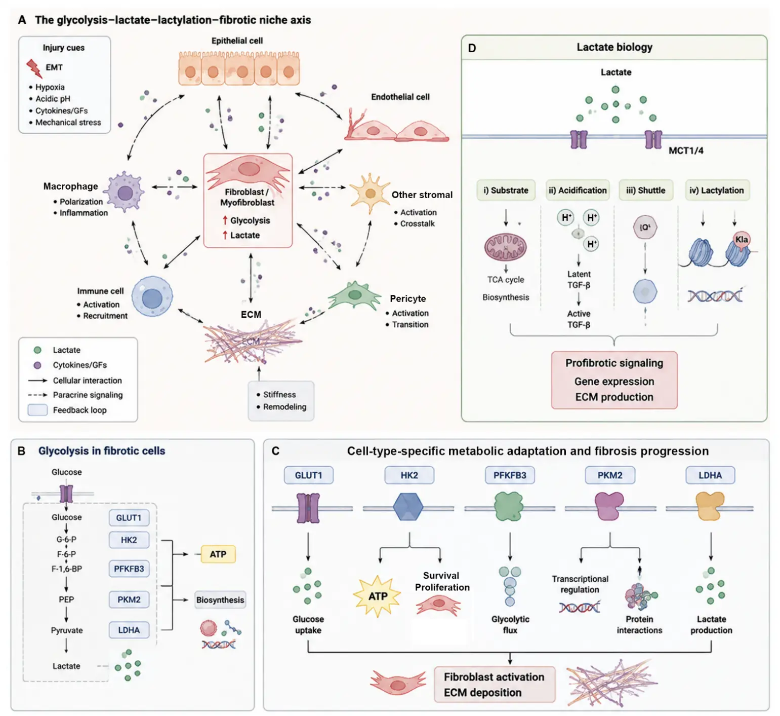
Fibrosis is a common pathological consequence of chronic tissue injury, characterized by persistent fibroblast activation, excessive extracellular matrix (ECM) accumulation, and progressive impairment of organ function. Although classical fibrogenic pathways, particularly transforming growth factor-β (TGF-β) signaling, have been extensively studied, growing evidence suggests that metabolic adaptation is an essential component that supports the persistence of the fibrotic phenotype. Among metabolic pathways, enhanced glycolytic activity can support fibrotic remodeling in specific cellular contexts while also participating in adaptive or reparative responses depending on cell identity and disease stage. The glycolysis–lactate–lactylation axis links metabolic alterations with transcriptional reprogramming, immune regulation, and extracellular matrix remodeling. This review summarizes the roles of key glycolytic regulators, including GLUT1, HK2, PFKFB3, PFKM/PFKP, PKM2, LDHA, MCT1/4, and PDK1, across pulmonary, hepatic, renal, cardiac, and cutaneous fibrosis. We further discuss the interactions among glycolytic metabolism, TGF-β signaling, HIF-1α activation, YAP/TAZ-mediated mechanotransduction, and immune-metabolic communication, highlighting emerging therapeutic strategies and challenges in targeting metabolic vulnerabilities in fibrosis.
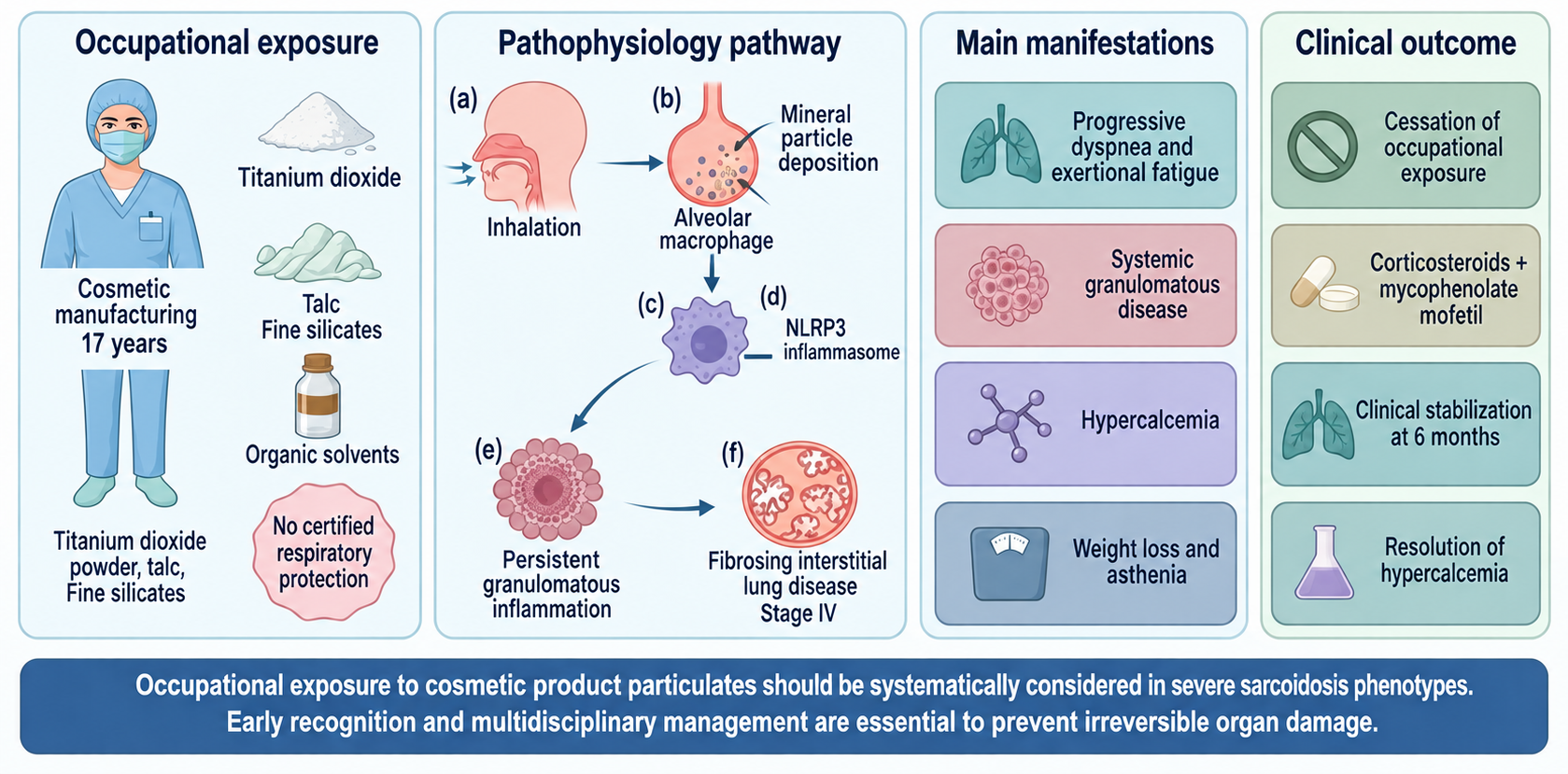
(1) Sarcoidosis is a systemic granulomatous disease of multifaceted etiology, characterized predominantly by pulmonary parenchymal involvement. While genetic susceptibility represents a foundational prerequisite, environmental and occupational xenobiotics are increasingly recognized as pivotal etiologic triggers. In cosmetic formulation environments, prolonged inhalation of mineral micro- and nanoparticles—such as titanium dioxide, talc, and fine silicates—alongside volatile organic solvents, provides a persistent antigenic stimulus that can drive aberrant granulomatous cascades toward aggressive, fibrosing phenotypes. (2) A 54-year-old female industrial worker presented with a 17-year history of occupational exposure in cosmetic manufacturing (handling airborne inorganic powders, titanium dioxide, talc, and organic vehicles without certified particulate respiratory protection), exhibiting progressive dyspnea, marked asthenia, and weight loss. Examination revealed inspiratory crackles at the pulmonary apices, violaceous nasal plaques consistent with lupus pernio, subcutaneous nodules, and sarcoid acropathy. Imaging and histology confirmed stage IV systemic sarcoidosis with fibrosing interstitial lung disease and multiple deep lymphadenopathies. Biologically and histologically, sarcoidosis was confirmed. Complete cessation of occupational exposure was achieved through workplace reassignment. Following combination therapy with oral corticosteroids and mycophenolate mofetil, the patient demonstrated clinical stabilization, resolution of hypercalcemia, and static lung function at 6-month follow-up. (3) This case underscores the importance of considering occupational exposures, including handling cosmetic products, in the pathogenesis and progression of fibrosing sarcoidosis. Early recognition and multidisciplinary management, integrating immunosuppressive therapy and occupational health interventions, are critical to limit irreversible organ damage.