
Organoid Models of Liver Fibrosis: Bridging Genetic and Epigenetic Mechanisms with Biomarker Discovery
Organoid Models of Liver Fibrosis: Bridging Genetic and Epigenetic Mechanisms with Biomarker Discovery

Received: 30 April 2026 Revised: 22 May 2026 Accepted: 11 June 2026 Published: 01 July 2026

© 2026 The authors. This is an open access article under the Creative Commons Attribution 4.0 International License (https://creativecommons.org/licenses/by/4.0/).

Graphical Abstract
1. Introduction
Fibrosis is a pathological process that occurs when normal tissue repair mechanisms become dysregulated following an injury [1]. Under physiological conditions, wound healing involves a tightly controlled sequence of events that restore tissue integrity after damage. However, if the injury is chronic or the repair process is improperly regulated, there is an excessive accumulation of extracellular matrix (ECM) components, leading to progressive scarring and disruption of tissue architecture. Over time, this fibrotic remodeling can impair organ function and eventually lead to organ failure. Fibrosis can affect almost any tissue in the body and is a key feature of many chronic diseases [2,3]. For example, liver fibrosis can progress to cirrhosis and liver failure [4], while pulmonary fibrosis can lead to a gradual loss of lung function and respiratory insufficiency [5]. In the kidney, fibrotic remodeling contributes to chronic kidney disease, and in the heart, fibrosis disrupts electrical conduction and impairs contractile function. Despite variations in the causes and specific characteristics of different fibrotic diseases, many of them share common molecular pathways and cellular mechanisms [5].
Fibrosis associated with metabolic liver disease demonstrates how fibrotic processes in various organs can arise from shared systemic mechanisms. In metabolic dysfunction-associated steatotic liver disease (MASLD), formerly known as non-alcoholic fatty liver disease (NAFLD), hepatic fibrosis often occurs alongside vascular remodeling and arterial wall fibrosis [6]. These processes are interconnected through common metabolic and inflammatory pathways. Insulin resistance, a key factor in MASLD, not only results in hepatic steatosis and stellate cell activation but also contributes to vascular dysfunction due to chronic hyperglycemia and hyperinsulinemia. These conditions activate the renin-angiotensin-aldosterone system and increase angiotensin receptor signaling, leading to hypertrophy and fibrosis of the arterial wall. Moreover, TGF-β signaling, a significant mediator of hepatic stellate cell activation and liver fibrosis, is also associated with vascular remodeling and intimal thickening. Oxidative stress from excessive lipid accumulation in hepatocytes can exacerbate these effects by promoting inflammatory activation of Kupffer cells and stellate cells, while also contributing to endothelial dysfunction. As a result, systemic inflammatory mediators may act as a mechanistic link between hepatic fibrosis and cardiovascular disease, with markers of advanced MASLD fibrosis correlating with more severe coronary artery disease and vascular damage [6]. These findings highlight the systemic nature of fibrotic diseases and underscore the importance of integrated experimental models for studying cross-organ mechanisms of fibrosis [6].
At the core of fibrosis is the activation of fibroblasts into myofibroblasts, specialized cells that produce large amounts of ECM proteins, such as collagen and fibronectin. These cells accumulate at the injury sites and contribute to tissue stiffening and structural remodeling. Various signaling pathways, such as transforming growth factor-β (TGF-β), Wnt signaling, platelet-derived growth factor (PDGF), and different inflammatory mediators, regulate the activation of fibroblasts [7]. The interactions between epithelial cells, stromal cells, immune populations, and the extracellular matrix also shape the fibrotic response.
Despite significant progress in identifying these pathways, many aspects of fibrosis remain poorly understood. One major challenge is the lack of experimental models that accurately mimic human disease. Furthermore, conventional cell culture plastic exhibits supraphysiological stiffness compared with most native tissues, which can profoundly influence cellular signaling and mechanotransduction pathways and therefore limit the physiological relevance of two-dimensional culture systems. Animal models, while providing physiological context, often differ significantly from human biology and may only partially recapitulate the biological processes underlying disease development; therefore, they may note reliably predict clinical outcomes [5].
Organoid technology has emerged as a powerful alternative for modeling human tissues in vitro [8]. Organoids are three-dimensional cell culture systems derived from stem cells or primary tissues that self-organize into structures resembling native organs [9]. These systems retain many features of native tissues, including cell type diversity, spatial organization, and functional characteristics. Because organoids can be generated from human cells and manipulated experimentally, they offer unique opportunities for studying disease mechanisms in a controlled environment.
In recent years, organoid systems have been used to study fibrotic diseases [10]. By including stromal cells, applying fibrotic stimuli, or modifying ECM conditions, researchers have begun to recreate key aspects of fibrosis in vitro. As a result, these models provide new insights into the development of fibrotic processes and may facilitate the discovery of novel therapeutic strategies.
Recent advances in genomic and epigenomic profiling reveal that fibrotic diseases are not solely driven by environmental injury but are also influenced by inherited genetic variants and dynamic epigenetic regulation. Genome-wide association studies and large cohort analyses have identified several polymorphisms that impact susceptibility to liver fibrosis and the rate of disease progression. Variants in genes such as PNPLA3, TM6SF2, and MBOAT7 are strongly associated with fibrotic progression in metabolic and viral liver diseases [11,12,13]. In parallel, epigenetic mechanisms including DNA methylation, histone modifications, and non-coding RNAs are increasingly recognized as key regulators of hepatic stellate cell activation and ECM production. These discoveries have sparked interest in identifying molecular biomarkers that reflect fibrogenic activity and may enable earlier diagnosis or prediction of disease progression. Organoid systems provide a promising experimental platform for integrating genetic background, epigenetic regulation, and functional readouts of fibrotic remodeling in a human tissue context. The conceptual integration of genetic susceptibility, epigenetic regulation, organoid modeling, and biomarker discovery in liver fibrosis research is summarized in Figure 1.
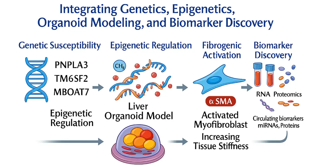
Figure 1. Integration of genetic, epigenetic, organoid modeling, and biomarker discovery approaches in liver fibrosis. The schematic illustrates a conceptual workflow linking inherited risk factors to disease modeling and translational readouts in liver fibrosis. Moving from left to right, genetic susceptibility is represented by key fibrosis- and steatosis-associated variants (e.g., PNPLA3, TM6SF2, MBOAT7). These genetic factors interact with epigenetic regulation, including DNA methylation (CH3), chromatin modifications, and non-coding RNA-mediated control of gene expression. Genetic and epigenetic inputs are integrated in a liver organoid model, where hepatocyte-like cells and hepatic stellate cells are cultured in a three-dimensional extracellular matrix environment. This system allows for the recapitulation of fibrogenic activation, characterized by stellate cell activation into α-SMA-positive myofibroblasts, extracellular matrix production, and increasing tissue stiffness. Ultimately, signals derived from organoids and disease-associated information contribute to biomarker discovery, highlighting circulating RNA species, proteins, and other molecular signatures detectable in blood or plasma.
2. Cellular and Molecular Basis of Fibrosis
Fibrosis represents a complex and dynamic process involving multiple cell types and signaling pathways [1]. Although the precise mechanisms vary between organs and disease contexts, several core features are shared across fibrotic conditions. Fibrotic responses are typically initiated by tissue injury. Damage to epithelial or parenchymal cells triggers the release of cytokines, growth factors, and damage-associated molecular signals that activate local immune responses. Immune cells, particularly macrophages and lymphocytes, accumulate at the site of injury and secrete additional mediators that promote tissue repair [1]. In acute injury, these signals eventually subside as the tissue heals. However, when injury persists, inflammatory signaling remains active, driving pathological remodeling. A central event in fibrosis is the activation of fibroblasts into myofibroblasts. These cells acquire contractile properties and express markers such as α-smooth muscle actin (α-SMA) [1]. Myofibroblasts produce large quantities of ECM components, including collagen type I and III, fibronectin, and hyaluronan. Excessive matrix deposition disrupts normal tissue architecture and increases tissue stiffness. The coordinated interaction of inflammatory signaling, fibroblast activation, and ECM accumulation ultimately drives progressive tissue remodeling and organ dysfunction. The key cellular events and signaling pathways involved in this fibrotic cascade are summarized in Figure 2.
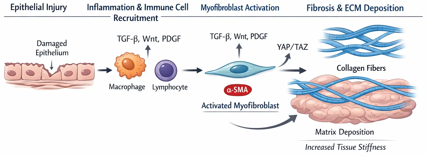
Figure 2. Cellular and molecular mechanisms driving fibrosis. Tissue injury triggers inflammatory signaling, which attracts immune cells like macrophages and lymphocytes. These cells release cytokines and growth factors that stimulate resident fibroblasts, causing them to transform into myofibroblasts. Activated myofibroblasts produce large quantities of extracellular matrix components (ECM), including collagen type I and III, fibronectin, and glucosaminoglycans such as hyaluronan, which contribute to matrix accumulation and tissue stiffness, eventually resulting in organ dysfunction. Key signaling pathways involved in this process include TGF-β, Wnt, PDGF, and mechanotransduction pathways like Yes-associated protein (YAP)/Transcription coactivator with PDZ-binding motif (TAZ).
TGF-β is widely considered the master regulator of fibrotic signaling [14]. This cytokine promotes fibroblast activation, stimulates ECM production, and suppresses matrix degradation. TGF-β signaling operates through both canonical SMAD-dependent pathways and non-canonical pathways involving MAP kinase and PI3K signaling [14]. Because of its central role, TGF-β signaling has become a major target for anti-fibrotic drug development. Mechanical signals also play a critical role in fibrosis progression. As ECM accumulates, tissues become progressively stiffer, altering cell behavior and promoting disease progression [15,16]. Cells sense these mechanical changes through integrins and cytoskeletal structures, activating mechanotransduction pathways such as YAP and TAZ [17,18]. These pathways reinforce fibroblast activation and promote further matrix deposition, creating a positive feedback loop that perpetuates fibrosis. However, in experimental models of liver injury, increases in liver stiffness have been observed to occur before detectable extracellular matrix accumulation, suggesting that early mechanical alterations may contribute to the activation of hepatic stellate cells and the initiation of fibrogenesis [19].
Beyond classical signaling pathways, genetic and epigenetic mechanisms are increasingly recognized as critical determinants of fibrotic susceptibility and progression. This is well-known for idiopathic pulmonary fibrosis [20,21]. Genome-wide association studies have identified several genetic variants that influence susceptibility to fibrotic liver disease and modify disease progression, including variants in PNPLA3, TM6SF2, and MBOAT7 [11,12,13]. These genetic factors may affect pathways involved in lipid metabolism, inflammation, and ECM remodeling, thereby altering the cellular response to chronic injury. Epigenetic regulation further contributes to the dynamic control of fibrogenic gene expression [22]. DNA methylation, histone modifications, and chromatin remodeling influence the transcriptional programs that govern activation of hepatic stellate cells and other stromal populations [23,24]. In addition, non-coding RNAs, including microRNAs and long non-coding RNAs, have emerged as key regulators of fibrotic signaling networks [25,26]. Several microRNAs modulate TGF-β signaling, ECM synthesis, and inflammatory pathways, thereby shaping the balance between tissue repair and pathological fibrosis. These genetic and epigenetic layers of regulation add considerable complexity to the fibrotic response and represent promising targets for therapeutic intervention and biomarker development.
In addition to fibroblasts, several other cell types contribute to fibrotic processes. Injured epithelial cells can adopt aberrant phenotypes and secrete pro-fibrotic factors. Macrophages can assume distinct activation states that either promote or resolve fibrosis. Endothelial cells may undergo endothelial-to-mesenchymal transition (EndMT) in some contexts, further contributing to the pool of matrix-producing cells [1]. The interplay among these diverse cell populations ultimately determines whether tissue repair proceeds normally or progresses toward chronic fibrosis.
Understanding these complex interactions requires experimental systems that preserve multicellular architecture and signaling networks. Among emerging approaches, organoid models provide a promising platform for capturing these features in vitro.
3. Organoid Technology: An Overview
Organoids are three-dimensional cellular structures that arise from the intrinsic self-organizing ability of stem or progenitor cells [8]. However, in some cases, mature differentiated cells may regain proliferative capacity through dedifferentiation or reprogramming under defined culture conditions [27]. For example, mature epithelial cells from tissues like liver, intestine, or airway have been shown to generate organoid-like structures after injury-mimicking culture environments or genetic reprogramming. Nevertheless, the efficiency of organoid formation from terminally differentiated cells is generally lower than from stem or progenitor cells, and the mechanisms enabling this cellular plasticity remain incompletely understood. When cultured under suitable conditions, these cells proliferate and differentiate while organizing themselves into structures that mimic native tissues. Organoids typically consist of multiple cell types and often exhibit functional characteristics of the organ they originated from [28]. The advancement of organoid technology is propelled by progress in stem cell biology and tissue engineering. Initial studies showed that adult stem cells taken from intestinal crypts could create self-organizing epithelial structures when grown in a three-dimensional ECM environment [29]. These intestinal organoids replicated essential features of the intestinal epithelium, such as crypt-like regions and specialized cell types. Organoids can be produced from various sources. Organoids derived from adult stem cells are obtained directly from patient biopsies or surgical specimens [30]. These cultures frequently retain the genetic and epigenetic traits of the original tissue, making them valuable for investigating patient-specific disease mechanisms. Alternatively, organoids can be generated from pluripotent stem cells, including embryonic stem cells or induced pluripotent stem cells. By exposing these cells to specific combinations of developmental signaling factors, researchers can direct their differentiation towards particular organ lineages. A diagram outlining the creation of organoid systems from stem or patient-derived cells and their use in experimental disease modeling is depicted in Figure 3.
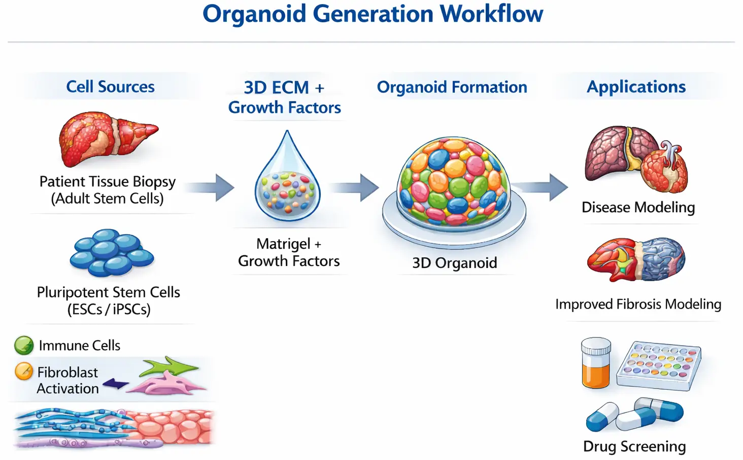
Figure 3. Generation and experimental use of organoid systems. Organoids can be generated from adult stem cells obtained from patient biopsies or from pluripotent stem cells, including embryonic stem cells or induced pluripotent stem cells. These cells are grown in a three-dimensional extracellular matrix scaffold, usually Matrigel, with specific growth factors that promote self-organization and differentiation. The resulting organoids consist of various cell types and mimic aspects of tissue architecture. These systems can then be utilized for genetic manipulation, disease modeling, drug screening, and mechanistic studies.
The culture environment is a crucial component of organoid formation. Most organoids are grown within a three-dimensional ECM matrix scaffold, commonly Matrigel, which provides mechanical support and biochemical cues that promote cell growth and organization [30]. Growth factors and signaling molecules are added to the culture medium to mimic developmental signaling pathways that drive tissue formation. Organoids recapitulate aspects of tissue architecture and cellular diversity, providing experimental advantages over conventional cell cultures. They allow researchers to study interactions between different cell types in a controlled environment and to perform genetic or pharmacological perturbations that would be difficult to perform in vivo. These properties make organoids particularly attractive for modeling complex diseases that involve multiple cell populations and structural changes, including fibrosis.
4. Organoid Models of Fibrosis Across Organs
The application of organoid technology to fibrosis research is relatively recent, but several organ systems have already been explored. These models aim to capture key aspects of fibrotic pathology by recreating interactions between epithelial cells, fibroblasts, and other stromal components. Organoid-based modeling approaches have been developed for various organ systems affected by fibrotic disease, allowing for comparative investigation of shared and tissue-specific mechanisms of fibrosis. Representative examples of fibrosis-related organoid models across different organs are shown in Figure 4.
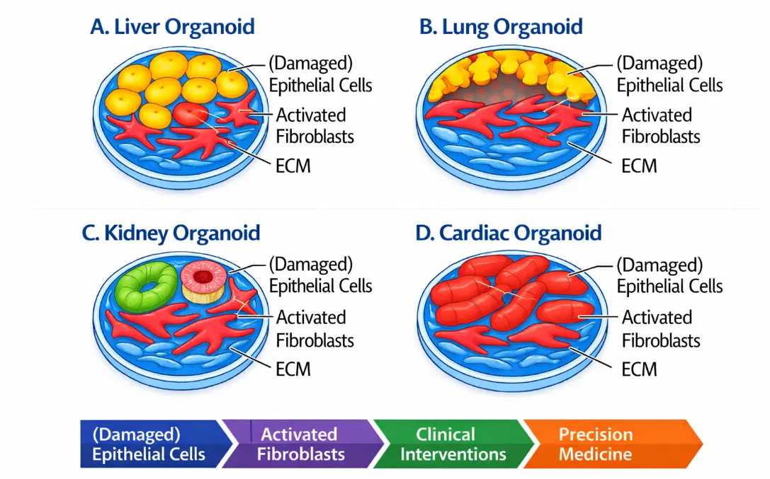
Figure 4. Organoid models are used to study fibrosis in various organs. Organoid technologies have been utilized across multiple organ systems to simulate fibrotic diseases. (A) Liver organoids can replicate hepatocyte injury and hepatic stellate cell activation, ultimately leading to extracellular matrix (ECM) deposition. (B) Lung organoids provide the opportunity to investigate epithelial injury and fibroblast activation, which are associated with pulmonary fibrosis. (C) Kidney organoids, which contain nephron-like structures, can mimic epithelial injury and interstitial matrix accumulation. (D) Cardiac organoids, consisting of cardiomyocytes, fibroblasts, and endothelial cells, allow for the analysis of fibroblast activation and fibrotic remodeling that impacts cardiac function.
4.1. Liver Fibrosis
Liver fibrosis is one of the most intensively studied fibrotic diseases and has served as an early testbed for organoid-based modeling approaches. Chronic liver injury caused by viral hepatitis, alcohol abuse, or metabolic disorders triggers activation of hepatic stellate cells, which are the primary drivers of fibrotic matrix deposition in the liver [31]. Organoid systems derived from liver progenitor cells or pluripotent stem cells have been used to recreate aspects of hepatic tissue architecture, including hepatocyte-like cells and cholangiocyte populations [32]. To model fibrosis, researchers often incorporate hepatic stellate cells or fibroblasts into liver organoid cultures [33]. Exposure to pro-fibrotic stimuli such as TGF-β or inflammatory cytokines induces activation of these stromal cells and promotes ECM deposition. In some models, lipotoxic stress or metabolic perturbations are used to mimic non-alcoholic fatty liver disease, a major cause of liver fibrosis. These systems have revealed important insights into how hepatocyte injury signals can activate stellate cells and initiate fibrotic remodeling.
Recent work has further advanced liver fibrosis modeling by integrating multiple liver cell types into microphysiological systems. For example, a liver-on-a-chip model combining human adult liver stem cell-derived hepatobiliary organoids with induced pluripotent stem cell-derived Kupffer cells and hepatic stellate cells successfully recapitulated key features of metabolic dysfunction–associated steatohepatitis (MASH/NASH), including steatosis, inflammation, hepatocellular injury, and stellate cell activation leading to fibrosis [34]. By exposing the system to metabolic and gut-derived factors such as free fatty acids, fructose, lipopolysaccharides, and phenylacetic acid, the model reproduced disease hallmarks and enabled drug testing with responses that paralleled clinical trial outcomes. This approach highlights the potential of complex organoid-based platforms for studying fibrotic liver disease and evaluating therapeutic strategies.
Other studies have highlighted the importance of genetic and epigenetic factors in shaping the progression of liver fibrosis. Variability in disease susceptibility among individuals with similar environmental exposures suggests that host genetic background plays a critical role in determining fibrotic outcomes. Several polymorphisms associated with lipid metabolism, immune regulation, and inflammatory signaling are linked to accelerated fibrosis progression in chronic liver diseases [11,12,13]. Understanding how these genetic variants influence hepatic stellate cell activation and hepatocyte stress responses remains an active area of investigation.
Large-scale genetic studies provide important insights into interindividual variability in fibrosis progression. Among the most robustly replicated findings is the association between the PNPLA3 I148M variant and increased risk of steatohepatitis, accelerated fibrosis progression, and hepatocellular carcinoma. Additional variants in TM6SF2, MBOAT7, and HSD17B13 further modulate susceptibility to metabolic liver disease and fibrotic outcomes [11,12,13]. These genetic determinants influence pathways such as lipid metabolism, hepatocellular stress responses, and inflammatory signaling, thereby shaping the microenvironment that promotes hepatic stellate cell activation. Incorporating these variants into experimental organoid systems, either through the use of patient-derived tissues or CRISPR-based genome editing, offers a powerful strategy to dissect how specific genetic backgrounds influence fibrogenic signaling and therapeutic responsiveness.
Epigenetic mechanisms also profoundly influence fibrogenic pathways in the liver. Activation of hepatic stellate cells is accompanied by extensive chromatin remodeling and alterations in DNA methylation patterns that promote expression of genes involved in ECM production [35]. Histone modifications can regulate the accessibility of profibrotic transcriptional programs, while non-coding RNAs modulate post-transcriptional gene regulation. Increasing evidence suggests that specific microRNAs act either as drivers or suppressors of fibrosis by targeting key signaling molecules within the TGF-β and Wnt pathways [36,37]. Organoid models provide a powerful platform for investigating these regulatory mechanisms in a human cellular context. Because organoids can be derived from patient tissues, they allow researchers to study how individual genetic backgrounds influence fibrotic responses. Integration of CRISPR-based gene editing with organoid systems further enables functional testing of candidate genes and regulatory elements implicated in fibrosis susceptibility.
4.2. Lung Fibrosis
Pulmonary fibrosis, especially idiopathic pulmonary fibrosis, is characterized by progressive scarring of the lung interstitium and destruction of alveolar architecture. Modeling this disease has historically been challenging due to the structural complexity of lung tissue. Recent advances in lung organoid technology enable the generation of airway and alveolar structures containing multiple epithelial cell types. In lung fibrosis models, epithelial organoids are often co-cultured with fibroblasts or mesenchymal cells. Injury to epithelial cells can be simulated through exposure to chemical toxins, inflammatory mediators, or genetic perturbations associated with idiopathic pulmonary fibrosis. Hyaluronan (HA) is a major component of the ECM that becomes strongly upregulated in injured or remodeling tissues and has been increasingly recognized as an important regulator of pulmonary fibrosis. In fibrotic lung diseases such as idiopathic pulmonary fibrosis (IPF), HA accumulates in the lung interstitium and contributes to inflammation, fibroblast activation, and excessive extracellular matrix deposition [38]. A key mechanism underlying these effects involves interactions between HA and its principal cell-surface receptor CD44. The binding of HA to CD44 can regulate fibroblast migration, activation, and survival, thereby promoting the accumulation of myofibroblasts that produce collagen and other matrix components. In addition, HA-CD44 signaling has been linked to the acquisition of an invasive fibroblast phenotype, enabling fibroblasts to infiltrate damaged tissue and contribute to progressive matrix remodeling and fibrotic expansion [38]. These findings support the concept that HA is not merely a structural matrix component but also an active signaling molecule that modulates fibroblast behavior and tissue remodeling. These interactions are well suited for experimental interrogation in organoid-based co-culture systems. Lung organoids have also been used to investigate how aging, environmental exposures, and genetic risk factors influence susceptibility to fibrosis [39,40].
4.3. Kidney Fibrosis
Kidney fibrosis is a hallmark of chronic kidney disease and is closely associated with progressive loss of renal function. Organoid models of the kidney are typically generated from pluripotent stem cells and contain structures that resemble developing nephrons, including glomerular and tubular components [41]. While these organoids mainly represent early stages of kidney development, they have proven to be valuable for investigating the initial mechanisms of renal injury and fibrosis. By inducing injury in kidney organoids experimentally, researchers can stimulate the activation of stromal cells and the increased deposition of ECM components. These responses mimic some aspects of interstitial fibrosis seen in chronic kidney disease. These models allow exploration of how damage to the epithelial cells in the nephron triggers activation of surrounding stromal cells and the process of fibrotic remodeling. Fibrotic remodeling is also a feature of several inherited cystic kidney diseases, such as autosomal dominant and autosomal recessive polycystic kidney disease, where progressive cyst expansion is accompanied by interstitial fibrosis.
4.4. Cardiac Fibrosis
Cardiac fibrosis occurs in response to myocardial infarction, pressure overload, or other forms of cardiac stress. It is characterized by the accumulation of collagen and other extracellular matrix components within the myocardium, which disrupts electrical conduction and impairs mechanical function. Cardiac organoids and engineered heart tissues composed of cardiomyocytes, fibroblasts, and endothelial cells have been used to model aspects of cardiac fibrosis [42]. In these systems, fibroblast activation can be induced through exposure to TGF-β or mechanical stress. Activated fibroblasts deposit ECM within the organoid, leading to increased tissue stiffness and altered electrical signaling. Such models provide a platform for studying how fibrotic remodeling affects cardiac function and for evaluating potential anti-fibrotic therapies.
5. Advantages of Organoid Models for Fibrosis Research
Organoid systems offer several advantages that make them particularly valuable for studying fibrotic diseases. One of the most important strengths of organoids is their ability to capture aspects of human tissue biology that are difficult to reproduce in conventional experimental models. Because organoids are derived from human cells, they retain species-specific gene expression patterns, signaling pathways, and cellular responses that may differ significantly from those observed in animal models. Another major advantage is the preservation of three-dimensional architecture. Tissue structure plays a critical role in regulating cell behavior, and the spatial arrangement of cells influences how signals are transmitted and interpreted. In organoids, cells interact with each other and with the surrounding extracellular matrix in ways that more closely resemble their natural environment. This spatial organization allows researchers to study processes such as epithelial injury, fibroblast activation, and ECM remodeling within a context that approximates real tissue. Organoid cultures also allow controlled manipulation of the cellular microenvironment. Researchers can introduce specific growth factors, cytokines, or mechanical stimuli to probe the mechanisms that drive fibrosis. Genetic manipulation using CRISPR-based approaches further enables the investigation of disease-associated genes and signaling pathways [43]. These capabilities make organoids powerful tools for dissecting complex molecular networks involved in fibrotic disease.
A particularly exciting aspect of organoid technology is the possibility of generating patient-specific models from different tissues such as liver, prostate, and kidney [44,45,46,47]. Organoids derived from individual patients retain many of the genetic and epigenetic features of the original tissue. This opens the possibility of studying how different individuals respond to injury or therapeutic interventions. In the future, patient-derived organoids may be used to test candidate treatments and guide personalized therapeutic strategies for fibrotic diseases.
An additional advantage of organoid platforms is their potential for biomarker discovery and for validating drug sensitivity and resistance [48]. Organoids can be generated from patient-derived tissues and cultured under controlled experimental conditions, providing a unique opportunity to identify molecular signatures associated with fibrotic progression. Transcriptomic, epigenomic, and proteomic analyses performed on organoid cultures can reveal candidate biomarkers that reflect early stages of disease or predict therapeutic responses. Moreover, integrating organoid systems with emerging analytical technologies such as single-cell sequencing and spatial transcriptomics enables detailed characterization of cell-type-specific signaling pathways and cellular diversity involved in fibrosis [49]. These approaches may facilitate the identification and validation of novel circulating biomarkers derived from extracellular vesicles, secreted proteins, or nucleic acids that could improve non-invasive diagnosis and monitoring of liver fibrosis.
An important aspect of validating organoid models is determining how closely their molecular profiles resemble those of human tissues affected by disease. Recent transcriptomic and single-cell sequencing studies have begun to compare gene expression signatures of organoids with those observed in patient-derived fibrotic tissues. These analyses indicate that organoid cultures can reproduce key transcriptional programs associated with epithelial injury, inflammatory signaling, and fibrogenic activation. At the same time, such comparisons also reveal differences in cellular composition and maturation states between organoids and native tissues, highlighting areas where further model refinement is required. Systematic integration of organoid transcriptomics with datasets derived from human fibrotic tissue will therefore be important for benchmarking model fidelity and identifying cell-type-specific disease signatures.
In addition to providing mechanistic insights, organoid systems may also help in identifying clinically relevant biomarkers. Profiling the secretome of organoid cultures can reveal proteins, extracellular vesicles, and nucleic acids released during fibrotic remodeling [34,50]. These molecules could potentially serve as candidate circulating biomarkers that reflect disease activity in patients. For example, microRNAs associated with fibrosis, collagen fragments, and enzymes involved in matrix remodeling are found in patient serum and are currently being studied as non-invasive markers of liver fibrosis. By combining organoid models with high-throughput multi-omics approaches, such as transcriptomics, epigenomics, proteomics, and metabolomics, researchers will be able to systematically identify molecular signatures linked to early fibrogenesis and treatment response. These integrative analyses have the potential to expedite the translation of experimental findings into clinically valuable diagnostic tools.
6. Limitations of Current Organoid Models
Despite their considerable promise, organoid models still face several important limitations that must be addressed before they can fully recapitulate fibrotic disease. One major challenge is the incomplete representation of the cellular diversity found in native tissues. Many organoid systems are primarily composed of epithelial cells and lack the full complement of stromal, immune, and vascular components that participate in fibrosis in vivo. Because these cell populations interact extensively during fibrotic remodeling, their absence can limit the physiological relevance of the model. Another limitation involves the ECM environment used for organoid culture. Most organoids are grown in Matrigel, a gelatinous basement membrane-like ECM derived from Engelbreth-Holm-Swarm mouse sarcoma cells that contains laminin, collagen IV, entactin, heparan sulfate proteoglycans, and various growth factors [51]. Although Matrigel provides a supportive scaffold for organoid growth, its composition is complex, poorly defined, and can exhibit significant batch-to-batch variability [51]. Moreover, its mechanical properties do not necessarily match those of fibrotic tissues, which are typically much stiffer [52]. As a result, conventional organoid cultures may fail to capture the mechanical cues that strongly influence fibroblast activation and matrix deposition. Reproducibility and standardization also present challenges. Organoid cultures can vary significantly depending on factors such as the source of cells, culture conditions, and handling procedures. This variability can complicate comparisons between studies and hinder the development of standardized experimental protocols. Consequently, efforts are underway to establish systematic workflows to improve functionality and reproducibility in work with organoids. These will allow the transfer of organoid technology from an artisanal laboratory method into a more standardized, high-throughput platform for realizing practical clinical and industrial applications with full Good Manufacturing Practice (GMP) compliance [53].
Additionally, many organoid systems remain difficult to scale for high-throughput applications. Finally, fibrosis is typically a chronic process that develops over long periods of time in vivo. Replicating such long-term disease progression in organoid cultures is challenging, as these systems are often maintained for relatively short durations. Developing culture conditions that support long-term stability and maturation will be important for more accurately modeling chronic fibrotic diseases. Moreover, generating organoids directly from fibrotic tissues would be a valuable complementary apporoach for investigating the fibrotic environment in detail.
7. Emerging Technologies Enhancing Fibrosis Organoid Models
Recent technological advances are helping to overcome some of the limitations of current organoid systems [53]. One promising approach involves incorporating additional cell types through co-culture strategies. By introducing fibroblasts, endothelial cells, or immune cells into organoid cultures, researchers can recreate more complex tissue microenvironments [54]. These multicellular systems enable the investigation of how interactions among different cell populations contribute to fibrosis [55]. Recent technological developments are further expanding the capabilities of organoid systems by enabling more physiologically relevant microenvironments and more detailed molecular analyses. An overview of several emerging technologies that enhance organoid-based fibrosis models is presented in Figure 5.
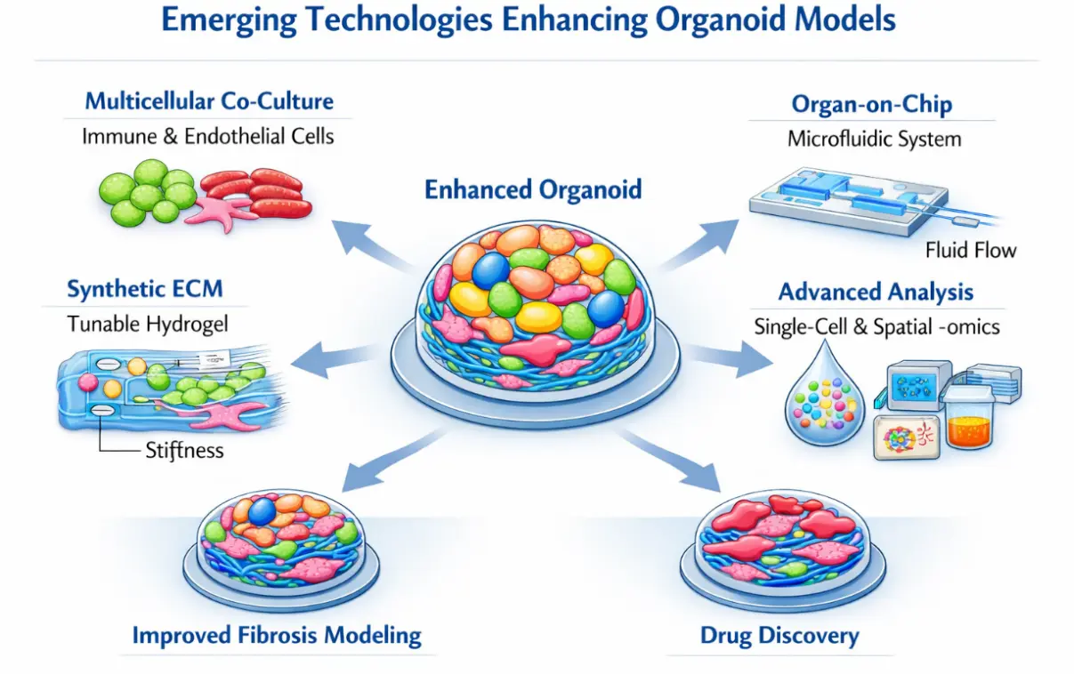
Figure 5. Emerging technologies enhancing organoid models for fibrosis research. Several technological innovations are improving the physiological relevance of organoid systems. Co-culture strategies incorporate stromal, endothelial, and immune cells to recreate complex tissue microenvironments. Microfluidic organ-on-chip devices enable controlled mechanical forces, fluid flow, and chemical gradients. Synthetic extracellular matrices allow precise tuning of biochemical composition and stiffness to mimic fibrotic tissue. Advanced analytical approaches such as single-cell RNA sequencing, spatial transcriptomics, and high-resolution imaging provide detailed insights into cellular interactions and molecular pathways within organoids. Together, these technologies expand the utility of organoid platforms for studying fibrotic diseases and developing anti-fibrotic therapies.
Microengineering technologies are transforming organoid research. Organ-on-chip platforms use microfluidic devices to control fluid flow, hydrodynamic parameters, mechanical forces, and chemical gradients within cultured tissues. Integrating organoids with these systems allows researchers to mimic aspects of physiological circulation and mechanical stress that are difficult to reproduce in static cultures [56]. Such platforms may be particularly valuable for studying how mechanical forces influence fibrotic remodeling. Another area of innovation involves the development of synthetic ECMs. Unlike Matrigel, synthetic hydrogels can be engineered with precisely defined biochemical and mechanical properties [53]. By tuning matrix stiffness or incorporating specific signaling molecules, researchers can create environments that more closely resemble fibrotic tissues. This capability enables systematic investigation of how mechanical and biochemical cues regulate fibroblast activation and ECM production. Advances in imaging and molecular profiling are also enhancing the study of fibrosis in organoids. Techniques such as single-cell RNA sequencing, spatial transcriptomics, and high-resolution microscopy allow detailed characterization of cellular states and interactions within organoid cultures. These tools provide unprecedented insight into the cellular dynamics that drive fibrotic remodeling.
Epigenomic profiling technologies are beginning to play an important role in organoid-based fibrosis research. Methods such as ATAC-seq, ChIP-seq, and single-cell DNA methylation profiling allow detailed mapping of chromatin accessibility and regulatory elements that control fibrogenic gene expression [57,58,59]. Applying these techniques to organoid systems can reveal how epigenetic landscapes change during hepatic stellate cell activation and fibrotic remodeling. Organoids can be derived from genetically diverse patients, making comparative epigenomic analyses valuable in identifying regulatory networks that distinguish progressive from non-progressive disease states. This information could ultimately support the discovery of epigenetic biomarkers and therapeutic targets.
Another emerging direction involves the characterization of biomechanical properties in organoid systems. Tissue stiffness is a defining feature of fibrotic remodeling and plays a central role in activating mechanotransduction pathways such as YAP/TAZ signaling. While stiffness has been extensively studied in fibrotic tissues in vivo, quantitative measurements of mechanical properties in fibrosis-related organoid models remain relatively limited. Incorporating techniques such as atomic force microscopy, traction force microscopy, or tunable synthetic hydrogels could enable systematic investigation of how mechanical cues regulate fibroblast activation and extracellular matrix accumulation within organoid systems. Developing such approaches will be important for determining whether organoids faithfully recapitulate the biomechanical environment characteristic of fibrotic tissues.
8. Future Directions
As organoid technology continues to evolve, several developments are likely to further expand its utility for fibrosis research. One important goal is to incorporate immune components into organoid systems. Immune cells play essential roles in both promoting and resolving fibrosis, yet they are largely absent from most current models. Developing strategies to integrate macrophages, lymphocytes, and other immune populations into organoid cultures will be critical for capturing the full complexity of fibrotic responses. Another key objective is the generation of vascularized organoids [60,61,62]. Blood vessels play central roles in tissue homeostasis and repair, and endothelial dysfunction is increasingly recognized as an important contributor to fibrosis. Incorporating vascular networks into organoid systems could improve nutrient delivery, enable modeling of vascular-stromal interactions, and enhance the physiological relevance of these models. Developing culture systems that allow organoids to mature and persist over extended periods will also be important. Such systems would enable modeling of chronic disease progression and allow researchers to study how fibrotic processes evolve over time. Improvements in biomaterials, culture media, and bioreactor technologies may help achieve this goal. In parallel, organoid models are likely to play an expanding role in drug discovery [63]. High-throughput screening platforms based on organoids could enable systematic testing of candidate anti-fibrotic compounds in human tissue contexts. Because organoids can be derived from individual patients, they also offer opportunities for personalized medicine approaches in which therapeutic responses are evaluated in patient-specific models. Finally, integrating organoid experiments with computational modeling and systems biology approaches may help unravel the complex regulatory networks that govern fibrosis. By combining experimental and computational methods, researchers may be able to identify key molecular drivers of fibrotic disease and develop more effective therapeutic strategies.
9. Conclusions
Fibrosis represents a significant clinical challenge affecting multiple organs and contributing significantly to the global disease burden. Although substantial progress has been made in understanding the molecular pathways involved in fibrotic remodeling, the development of effective therapies has been hindered by limitations in existing experimental models. Organoid technology offers a powerful new approach for modeling fibrotic diseases in vitro. By recreating aspects of tissue architecture and cellular diversity, organoids provide a more physiologically relevant platform than traditional cell culture systems while maintaining the experimental accessibility of in vitro models. Early studies across the liver, lung, kidney, and cardiac systems show that organoids reproduce key features of fibrotic pathology, including fibroblast activation, ECM deposition, and altered tissue function. Despite these advances, current organoid models remain imperfect representations of native tissues. Challenges such as incomplete cellular complexity, lack of vascularization, and difficulties in modeling long-term disease progression must be addressed. Ongoing developments in bioengineering, microfluidics, biomaterials, and single-cell analysis are likely to overcome many of these limitations. In particular, integrating organoid technologies with genomic and epigenomic profiling approaches may open new avenues for identifying molecular drivers and biomarkers of fibrosis. Patient-derived organoids, combined with high-throughput sequencing technologies, could enable a systematic investigation of genetic susceptibility, epigenetic regulation, and treatment response in fibrotic disease. Such strategies may help bridge the gap between mechanistic studies and clinical translation, ultimately supporting the development of precision medicine approaches for patients with chronic liver disease. Beyond their application to individual organs, future organoid platforms may also help elucidate systemic interactions between metabolic, inflammatory, and biomechanical processes that drive fibrotic remodeling across multiple tissues. Importantly, organoid systems provide a promising bridge between mechanistic studies and clinical biomarker discovery. Integrating patient-derived organoids with genomic and epigenomic profiling may enable the identification of molecular signatures that predict disease progression or therapeutic response.
As these technologies mature, organoid models are poised to become indispensable tools for studying fibrosis. They hold promise not only for elucidating fundamental mechanisms of disease but also for accelerating drug discovery and enabling personalized therapeutic strategies. The ability to recapitulate fibrosis in controlled laboratory systems may ultimately transform our understanding of fibrotic diseases and accelerate the development of new therapeutic strategies.
Statement of the Use of Generative AI and AI-Assisted Technologies in the Writing Process
During the preparation of this manuscript, the author used EditMyEnglish to polish the language. After using this tool/service, the author reviewed and edited the content as needed and takes full responsibility for the content of the published article.
Ethics Statement
Not applicable.
Informed Consent Statement
Not applicable.
Data Availability Statement
No new data were generated or analyzed in this study.
Funding
The author receives support from the German Cancer Aid (grant 70115581).
Declaration of Competing Interest
The author is one of the Editors-in-Chief of Fibrosis. However, he was not involved in the peer-review process, the selection of reviewers, or the editorial decision regarding the publication of this manuscript. The author declares that he has no known competing financial interests or personal relationships that could have influenced the work reported in this paper.
References
- Lurje I, Gaisa NT, Weiskirchen R, Tacke F. Mechanisms of organ fibrosis: Emerging concepts and implications for novel treatment strategies. Mol. Asp. Med. 2023, 92, 101191. DOI:10.1016/j.mam.2023.101191 [Google Scholar]
- Chen X, Zhang J, Guo L, Wu C, Zhou J, Xu M, et al. Decoding organ fibrosis: Mechanistic insights and emerging therapeutic strategies. Signal Transduct. Target. Ther. 2026, 11, 82. DOI:10.1038/s41392-025-02532-0 [Google Scholar]
- Koda Y, Nakamoto N, Kanai T. Immunological mechanisms underlying fibrotic diseases via single-cell technologies. Front. Immunol. 2026, 16, 1720047. DOI:10.3389/fimmu.2025.1720047 [Google Scholar]
- Acharya P, Chouhan K, Weiskirchen S, Weiskirchen R. Cellular Mechanisms of Liver Fibrosis. Front. Pharmacol. 2021, 12, 671640. DOI:10.3389/fphar.2021.671640 [Google Scholar]
- Rieder F, Nagy LE, Maher TM, Distler JHW, Kramann R, Hinz B, et al. Fibrosis: Cross-organ biology and pathways to development of innovative drugs. Nat. Rev. Drug Discov. 2025, 24, 543–569; Erratum in Nat. Rev. Drug Discov. 2025, 24, 399. DOI:10.1038/s41573-025-01190-9 [Google Scholar]
- Tarantino G, Costantini S, Finelli C, Capone F, Guerriero E, La Sala N, et al. Carotid intima-media thickness is predicted by combined eotaxin levels and severity of hepatic steatosis at ultrasonography in obese patients with Nonalcoholic Fatty Liver Disease. PLoS ONE 2014, 9, e105610. DOI:10.1371/journal.pone.0105610 [Google Scholar]
- Chen F, Lyu L, Xing C, Chen Y, Hu S, Wang M, et al. The pivotal role of TGF-β/Smad pathway in fibrosis pathogenesis and treatment. Front. Oncol. 2025, 15, 1649179. DOI:10.3389/fonc.2025.1649179 [Google Scholar]
- Navik U, Singh SK, Khurana A, Weiskirchen R. Revolutionizing liver fibrosis research: The promise of 3D organoid models in understanding and treating chronic liver disease. Expert Rev. Gastroenterol. Hepatol. 2025, 19, 105–110. DOI:10.1080/17474124.2025.2455581 [Google Scholar]
- Nair DG, Weiskirchen R. Advanced In Vitro Models for Preclinical Drug Safety: Recent Progress and Prospects. Curr. Issues Mol. Biol. 2024, 47, 7. DOI:10.3390/cimb47010007 [Google Scholar]
- Nair DG, Nair DB, Weiskirchen R. Reconstructing Liver Fibrosis: 3D Human Models, Microbiome Interfaces, and Therapeutic Innovation. Curr. Issues Mol. Biol. 2026, 48, 165. DOI:10.3390/cimb48020165 [Google Scholar]
- Saliba-Gustafsson P, Härdfeldt J, Pedrelli M, Parini P. Genomic Signatures of MASLD: How Genomics Is Redefining Our Understanding of Metabolic Liver Disease. Int. J. Mol. Sci. 2025, 26, 10881. DOI:10.3390/ijms262210881 [Google Scholar]
- Weiskirchen R, Lonardo A. PNPLA3 as a driver of steatotic liver disease: Navigating from pathobiology to the clinics via epidemiology. J. Transl. Genet. Genom. 2024, 8, 355–377. DOI:10.20517/jtgg.2024.70 [Google Scholar]
- Chandrasekaran P, Weiskirchen R. The Pivotal Role of the Membrane-Bound O-Acyltransferase Domain Containing 7 in Non-Alcoholic Fatty Liver Disease. Livers 2024, 4, 1–14. DOI:10.3390/livers4010001 [Google Scholar]
- Huang KL, Wang LK, Tsai FM. TGF-β Signaling as a Pathological Continuum Linking Idiopathic Pulmonary Fibrosis and Lung Cancer. Cells 2026, 15, 480. DOI:10.3390/cells15050480 [Google Scholar]
- Zha B, Zhang C, Wu C. The stiffness of extracellular matrix in regulating cellular metabolism. Am. J. Physiol. Cell Physiol. 2025, 329, C298–C306. DOI:10.1152/ajpcell.00913.2024 [Google Scholar]
- Mayorca-Guiliani AE, Leeming DJ, Henriksen K, Mortensen JH, Nielsen SH, Anstee QM, et al. ECM formation and degradation during fibrosis, repair, and regeneration. NPJ Metab. Health Dis. 2025, 3, 25. DOI:10.1038/s44324-025-00063-4 [Google Scholar]
- Chu CQ, Quan T. Fibroblast Yap/Taz Signaling in Extracellular Matrix Homeostasis and Tissue Fibrosis. J. Clin. Med. 2024, 13, 3358. DOI:10.3390/jcm13123358 [Google Scholar]
- Lei C, Wu D, Zhang H, Yang Z, Huang B, Wang J, et al. The mechanotransduction-immune axis in organ fibrosis: Dual regulatory mechanisms and translational therapeutic perspectives. Front. Immunol. 2025, 16, 1670213. DOI:10.3389/fimmu.2025.1670213 [Google Scholar]
- Georges PC, Hui JJ, Gombos Z, McCormick ME, Wang AY, Uemura M, et al. Increased stiffness of the rat liver precedes matrix deposition: Implications for fibrosis. Am. J. Physiol. Gastrointest. Liver Physiol. 2007, 293, G1147–G1154. DOI:10.1152/ajpgi.00032.2007 [Google Scholar]
- Zhumagaliyeva A, Chorostowska-Wynimko J, Jezela-Stanek A. Genetic Determinants of Progressive Pulmonary Fibrosis: A Comprehensive Review. Int. J. Mol. Sci. 2025, 26, 11846. DOI:10.3390/ijms262411846 [Google Scholar]
- Li RM, Stentenbach M, Tan DBA, Ludewick HP, Walters EH, Clynick B, et al. The role of epigenetic regulation in idiopathic pulmonary fibrosis. Am. J. Physiol. Lung Cell Mol. Physiol. 2026, 330, L13–L32. DOI:10.1152/ajplung.00030.2025 [Google Scholar]
- Lu W, Xu J, Xu Y, Zhou Y, Que S, Liu Z. Liver fibrosis and cirrhosis in the multi-omics era: Mechanisms and therapeutic perspectives from human and animal models. Front. Cell. Infect. Microbiol. 2026, 15, 1686649. DOI:10.3389/fcimb.2025.1686649 [Google Scholar]
- Kong M, Zhou J, Kang A, Kuai Y, Xu H, Li M, et al. Histone methyltransferase Suv39h1 regulates hepatic stellate cell activation and is targetable in liver fibrosis. Gut 2024, 73, 810–824. DOI:10.1136/gutjnl-2023-329671 [Google Scholar]
- Ding K, Xie R, Han B, Zheng H, Tian T. Histone methyltransferase SMYD2 regulates the activation of hepatic stellate cells by activating TLR4 signaling. Sci. Rep. 2025, 15, 13166. DOI:10.1038/s41598-025-96699-9 [Google Scholar]
- Rowe MM, Kaestner KH. The Role of Non-Coding RNAs in Liver Disease, Injury, and Regeneration. Cells 2023, 12, 359. DOI:10.3390/cells12030359 [Google Scholar]
- Wang H, Sun K, Peng H, Wang Y, Zhang L. Emerging roles of noncoding RNAs in idiopathic pulmonary fibrosis. Cell Death Discov. 2024, 10, 443. DOI:10.1038/s41420-024-02170-5 [Google Scholar]
- Unagolla JM, Jayasuriya AC. Recent advances in organoid engineering: A comprehensive review. Appl. Mater. Today 2022, 29, 101582. DOI:10.1016/j.apmt.2022.101582 [Google Scholar]
- Verstegen MMA, Coppes RP, Beghin A, De Coppi P, Gerli MFM, de Graeff N, et al. Clinical applications of human organoids. Nat. Med. 2025, 31, 409–421. DOI:10.1038/s41591-024-03489-3 [Google Scholar]
- Tian CM, Yang MF, Xu HM, Zhu MZ, Yue NN, Zhang Y, et al. Stem cell-derived intestinal organoids: A novel modality for IBD. Cell Death Discov. 2023, 9, 255. DOI:10.1038/s41420-023-01556-1 [Google Scholar]
- Hardt KS, Pohlberger RF, Keller DT, Buhl EM, Vondran FWR, Roeth AA, et al. Human Liver Organoids as an Experimental Tool to Investigate Lipocalin-2 in Hepatic Inflammation. Cells 2026, 15, 216. DOI:10.3390/cells15030216 [Google Scholar]
- Friedman SL, Roll FJ, Boyles J, Bissell DM. Hepatic lipocytes: The principal collagen-producing cells of normal rat liver. Proc. Natl. Acad. Sci. USA 1985, 82, 8681–8685. DOI:10.1073/pnas.82.24.8681 [Google Scholar]
- Guan Y, Fang Z, Hu A, Roberts S, Wang M, Ren W, et al. Live-cell imaging of human liver fibrosis using hepatic micro-organoids. JCI Insight 2024, 10, e187099. DOI:10.1172/jci.insight.187099 [Google Scholar]
- Brovold M, Keller D, Soker S. Differential fibrotic phenotypes of hepatic stellate cells within 3D liver organoids. Biotechnol. Bioeng. 2020, 117, 2516–2526. DOI:10.1002/bit.27379 [Google Scholar]
- Balachander GM, Ng IC, Pai RR, Mitra K, Tasnim F, Lim YS, et al. LEADS—A comprehensive human liver-on-a-chip for non-alcoholic steatohepatitis (NASH) drug testing. Lab. Chip. 2025, 25, 3444–3466. DOI:10.1039/d5lc00221d [Google Scholar]
- Barcena-Varela M, Colyn L, Fernandez-Barrena MG. Epigenetic Mechanisms in Hepatic Stellate Cell Activation During Liver Fibrosis and Carcinogenesis. Int. J. Mol. Sci. 2019, 20, 2507. DOI:10.3390/ijms20102507 [Google Scholar]
- Wang S, Yu H, Liu S, Liu Y, Gu X. Regulation of idiopathic pulmonary fibrosis: A cross-talk between TGF-β signaling and MicroRNAs. Front. Med. 2024, 11, 1415278. DOI:10.3389/fmed.2024.1415278 [Google Scholar]
- Zhang H, Zhou Y, Wen D, Wang J. Noncoding RNAs: Master Regulator of Fibroblast to Myofibroblast Transition in Fibrosis. Int. J. Mol. Sci. 2023, 24, 1801. DOI:10.3390/ijms24021801 [Google Scholar]
- Li Y, Jiang D, Liang J, Meltzer EB, Gray A, Miura R, et al. Severe lung fibrosis requires an invasive fibroblast phenotype regulated by hyaluronan and CD44. J. Exp. Med. 2011, 208, 1459–1471. DOI:10.1084/jem.20102510 [Google Scholar]
- Wang H, Han Z, Yang Y, Liu L, Huang Y, Chen J, et al. Modeling of lung organoid-based fibrosis for testing the sensitivity of anti-fibrotic drugs. Stem Cell Res. Ther. 2025, 16, 132. DOI:10.1186/s13287-025-04251-3 [Google Scholar]
- Park HR, Kwon Y, Ji HJ, Kim SY, Kim MK, Ahn KB, et al. A human lung organoid platform for studying radiation-induced pulmonary fibrosis and antifibrotic drug screening. Sci. Rep. 2025, 16, 1905. DOI:10.1038/s41598-025-31582-1 [Google Scholar]
- Nakanoh H, Tsuji K, Fukushima K, Uchida N, Haraguchi S, Kitamura S, et al. Kidney Organoids: Current Advances and Applications. Life 2025, 15, 1680. DOI:10.3390/life15111680 [Google Scholar]
- Song M, Choi DB, Im JS, Song YN, Kim JH, Lee H, et al. Modeling acute myocardial infarction and cardiac fibrosis using human induced pluripotent stem cell-derived multi-cellular heart organoids. Cell Death Dis. 2024, 15, 308. DOI:10.1038/s41419-024-06703-9 [Google Scholar]
- Geurts MH, Gandhi S, Boretto MG, Akkerman N, Derks LLM, van Son G, et al. One-step generation of tumor models by base editor multiplexing in adult stem cell-derived organoids. Nat. Commun. 2023, 14, 4998. DOI:10.1038/s41467-023-40701-3 [Google Scholar]
- Pohlberger RF, Hardt KS, Kühnel MP, Palzer J, Reinhardt JL, Beetz O, et al. From Patient Liver Tissue to Organoids: Establishment of a Translational Platform Using Healthy, Steatotic, and Cirrhotic Tissue Sources. Cells 2026, 15, 432. DOI:10.3390/cells15050432 [Google Scholar]
- Thielen N, Kuang C, Chu E, Wei N. High-Throughput screening for novel drug combinations using Patient-Derived organoids. Biochem. Pharmacol. 2026, 251, 118090. DOI:10.1016/j.bcp.2026.118090 [Google Scholar]
- Kitano H, Sekino Y, Yukihiro K, Okazaki M, Nomura N, Hatayama T, et al. Patient-derived prostate organoids identify MAOA as a disease severity-associated molecular marker in chronic pelvic pain syndrome. Sci. Rep. 2026, Epub ahead of print. DOI:10.1038/s41598-026-53351-4 [Google Scholar]
- Guo L, Fan Y, Sun H, Du H, Xiong H, Li Y, et al. Patient-derived kidney organoids recapitulate ADPKD and facilitate the identification of Rho pathway inhibitors as candidate therapeutics. Cell Rep. Med. 2026, 7, 102720. DOI:10.1016/j.xcrm.2026.102720 [Google Scholar]
- Acar A. Patient-derived tumor organoids: Advances, applications, and future directions in biomedical research. Front. Med. 2026, 12, 1733668. DOI:10.3389/fmed.2025.1733668 [Google Scholar]
- Ma Q, Zhang X, Pan J. A cell atlas of multiple liver organoids and the fetal liver based on scRNA-seq. iScience 2026, 29, 114955. DOI:10.1016/j.isci.2026.114955 [Google Scholar]
- Niklinska-Schirtz BJ, Venkateswaran S, Anbazhagan M, Kolachala VL, Prince J, Dodd A, et al. Ileal Derived Organoids from Crohn’s Disease Patients Show Unique Transcriptomic and Secretomic Signatures. Cell Mol. Gastroenterol. Hepatol. 2021, 12, 1267–1280. DOI:10.1016/j.jcmgh.2021.06.018 [Google Scholar]
- Giri S, Rajesh P. The Synthetic Extracellular Matrix as a Maestro of the In Vitro Stem Cell Niche: Orchestrating Fate and Function. Biomedicines 2026, 14, 485. DOI:10.3390/biomedicines14020485 [Google Scholar]
- Wolff L, Hendrix S. Rethinking Matrigel: The Complex Journey to Matrix Alternatives in Organoid Culture. Adv. Sci. 2025, 12, e08734. DOI:10.1002/advs.202508734 [Google Scholar]
- Kim D, Youn J, Kim J, Lee J, Yoon J, Kim DS. From organoid culture to manufacturing: Technologies for reproducible and scalable organoid production. NPJ Biomed. Innov. 2026, 3, 12. DOI:10.1038/s44385-025-00054-6 [Google Scholar]
- Rashidan K, Ayadilord M, Hazrati A, Nazerian A, Shafiee A, Hashemi SM. Immune and Immune-Integrated Organoids as NextGeneration Platforms for Disease Modeling. MedComm 2025, 6, e70531. DOI:10.1002/mco2.70531 [Google Scholar]
- Guan Y, Enejder A, Wang M, Fang Z, Cui L, Chen SY, et al. A human multi-lineage hepatic organoid model for liver fibrosis. Nat. Commun. 2021, 12, 6138. DOI:10.1038/s41467-021-26410-9 [Google Scholar]
- Li B, Tang Y, Huang Z, Ma L, Song J, Xue L. Synergistic innovation in organ-on-a-chip and organoid technologies: Reshaping the future of disease modeling, drug development and precision medicine. Protein Cell 2025, pwaf058. DOI:10.1093/procel/pwaf058 [Google Scholar]
- Booth GT, Daza RM, Srivatsan SR, McFaline-Figueroa JL, Gladden RG, Mullen AC, et al. High-capacity sample multiplexing for single cell chromatin accessibility profiling. BMC Genomics 2023, 24, 737. DOI:10.1186/s12864-023-09832-1 [Google Scholar]
- Shevade K, Yang YA, Feng K, Mader K, Sevim V, Parsons J, et al. Simultaneous capture of single cell RNA-seq, ATAC-seq, and CRISPR perturbation enables multiomic screens to identify gene regulatory relationships. Cell Rep. Methods 2025, 5, 101222. DOI:10.1016/j.crmeth.2025.101222 [Google Scholar]
- Dickmänken H, Wojno M, Mahieu L, Theunis K, Ekşi EC, Christiaens V, et al. Evaluating single-cell ATAC-seq atlasing technologies using sequence-to-function modeling. Nat. Commun. 2026, 17, 1951. DOI:10.1038/s41467-026-68742-4 [Google Scholar]
- Quintard C, Tubbs E, Jonsson G, Jiao J, Wang J, Werschler N, et al. A microfluidic platform integrating functional vascularized organoids-on-chip. Nat. Commun. 2024, 15, 1452. DOI:10.1038/s41467-024-45710-4 [Google Scholar]
- Cheruku GR, Wilson CV, Raviendran S, Xiao Q. Recent Advances and Future Perspectives in Vascular Organoids and Vessel-on-Chip. Organoids 2024, 3, 203–246. DOI:10.3390/organoids3030014 [Google Scholar]
- Nwokoye PN, Abilez OJ. Bioengineering methods for vascularizing organoids. Cell Rep. Methods 2024, 4, 100779. DOI:10.1016/j.crmeth.2024.100779 [Google Scholar]
- Wang D, Villenave R, Stokar-Regenscheit N, Clevers H. Human organoids as 3D in vitro platforms for drug discovery: Opportunities and challenges. Nat. Rev. Drug Discov. 2026, 25, 204–226. DOI:10.1038/s41573-025-01317-y [Google Scholar]