1. Introduction
Nanotechnology is the scientific field in which the nanoscale-materials are studied between 1 and 100 nm. It is a research area that works at the nanoscale and provides a number of focal points for a variety of scientific disciplines, including pharmacology, dentistry, and bioengineering [
1]. The prospects for nanomaterials in the future are significantly impacted by green chemistry. The development of secure, environment-friendly NPs should be the ultimate goal of this area, and it should be widely accepted in the world of nanotechnology [
2]. The properties of the integrated particles, such as their physicochemical properties, shape and size, are greatly affected by the solvents and reducing entities used for the reduction of nanoparticles, and these properties have also affected the utilization of nanoparticles. For a long time, the traditional methods have been utilized, but studies have revealed that the green methods are widely important for the synthesis of nanoparticles because they are environment friendly, less expensive, simpler to characterize and have less failure risks [
3]. The chemical and physical methods of synthesizing nanoparticles have various consequences for the environment due to their hazardous metabolites. The synthesis process of NPs from plants can be done by using plant extract. A metal salt is created, and the reaction is completed in only a few minutes to a few hours at room temperature. Green synthesis is incredibly attractive due to its toxicity minimizing potential. Because of these consequences, using plant extracts, amino acids, and vitamins is popular nowadays [
4].
The potential of the biological entities for the reduction of metal precursors has been widely known to scientists since the nineteenth century. Researchers become further interested in biological processes because of the development of green synthesis, which uses natural capping, stabilizing and reducing agents instead of hazardous, expensive, and energy-intensive chemicals [
5]. A significant number of undesired and dangerous compounds are being released, and rapid urbanization, industrialization and growth of population are all responsible for the destruction of the atmosphere of the earth. Additionally, there is an excessive need to develop synthesis methods that do not involve harmful hazardous substances. Biological/Green synthesis of nanoparticles is thus a valid replacement for physical and chemical approaches. The biological entities have enormous potential for the synthesis of the nanoparticles. Reduction of precursors of metal biogenically to corresponding nanoparticles is cost-effective, chemically free, sustainable, and suitable for mass production [
5,
6]. Cadmium Sulphide (CdS) belong to the group II-VI of the semiconductor nanoparticles. The distinctive optoelectronic capabilities of nanoparticles, which are very different from those of the bulk material, are the result of quantum confinement. They are utilized as biological sensors, light-emitting diodes, and the subject of substantial research in photocatalysis. They are also used to evaluate alternative water purifying techniques, which are a result of the growth of industry and growing concern over the environmental effects of organic dyes in water bodies. For the photocatalytic degradation of organic dyes under UV or visible illumination, semiconductor nanoparticles like CdS are excellent due to their distinctive photophysical and photochemical features [
7,
8,
9].
In recent years, various attempts to synthesized CdS nanoparticles with a green synthesis approach have been made in an effort to meet the high demand for environment-friendly CdS nanoparticles. Wei et al. report the use of starch as a capping agent in the synthesis of the CdS nanoparticles (15–18 nm). Sanghi et al. reported the immobilized fungus Coriolus versicolor was used to synthesize spherical-shaped CdS nanoparticles (100–200 nm) in ambient settings. According to Prasad et al., reproducible
Saccharomyces cerevisiae and
Lactobacillus sp. were used to synthesize CdS NPs with sizes of 4.93 and 3.75 nm, respectively. Immobilized Rhodobacter sphaeroides was employed by Bai et al. to synthesize CdS nanoparticles having an average size from 2.3 to 36.8 nm. Bacillus licheniformis bacterium was used to synthesize spherical CdS NPs with a size of less than ~5.1 nm. Lakshmipathy et al. employed the extract of watermelon rind as a stabilizing and capping agent to produce CdS nanoparticles (size ~90 nm). According to Srinivasa et al., the tea decoction served as a natural stabilizer in the green synthesis of CdS nanoparticles, which have an average size of 9 nm [
10].
The semiconductor nanoparticles such as CdS, ZnS, CuO, and CdSe, among others, have gained more attention in the recent two decades due to their distinct size-dependent electrical and optical properties. CdS nanostructures are given significant precedence over other semiconducting materials due to their tunable band gap, superior chemical stability, and simple and affordable fabrication methods. Researchers have also reported the use of various techniques to synthesize CdS NPs. Some of these are chemical precipitation, microwave heating, laser ablation, salvo thermal, hydrothermal, photochemical, and ultrasonic irradiation
etc. [
7,
11].
Although many other types of nanoparticles have been synthesized and are being used for various purposes, semiconductor nanoparticles are of particular importance. The interest of researchers in semiconductors in nanocrystalline forms and the processes for synthesizing those NPs has increased significantly over the past few years due to their unique and distinctive spectroscopic and optical features and their uses in environmental remediation. Semiconductor nanoparticles can be utilized to remediate polluted water in addition to sensing and inactivating environmentally harmful gasses. Several semiconductor nanoparticles, including CdS, CdSe, ZnO, ZnSe, and CuO, can be synthesized using various bottom-up manufacturing procedures, but CdS-NPs are the ones that have been the subject of the most research due to their unique physical and chemical characteristics. Since most optical-electronic devices use CdS semiconductor nanoparticles as the sample photoconductor because they are extremely sensitive to the detection of visible light. CdS nanoparticles play a key role in water filtration as well as water splitting as an electrocatalyst for the analysis of O
2 and H
2 gases. Due to their improved fluorescence and optical qualities, they can be used to improve solar cell efficacy, detect and cure cancer, and for a variety of biological purposes [
12,
13].
Fabaceae family Comprises 257 genera, one of them is
Lathyrus, which is further comprised of 62 species, one of them is
Lathyrus aphaca L. The
Lathyrus is a Neolithic plant genus whose species has spread across three continents and endured millennia of cultivation. It is a tough legume crop that is regarded as one of the most resistant to climate change and as a source of survival food during famines brought on by drought [
14].
Lathyrus maritimus L. (beach pea) seeds and parts of the plant were examined for the presence and concentration of the phytochemicals, including phenolics, trypsin inhibitors, various types of phosphorus, non-protein nitrogen, oligosaccharides, phytic acid, b-N-oxalylamino-l-alanine (BOAA) and condensed tannins [
15]. The
Lathyrus aphaca L. species is an annual herbaceous weed that flourishes in Jammu and Kashmir (J&K), India’s fertile and wastelands. The plants have a lifespan of 4 to 5 months and are known to lower saffron and wheat yields. Flowers are little, papilionoid, cream-colored, flawless, odorless, and nectarless. These characteristics define the Fabaceae family, to which this species belongs. In December, the seeds of this plant begin to germinate, beginning its life cycle. After a two-month vegetative phase, the plants spend February and March in the reproductive period, which is when fruit development starts. The acetone extract of the
Lathyrus aphaca L. stem shows the highest radical scavenging activity, so it may be used as an antioxidant agent [
16]. The two novel triterpenoid saponins were isolated from0020methanolic extract of
Lathyrus aphaca L. stem
, the one compound is the 3-O-[β-D-galactopyranosyl-(14)]-β-D-xylopyranosyl-3β,11α,28-trihydroxy-olean-12-ene-28-O-α-L-rhamnopyranoside, while compound two is 3-O-[β-D-xylopyranosyl-(1→4)]-β-D-glucopyranosyl-(1→4) α-L-arabinofuranosyl 2α,3β,19α-trihydroxy-olean-12-ene which is characterized through spectral analysis, different colour reactions, and chemical degradations. Based on the results of the experiment, it was determined that both substances had an allelopathic effect on the development of shoots from Zea mays seeds. Alkaloids, triterpenes, glycosides, tannins, and volatile bases have all been found in the leaves of
Lathyrus aphaca L. species [
17].
Although various green synthesis methods for CdS nanoparticles have been reported, comparative evaluations of their synthesis efficiency and antimicrobial efficacy are relatively scarce. Conducting such comparisons is crucial to identify the most effective green routes and better understand their practical potential. In this study, we not only explore the green synthesis of CdS nanoparticles using
Lathyrus aphaca L. extract but also provide a comparative analysis of their antimicrobial activity and synthesis efficiency with those reported in the existing literature. This approach highlights the unique advantages and limitations of our method and positions our findings within the broader context of green nanoparticle synthesis.
2. Materials and Methods
2.1. Materials
Grinded powder of
Lathyrus aphaca L., Na
2S.H
2O, (CH
3-COO)
2 Cd.2H
2O, methanol, ethanol, and distilled water were used as received. Bacterial species, Fungi species, Gentamicin sulphate (40 µg/mL) and Fluconazole (20 µg/mL) were all obtained from the Department of Zoology, Government Post Graduate College Mardan.
2.2. Extraction
The preparation of
Lathyrus aphaca L. extract via Soxhlet apparatus was adopted from the literature procedures. 20 g of the grinded powder of the
Lathyrus aphaca L. plant, which was kept in the thimble prepared manually from porous paper and then inserted in the chamber of the Soxhlet. Extraction was performed in the 200 mL ethanol filled in the flask of Soxhlet fitted at the bottom. The upper part of the Soxhlet was fixed in a condenser by the water out and inflow. The evaporation of solvent occurs at the temperature 80 °C over a heat plate in a sand bath, the cycling of solvent occurs by vaporization and going back to the chamber for extraction from the sample in the thimble, and emptying into the flask by reaching the level of liquid extract to siphon arm, this cycle takes place again and again. The process continued to occur until the solvent could not leave the residue, and then the solvent was evaporated to get dried extract, which is stored until further usage [
18].
2.3. Green Synthesis of CdS-LA Hybrid Nanoparticles
Dissolve 1 g of
Lathyrus aphaca L. extract in 90 mL of distilled water, then filtered the aquas extract to remove undissolved residue. Then 1.33 g of Cadmium Acetate was completely dissolved in 35 mL of aquas extract and stirred at room temperature for 10 min to form a homogeneous mixture. Then 0.4 g of Sodium Sulphide was completely dissolved in 10 mL of aquas extract and added dropwise to the homogeneous mixture of Cadmium Acetate, the resultant solution was stirred for 1 h. The appearance of yellow coloration is the visual confirmation of CdS nanoparticles formation. After stirring, the solution is then centrifuged and washed with plenty of distilled water 3 times and with ethanol 3 times. Then, the obtained product is dried and then annealed at 100 °C to get the final CdS nanoparticles. The dried CdS-LA hybrid nanoparticles were then collected and stored for further characterization and utilization for antimicrobial assay [
19].
2.4. Preparation of Sample Solution for Antimicrobial Assay
The dried nanoparticles were dissolved in dimethylsulfoxide (DMSO) for a solution of the concentration 20 mg/mL. For mixing properly, the solution was sonicated for 25 min at ambient temperature.
2.5. Antibacterial Activity
The antibiotic gentamicin (40 µg/discs) was used for comparison of the activity. The bacteria used were Bacillus subtilis, Staphylococcus aureus, Streptococcus species, Escherichia coli, and Klebsiella pneumonia. The sensitivity test analysis for antibacterial, the Muller Hinton Agar (MHA), is utilized for the preparation of bacterial media. Culture was prepared in a media of 250 mL distilled water by dissolving the 9.5 g of the MHA. Amber color solution obtained was mixed carefully and boiled with proper agitation for dissolving the agar powder, and a clear to slight opalescent type gel was obtained. Then, the media was autoclaved for sterilization at 121 °C temperature for about 15 min. The sterilized media was then allowed to cool down at ambient temperature in the laminar flow hood, and then pour 25 mL of the media was placed into each petri plate and left for some time to allow the media to solidification. Then spread the microbes’ culture on the media by using a swab of cotton that the whole media was covered by turning 90° inclination to avoid leaving any space. About 6 bores were made in each petri plate, which was separated from one another by about 2.5 cm distance. 10 µL of each sample (125, 250, 500, and 1000 µg/mL concentration) was poured into the first four bores, antibiotic in the second last bore and the solvent in the last bore. Besides this, for the positive control, five petri plates were placed for all the microbes having no antibiotic sample added, while for the sterility of the media negative control, a 1 petri plate was placed having no microbes. All the petri plates were stored in a biochemical oxygen demand (BOD) incubator at about 37 °C for 24 h.
2.6. Antifungal Activity
Antifungal activity of the samples was executed on a nutrient agar having fungus species Aspergillus flavus, Alternaria alternata, Collatotrichum dematium, Fusarium roseum, and Curvularia lunata. The culturing was done as the McFarland standard sterilized media, which was prepared at the 121 °C temperature for about 14 min in the autoclave. A well-diffusion method is applied as per requirement after steaking cultured for about 12 to 14 h. The 10 µL of each sample (125, 250, 500, and 1000 µg/mL concentration) was utilized for activity analysis, and 10 µL of Fluconazole (20 µg/mL) was used as the standard. For the positive control, five plates were placed for all the microbes having no antifungal sample, while for the sterility of the media, one petri plate was placed having no microbes as a negative control. Then, all the petri plates were stored for an incubation period of about 72 h at about 20 °C temperature.
2.7. Characterization
The characterization of synthesized CdS-LA hybrid nanoparticles was performed according to the previous studies with minimal changes. The absorbance spectrum from 200 to 800 nm of the CdS-LA hybrid nanoparticles is recorded by the UV-visible spectrophotometer. Morphology of nanoparticles was characterized by utilizing the SEM (scanning electron microscopy) technique. Elemental analysis was carried out to check the purity and the elemental content of the sample by using the EDX (Energy Dispersive X-ray spectroscopy). The FTIR (Fourier-transform infrared spectroscopy) analysis was performed in the infrared region of 400 to 4000 cm
−1 for analyzing functional groups in the sample and the presence of the phytochemicals on the surface of nanoparticles. X-ray diffraction (XRD) was carried out to analyze the crystallinity of the sample whose intensities were recorded from 10 to 80° at the 2 Theta angles [
13].
3. Results and Discussion
The plant powder in the water or another solvent was used for the extraction of chemical constituents or phytochemicals from the plant. Here, the Soxhlet method was opted for extraction. The solution of extract prepared in the distilled water was directly utilized for further synthesis of CdS-LA hybrid nanoparticles. This approach allowed the direct attachment of phytochemicals to the surface of nanoparticles. The studies of the phytochemicals of different plants have revealed that all plant extracts contain some chemical constituents such as flavonoids, alkaloids, glycosides, saponins, tannins, carbohydrates, proteins, and steroids. The phenolic metabolites (flavonoids, polyphenols, phenolic acid) showed extensive potential due to their biological applications. These components protect the plants from damaging oxidatively and act as good antioxidants for the animals. From a historical point of view, humans were using medicinal plants by taking in food to prevent and cure diseases. Since extraction of the plant is alcohol-based, it results in active phenolic compounds as compared to extracts based on water. The decomposition temperatures of active chemical components of plants are not the same. Thus, some active components are assumed to cover the surface of the nanoparticles. Synthesis of nanoparticles with the addition of plant extract may result in the growth of nuclei in a manner that achieves the lower surface energy to form the spherical nanoparticles. Hence, the method adopted for growth based on solution leads to the aggregation of the nanoparticles and reduces the high surface energy of the nanoparticles. Phytochemicals’ presence of plant extract can be controlled, which limits the rate of growth. Finally, nuclei of the CdS or aggregated structure of CdS were expected to be capped/covered by phytochemicals of plant extract. The hybrid structures synthesized in this manner are protected against the attack of oxygen and lead to the synthesis of stable CdS-LA hybrid structures [
20].
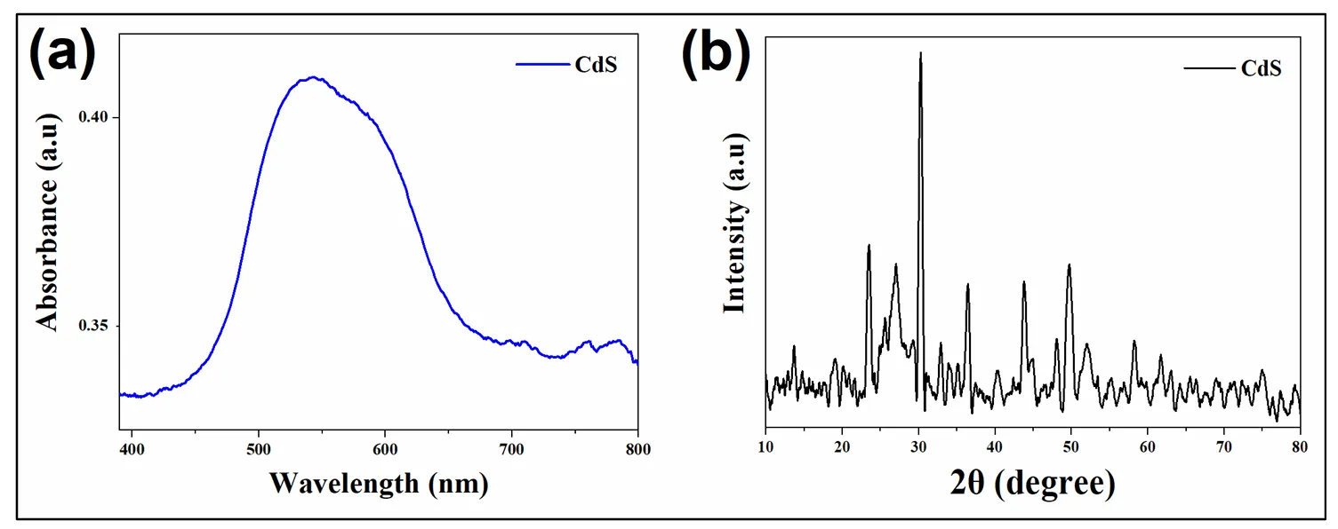
The synthesized CdS-LA hybrid nanoparticles (
Lathyrus aphaca L. extract capped CdS nanoparticles) were analyzed structurally by XRD. The XRD pattern of CdS-LA nanoparticles (JCPDS Card No. 89-0440) reveals multiple peaks corresponding to a face-centered cubic structure. Additionally, the presence of a minor wurtzite hexagonal phase is indicated by peaks matching JCPDS Card No. 80-006. The patterns recorded are given in
. CdS-LA hybrid nanoparticles showed a hexagonal crystal structure. The nanoparticles having a plant extract on their surface showed a broad hexagonal peak, which evidenced the presence of nanoparticles of smaller size in the sample. The average crystallite size, calculated using the Scherrer equation, was found to be approximately 58 nm. The results interpreted are as follows: the presence of phytochemicals limits the rate of fast growth of the CdS-LA hybrid nanoparticles, and a slight variation in peak values arises due to the modification of surface induced by plant constituents. The studies reported that plant extract is a reducing/capping agent for the metal nanoparticles and acts as a surfactant to lower the growth rate of nanoparticles. Here, plant extract, as a source of different chemical constituents, is expected to act as a surfactant by decreasing the growth kinetics and might cover the surface of CdS-LA hybrid nanoparticles [
20].
. (<b>a</b>) Absorption spectrum, (<b>b</b>) XRD Spectrum of CdS-LA hybrid nanoparticles.
The UV-visible spectroscopy was used to record the data of absorbance at different wavelengths (
). The peak values of absorption of the sample are blue-shifted by the addition of plant extract. This shift in peaks is caused by the effect of quantum confinement, which results in nanoparticles of small size in a sample of CdS-LA hybrid. The peak value is 543 nm for the sample CdS-LA hybrid nanoparticles. Thus, they clearly confirm the presence of small-size nanoparticles, and the result supported the X-ray diffraction analysis [
20].
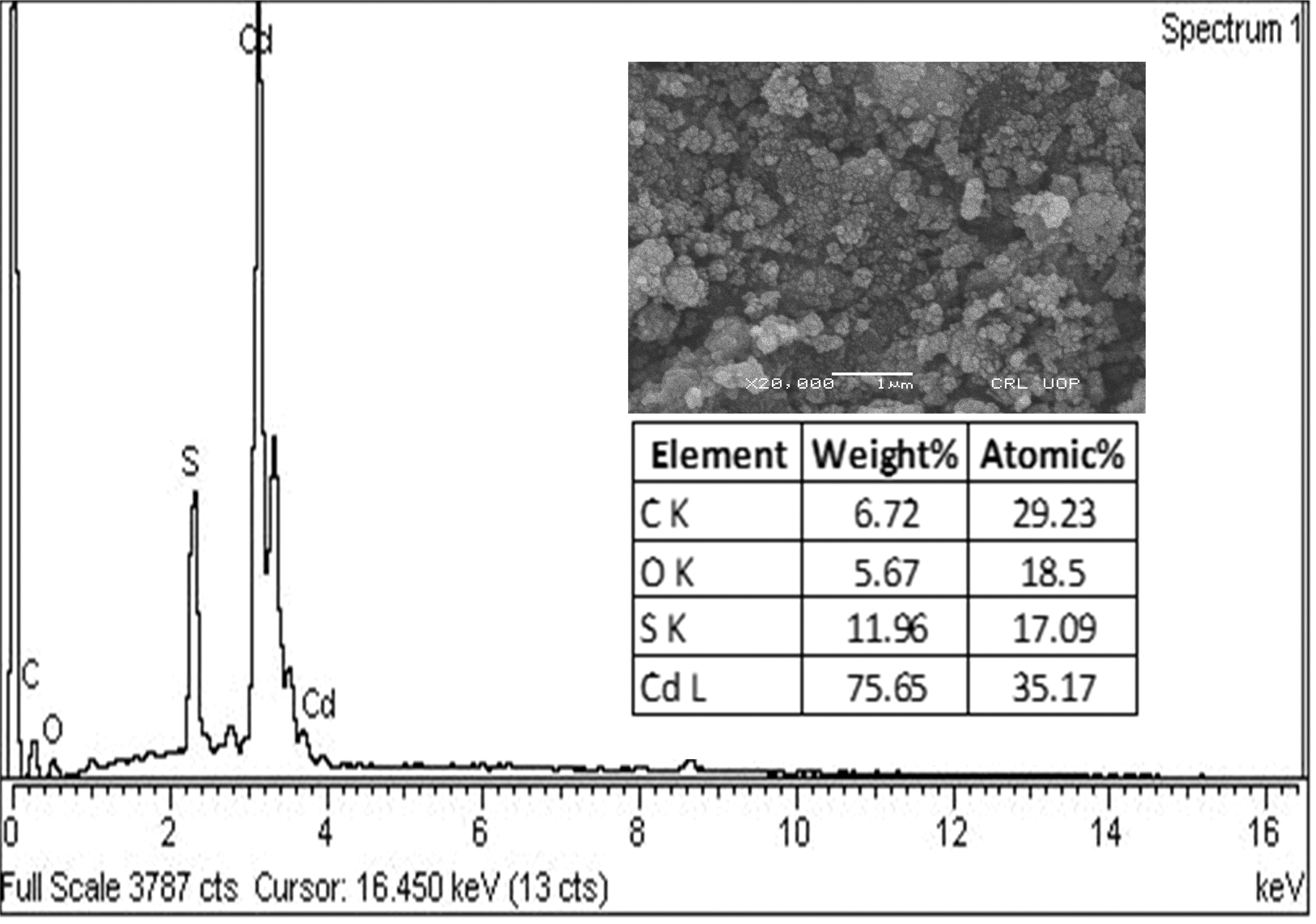
SEM analysis exposed that the surface appearance of CdS-LA hybrid nanoparticles was homogenous. The morphology of synthesized nanoparticles also showed that the CdS-LA hybrid nanoparticles have a spherical shape. Furthermore, our results complied with previous previously reported studies [
21]. Hence, the hexagonal geometry of CdS-LA hybrid nanoparticles made it a potent antibacterial agent due to the nanoparticle’s entry into microbial cells based on their structural morphology [
22]. The scanning electron micrograph of CdS-LA hybrid nanoparticles is shown in
.
. EDX spectrum and SEM image of the CdS-LA hybrid nanoparticles and the inset table comprises the percentage composition from the EDX analysis.
EDX analysis was executed for verification of the CdS-LA hybrid formation. The spectrum of the selected area of the sample is shown in
. The elemental constituents identified in the EDX were Cd (cadmium), S (sulphur), C (carbon), and O (oxygen). The weight percentages of the elements other than Cd and S confirmed the presence of the phytochemicals of plant extract. Thus, EDX analysis confirmed the attachment of the different chemical constituents of plant extract on the surface of the CdS nanoparticles [
20].
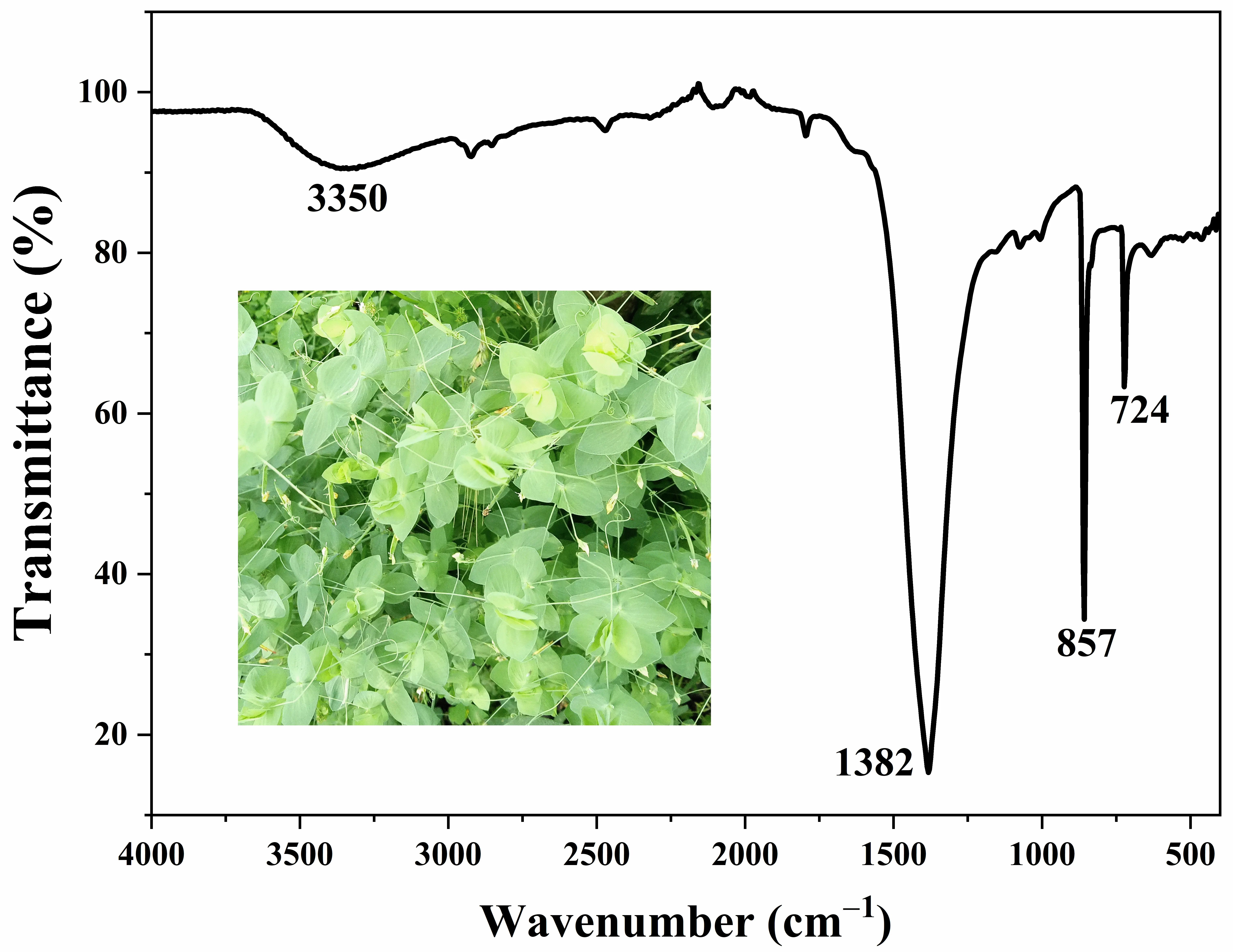
The FTIR (Fourier Transform Infrared) spectrum of CdS-LA hybrid was demonstrated in
. The peaks at 3150–3350 cm
−1 are due to stretching of the -OH group, which indicates the presence of the water content on the surface of nanoparticles. The peak given at 1382 cm
−1 was evolved from the vibration of the C-N stretch vibration. Phytochemicals of plant extract contain H, O, and C as the basic elements in its structure. Hence, a vibration of the H and C is an indication of the presence of the phytochemicals on the surface of the nanoparticles. The peaks were obtained at 857, 724, 614, and 559 cm
−1 for vibrations of Cd-S bonds. Thus, the results of FTIR agreed with the elemental analysis of sample and surface modification of CdS nanoparticles were confirmed by FTIR spectra of CdS-LA hybrid nanoparticles samples [
20].
. FTIR spectrum of CdS-LA hybrid nanoparticles.
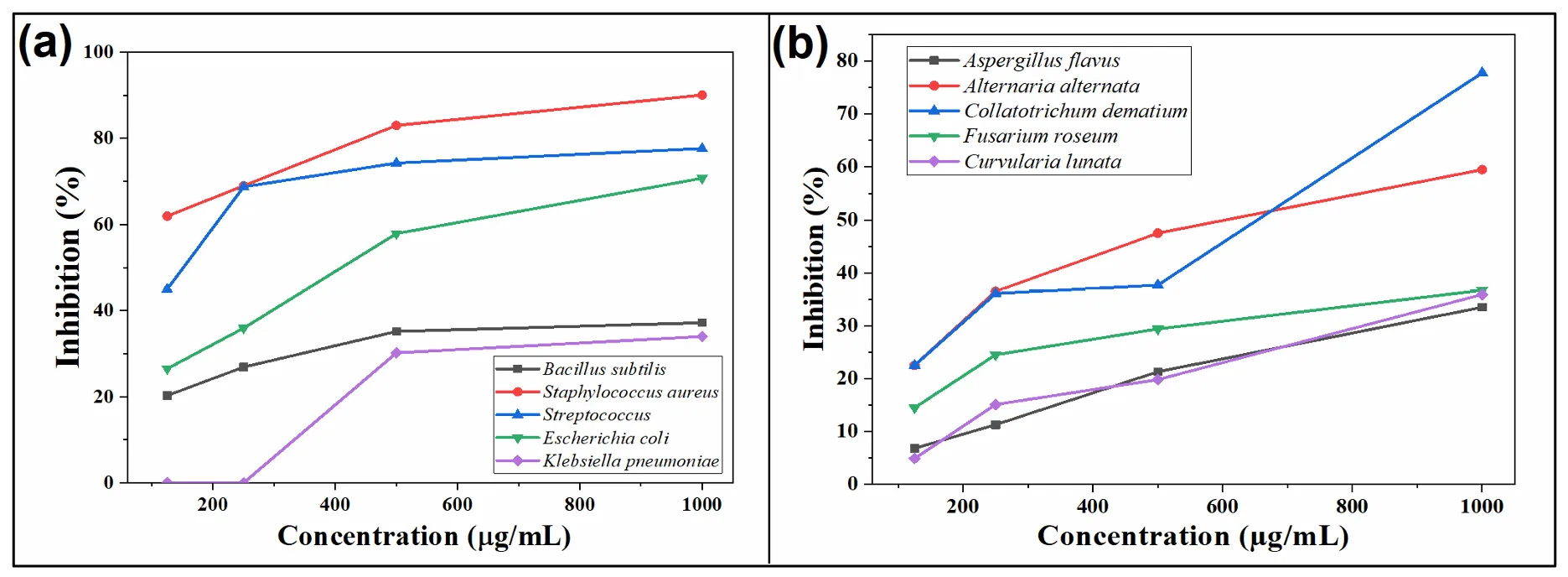
4. Antibacterial Activity
The antibacterial activity of CdS-LA hybrid nanoparticles was performed on five different species of bacteria. A comparison of the antibacterial activity of CdS-LA hybrid nanoparticles and standard Gentamicin sulphate against different species of bacteria was performed in which the inhibition zone of Gentamicin sulphate was taken as 100%, as shown in
. The antibacterial activity of CdS-LA hybrid nanoparticles recorded increases with an increase in their concentration. The antibacterial activity was recorded by different concentrations of CdS-LA hybrid nanoparticles against the five given species of bacteria, as shown in
, but maximum antibacterial activity was recorded against the
Staphylococcus aureus specie. The
Bacillus subtilis,
Staphylococcus aureus,
Streptococcus,
Escherichia coli, and
Klebsiella pneumoniae which has 13.02, 28.82, 31.06, 26.89, and 10.2 mm of inhibition zone, respectively, by 1000 µg/mL of CdS-LA hybrid nanoparticles as compared to the inhibition shown by standard Gentamicin sulphate (40 µg/mL) (
). The activity shown by CdS-LA hybrid nanoparticles is due to the phytochemicals of the plant extract that are present on the surface of CdS nanoparticles as a reducing and capping agent, which is also responsible for the antifungal activity of the CdS-LA hybrid nanoparticles which was confirmed by the previous study on the antifungal activity of
Lathyrus plant seeds [
23].
. Comparison of percent inhibition of CdS-LA hybrid nanoparticles against different species of (<b>a</b>) Bacteria and (<b>b</b>) Fungi.
.
Antibacterial activity of CdS-LA hybrid nanoparticles.
| CdS Nanoparticles |
Bacteria |
| Concentration (µg/mL) |
Bacillus subtilis |
Staphylococcus aureus |
Streptococcus species |
Escherichia coli |
Klebsiella pneumoniae |
| 125 |
7.1 |
19.83 |
18 |
10.07 |
NA |
| 250 |
9.44 |
22.08 |
27.5 |
13.66 |
NA |
| 500 |
12.31 |
26.56 |
29.71 |
22 |
9.06 |
| 1000 |
13.02 |
28.82 |
31.06 |
26.89 |
10.2 |
| Standard |
35 |
32 |
40 |
38 |
30 |
5. Antifungal Activity
The antifungal activity was performed on the five human and plant pathogenic fungi by ‘poisoned food technique. The comparison of the activity of CdS-LA hybrid nanoparticles and standard Fluconazole against five different species of fungi are given in
. The inhibition potential of CdS-LA hybrid nanoparticles increases with an increase in their concentration. The maximum inhibition was shown by 1000 µg/mL of CdS-LA hybrid nanoparticles against the
Collatotrichum dematium fungi, which was 77.8%. The
Aspergillus flavus,
Alternaria alternata,
Fusarium roseum, and
Curvularia lunata which was 33.5, 59.5, 36.7, 35.9%, respectively by 1000 µg/mL of CdS-LA hybrid nanoparticles as compared to the standard Fluconazole (20 µg/mL) (
). The activity shown by CdS-LA hybrid nanoparticles is due to the phytochemicals of the plant extract that are present on the surface of CdS nanoparticles as a reducing and capping agent, which was confirmed by the previous study on the antibacterial activity of the extract of
Lathyrus specie [
24].
.
Antifungal activity of CdS-LA hybrid nanoparticles.
| CdS Nanoparticles |
Fungi |
| Concentration (µg/mL) |
Aspergillus flavus |
Alternaria alternata |
Collatotrichum dematium |
Fusarium roseum |
Curvularia lunata |
| 125 |
6.8 |
22.5 |
22.5 |
14.5 |
4.9 |
| 250 |
11.3 |
36.5 |
36.1 |
24.5 |
15.1 |
| 500 |
21.3 |
47.5 |
37.7 |
29.4 |
19.8 |
| 1000 |
33.5 |
59.5 |
77.8 |
36.7 |
35.9 |
| Standard |
100 |
100 |
100 |
100 |
100 |
6. Proposed Mechanism of Antimicrobial Activity
The antimicrobial activity observed in our study can be attributed to several possible mechanisms by which the green-synthesized CdS nanoparticles (CdS-LA NPs) act against microbial cells. One common way where bacterial and fungal cells experience oxidative stress is through the creation of ROS, which can damage their lipids, proteins and DNA [
25]. Also, because nanoparticles are small and have much more surface area than large particles, they can come into direct contact with microbial membranes, causing them to rupture, become more permeable and allow cell contents to escape [
25]. The flavonoids, alkaloids and phenolic compounds in
Lathyrus aphaca L. extract might add to its antimicrobial power by either working together or disabling metabolic enzymes and pathways of microbes [
25]. It may be these joint effects that explain the strong dose-dependent inhibition against both bacterial and fungal strains. Molecular studies in the future could give us clearer knowledge about these mechanisms.
7. Conclusions
It was concluded that green synthesis was employed for CdS nanoparticle synthesis via a simple wet chemical approach. The plant extract was employed for modification of the surface of the CdS nanoparticles. The growth dynamics of the CdS nanoparticles could be controlled by using plant extract in the synthesis of nanoparticles. The antibacterial and antifungal activity was performed for five different species of bacteria and fungi to evaluate the biological applications of the CdS-LA hybrid nanoparticles. The EDX analysis of the sample confirmed the attachment of the phytochemicals of plant extract with the CdS surface, as evident from the elemental analysis. The peak in the visible region of the absorption spectrum is between 500–600 nm, which was attributed to the CdS nanoparticles. The results of the biological activity revealed that the CdS nanoparticles capped by Lathyrus aphaca extract have inhibition potential against various species of bacteria and fungi, which shows the biological importance of the green synthesis of nanoparticles using different biological moieties.
Acknowledgments
We gratefully acknowledge the administration of Peshawar Model Degree College Boys, Mardan, for their valuable support and for providing a conducive research environment, including access to their laboratory facilities for carrying out some of the experimental reactions.
Author Contributions
S.F. and H.U. are credited for conceptualization, data curation, formal analysis, investigation, methodology, writing—original draft; N.A. and N.Z.K.M. are credited for conceptualization, project administration, supervision; M.A. is credited for conceptualization, data curation, formal analysis, investigation, methodology; I.K. is credited for conceptualization, data curation, formal analysis, investigation, methodology; F.A. is credited for formal analysis, investigation, methodology; N.A. are credited for funding acquisition, writing—review, editing, software validation.
Ethics Statement
Not Applicable.
Informed Consent Statement
Not Applicable.
Data Availability Statement
All data generated or analyzed during this study are included in this published article.
Funding
This research was self-funded by Naveed Akhtar.
Declaration of Competing Interest
The authors declare no conflicts of interest.
References
-
1.
Rafique M, Sadaf I, Rafique MS, Tahir MB. A review on green synthesis of silver nanoparticles and their applications.
Artif. Cells Nanomed. Biotechnol. 2017,
45, 1272–1291.
[Google Scholar]
-
2.
Varma RS. Greener approach to nanomaterials and their sustainable applications.
Curr. Opin. Chem. Eng. 2012,
1, 123–128.
[Google Scholar]
-
3.
Abdelghany TM, Al-Rajhi AMH, Al Abboud MA, Alawlaqi MM, Ganash Magdah A, Helmy EAM, et al. Recent Advances in Green Synthesis of Silver Nanoparticles and Their Applications: About Future Directions.
Rev. Bionanosci. 2018,
8, 5–16.
[Google Scholar]
-
4.
Gour A, Jain NK. Advances in green synthesis of nanoparticles.
Artif. Cells Nanomed. Biotechnol. 2019,
47, 844–851.
[Google Scholar]
-
5.
Jayaseelan C, Rahuman AA, Kirthi AV, Marimuthu S, Santhoshkumar T, Bagavan A, et al. Novel microbial route to synthesize ZnO nanoparticles using Aeromonas hydrophila and their activity against pathogenic bacteria and fungi.
Spectrochim. Acta A Mol. Biomol. Spectrosc. 2012,
90, 78–84.
[Google Scholar]
-
6.
Hussain I, Singh NB, Singh A, Singh H, Singh SC. Green synthesis of nanoparticles and its potential application.
Biotechnol. Lett. 2016,
38, 545–560.
[Google Scholar]
-
7.
Rao MD, Pennathur G. Green synthesis and characterization of cadmium sulphide nanoparticles from Chlamydomonas reinhardtii and their application as photocatalysts.
Mater. Res. Bull. 2017,
85, 64–73.
[Google Scholar]
-
8.
Hassan A, Haris M, Khan SU, Khan I, Akif M, Akhtar N. Lathyrus aphaca Extract MnO Nanoparticles: Synthesis, Characterization, and Photocatalytic Degradation of Methylene Blue Dye.
Photocat. Res. Potent. 2024,
1, 10004.
[Google Scholar]
-
9.
Akhtar N, Choi C, Ateeq M, Fazil P, Shah NS, Khan JA, et al. Visible light active CdS/CuO nanocomposites for photocatalytic degradation of ciprofloxacin, H2 production and antimicrobial activity.
Chem. Eng. J. 2025,
507, 160336.
[Google Scholar]
-
10.
Tudu SC, Zubko M, Kusz J, Bhattacharjee A. CdS nanoparticles (<5 nm): green synthesized using Termitomyces heimii mushroom–structural, optical and morphological studies.
Appl. Phys. A Mater. Sci. Process. 2021,
127, 1–9.
[Google Scholar]
-
11.
Akhtar N, Malik S, Muhammad F, Ullah Z, Mardan U, Pakhtunkhwa K, et al. CuO Nanoparticles: Tuning Properties for Energy and Optoelectronic Applications.
Nano. Select. 2025,
8, e70015.
[Google Scholar]
-
12.
Ullah A, Rasheed S, Ali I, Ullah N. Plant Mediated Synthesis of CdS Nanoparticles, their characterization and application for photocatalytic degradation of toxic organic dye.
Chem. Rev. Lett. 2021,
4, 98–107.
[Google Scholar]
-
13.
Shivashankarappa A, Sanjay KR. Escherichia coli-based synthesis of cadmium sulfide nanoparticles, characterization, antimicrobial and cytotoxicity studies.
Brazil. J. Microb. 2020,
51, 939–948.
[Google Scholar]
-
14.
Lambein F, Travella S, Kuo YH, Van Montagu M, Heijde M. Grass pea (
Lathyrus sativus L.): orphan crop, nutraceutical or just plain food?
Planta 2019,
250, 821–838.
[Google Scholar]
-
15.
Chavan UD, McKenzie DB, Amarowicz R, Shahidi F. Phytochemical components of beach pea (
Lathyrus maritimus L.).
Food Chem. 2003,
81, 61–71.
[Google Scholar]
-
16.
Yadava RN, Asati N. Antioxidant Activity Of Extracts Of Lathyrus Aphaca Linn.
World J. Pharm. Res. 2018,
7, 995.
[Google Scholar]
-
17.
Iqbal J, Rehmani MIA, Sagheer S, Kaleem N, Muneer J. Herbicidal potential of some dry land plants against
Lathyrus aphaca (L.), winter season weed.
Planta Daninha 2020,
38, e020171297.
[Google Scholar]
-
18.
Hassan A, Ullah H, Bonomo MG. Antibacterial and Antifungal Activities of the Medicinal Plant Veronica biloba.
J. Chem. 2019,
1, 5264943.
[Google Scholar]
-
19.
Mullamuri B, Mosali VSS, Maseed H, Majety SS, Chandu B. Photocatalytic Activity of Heavy Metal Doped CdS Nanoparticles Synthesized by Using Ocimum sanctum Leaf Extract.
Biointerf. Res. Appl. Chem. 2021,
11, 12547–12559.
[Google Scholar]
-
20.
Naranthatta S, Janardhanan P, Pilankatta R, Nair SS. Green Synthesis of Engineered CdS Nanoparticles with Reduced Cytotoxicity for Enhanced Bioimaging Application.
ACS Omega 2021,
6, 8646–8655.
[Google Scholar]
-
21.
Kumar A, Sharma RK, Goyal N, Gautam S. Synthesis, characterization & study of Ni-doped CdS nanoparticle for high voltage application.
Vacuum 2019,
160, 75–80.
[Google Scholar]
-
22.
Azam Z, Ayaz A, Younas M, Qureshi Z, Arshad B, Zaman W, et al. Microbial synthesized cadmium oxide nanoparticles induce oxidative stress and protein leakage in bacterial cells.
Microb. Pathog. 2020,
144, 104188.
[Google Scholar]
-
23.
Khan NA. Two antifungal active triterpenoid saponins from the seeds of Lathyrus plants.
Nat. Prod. Res. 2011,
25, 1687–1694.
[Google Scholar]
-
24.
Khan NA, Quereshi S, Pandey A, Srivastava A. Antibacterial activity of seed extracts of commercial and wild Lathyrus Species.
Turkish J. Biol. 2009,
33, 165–169.
[Google Scholar]
-
25.
Rukh G, Ullah A, Akhtar N, Yasmeen S, Zada A, Fazil P, et al. Exceptional synergistic photodynamic antimicrobial chemotherapy of graphene oxide decorated with nicotinamide zinc phthalocyanine potentiated by potassium iodide.
Diam. Relat. Mater. 2025,
156, 112452.
[Google Scholar]