
Engineering Extracellular Vesicles for Direct and Cross-Presentation in Tumor Vaccines

Engineering Extracellular Vesicles for Direct and Cross-Presentation in Tumor Vaccines
Zhou Zhou
1,2
Chenchen Zhao
1,2,*

Received: 21 September 2026 Revised: 08 October 2026 Accepted: 19 October 2026 Published: 29 October 2026

© 2026 The authors. This is an open access article under the Creative Commons Attribution 4.0 International License (https://creativecommons.org/licenses/by/4.0/).
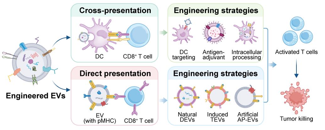
Graphical Abstract
1. Introduction
Tumor vaccines activate the host immune system by delivering tumor-specific antigens (TSAs), inducing expansion and functional differentiation of tumor-specific CD8+ cytotoxic T lymphocytes (CTLs), and ultimately eliminating tumor cells [1]. Ideally, therapeutic tumor vaccines deliver tumor antigens to professional antigen-presenting cells (APCs) in lymph nodes, where antigens are processed and presented as peptide-major histocompatibility complex (pMHC) to activate naive CD8+ T cells [2]. Thus, inducing sufficient and highly functional tumor-specific CD8+ T cell responses is a central challenge in tumor vaccine design. Despite substantial progress in antigen discovery, vaccine platforms, and delivery systems in recent years, the clinical translation of tumor vaccines remains constrained by several obstacles [3]. Most tumor antigens, particularly tumor-associated antigens (TAAs), exhibit low immunogenicity, making it difficult to overcome immune tolerance. Conventional vaccine platforms also suffer from suboptimal antigen delivery efficiency. Moreover, immunosuppressive factors within the tumor microenvironment can restrict the infiltration, proliferation, and effector functions of vaccine-induced T cells [4,5]. These challenges highlight the need for strategies that improve antitumor immunity by regulating antigen-presentation pathways.
Antigen presentation connects vaccine antigens to CD8+ T cell activation through two major pathways [6]. Cross-presentation involves the uptake of exogenous antigens by professional APCs, particularly dendritic cells (DCs), followed by intracellular processing, loading onto MHC-I, and presentation to CD8+ T cells [7]. This pathway underlies the design of many conventional tumor vaccine platforms. However, cross-presentation can be limited by inefficient endosomal escape, excessive lysosomal degradation, and impaired DC function within the TME [8]. Direct presentation refers to the direct engagement of pre-formed pMHC complexes with cognate T-cell receptors (TCRs), enabling CD8+ T-cell activation without requiring antigen processing by a conventional APC [9]. In tumor immunity, tumor cells often downregulate MHC-I to evade immune recognition, while DC cross-presentation can be impaired by the TME, creating complementary barriers to effective antitumor immunity. Accordingly, strategies that exploit and integrate these two pathways may offer additional opportunities to improve tumor vaccine efficacy.
Extracellular vesicles (EVs) are nanoscale, lipid-bilayer-enclosed particles secreted by virtually all cell types and play pivotal roles in intercellular communication [10,11]. Based on biogenesis, EVs are broadly classified into exosomes and microvesicles, with apoptotic bodies representing another class of cell-derived vesicles [12,13]. As cancer vaccine carriers, EVs offer several potential advantages: their endogenous origin confers biocompatibility; surface molecules such as CD47 may influence circulation and cellular interactions; EVs can traffic to draining lymph nodes and be internalized by APCs; and their surface molecules and cargo can be tailored through genetic engineering, chemical modification, or membrane fusion [14,15,16]. Importantly, EVs can support multiple modes of antigen presentation. EV-associated pMHC can directly engage TCRs on CD8+ T cells, whereas EV-associated tumor antigens can be internalized and processed by APCs for cross-presentation. EVs can also transfer intact pMHC complexes to APC surfaces through cross-dressing, enabling T-cell recognition without reprocessing of the transferred complexes [6]. These complementary antigen-presentation pathways provide the conceptual basis for engineering EVs to enhance direct presentation, cross-presentation, or both within an integrated vaccine platform. Figure 1 summarizes the overall framework of this review, illustrating the two major antigen-presentation pathways and the corresponding engineering strategies.
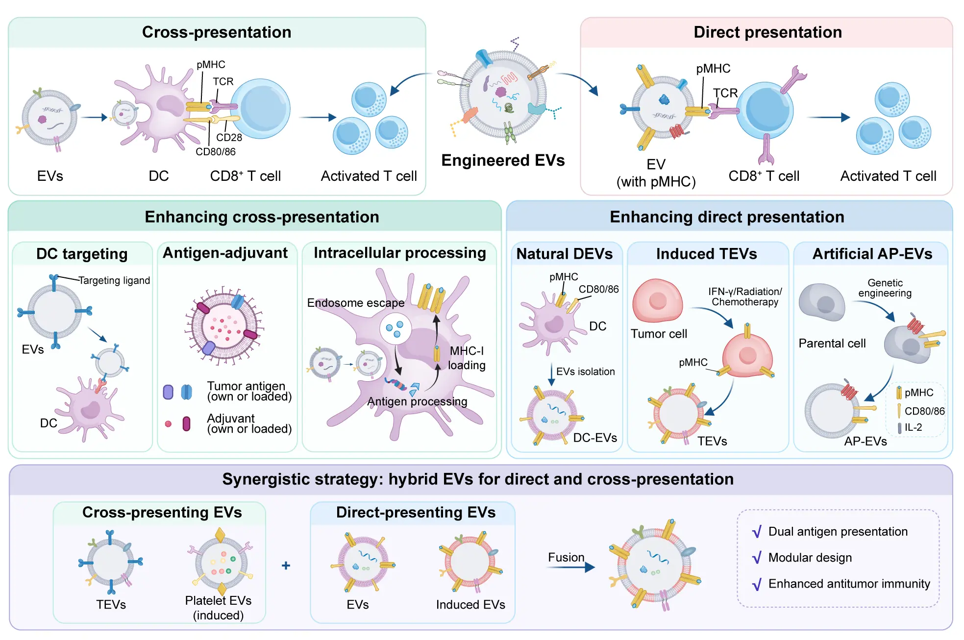
Figure 1. Schematic overview of EV-mediated direct antigen presentation and cross-presentation in tumor vaccines and the corresponding engineering strategies. EVs can directly stimulate CD8+ T cells through surface pMHC–TCR interactions or undergo DC-mediated cross-presentation following antigen uptake and intracellular processing. Engineering strategies for enhancing cross-presentation include DC targeting, antigen–adjuvant co-delivery, and modulation of intracellular antigen processing, whereas strategies for enhancing direct antigen presentation include DC-derived EVs (DEVs), pMHC-enriched tumor-derived EVs, and engineered artificial antigen-presenting EVs (AP-EVs). Hybrid EVs represent complementary strategies that integrate direct antigen presentation and cross-presentation within a dual-pathway vaccine platform.
2. Biological Basis of EVs as Antigen Presentation Platforms
As endogenous mediators of intercellular communication, EVs transport proteins, lipids, and nucleic acids, and regulate immune responses through surface molecules such as MHC family molecules, co-stimulatory molecules, and immune checkpoint regulators [17,18]. Understanding EV classification, biogenesis, natural antigen presentation machinery, immune interactions, and functional heterogeneity is a prerequisite for engineering EVs for direct antigen presentation and cross-presentation. In this section, we systematically elaborate on the fundamental biological characteristics of EVs as a next-generation multifunctional antigen-presenting nanoplatforms.
2.1. Classification and Biogenesis of EVs
EVs are highly heterogeneous membrane-enclosed vesicles categorized into exosomes, microvesicles, and apoptotic bodies [19]. Exosomes (30–150 nm) originate from the endosomal pathway: plasma membrane invagination forms early endosomes, which mature into multivesicular bodies (MVBs) containing intraluminal vesicles; upon MVB fusion with the plasma membrane, vesicles are released [20]. Microvesicles (50 nm–5 μm) bud directly from the plasma membrane via cytoskeletal remodeling and calcium signaling. Apoptotic bodies (>1000 nm) are produced during programmed cell death and are generally excluded from EV vaccine research. EV heterogeneity also extends to molecular cargo, which varies according to the cell of origin and the mechanisms of vesicle biogenesis. However, the relatively limited yield of naturally secreted EVs, together with challenges in isolation, purification, and large-scale production, can constrain their broader application. To address these limitations, cell-derived biomimetic nanovesicles have been developed as complementary delivery platforms. These vesicles are generally produced from parental cells or their membranes through artificial processes, such as mechanical disruption and membrane extrusion, rather than through native vesicle biogenesis. Although they share certain structural features with natural EVs, they may differ in molecular composition, cargo distribution, and biological functions. Nevertheless, owing to their cell-derived components and potential for scalable preparation, they can provide complementary opportunities for antigen delivery and immune modulation in EV-related vaccine platforms [21]. Natural EVs from different parental cells also selectively enrich specific proteins, mRNAs, microRNAs, and lipids, contributing to functional differences in antigen presentation and immune regulation [6]. Understanding both the biogenesis of natural EVs and the characteristics of biomimetic nanovesicles is therefore important for interpreting their biological functions and informing the design of antigen-presentation platforms.
2.2. Surface Molecules and Immune Interactions of EVs
EV functions depend on their surface molecular repertoire. EVs inherit membrane proteins from their parental cells and may selectively incorporate functional molecules, potentially contributing to their antigen-presentation-related properties [22]. Some EVs carry MHC-I and/or MHC-II molecules, which can participate in antigen presentation depending on their molecular composition and biological context [23]. When parental cells (especially DCs) express pMHC, these complexes integrate into EV surfaces via MVB or plasma membrane budding, enabling T cell activation. EVs also carry co-stimulatory molecules (CD80, CD86) and adhesion molecules (ICAM-1), providing second signals that determine T-cell activation versus anergy [17]. After administration, EVs interact with immune cell surface receptors, initiating immunomodulatory cascades. Efficient recognition and internalization of EVs by APCs such as DCs are critical prerequisites for cross-presentation of exogenous antigens. DCs and other professional APCs can recognize and internalize EVs through multiple surface pattern recognition receptors [6]. Receptor-mediated uptake determines EV capture efficiency and intracellular trafficking [24]. Different internalization routes direct antigens toward MHC-I cross-presentation or MHC-II classical presentation, shaping T cell response type and intensity [25]. EV immunomodulation exhibits duality: EVs bearing pMHC and co-stimulatory molecules activate antigen-specific T cells, whereas tumor-derived EVs (TEVs) overexpressing PD-L1 engage PD-1, inducing T cell exhaustion or apoptosis [26]. Together, these intrinsic surface properties and cellular interactions provide the biological basis for engineering EVs to enhance direct antigen presentation and cross-presentation.
3. Engineering Strategies to Enhance EV-Mediated Cross-Presentation
Cross-presentation involves the uptake of exogenous antigens by professional APCs, particularly DCs, followed by their processing and presentation on MHC-I to CD8+ T cells [27]. Two major pathways have been described for MHC-I cross-presentation: the cytosolic and vacuolar pathways. In the cytosolic pathway, internalized EVs enter the cytosol and are subsequently degraded by the proteasome into peptide fragments, which are transported by the transporter associated with antigen processing (TAP) to the endoplasmic reticulum (ER), where they are loaded onto MHC-I molecules. The resulting pMHC-I complexes are transported to the DC surface, where CD8+ T cells recognize them and initiate CTL responses [6]. In the vacuolar pathway, by contrast, internalized antigens are processed by endosomal or phagosomal proteases and the resulting peptides are loaded onto MHC-I within these compartments [28]. These pathways are not necessarily mutually exclusive, and their relative contributions may vary with antigen properties, uptake routes, and the intracellular trafficking environment. Cross-presentation is particularly important for initiating effective antitumor CD8+ T-cell responses when tumor cells exhibit limited MHC-I expression [29].
Despite its central role in antitumor immunity, EV-mediated cross-presentation can be constrained by multiple biological barriers, including preferential trafficking toward endo-lysosomal compartments, inefficient endosomal escape, and limited access of EV-associated antigens to the cytosolic proteasome-TAP-dependent MHC-I processing pathway [28,30,31]. These limitations have motivated the development of engineering strategies that improve EV delivery to DCs, coordinate antigen delivery with immunostimulatory signals, and optimize intracellular antigen trafficking and processing. The major strategies for enhancing EV-mediated cross-presentation are summarized in Table 1 and discussed in the following sections.
3.1. DC Targeting of EVs
Efficient capture and internalization of EVs by DCs are critical for initiating EV-mediated cross-presentation [32]. Surface engineering of EVs with ligands or targeting moieties that engage DC endocytic receptors can enhance EV-DC interactions and cellular uptake, thereby increasing antigen delivery and MHC-I presentation [33]. Among available approaches, receptor-directed targeting with antibodies promotes EV recognition by selected DC populations. DEC205 (CD205) is a highly efficient endocytic receptor on the surface of DCs [34]. Dang et al. modified anti-DEC205 antibodies onto the surface of red blood cell-derived EVs (RBCEVs), achieving specific targeting of DEC-205+ DCs in the spleen [35]. RBCEVs loaded with ovalbumin (OVA) promoted OVA peptide presentation on MHC-I and elicited robust CD8+ T cell responses. This platform also offers a potential alternative to tumor- or DC-derived EVs, which may present challenges related to heterogeneity and large-scale production. In a complementary approach, Bhatta et al. used metabolic labeling with Ac4ManAz to introduce azide groups onto TEVs, followed by conjugation of DBCO-modified anti-DEC205 through click chemistry [36]. This strategy enhanced TEV accumulation in draining lymph node DCs and improved antigen processing and presentation. In both prophylactic and therapeutic E.G7-OVA tumor models, anti-DEC205-EVs induced stronger antigen-specific CTL responses and more pronounced tumor growth inhibition. Together, these studies illustrate how antibody-mediated targeting can be combined with different EV sources and surface-engineering methods to improve DC delivery.
Table 1. Representative engineering strategies for enhancing EV-mediated cross-presentation.
|
Strategy |
Engineering Approach |
EV Source |
Key Mechanism |
Refs. |
|---|---|---|---|---|
|
DC targeting |
Anti-DEC205 modification |
Red blood cell, tumor cell |
DC-targeted delivery |
|
|
Mannose modificationn |
Serum |
DC-targeted delivery |
[37] |
|
|
LewisY modification |
Glioblastoma cell |
Enhanced cellular uptake |
[38] |
|
|
Antigen-adjuvant co-delivery |
OMV–TEV hybridization |
Bacteria, tumor cell |
Co-delivery of intrinsic adjuvant and antigen |
|
|
Pyroptosis-induced EVs |
Pyroptotic tumor cell |
Enriched DAMPs and antigens |
[42] |
|
|
“Waste” EVs |
Metformin/PLGA-treated tumor cell |
Intrinsic adjuvant activity via dsDNA |
[43] |
|
|
Exogenous antigen/adjuvant loading |
Bacteria, tumor cell |
Exogenous antigen/adjuvant incorporation and in situ vaccine generation |
||
|
Intracellular processing |
GALA peptide modification |
Tumor cell |
Endosomal escape |
[52] |
|
LLO-mediated endosomal escape |
Bacteria |
Endosomal escape |
[53] |
|
|
Autophagy modulation |
Melanoma cell, DC |
Enhanced cross-presentation |
[54] |
|
|
YTHDF1 inhibition |
Tumor cell |
Reduced antigen degradation |
[55] |
Beyond antibody-mediated targeting, mannose modification represents a widely explored strategy for DC targeting [56]. The mannose receptor (CD206), an endocytic C-type lectin receptor expressed by subsets of DCs, can recognizes mannose moieties and mediates receptor-dependent internalization, and decorating EV surfaces with mannose has therefore been explored to enhance their recognition and uptake by DCs [57]. Choi et al. prepared EXO-PEG-mannose complexes (EXO-PEG-man) by incorporating DSPE-PEG-mannose conjugates into bovine serum-derived exosome (EXO) membranes [37]. In vitro, EXO-PEG-man showed approximately 1.4-fold greater uptake by DC2.4 cells than unmodified EXOs and 2.1-fold greater uptake than PEGylated-only controls. No significant uptake enhancement was observed in mannose receptor-negative NIH3T3 fibroblasts, supporting the receptor specificity of this approach. Following subcutaneous injection, EXO-PEG-mannose also showed approximately 1.8-fold greater accumulation in draining lymph nodes than the PEGylated control. These findings suggest that mannose modification can improve both receptor-associated cellular uptake and lymphatic delivery, although enhanced uptake alone does not establish more efficient antigen cross-presentation or stronger antitumor immunity.
A related but more explicitly glycan-guided approach targets DC-SIGN (CD209), another C-type lectin receptor involved in antigen uptake and presentation [58]. Dusoswa et al. systematically investigated the glycocalyx composition of glioblastoma-derived EVs and found that although natural EVs are rich in sialylated N-glycans, they lack natural ligands for DC-SIGN [38]. To address this limitation, they synthesized a high-affinity DC-SIGN ligand, LewisY (LeY), conjugated to a palmitic acid tail, and integrated palmitoyl-LeY into the EV membrane via physical insertion. LeY modification increased EV uptake by human monocyte-derived DCs by approximately fourfold, as further supported by imaging flow cytometry. Beyond demonstrating enhanced uptake, this study highlights the importance of the pre-existing EV glycocalyx in determining receptor recognition and illustrates how glycan engineering can compensate for an unfavorable native surface composition.
Collectively, these studies demonstrate that DC targeting can be achieved through distinct but complementary design principles: antibody-mediated recognition of selected receptors, ligand-mediated engagement of carbohydrate-recognition pathways, and rational glycan engineering guided by the native EV surface composition. However, increased DC uptake is an intermediate step rather than a sufficient indicator of successful cross-presentation. The efficiency of cross-presentation also depends on antigen availability, intracellular trafficking, processing, and MHC-I loading.
3.2. Antigen–Adjuvant Co-Delivery
Efficient induction of potent CTL responses requires antigen presentation in an appropriate immunostimulatory context, in which tumor antigens are accompanied by signals that promote DC maturation and T-cell priming [59]. A central design principle of many tumor vaccine platforms is therefore the co-delivery of tumor antigens and immune adjuvants, ideally ensuring that DCs receive antigenic and activation signals within the same immunological context [60,61]. For EV-based vaccines, these strategies can be broadly categorized according to the source and mode of antigen–adjuvant provision into three groups: exploiting the intrinsic antigen or adjuvant function of EVs, enhancing endogenous EV functions through the pre-processing of parental cell, and loading exogenous antigens or adjuvants onto or into EVs.
3.2.1. Harnessing and Enhancing the Intrinsic Antigenic and Adjuvant Functions of EVs
Bacterial outer membrane vesicles (OMVs) are naturally enriched with pathogen-associated molecular patterns (PAMPs), including lipopolysaccharide (LPS), lipoproteins, and flagellin on their surface, making them ideal natural immune adjuvants [62]. TEVs, in turn, can carry diverse tumor-associated antigens (TAAs) and tumor neoantigens, providing a broad source of tumor antigens [63]. Therefore, TEVs themselves serve as a broad and physiologically relevant antigen source. Building upon these complementary properties, researchers have fused OMVs with tumor cell-derived EVs to construct eukaryotic-prokaryotic hybrid vesicle platforms, achieving integrated co-delivery of endogenous adjuvants and antigens on a single nanoparticle [41]. Zou et al. hybridized bacterial OMVs with autologous tumor cell membranes to form mTOMV, demonstrating enhanced activation of innate immune cells and improved specific lysis of homologous tumor cells by T cells in vitro [39]. Similarly, Guo et al. further fused Staphylococcus aureus-derived EVs with B16F10 melanoma cell membranes (CM) to construct EV-CM hybrid vesicles [40]. In this system, S. aureus-derived EVs served as natural adjuvants to induce the production of pro-inflammatory cytokines such as IFN-γ, IL-1β, and IL-6, while the CM provided broad-spectrum tumor antigens. The combination of the two components significantly activated systemic antitumor immunity and potently suppressed melanoma progression. By integrating antigenic and immunostimulatory components, these hybrid platforms can reduce the need for separate adjuvant-loading procedures. However, the immunostimulatory activity of OMVs, particularly that associated with LPS, may also cause excessive inflammation or toxicity, highlighting the need to balance adjuvant potency with safety.
Based on the direct utilization of endogenous EV functions, researchers have further explored the possibility of enhancing the inherent properties of EVs through pre-processing, thereby amplifying their natively carried adjuvant or antigen signals [64]. Pyroptosis is gasdermin-mediated lytic cell death with pore formation and content release [65]. Li et al. isolated pyroptotic vesicles from pyroptotic tumor cells and found that these vesicles not only carried the full-spectrum antigens of parental tumor cells, but were also enriched with abundant endogenous damage-associated molecular patterns (DAMPs), including HMGB1, ATP, and the pro-inflammatory cytokines IL-1β and IL-18 [42]. Proteomics showed higher immunostimulatory protein content than conventional exosomes. After DC uptake, DAMPs activated DCs via TLR/IL-1R, promoting maturation and cross-priming. Thus, rather than introducing an exogenous adjuvant, this strategy enhances the endogenous immunostimulatory content of TEVs by altering the mode of tumor cell death.
A complementary strategy exploits cellular stress responses to generate EVs with intrinsic adjuvant activity. Gong et al. reported that when tumor cells are treated with metformin/positively charged PLGA nanoparticles (Met-PC-NPs) to induce elevated intracellular ROS levels, the cellular DNA damage response pathway is triggered. The resulting dsDNA was electrostatically captured by the nanoparticles and ultimately expelled from the cells via “waste” EVs [43]. Met-PC-EVs carried tumor antigens and dsDNA, a natural cGAS-STING agonist, acting as self-adjuvant to promote DC maturation and cross-presentation without exogenous adjuvants. The uniqueness of this strategy lies in exploiting the cellular stress-response and detoxification mechanism, enabling tumor EVs to acquire dual functions of antigen delivery and self-adjuvancy without introducing exogenous adjuvants. This approach therefore exploits cellular stress and waste-disposal processes to confer self-adjuvanting properties on TEVs.
Collectively, these studies illustrate two complementary routes to antigen–adjuvant co-delivery: combining antigen-bearing tumor components with naturally immunostimulatory vesicles, and enhancing the endogenous danger signals of EVs through parental-cell manipulation. Both approaches can simplify adjuvant integration, but their effectiveness depends on the balance between antigen availability and immune stimulation. In particular, the broad antigenic content of TEVs does not necessarily guarantee effective antitumor immunity, as TEVs may also carry immunosuppressive molecules such as PD-L1. Engineering strategies that reduce PD-L1 expression in parental tumor cells or otherwise limit PD-1/PD-L1-mediated immunosuppression may help improve the immunological profile of tumor-derived vesicles [66]. Likewise, the inflammatory activity of OMVs requires careful control to avoid excessive immune activation. Genetic modification of lipid A biosynthesis or LPS depletion can reduce OMV-associated endotoxin toxicity, although the effects of these modifications on vesicle integrity and adjuvant activity must also be considered [67]. Therefore, these strategies should be evaluated not only by their antigen and adjuvant content, but also by their ability to promote productive MHC-I cross-presentation and antigen-specific T-cell responses while maintaining an acceptable safety profile.
3.2.2. Exogenous Antigens and Adjuvants Loading
When the intrinsic antigenic or adjuvant functions of EVs are insufficient to achieve the desired immune response, engineering strategies can be used to incorporate additional antigens or immunostimulatory molecules. These approaches expand the functional versatility of EV-based vaccines by combining antigen delivery with enhanced immune activation or modulation.
One strategy is to display exogenous antigens on EV surfaces while retaining the intrinsic adjuvant activity of OMVs. Lu et al. rapidly displayed OVA antigens on the surface of OMVs through the ClyA–SpyCatcher-based conjugation system, and further conjugated αPD-L1 antibodies to construct an Antigen Presentation Signal Enhancer (APSE) [44]. The natural PAMPs of OMVs activated DC co-stimulatory signals, while αPD-L1 simultaneously blocked co-inhibitory signals on the DC surface, significantly enhancing antigen-specific CTL responses. Furthermore, Miao et al. engineered E. coli to express the ClyA-OVA fusion protein through genetic engineering and isolated OMVs to construct an inhalable nanovaccine termed BMVax [45]. By displaying OVA on the vesicle surface while retaining the intrinsic adjuvant activity of OMVs, BMVax integrated antigen delivery and innate immune stimulation in a single platform. Following respiratory administration, it induced robust immune responses in tracheobronchial lymph nodes and achieved an 83.3% complete prevention rate in a B16-OVA lung metastasis model. Together, these studies illustrate how antigen display can be combined with the intrinsic immunostimulatory properties of OMVs.
Beyond antigen incorporation, EVs can be engineered to provide additional immune-activating signals [68]. Pan et al. displayed IFNα on the surface of E. coli-derived bacterial membrane vesicles (BMVs), generating IFNα-BMVs [46]. In this system, intrinsic PAMPs provided innate immune stimuli, while surface-displayed IFNα supplied an additional cytokine signal, enhancing DC maturation and antigen cross-presentation. In a related approach, tumor cells were engineered to overexpress the co-stimulatory molecule CD40L, and the resulting cell-derived vesicles, termed PD1/CD40L-NVs, were isolated [47]. CD40L provided an early activation signal for DC maturation, which synergized with the intrinsic lymph node-homing ability of the tumor membrane to induce a 2-fold increase in DC maturation within lymph nodes. These examples demonstrate that engineering EVs to display cytokines or co-stimulatory molecules can extend their function beyond antigen transport by providing additional signals that support antigen presentation and T-cell priming.
Loading synthetic adjuvant molecules into EVs represents another important direction. Tian et al. covalently conjugated the STING agonist DMXAA to a cationic polymer backbone to form CPTS complexes, which were then self-assembled with OVA antigens and encapsulated within OMVs [48]. Miao et al. conjugated biotinylated CpG DNA to the surface of tumor cell-derived EVs, constructing a vaccine platform that simultaneously carries tumor antigens and CpG adjuvants [50]. Extending this strategy beyond immune stimulation, Yang et al. further engineered autologous tumor-derived microvesicles (TMVs) loaded with manganese phosphate nanoparticles (MnPs) to amplify cGAS-STING signaling, while co-delivering PD-L1-targeting siRNA (siPD-L1), inducing durable protective immunity in melanoma and colorectal cancer models [51]. These platforms illustrate the flexibility of EV engineering in integrating different functional cargos, including innate immune agonists and molecules targeting immune-inhibitory pathways.
An alternative strategy is to generate tumor antigens in situ rather than load pre-existing antigens onto EVs. Nie et al. assembled metal-phenolic network prisons on the OMV surface through coordination between ferric ions and tannic acid, significantly reducing the systemic toxicity of OMVs [49]. Upon exposure to the low pH and high ATP conditions characteristic of the TME, the coating dissociated to release OMVs, inducing Fenton reaction-mediated immunogenic cell death (ICD) and generating tumor antigens in situ, which then converged with OMV adjuvants to form an in situ vaccine. This approach therefore shifts the emphasis from exogenous antigen loading to local antigen generation and subsequent immune stimulation, potentially simplifying vaccine preparation while linking antigen release to the tumor microenvironment.
Collectively, these studies demonstrate that EV-based antigen–adjuvant co-delivery can be achieved through antigen surface display, the incorporation of additional immune-activating signals, the loading of synthetic immunomodulatory cargos, or the induction of antigen generation in situ. Although these strategies offer complementary ways to coordinate antigen delivery and immune activation, their outcomes depend on the nature of the incorporated components, their accessibility to the relevant cellular pathways, and the biological context in which they are delivered. Accordingly, successful co-delivery should be evaluated not simply by cargo incorporation or DC activation, but also by evidence of enhanced MHC-I cross-presentation, antigen-specific T-cell responses, and antitumor efficacy, alongside appropriate safety assessment.
3.3. Modulation of Intracellular Antigen Processing
Following uptake by DCs, EVs enter the endocytic system and are progressively trafficked through early and late endosomal compartments, with a substantial fraction ultimately reaching lysosomes, where internalized cargo can undergo proteolytic degradation [69]. Such trafficking can limit the availability of intact tumor antigens for cytosolic MHC-I processing, whereas excessive lysosomal degradation may further compromise cross-presentation [70,71]. Accordingly, engineering strategies that redirect EV intracellular trafficking, facilitate endosomal escape, or limit excessive lysosomal degradation represent important approaches for enhancing MHC-I cross-presentation and CD8+ T cell priming [72].
Surface modification with pH-sensitive fusogenic peptides is a direct strategy to promote EV endosomal escape [73]. These peptides undergo pH-dependent conformational transitions within acidic endosomal compartments, enabling interaction with and destabilization of the endosomal membrane and thereby facilitating the release of EV-associated cargo into the cytosol. Morishita et al. genetically engineered B16BL6 melanoma cells to express a streptavidin-lactadherin (SAV-LA) fusion protein, enabling the secreted exosomes to display SAV on their surface [52]. They then exploited the high affinity between SAV and biotin to conjugate biotinylated GALA peptide to the exosome surface, constructing GALA-modified exosomes (GALA-exo). GALA is a 30-amino-acid peptide containing repetitive EALA motifs that adopts an amphipathic α-helical conformation upon pH reduction from 7 to 5, thereby disrupting endosomal membranes. Calcein leakage assays demonstrated that GALA-exo exhibited concentration-dependent membrane lytic activity under acidic conditions (pH 5.0), whereas no activity was observed at neutral pH (7.4). Confocal microscopy revealed that following DC uptake, the cargo of GALA-exo successfully escaped from lysosomes into the cytosol, whereas unmodified exosomes remained colocalized with lysosomal compartments. Importantly, GALA-exo-treated DCs exhibited significantly enhanced MHC-I presentation of tumor antigens, leading to effective activation of antigen-specific CD8+ T cells. Collectively, this study established that pH-sensitive peptide-mediated modification of exosomes enables cytosolic antigen delivery and enhanced MHC-I cross-presentation in DCs.
An alternative strategy involves genetically engineering the EV surfaces to directly display endosomal escape proteins [74]. Li et al. constructed the OMV-LL platform by simultaneously fusing listeriolysin O (LLO) and the RNA-binding protein L7Ae to the C-terminus of the OMV surface protein ClyA [53]. L7Ae specifically recognizes the box C/D sequence, enabling rapid “plug-and-display” adsorption of mRNA antigens onto the OMV surface (OMV-LL-mRNA), while LLO forms pores within the acidic endosomal environment of DCs to mediate endosomal escape, allowing mRNA to enter the cytosol for translation. In bone marrow-derived DCs (BMDCs), OMV-LL-associated fluorescence was initially colocalized with lysosomes but became more diffusely distributed in the cytosol over time, consistent with endosomal escape. Compared with the LLO-deficient control, OMV-LL-mRNA enhanced the expression of MHC-I-OVA complexes, with antigen presentation sustained for up to 48 h. The platform also inhibited pulmonary melanoma metastasis and induced complete tumor regression in a subset of animals in a colon cancer model. Unlike GALA-mediated delivery of EV-associated cargo, this strategy combines endosomal escape with intracellular mRNA delivery and antigen expression, thereby providing a distinct route to MHC-I presentation.
Beyond directly facilitating endosomal escape, modulating the autophagic pathway in host DCs offers a distinct strategy to enhance cross-presentation. Yan et al. generated a biomimetic hybrid nanovesicle by fusing UVC-induced pyroptotic B16 tumor cells with DC2.4 cells through gradient extrusion, and further loaded the autophagy-inducing peptide beclin-1 to construct hDMVac [54]. Beclin-1 is a critical initiator of autophagosome formation, with its 267–284 amino acid sequence activating the class III PI3K complex to trigger autophagosome biogenesis. The study found that after uptake by DCs, hDMVac initially co-localized with lysosomes, but subsequently escaped from lysosomes and co-localized with the endoplasmic reticulum and Golgi apparatus, a trafficking pattern consistent with the classical antigen cross-presentation pathway. hDMVac-treated BMDCs displayed significantly upregulated MHC-I surface expression, a 2.9-fold increase in CD80/CD86 maturation markers, and enhanced cross-presentation capacity of cDC1s. In therapeutic B16 melanoma models, hDMVac significantly inhibited tumor growth and prolonged mouse survival. In prophylactic models, hDMVac delayed tumor onset and protected a subset of mice from tumor formation. This platform combines the antigenic and danger signals associated with pyroptotic tumor cells with autophagy-related modulation of intracellular trafficking. Thus, rather than relying solely on membrane disruption, it seeks to improve the intracellular handling of antigen-bearing EVs.
An alternative approach is to limit excessive antigen degradation by modulating the host DCs themselves. N6-methyladenosine (m6A) modification is the most prevalent chemical modification of eukaryotic mRNAs, and YTHDF1 serves as an m6A reader protein that recognizes m6A-modified mRNAs and promotes their translation [75]. Han et al. showed that YTHDF1 promotes the translation of mRNAs encoding lysosomal proteases in DCs, thereby enhancing lysosomal antigen degradation and restricting cross-presentation [76]. Building on this, You et al. designed the NICER nanovaccine by co-assembling ALDH1A1-overexpressing TEVs with an epigenetic nanoregulator (ENR) loaded with siYthdf1, and further modified the surface with a DC-SIGN-targeting aptamer [55]. YTHDF1 suppression reduced lysosomal protease abundance and activity without a corresponding decrease in protease mRNA levels, indicating post-transcriptional regulation. This intervention reduced antigen degradation, increased the availability of intact antigen in the cytosol, and enhanced MHC-I cross-presentation and OT-I CD8+ T-cell activation in vitro. NICER also inhibited tumor growth and prolonged survival in 4T1 breast cancer and B16 melanoma models. The distinctive feature of this strategy lies in its modulation of the translational program of host APCs rather than direct EVs engineering, thereby reducing antigen lysosomal degradation at the source and providing a novel paradigm for intracellular processing modulation.
Collectively, these studies address distinct intracellular barriers to MHC-I presentation. Fusogenic peptides and endosomal escape proteins promote cytosolic delivery, autophagy-related modulation seeks to improve intracellular trafficking, and YTHDF1 inhibition limits excessive lysosomal antigen degradation. These approaches are complementary because their effects depend on the cargo type, intracellular destination, and antigen-processing route involved. Importantly, enhanced endosomal escape or reduced antigen degradation does not by itself establish improved cross-presentation; evidence of pMHC-I complex formation and antigen-specific CD8+ T-cell responses remains essential.
4. Engineering Strategies for Direct Antigen Presentation by EVs
EVs can activate CD8+ T cells not only via cross-presentation but also through direct engagement of surface pMHC with TCRs, without requiring antigen uptake, intracellular processing, or de novo MHC-I loading by a conventional APC [77]. This direct pathway enables rapid immune initiation within hours and may reduce dependence on the antigen-processing capacity of recipient APCs. By contrast, cross-presentation involves the uptake of exogenous antigens by APCs, followed by intracellular processing and presentation of the resulting peptides on the APCs’ own MHC-I molecules. A related mechanism, termed cross-dressing or semi-direct antigen presentation, occurs when APCs acquire intact pMHC complexes from other cells or EVs and subsequently display pMHC complexes to T cells without reprocessing the transferred antigens [78]. Thus, the key distinction is whether the antigen is processed and loaded onto MHC-I by the recipient APC, transferred as an intact pMHC complex, or presented directly by EV-associated pMHC. These complementary mechanisms provide a rationale for engineering EVs to support direct T-cell stimulation and APC-mediated cross-presentation.
EV-mediated direct antigen presentation exhibits unique biological advantages and translational value in tumor immunotherapy. First, it enables a more direct route of antigen recognition by T cells by bypassing the intracellular antigen-processing and MHC-loading steps required for conventional cross-presentation. This may accelerate T-cell stimulation and reduce dependence on antigen-processing capacity within recipient APCs. Second, engineered pMHC-bearing EVs can directly deliver antigen-specific stimulatory signals to T cells within the TME, providing a potential strategy to complement conventional APC-dependent antigen presentation. A summary of current strategies for enhancing EV-mediated direct antigen presentation is provided in Table 2.
Table 2. Representative engineering strategies for enhancing EV-mediated direct antigen presentation.
|
Strategy |
Engineering Approach |
EV Source |
Key Mechanism |
Refs. |
|---|---|---|---|---|
|
DC-derived EVs |
Antigen loading |
DC |
Integration of antigen presentation, co-stimulation, and checkpoint blockade |
[79] |
|
IL-12 and anti-CTLA-4 functionalization |
DC |
IL-12-mediated cytokine signaling |
[80] |
|
|
In situ generation of DEVs |
In situ DC-derived EVs |
In situ generation of immunostimulatory DEVs |
[81] |
|
|
pMHC-enriched tumor-derived EVs |
STX11 overexpression |
Tumor cell |
Enhanced pMHC-I display and co-stimulatory capacity |
[82] |
|
IFN-γ stimulation |
Tumor cell |
Enhanced pMHC-I display |
[83] |
|
|
CAP treatment |
Tumor cell |
CAP-mediated induction of antigen-presentation features |
[84] |
|
|
Radiotherapy induction |
Senescent tumor cells |
Enhanced pMHC-I display and co-stimulatory capacity |
[85] |
|
|
Artificial AP-EVs |
pMHC-I construct |
HEK293 cells |
EABR-mediated APV release and pMHC-I display |
[86] |
|
Co-display pMHC-I, co-stimulatory molecules, and IL-2 |
HEK293 cells |
Integration of antigen-specific, co-stimulatory, and cytokine signals |
[87] |
4.1. DC-Derived EVs
DC-derived EVs (DEVs) naturally inherit pMHC complexes and co-stimulatory molecules (CD80/CD86) from parental DCs, enabling direct antigen presentation to T cells. However, their antigen repertoire and stimulatory capacity may be limited by the properties and activation state of the parental DCs. Accordingly, current strategies aim to enhance the antigenic content of DEVs, supplement T-cell activation signals, or promote their generation in situ. For example, Liu et al. engineered DCs with recombinant adenovirus to express OVA or multiple tumor antigens and subsequently isolated the resulting DEVs to generate the ASPIRE nanovaccine [79]. This platform directly presents neoantigens to CD8+ T cells, provides co-stimulatory signals through B7 molecules, and incorporates anti-PD-1 activity to counter inhibitory signaling. Although DEVs inherently contain key components required for T-cell activation, their limited capacity for cytokine secretion can constrain sustained T-cell expansion and differentiation in vivo. Chen et al. therefore engineered DEVs with surface-conjugated IL-12 and αCTLA-4 (DEV@IL-12-αCTLA-4) [80]. In this design, IL-12 provides an additional cytokine signal, whereas αCTLA-4 blocks CTLA-4-mediated inhibitory signaling, extending the function of DEVs beyond antigen presentation alone.
Although ex vivo DEV production allows control over parental DC activation and EV engineering, it requires cell culture and isolation procedures and may be constrained by the functional status of the source DCs. To address these limitations, Zhang et al. developed IGNITE, an in situ amplification strategy that combines ginseng-derived nanoparticles (GENs) with nanohydroxyapatite to activate tumor-infiltrating DCs and promote DEV generation, increasing DEV yield by up to 30-fold [81]. The resulting in situ-generated DEVs were enriched in immunostimulatory molecules and could transport tumor antigens to lymphoid tissues, enhancing systemic antitumor immune responses. This strategy illustrates the potential of in situ activation and release to amplify EV-mediated antigen presentation while reducing the need for ex vivo DC manipulation.
Collectively, these strategies address different limitations of DEV-based vaccines: antigen engineering broadens or specifies the antigenic repertoire, surface functionalization supplements immunostimulatory signals, and in situ amplification may improve local production. Nevertheless, increased DEV yield or enhanced immunostimulatory activity does not necessarily establish effective direct antigen presentation. Further evaluation should distinguish T-cell activation mediated by EV-associated pMHC from responses arising through EV uptake and cross-dressing by recipient DCs, while also assessing antigen-specific CD8+ T-cell responses, systemic antitumor efficacy, and safety.
4.2. pMHC-Enriched Tumor-Derived EVs
Unlike professional APCs, tumor cells often exhibit insufficient co-stimulatory activity and may downregulate MHC-I expression, limiting their capacity to activate T cells. However, they naturally retain a broad and patient-specific repertoire of tumor antigens, making them an attractive source for personalized antigen-presenting EVs. Accordingly, various strategies have been explored to enhance the antigen-presenting properties of TEVs, including genetic engineering, cytokine stimulation, physical intervention, and cellular stress or pathological-state induction. These approaches can increase MHC-I/pMHC abundance and/or immunostimulatory signals in TEVs, thereby enhancing their potential for direct antigen presentation and T-cell activation [88].
Genetic engineering offers a means of reprogramming tumor cells to acquire APC-like features. Yu et al. found that upregulation of syntaxin 11 (STX11) significantly enhances the expression of MHC-I and co-stimulatory molecules (CD80/CD86) on the tumor cell surface, endowing them with DC-like antigen presentation characteristics [82]. Based on this, they extracted EVs from STX11-overexpressing tumor cells and constructed a nanovaccine named RP@sMs. By directly presenting tumor antigens to CD8+ T cells, this vaccine effectively activates antigen-specific cytotoxic T-cell responses, providing proof of concept for enhancing the antigen-presenting properties of TEVs through genetic modification.
In addition to genetic engineering, cytokine stimulation represents a broadly applicable approach to enhance the antigen presentation capacity of tumor cells [89]. Li et al. treated multiple tumor cell lines with IFN-γ to broadly upregulate surface MHC-I expression, subsequently extracted antigen-enriched cell membranes (AECM) and coated them onto the STING agonist PC7A to construct the AECM@PC7A nanovaccine [83]. Notably, this strategy not only enhances direct presentation by the resulting membrane-derived platform but also illustrates that DCs can acquire intact pMHC complexes through cross-dressing, thereby providing an additional route for T-cell activation.
Physical intervention offers another route to enhancing tumor-cell antigen-presentation features. Li et al. developed a physical intervention strategy based on cold atmospheric plasma (CAP), termed CAPTURE [84]. CAP treatment of tumor cells upregulates MHC-I expression through the KEAP1–Nrf2–p62 autophagy pathway. Meanwhile, tumor cells were engineered to display membrane-anchored anti-CD28 antibodies. The resulting CAPTURE platform exhibited increased surface MHC-I density, while membrane-bound anti-CD28 provided co-stimulatory signals through CD28 engagement on T cells. This strategy offers a non-genetic, drug-free physical approach to enhancing the direct-presentation capacity of TEVs.
Beyond active intervention, certain pathological states can also endow tumor cells with APC-like characteristics. Pan et al. observed that radiotherapy-induced senescent tumor cells (STCs) naturally upregulate MHC-I and co-stimulatory molecules (CD80/CD86) [85]. Mechanistically, STCs acquire APC-like functions through the upregulation of antigen processing and presentation-related genes (H-2D1, H-2K1, Tap1, Tap2, etc.). Based on this, they extracted EVs from senescent tumor cells and found that these EVs could directly prime STC-specific CD8+ T cells in the spleen, generating robust systemic antitumor effects. This study not only revealed the association between cellular senescence and antigen presentation capacity but also provided a theoretical basis for combining radiotherapy with EV-based vaccine strategies.
Collectively, these studies illustrate complementary routes to enhancing tumor-derived antigen-presentation platforms: genetic engineering can reprogram antigen-presentation and co-stimulatory machinery, cytokine or physical stimulation can increase MHC-I expression, and therapy-induced senescence can establish an APC-like phenotype. However, increased MHC-I abundance alone does not necessarily establish enhanced presentation of relevant tumor-derived peptides or effective T-cell activation. Functional evaluation should therefore include pMHC expression, antigen-specific CD8+ T-cell responses, and antitumor efficacy.
4.3. Artificial Antigen-Presenting EVs
Although tumor-cell-based strategies can provide a broad repertoire of tumor antigens and enhance antigen-presentation features, their complex and heterogeneous molecular composition may complicate precise control over EV composition and function. Artificial antigen-presenting EVs (AP-EVs) offer an alternative approach by enabling more defined and modular construction of antigen-presenting platforms. Olson et al. reported a strategy for constructing AP-EVs in non-immune HEK293 cells [86]. They designed a single-chain trimer (SCT) pMHC fusion protein linked to an ESCRT-recruitment sequence (EABR). Expression of this fusion protein recruited endogenous ESCRT machinery and promoted vesicle budding, generating AP-EVs with high pMHC valency. These AP-EVs selectively stimulated T cells expressing the corresponding TCR, while their defined composition and cell-independent antigen-presenting design provided a potential basis for scalable production. By enabling pMHC display in a non-immune cell background, this strategy offers a potential route to more controlled, scalable production of antigen-specific EVs.
Beyond pMHC display alone, Lyu et al. developed engineered EVs incorporating pMHC-I, co-stimulatory molecules, and IL-2 to integrate the three signals required for T-cell activation [87]. In this design, pMHC-I provides antigen-specific TCR stimulation, co-stimulatory molecules support T-cell activation, and IL-2 promotes T-cell proliferation. Single-EV level analysis revealed that over 50% of the EVs simultaneously expressed IL-2 and CD80. AP-EVs directly activated OT-I CD8+ T cells in the absence of APCs in a dose-dependent proliferative manner. AP-EVs selectively expanded OVA-specific CD8+ T cells in vivo and, combined with anti-PD-1 therapy, significantly inhibited tumor progression in the E.G7 tumor model. This strategy illustrates how integrating antigen-specific recognition with co-stimulatory and cytokine signals can enhance the functional capacity of artificial EV-based platforms.
5. Synergistic Strategies: Hybrid EVs for Direct and Cross-Presentation
Direct presentation and cross-presentation provide complementary routes for activating antitumor T-cell responses. Direct presentation enables antigen recognition through pre-formed surface pMHC, whereas cross-presentation supports antigen processing by professional antigen-presenting cells and can broaden and sustain antitumor T-cell responses. Hybrid EVs that integrate pMHC-bearing components with tumor-antigen-rich or immunostimulatory components therefore offer a potential strategy to coordinate these two complementary pathways within a single vaccine platform.
DEVs naturally carry pMHC complexes and co-stimulatory molecules inherited from parental DCs, conferring the potential to directly activate T cells, whereas TEVs provide a broad repertoire of tumor antigens that can serve as substrates for cross-presentation. By combining these components through membrane fusion techniques (e.g., repeated extrusion, sonication), direct and cross-presentation functions can be integrated into a single vaccine platform. Liu et al. generated hybrid cells by PEG-mediated fusion of DCs and 4T1 tumor cells, extracted the fused cell membranes, and coated them onto MOF nanoparticles to construct the NP@FM nanovaccine [90]. NP@FM inherited DC-derived pMHC complexes and co-stimulatory molecules, enabling direct activation of CD8+ T cells, while simultaneously carrying the full tumor antigen spectrum from 4T1 cells to be recognized and internalized by DCs, thereby promoting DC maturation and cross-presentation. In vitro, the fused-membrane group induced a significantly higher proportion of CD8+ CTLs than groups receiving tumor-membrane or DC-membrane alone. In a 4T1 tumor prophylaxis model, MOF@FM achieved a tumor-free rate of up to 60%.
Li et al. adopted a more modular fusion strategy by fusing DC2.4-derived exosomes (DEX) with glioblastoma cell membranes (GM) to construct the DEX/GM hybrid vaccine [91]. DEX naturally carry MHC-I/II complexes and co-stimulatory molecules from parental DCs, enabling direct antigen presentation, whereas GM provide a broad spectrum of tumor antigens that DCs can take up and process through the cross-presentation pathway. Moreover, T cells activated by DEX/GM can kill tumor cells and release additional antigens that are subsequently captured by DCs, potentially establishing a positive feedback loop. In vivo, DEX/GM promoted DC maturation, enhanced antigen cross-presentation, and induced durable systemic immune memory. These findings illustrate how spatial integration of direct-presentation and cross-presentation components can coordinate complementary antigen-presentation functions within a single vaccine platform.
Although hybridization of DEVs and TEVs can achieve dual-pathway synergy, its efficacy is still limited by the immune status of the parental cells. Because cellular physiological status is closely linked to immunoregulatory function, researchers have begun to utilize physiological stimuli (e.g., senescence, activation) to actively amplify the direct and cross-presentation capabilities of EVs, thereby constructing non-engineered dual-pathway nanovaccines without relying on genetic engineering or chemical conjugation. For example, the ASCENT platform developed by Fan et al. uses senescent tumor cells and activated platelets as EV sources [92]. Doxorubicin-induced senescent B16-F10 tumor cells exhibit increased MHC-I expression, enabling senescent tumor cell membrane-derived nanovesicles (STNVs) to carry higher levels of pMHC-I and enhance direct antigen presentation. In parallel, low-dose thrombin-activated platelets express high levels of CD40L and OX40L, which can promote DC activation and cross-presentation. Fusion of STNVs with activated platelet-derived nanovesicles (aPNVs) generated ASCENT hybrid EVs that displayed pMHC-I along with CD40L and OX40L without additional molecular modification. In an immunosuppressive 4T1 tumor model, ASCENT combined with αPD-L1 therapy increased the complete tumor regression rate to approximately 60%. This approach illustrates how manipulating the physiological states of source cells can complement EV hybridization, potentially simplifying the engineering process while integrating direct-presentation and APC-activating functions.
6. Conclusions and Perspectives
As natural mediators of intercellular communication, EVs provide a versatile platform for tumor vaccine development owing to their biocompatibility, lymphatic trafficking, and engineering flexibility. Importantly, EVs can support both direct antigen presentation through surface pMHC and cross-presentation of tumor antigens by DCs. This review summarized engineering strategies targeting these two pathways, including DC targeting, antigen–adjuvant co-delivery, intracellular processing modulation, pMHC enrichment of DEVs and TEVs, and the construction of artificial AP-EVs. We further discussed synergistic strategies that integrate direct and cross-presentation through EV hybridization and physiological modulation. Together, these strategies highlight the potential of EV-based vaccines to coordinate direct T-cell stimulation with APC-mediated antigen amplification.
Despite the promising prospects, the clinical translation of EV-based tumor vaccines still faces multiple challenges. First, EVs are highly heterogeneous, and their isolation, purification, characterization, and quality control lack unified standards, resulting in significant batch-to-batch variation. Second, efficiently and stably loading functional molecules onto or into EVs while preserving their native structure and biological activity remains technically challenging. The in vivo distribution, pharmacokinetics, and clearance of administered EVs also remain incompletely understood, while long-term safety, immunogenicity, and scalable manufacturing require further evaluation [93]. In addition to these general challenges, the functional validation of antigen presentation is particularly important. Increased antigen loading, cellular uptake, or surface MHC-I expression does not necessarily demonstrate effective antigen-specific presentation. Future studies should assess pMHC display, antigen-specific CD8+ T-cell activation, antitumor efficacy, and safety using complementary assays. For clinical translation, standardized potency assays should ideally capture both the relevant antigen-presentation function and its consistency across batches.
Future EV-based tumor vaccines should prioritize controllable, tumor-selective, and functionally validated designs. Stimuli-responsive engineering may enable antigen-presentation activity to be regulated by signals in the tumor microenvironment, potentially improving local immune activation while limiting systemic toxicity. Personalized strategies using patient-derived tumor antigens may help address interpatient variability and tumor heterogeneity, although their feasibility will depend on reproducible manufacturing and reliable antigen selection. In addition, modular and orthogonal engineering could facilitate the integration of antigen loading, targeting, intracellular delivery, and immune stimulation while allowing each function to be optimized and evaluated.
Notably, integrating artificial intelligence and machine learning with EV physicochemical, engineering, and immunological datasets may facilitate the prediction and prioritization of promising EV vaccine designs [94,95,96]. However, these applications depend on standardized, high-quality datasets and a stronger understanding of EV biology. Given the heterogeneity of EV preparations and the complexity of antigen-presentation pathways, AI-derived predictions will require experimental validation. With continued advances in engineering, functional characterization, and manufacturing, EV-based vaccines may contribute to the development of more precise and effective cancer immunotherapies.
Statement of the Use of Generative AI and AI-Assisted Technologies in the Writing Process
During the preparation of this manuscript, the authors used DeepSeek (web version) in order to assist with language refinement. After using this tool/service, the authors reviewed and edited the content as needed and take full responsibility for the content of the published article.
Acknowledgments
We would like to thank the library staff of Henan Medical University for their support with literature retrieval.
Author Contributions
Conceptualization, C.Z.; Investigation, C.Z. and Z.Z.; Writing—Original Draft Preparation, C.Z. and Z.Z.; Writing—Review & Editing, C.Z. and Z.Z.; Visualization, C.Z.; Supervision, C.Z.; Project Administration, C.Z.; Funding Acquisition, C.Z. All authors have read and agreed to the published version of this manuscript.
Ethics Statement
Not applicable.
Informed Consent Statement
Not applicable.
Data Availability Statement
No new data were generated or analyzed in support of this review. All data discussed or cited are available from the original publications provided in the reference list.
Funding
This work was supported by Key Scientific Research Projects of Higher Education Institutions in Henan Province (Grant No.27AQ430009), Key Scientific and Technological Projects of Henan Province (Grant No.262102310038), the Program for Ph.D. Starting Research Funding from Henan Medical University (Grant No. 505585), and the 111 Project (No. D20036).
Declaration of Competing Interest
The authors declare that they have no known competing financial interests or personal relationships that could have appeared to influence the work reported in this paper.
References
- Zaidi N, Jaffee EM, Yarchoan M. Recent advances in therapeutic cancer vaccines. Nat. Rev. Cancer 2025, 25, 517–533. DOI:10.1038/s41568-025-00820-z [Google Scholar]
- Peng K, Zhao X, Fu YX, Liang Y. Eliciting antitumor immunity via therapeutic cancer vaccines. Cell Mol. Immunol. 2025, 22, 840–868. DOI:10.1038/s41423-025-01316-4 [Google Scholar]
- Gomerdinger VF, Nabar N, Hammond PT. Advancing engineering design strategies for targeted cancer nanomedicine. Nat. Rev. Cancer 2025, 25, 657–683. DOI:10.1038/s41568-025-00847-2 [Google Scholar]
- Liu P, Zhou Z, Su X, Xu Y, Li J. Opportunities and challenges for cancer immunotherapy based on antigen cross-presentation. Ann. Med. 2026, 58, 2635774. DOI:10.1080/07853890.2026.2635774 [Google Scholar]
- Hui X, Gao G, Ding S, Gao S, Tian J, Xu S, et al. Next-Generation Immunotherapy Strategies Driven by Tumor Microenvironment Modulation. Immune Discov. 2026, 2, 10003. DOI:10.70322/immune.2026.10003 [Google Scholar]
- Buzas EI. The roles of extracellular vesicles in the immune system. Nat. Rev. Immunol. 2023, 23, 236–250. DOI:10.1038/s41577-022-00763-8 [Google Scholar]
- Joffre OP, Segura E, Savina A, Amigorena S. Cross-presentation by dendritic cells. Nat. Rev. Immunol. 2012, 12, 557–569. DOI:10.1038/nri3254 [Google Scholar]
- Sharma P, Zhang X, Ly K, Kim JH, Wan Q, Kim J, et al. Hyperglycosylation of prosaposin in tumor dendritic cells drives immune escape. Science 2024, 383, 190–200. DOI:10.1126/science.adg1955 [Google Scholar]
- Kim BJ, Abdelfattah NS, Hostetler A, Irvine DJ. Progress in cancer vaccines enabled by nanotechnology. Nat. Nanotechnol. 2025, 20, 1558–1572. DOI:10.1038/s41565-025-02021-z [Google Scholar]
- Pragati L, Sarah H, Shruthi H, Emma R, Alperen A, Stefanie U, et al. A Multiplex Flow Cytometric Approach to Define Molecularly Distinct Extracellular Vesicle Subsets. Immune Discov. 2025, 1, 10016. DOI:10.70322/immune.2025.10016 [Google Scholar]
- Lin X, Yue L, Cheng K, Rao L. Engineering Cellular Vesicles for Immunotherapy. Acc. Mater. Res. 2025, 6, 327–339. DOI:10.1021/accountsmr.4c00362 [Google Scholar]
- Cheng L, Hill AF. Therapeutically harnessing extracellular vesicles. Nat. Rev. Drug Discov. 2022, 21, 379–399. DOI:10.1038/s41573-022-00410-w [Google Scholar]
- Zhou X, Huang J, Zhang D, Qian Z, Zuo X, Sun Y. Small extracellular vesicles: The origins, current status, future prospects, and applications. Stem Cell Res. Ther. 2025, 16, 184. DOI:10.1186/s13287-025-04330-5 [Google Scholar]
- Xu G, Jin J, Fu Z, Wang G, Lei X, Xu J, et al. Extracellular vesicle-based drug overview: Research landscape, quality control and nonclinical evaluation strategies. Signal Transduct. Target. Ther. 2025, 10, 255. DOI:10.1038/s41392-025-02312-w [Google Scholar]
- Hu X, Zhu H, Meng Q, He X, Shen Y, Xu Y, et al. Disrupting KAT8 Liquid–Liquid Phase Separation with Hybrid Vesicle–Liposome Platform for Enhanced PD‐L1 Blockade Treatment. Adv. Mater. 2026, 38, e23584. DOI:10.1002/adma.202523584 [Google Scholar]
- Pan Y, Liu X, Meng Q, Cao L, Li R, Xu Y, et al. Redirecting Monocyte Differentiation with Engineered Extracellular Vesicles for Glioma Immunotherapy. Adv. Sci. 2026, 13, e76910. DOI:10.1002/advs.76910 [Google Scholar]
- Wang J, Xing K, Zhang G, Li Z, Ding X, Leong DT. Surface Components and Biological Interactions of Extracellular Vesicles. ACS Nano 2025, 19, 8433–8461. DOI:10.1021/acsnano.4c16854 [Google Scholar]
- Li X, Chen W, Li W, Zhuang J, Liu Z, Wang J. Small Extracellular Vesicles-Mediated Immunoregulation in Melanoma Progression and Its Implications for Immunotherapy. Immune Discov. 2026, 2, 10007. DOI:10.70322/immune.2026.10007 [Google Scholar]
- Greening DW, Xu R, Rai A, Suwakulsiri W, Chen M, Simpson RJ. Clinical relevance of extracellular vesicles in cancer—Therapeutic and diagnostic potential. Nat. Rev. Clin. Oncol. 2025, 22, 924–952. DOI:10.1038/s41571-025-01074-2 [Google Scholar]
- Kalluri R, LeBleu VS. The biology, function, and biomedical applications of exosomes. Science 2020, 367, eaau6977. DOI:10.1126/science.aau6977 [Google Scholar]
- Lou S, Hu W, Wei P, He D, Fu P, Ding K, et al. Artificial Nanovesicles Derived from Cells: A Promising Alternative to Extracellular Vesicles. ACS Appl. Mater. Interfaces 2025, 17, 22–41. DOI:10.1021/acsami.4c12567 [Google Scholar]
- Morales-Sanfrutos J, Etxeberria-Ugartemendia J, Barroso-Gomila O, González E, Sendino M, Ximénez-Embún P, et al. Defining the reference proteomes for small extracellular vesicles and non-vesicular components. Nat. Cell Biol. 2026, 28, 622–639. DOI:10.1038/s41556-026-01878-z [Google Scholar]
- Marar C, Starich B, Wirtz D. Extracellular vesicles in immunomodulation and tumor progression. Nat. Immunol. 2021, 22, 560–570. DOI:10.1038/s41590-021-00899-0 [Google Scholar]
- Ripoll L, Zickler AM, Vader P, El Andaloussi S, Verweij FJ, van Niel G. Biology and therapeutic potential of extracellular vesicle targeting and uptake. Nat. Rev. Mol. Cell Biol. 2026, 27, 358–376. DOI:10.1038/s41580-025-00922-4 [Google Scholar]
- Luri-Rey C, Teijeira Á, Wculek SK, de Andrea C, Herrero C, Lopez-Janeiro A, et al. Cross-priming in cancer immunology and immunotherapy. Nat. Rev. Cancer 2025, 25, 249–273. DOI:10.1038/s41568-024-00785-5 [Google Scholar]
- Ma F, Liu X, Zhang Y, Tao Y, Zhao L, Abusalamah H, et al. Tumor extracellular vesicle–derived PD-L1 promotes T cell senescence through lipid metabolism reprogramming. Sci. Transl. Med. 2025, 17, eadm7269. DOI:10.1126/scitranslmed.adm7269 [Google Scholar]
- Wang X, Huang Z, Xing L, Shang L, Jiang J, Deng C, et al. STING agonist-based ER-targeting molecules boost antigen cross-presentation. Nature 2025, 641, 202–210. DOI:10.1038/s41586-025-08758-w [Google Scholar]
- Cruz FM, Chan A, Rock KL. Pathways of MHC I cross-presentation of exogenous antigens. Semin. Immunol. 2023, 66, 101729. DOI:10.1016/j.smim.2023.101729 [Google Scholar]
- Xie B, Yuan B, Zhao X, Xie T, Li R, Wei P, et al. Ms4a7 expression in cDC1s determines cross-presentation and antitumor immunity. Science 2025, 390, eady5362. DOI:10.1126/science.ady5362 [Google Scholar]
- Lu Z, Chen J, Yu P, Atherton MJ, Gui J, Tomar VS, et al. Tumor factors stimulate lysosomal degradation of tumor antigens and undermine their cross-presentation in lung cancer. Nat. Commun. 2022, 13, 6623. DOI:10.1038/s41467-022-34428-w [Google Scholar]
- Ribovski L, Joshi BS, Gao J, Zuhorn IS. Breaking free: Endocytosis and endosomal escape of extracellular vesicles. Extracell. Vesicles Circ. Nucleic Acids 2023, 4, 283–305. DOI:10.20517/evcna.2023.26 [Google Scholar]
- Chen L, Xu Y, Hu X, Pan Y, She P, Chen X, et al. In situ generation of dendritic cell vaccines in 3D printing scaffolds for cancer post-surgical therapy. Natl. Sci. Rev. 2026, 13, nwag037. DOI:10.1093/nsr/nwag037 [Google Scholar]
- Zheng W, He R, Liang X, Roudi S, Bost J, Coly P, et al. Cell‐specific targeting of extracellular vesicles through engineering the glycocalyx. J. Extracell. Vesicle 2022, 11, e12290. DOI:10.1002/jev2.12290 [Google Scholar]
- Kim Y, Park W, Kim S, Kim EH, Choi J, Jang H, et al. Dual-Targeting mRNA Cancer Vaccines for Simultaneous Antigen Presentation in Dendritic and Tumor Cells. ACS Nano 2026, 20, 9925–9939. DOI:10.1021/acsnano.5c20535 [Google Scholar]
- Dang XTT, Phung CD, Lim CMH, Jayasinghe MK, Ang J, Tran T, et al. Dendritic cell‐targeted delivery of antigens using extracellular vesicles for anti‐cancer immunotherapy. Cell Prolif. 2024, 57, e13622. DOI:10.1111/cpr.13622 [Google Scholar]
- Bhatta R, Xu W, Dai R, Liu Y, Zhou J, Xue Y, et al. Metabolic Tagging of Tumour Extracellular Vesicles for Targeted Modulation of Dendritic Cells. J. Extracell. Vesicle 2026, 15, e70304. DOI:10.1002/jev2.70304 [Google Scholar]
- Choi ES, Song J, Kang YY, Mok H. Mannose‐Modified Serum Exosomes for the Elevated Uptake to Murine Dendritic Cells and Lymphatic Accumulation. Macromol. Biosci. 2019, 19, 1900042. DOI:10.1002/mabi.201900042 [Google Scholar]
- Dusoswa SA, Horrevorts SK, Ambrosini M, Kalay H, Paauw NJ, Nieuwland R, et al. Glycan modification of glioblastoma‐derived extracellular vesicles enhances receptor‐mediated targeting of dendritic cells. J. Extracell. Vesicle 2019, 8, 1648995. DOI:10.1080/20013078.2019.1648995 [Google Scholar]
- Zou MZ, Li ZH, Bai XF, Liu CJ, Zhang XZ. Hybrid Vesicles Based on Autologous Tumor Cell Membrane and Bacterial Outer Membrane to Enhance Innate Immune Response and Personalized Tumor Immunotherapy. Nano Lett. 2021, 21, 8609–8618. DOI:10.1021/acs.nanolett.1c02482 [Google Scholar]
- Guo R, Du F, Xiang X, Feng Z, Huang J, Nie C, et al. Ultrasound-enhanced Pt-coordinated polymer immunopotentiators and heterogenic fusion membrane-based multifunctional tumor vaccine nanoplatforms for melanoma treatment. Signal Transduct. Target. Ther. 2025, 10, 278. DOI:10.1038/s41392-025-02355-z [Google Scholar]
- Chen Q, Huang G, Wu W, Wang J, Hu J, Mao J, et al. A Hybrid Eukaryotic–Prokaryotic Nanoplatform with Photothermal Modality for Enhanced Antitumor Vaccination. Adv. Mater. 2020, 32, 1908185. DOI:10.1002/adma.201908185 [Google Scholar]
- Li Z, Wang Y, Mo F, Wolter T, Hong R, Barrett A, et al. Engineering pyroptotic vesicles as personalized cancer vaccines. Nat. Nanotechnol. 2025, 20, 1108–1118. DOI:10.1038/s41565-025-01931-2 [Google Scholar]
- Gong J, Liang K, Sun Y, Xie L, Huang Y, Song Q, et al. Leveraging Naturally Assembled Tumor Extracellular Vesicles as Self-Adjuvanting Nanovaccines to Potentiate Cancer Immunotherapy via cGAS/STING Activation. ACS Nano 2026, 20, 6742–6756. DOI:10.1021/acsnano.5c15637 [Google Scholar]
- Lu Y, Ma N, Cheng K, Liu G, Liang J, Xu C, et al. An OMV‐Based Nanovaccine as Antigen Presentation Signal Enhancer for Cancer Immunotherapy. Adv. Mater. 2025, 37, 2413392. DOI:10.1002/adma.202413392 [Google Scholar]
- Miao Y, Zhang H, Wang C, Li P, Chen L, Kang Z, et al. Inhalable Nanovaccine Based on Bioengineered Bacteria‐Derived Membrane Vesicles Against Lung Metastasis. Adv. Mater. 2026, 38, e06174. DOI:10.1002/adma.202506174 [Google Scholar]
- Pan Y, Tang Y, Chen L, Xu Y, Hu X, Zhang J, et al. Modularly designed hybrid vesicles as dual-adjuvant nanovaccines for cancer immunotherapy. Biomaterials 2026, 335, 124320. DOI:10.1016/j.biomaterials.2026.124320 [Google Scholar]
- Pan Y, Wu X, Liu L, Zhao C, Zhang J, Yang S, et al. Genetically Engineered Cytomembrane Nanovaccines for Cancer Immunotherapy. Adv. Healthc. Mater. 2024, 13, 2400068. DOI:10.1002/adhm.202400068 [Google Scholar]
- Tian XL, Chen P, Hu Y, Zhang L, Li J, Zhang J. Bacterial outer membrane vesicles encapsulated cationic polymer with STING agonist as nanovaccines for enhanced immunotherapy. Chem. Eng. J. 2025, 521, 166687. DOI:10.1016/j.cej.2025.166687 [Google Scholar]
- Nie W, Jiang A, Ou X, Zhou J, Li Z, Liang C, et al. Metal-polyphenol “prison” attenuated bacterial outer membrane vesicle for chemodynamics promoted in situ tumor vaccines. Biomaterials 2024, 304, 122396. DOI:10.1016/j.biomaterials.2023.122396 [Google Scholar]
- Miao F, Lu J, Zhao Y, Wang W, Zhang T, Liu J, et al. A Biomimetic Nanovesicle‐Based Synthetic Vaccine Platform Through Co‐Anchoring Oxidized Phospholipid and CpG for Rejuvenating Antitumor Immunity in Aged Mice. Angew. Chem. Int. Ed. 2026, 65, e22249. DOI:10.1002/anie.202522249 [Google Scholar]
- Yang Z, Han H, Zhao Z, Ding L, Xu X, Ma M, et al. Personalized Autologous Vaccines Activate Stimulator of Interferon Gene and Suppress Dendritic Cell Programmed Death Ligand 1 to Potentiate Antitumor Immunity. ACS Nano 2026, 20, 21752–21766. DOI:10.1021/acsnano.6c03725 [Google Scholar]
- Morishita M, Takahashi Y, Nishikawa M, Ariizumi R, Takakura Y. Enhanced Class I Tumor Antigen Presentation via Cytosolic Delivery of Exosomal Cargos by Tumor-Cell-Derived Exosomes Displaying a pH-Sensitive Fusogenic Peptide. Mol. Pharm. 2017, 14, 4079–4086. DOI:10.1021/acs.molpharmaceut.7b00760 [Google Scholar]
- Li Y, Ma X, Yue Y, Zhang K, Cheng K, Feng Q, et al. Rapid Surface Display of mRNA Antigens by Bacteria‐Derived Outer Membrane Vesicles for a Personalized Tumor Vaccine. Adv. Mater. 2022, 34, 2109984. DOI:10.1002/adma.202109984 [Google Scholar]
- Yan H, Wu L, Liu J, Wang R, Lu J, Miao F, et al. Autophagy-enhanced hybrid pyroptotic vesicles as personalized vaccines for cancer immunotherapy. J. Nanobiotechnol 2026, 24, 598. DOI:10.1186/s12951-026-04496-x [Google Scholar]
- You Q, Wu G, Li H, Liu J, Cao F, Ding L, et al. A nanovaccine targeting cancer stem cells and bulk cancer cells for postoperative cancer immunotherapy. Nat. Nanotechnol. 2025, 20, 1298–1311. DOI:10.1038/s41565-025-01952-x [Google Scholar]
- Lei J, Qi S, Yu X, Gao X, Yang K, Zhang X, et al. Development of Mannosylated Lipid Nanoparticles for mRNA Cancer Vaccine with High Antigen Presentation Efficiency and Immunomodulatory Capability. Angew. Chem. Int. Ed. 2024, 63, e202318515. DOI:10.1002/anie.202318515 [Google Scholar]
- Zhou H, Shi Y, Wang Y, Bai G. Exosome-based vaccines in cancer immunotherapy: Antitumor mechanisms, engineering strategies, and challenges. J. Cancer Res. Clin. Oncol. 2026, 152, 68. DOI:10.1007/s00432-026-06435-7 [Google Scholar]
- Lefèbre J, Besch M, Csorba N, Garami K, Orgován Z, Schlosser G, et al. Covalent Activation of the C‐type Lectin DC‐SIGN. Angew. Chem. Int. Ed. 2026, 65, e20594. DOI:10.1002/anie.202520594 [Google Scholar]
- Wijfjes Z, van Dalen FJ, Le Gall CM, Verdoes M. Controlling Antigen Fate in Therapeutic Cancer Vaccines by Targeting Dendritic Cell Receptors. Mol. Pharm. 2023, 20, 4826–4847. DOI:10.1021/acs.molpharmaceut.3c00330 [Google Scholar]
- Meng J, Zhang P, Chen Q, Wang Z, Gu Y, Ma J, et al. Two‐Pronged Intracellular Co‐Delivery of Antigen and Adjuvant for Synergistic Cancer Immunotherapy. Adv. Mater. 2022, 34, 2202168. DOI:10.1002/adma.202202168 [Google Scholar]
- Xia J, Chen X, Dong M, Liu S, Zhang L, Pan J, et al. Antigen self-presenting dendrosomes swallowing nanovaccines boost antigens and STING agonists codelivery for cancer immunotherapy. Biomaterials 2025, 316, 122998. DOI:10.1016/j.biomaterials.2024.122998 [Google Scholar]
- Zhao C, Pan Y, Cao L, Tang Y, Chen X, Zhao X, et al. Metal-Phenolic Outer Membrane Vesicles for Cancer Radioimmunotherapy. J. Am. Chem. Soc. 2025, 147, 26195–26207. DOI:10.1021/jacs.5c02774 [Google Scholar]
- Chen Q, Jiang C, Du X, Tang M, Shang Q, Hu C, et al. Spatiotemporally engineered tumor-derived extracellular vesicle-based scaffold vaccine for personalized cancer immunotherapy. Nat. Commun. 2026, 17, 4310. DOI:10.1038/s41467-026-70924-z [Google Scholar]
- Jung I, Shin S, Baek MC, Yea K. Modification of immune cell-derived exosomes for enhanced cancer immunotherapy: Current advances and therapeutic applications. Exp. Mol. Med. 2024, 56, 19–31. DOI:10.1038/s12276-023-01132-8 [Google Scholar]
- Wein T, Millman A, Lange K, Yirmiya E, Hadary R, Garb J, et al. CARD domains mediate anti-phage defence in bacterial gasdermin systems. Nature 2025, 639, 727–734. DOI:10.1038/s41586-024-08498-3 [Google Scholar]
- Chen Y, Wang L, Zheng M, Zhu C, Wang G, Xia Y, et al. Engineered extracellular vesicles for concurrent Anti-PDL1 immunotherapy and chemotherapy. Bioact. Mater. 2022, 9, 251–265. DOI:10.1016/j.bioactmat.2021.07.012 [Google Scholar]
- Chen MY, Cheng TW, Pan YC, Mou CY, Chiang YW, Lin WC, et al. Endotoxin-Free Outer Membrane Vesicles for Safe and Modular Anticancer Immunotherapy. ACS Synth. Biol. 2025, 14, 148–160. DOI:10.1021/acssynbio.4c00483 [Google Scholar]
- Long Q, Zheng P, Zheng X, Li W, Hua L, Yang Z, et al. Engineered bacterial membrane vesicles are promising carriers for vaccine design and tumor immunotherapy. Adv. Drug Deliv. Rev. 2022, 186, 114321. DOI:10.1016/j.addr.2022.114321 [Google Scholar]
- Hagedorn L, Jürgens DC, Merkel OM, Winkeljann B. Endosomal escape mechanisms of extracellular vesicle-based drug carriers: Lessons for lipid nanoparticle design. Extracell. Vesicles Circ. Nucleic Acids 2024, 5, 344–357. DOI:10.20517/evcna.2024.19 [Google Scholar]
- Zhang T, Aipire A, Li Y, Guo C, Li J. Antigen cross-presentation in dendric cells: From bench to bedside. Biomed. Pharmacother. 2023, 168, 115758. DOI:10.1016/j.biopha.2023.115758 [Google Scholar]
- Ding X, Zhang J, Wan S, Wang X, Wang Z, Pu K, et al. Non-discriminating engineered masking of immuno-evasive ligands on tumour-derived extracellular vesicles enhances tumour vaccination outcomes. Nat. Nanotechnol. 2025, 20, 156–166. DOI:10.1038/s41565-024-01783-2 [Google Scholar]
- Bikorimana JP, Salame N, Beaudoin S, Balood M, Crosson T, Abusarah J, et al. Promoting antigen escape from dendritic cell endosomes potentiates anti-tumoral immunity. Cell Rep. Med. 2022, 3, 100534. DOI:10.1016/j.xcrm.2022.100534 [Google Scholar]
- Kim J, Hwang YH, Nam GH, Kim IS. Breaking barriers: Engineering extracellular vesicles for enhanced endosomal escape and therapeutic delivery. J. Control. Release 2026, 389, 114462. DOI:10.1016/j.jconrel.2025.114462 [Google Scholar]
- Jackson Cullison SR, Flemming JP, Karagoz K, Wermuth PJ, Mahoney MG. Mechanisms of extracellular vesicle uptake and implications for the design of cancer therapeutics. J. Extracell. Biol. 2024, 3, e70017. DOI:10.1002/jex2.70017 [Google Scholar]
- Xu L, Pan Y, Li G, She P, Meng QF, Liu Z, et al. Targeting m6A writer METTL3 with engineered nanovesicles reduces neuroinflammation in vitro and in vivo. Nat. Commun. 2026, 17, 9534. DOI:10.1038/s41467-026-75862-4 [Google Scholar]
- Han D, Liu J, Chen C, Dong L, Liu Y, Chang R, et al. Anti-tumour immunity controlled through mRNA m6A methylation and YTHDF1 in dendritic cells. Nature 2019, 566, 270–274. DOI:10.1038/s41586-019-0916-x [Google Scholar]
- Kalluri R. The biology and function of extracellular vesicles in immune response and immunity. Immunity 2024, 57, 1752–1768. DOI:10.1016/j.immuni.2024.07.009 [Google Scholar]
- MacNabb BW, Chen X, Tumuluru S, Godfrey J, Kasal DN, Yu J, et al. Dendritic cells can prime anti-tumor CD8+ T cell responses through major histocompatibility complex cross-dressing. Immunity 2022, 55, 2206–2208. DOI:10.1016/j.immuni.2022.09.015 [Google Scholar]
- Liu C, Liu X, Xiang X, Pang X, Chen S, Zhang Y, et al. A nanovaccine for antigen self-presentation and immunosuppression reversal as a personalized cancer immunotherapy strategy. Nat. Nanotechnol. 2022, 17, 531–540. DOI:10.1038/s41565-022-01098-0 [Google Scholar]
- Chen J, Tan Q, Yang Z, Chen W, Zhou E, Li M, et al. Dendritic Cell Derived‐Extracellular Vesicles Engineered to Express Interleukin‐12 and Anti‐CTLA‐4 on Their Surface for Combinational Cancer Immunotherapy. J. Extracell. Vesicle 2025, 14, e70068. DOI:10.1002/jev2.70068 [Google Scholar]
- Zhang J, Du J, Xing K, Ji X, Pu K, Wang M, et al. Amplifying tumour antigen presentations from intratumourally entrapped dendritic cells. Nat. Nanotechnol. 2026, 21, 1048–1059. DOI:10.1038/s41565-026-02204-2 [Google Scholar]
- Yu P, Jin Z, Meng L, Shi Z, Li M, Luo J, et al. Biomimetic vesicles engineered from modified tumour cells act as personalized vaccines for post-surgical cancer immunotherapy. Nat. Nanotechnol. 2026, 21, 443–454. DOI:10.1038/s41565-025-02113-w [Google Scholar]
- Li Y, Fang M, Yu H, Wang X, Xue S, Jiang Z, et al. Neoantigen enriched biomimetic nanovaccine for personalized cancer immunotherapy. Nat. Commun. 2025, 16, 4783. DOI:10.1038/s41467-025-59977-8 [Google Scholar]
- Li S, Wang P, Wu Z, Mu Q, Zhou R, Liu N, et al. Cold atmospheric plasma–engineered nanovaccine with spatiotemporal sequential immunization reprograms antitumor immunity. Sci. Adv. 2026, 12, eaeb5894. DOI:10.1126/sciadv.aeb5894 [Google Scholar]
- Pan J, Zhang S, Xu Y, Li Z, Luo M, Peng Z, et al. A senescent tumor cell-derived nanovesicle directly primes splenic T cells to potentiate cancer radiotherapy. Cell Rep. Med. 2026, 7, 102709. DOI:10.1016/j.xcrm.2026.102709 [Google Scholar]
- Olson BA, Huey-Tubman KE, Mao Z, Hoffmann MAG, Bjorkman PJ, Murray RM, et al. Recruiting ESCRT to single-chain heterotrimer peptide MHCI releases antigen-presenting vesicles that stimulate T cells selectively. Proc. Natl. Acad. Sci. USA 2025, 122, e2501046122. DOI:10.1073/pnas.2501046122 [Google Scholar]
- Lyu X, Yamano T, Nagamori K, Imai S, Van Le T, Bolidong D, et al. Direct delivery of immune modulators to tumour‐infiltrating lymphocytes using engineered extracellular vesicles. J. Extracell. Vesicle 2025, 14, e70035. DOI:10.1002/jev2.70035 [Google Scholar]
- Zhang X, Cui H, Zhang W, Li Z, Gao J. Engineered tumor cell-derived vaccines against cancer: The art of combating poison with poison. Bioact. Mater. 2023, 22, 491–517. DOI:10.1016/j.bioactmat.2022.10.016 [Google Scholar]
- Yang MQ, Du Q, Varley PR, Goswami J, Liang Z, Wang R, et al. Interferon regulatory factor 1 priming of tumour-derived exosomes enhances the antitumour immune response. Br. J. Cancer 2018, 118, 62–71. DOI:10.1038/bjc.2017.389 [Google Scholar]
- Liu WL, Zou MZ, Liu T, Zeng JY, Li X, Yu WY, et al. Cytomembrane nanovaccines show therapeutic effects by mimicking tumor cells and antigen presenting cells. Nat. Commun. 2019, 10, 3199. DOI:10.1038/s41467-019-11157-1 [Google Scholar]
- Li S, Zhang D, Wang S, Pang C, Zheng M, Shi B, et al. Biomimetic Nanovaccine Integrating Dendritic Cell Exosomes with Tumor Cell Membranes for Sustained Prophylaxis Against Glioblastoma. Adv. Funct. Mater. 2026, 36, e27324. DOI:10.1002/adfm.202527324 [Google Scholar]
- Fan X, Liu F, You Q, Wang Y, Du L, Zou X, et al. Non‐Engineered Physiologically Modulated Nanovesicles Augment Antitumor Immune Responses via Dual‐Pathway T Cell Activation. Adv. Mater. 2026, 38, e73231. DOI:10.1002/adma.73231 [Google Scholar]
- Zhang J, Pan Y, She P, Rao L. From bench to bedside: The promise and roadblocks of extracellular vesicle therapeutics. Theranostics 2026, 16, 5044–5064. DOI:10.7150/thno.131621 [Google Scholar]
- Rao L, Yuan Y, Shen X, Yu G, Chen X. Designing nanotheranostics with machine learning. Nat. Nanotechnol. 2024, 19, 1769–1781. DOI:10.1038/s41565-024-01753-8 [Google Scholar]
- Lu H, Zhang J, Shen T, Jiang W, Liu H, Su J. Harnessing artificial intelligence for engineering extracellular vesicles. Extracell. Vesicles Circ. Nucleic Acids. 2025, 6, 522–546. DOI:10.20517/evcna.2025.35 [Google Scholar]
- Liu H, Li S, Wang J, Su J. Artificial intelligence virtual extracellular vesicles (AIVEVs). Bioact. Mater. 2026, 61, 34–55. DOI:10.1016/j.bioactmat.2025.12.050 [Google Scholar]