
Innate Immune Sensing Pathways as Determinants of Viral Pathogenesis
Innate Immune Sensing Pathways as Determinants of Viral Pathogenesis

Received: 28 June 2026 Revised: 14 August 2026 Accepted: 29 September 2026 Published: 27 October 2026

© 2026 The authors. This is an open access article under the Creative Commons Attribution 4.0 International License (https://creativecommons.org/licenses/by/4.0/).
1. Introduction
The innate immune system represents the first line of defense against viral infections and plays a putative role in shaping disease outcome. Central to this response is the ability of host cells to rapidly detect invading viruses through germline-encoded pattern recognition receptors (PRRs) that sense conserved viral components, particularly nucleic acids. Among these, cytosolic receptors such as retinoic acid-inducible gene I (RIG-I) and melanoma differentiation-associated protein 5 (MDA5), as well as the cyclic GMP–AMP synthase (cGAS)–stimulator of interferon genes (STING) pathway and endosomal Toll-like receptors (TLRs), have emerged as key mediators of antiviral immunity [1,2]. Upon activation, these sensors initiate signaling cascades that culminate in the production of type I and III interferons (IFNs) and pro-inflammatory cytokines, thereby establishing an antiviral state and orchestrating downstream adaptive immune responses.
Recent advances have fundamentally reshaped our understanding of innate immune sensing, revealing that these pathways are not merely redundant or nonspecific but instead highly regulated systems that critically determine viral pathogenesis. In particular, the temporal dynamics of interferon responses have emerged as a key factor in disease outcome. During the early stage of virus infection, immune cells show enhanced mitochondrial respiration and glycolysis, resulting in elevated cytokine production [3].
Across both RNA and DNA virus infections, accumulating evidence suggests that the timing, magnitude, and cellular context of innate immune activation can influence viral control and disease outcome. While appropriately regulated early responses can contribute to antiviral protection, dysregulated or sustained innate signaling may also promote inflammatory responses, immune cell recruitment, and tissue damage. This paradigm has been exemplified in studies of SARS-CoV-2, where impaired type I IFN responses were linked to severe disease [4,5]. Similarly, in influenza virus infection, delayed interferon signaling has been shown to contribute to immunopathology and increased mortality [6]. Moreover, in infections with highly pathogenic viruses such as the dengue virus, aberrant cytokine production contributes to disease severity [7]. This principle is also evident in HSV-1 infection, where cGAS-dependent signaling can promote antiviral interferon responses but, under certain conditions, also drive inflammatory chemokine production, neutrophil recruitment, and viral dissemination during herpes simplex encephalitis [8,9]. Similarly, HCMV infection illustrates the importance of cellular and immunological context. Here, cGAS-STING contributes to early IFN-I production and restriction of viral replication, whereas excessive immune activation during severe infection has been associated with cytokine production and tissue damage [10,11]. Adenovirus studies further indicate that cGAS–STING contributes to early antiviral signaling, while its contribution to subsequent immune control can be more limited, emphasizing that the consequences of innate sensing depend on the virus, cell type, and stage of infection [12,13].
Concurrently, viruses have evolved sophisticated mechanisms to evade or subvert host sensing pathways. Viral proteins can directly inhibit PRR activation, interfere with downstream signaling components, or antagonize interferon responses, thereby modulating host immunity to favor viral replication and transmission [14]. For example, multiple coronaviral proteins have been shown to target key components of the RIG-I and STING pathways, underscoring the evolutionary pressure exerted by innate immune defenses [15,16].
Together, these insights support a unifying concept in which early innate immune sensing pathways act as critical determinants of viral pathogenesis. Rather than serving solely as an initial barrier to infection, these pathways integrate signals that define the trajectory of the host response, influencing whether infection is resolved or progresses to severe disease. In this review, we synthesize current knowledge on the molecular mechanisms of viral sensing and discuss how the timing, magnitude, and regulation of these pathways shape the balance between antiviral protection and immunopathology.
2. Core Viral Sensing Pathways
The detection of viral infections by the innate immune system relies on a limited but highly specialized set of pattern recognition receptors (PRRs) that survey distinct cellular compartments for the presence of viral nucleic acids. Among these, cytosolic RNA sensors of the RIG-I-like receptor (RLR) family, DNA-sensing pathways such as cyclic GMP–AMP synthase (cGAS)–stimulator of interferon genes (STING), and endosomal Toll-like receptors (TLRs) constitute the core machinery of antiviral sensing. These pathways enable host cells to discriminate between self and non-self nucleic acids and to initiate rapid antiviral responses.
Cytosolic sensing of viral RNA is primarily mediated by RIG-I and melanoma differentiation-associated protein 5 (MDA5), which recognize distinct RNA features generated during viral replication (Figure 1). RIG-I preferentially detects short double-stranded RNA (dsRNA) bearing 5′-triphosphate ends, a molecular signature commonly associated with negative-strand RNA viruses such as influenza A virus and vesicular stomatitis virus [17,18]. In contrast, MDA5 recognizes long dsRNA structures typically produced by picornaviruses, including encephalomyocarditis virus [19]. Upon ligand binding, both receptors undergo conformational changes that promote interaction with the mitochondrial adaptor protein MAVS, triggering downstream signaling cascades that activate interferon regulatory factors (IRF3/7) and nuclear factor κB (NF-κB), ultimately leading to the production of type I and III interferons. The non-redundant roles of RIG-I and MDA5 are underscored by genetic studies demonstrating increased susceptibility to specific viral infections upon loss of these sensors [19].
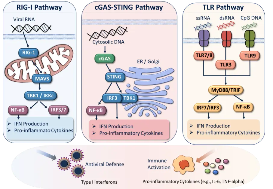
Figure 1. Core viral sensing pathways of the innate immune system. Innate immune detection of viral infection is mediated by a set of compartmentalized pattern recognition receptors (PRRs) that recognize distinct viral nucleic acid signatures. In the cytosol, RIG-I-like receptors (RIG-I and MDA5) detect viral RNA species and signal via the adaptor MAVS, leading to activation of TBK1 and IKKε and subsequent induction of interferon regulatory factors (IRF3/7) and the NF-ĸB pathway. In parallel, the cGAS–STING pathway senses cytosolic DNA and triggers STING-dependent signaling from the endoplasmic reticulum and Golgi compartments, culminating in IRF3 and NF-ĸB activation. Endosomal Toll-like receptors (TLR3, TLR7/8, and TLR9) recognize viral RNA and DNA in endosomal compartments and signal through adaptor proteins such as MyD88 and TRIF to activate both IRF and NF-κB pathways. These signaling cascades converge on the induction of type I and III interferons and pro-inflammatory cytokines, establishing an antiviral state and initiating downstream immune responses.
In parallel with RNA sensing, the cGAS–STING pathway is the central mechanism for detecting cytosolic DNA. cGAS recognizes double-stranded DNA irrespective of sequence and catalyzes the synthesis of the second messenger cyclic GMP–AMP (cGAMP), which subsequently activates STING on the endoplasmic reticulum membrane [2,20]. This pathway is essential for host defense against DNA viruses such as herpes simplex virus 1 (HSV-1), but has also been implicated in responses to certain RNA viruses through indirect mechanisms, including the release of mitochondrial DNA during infection [21]. Activation of STING leads to phosphorylation of IRF3 via TANK-binding kinase 1 (TBK1), culminating in interferon production. Notably, recent studies have revealed that dysregulation of cGAS–STING signaling can contribute not only to antiviral defense but also to inflammatory pathology, highlighting its dual role in host responses [22].
Complementing cytosolic sensing, endosomal Toll-like receptors provide an additional layer of viral recognition, particularly in specialized immune cells such as plasmacytoid dendritic cells. TLR3 detects double-stranded RNA, whereas TLR7 and TLR8 recognize single-stranded RNA, and TLR9 senses unmethylated CpG DNA motifs commonly found in viral genomes [23]. Engagement of these receptors initiates signaling via adaptor proteins such as TRIF or MyD88, leading to robust interferon production. For instance, TLR7-mediated sensing of viral RNA in plasmacytoid dendritic cells is a major source of systemic type I interferons during infections with viruses such as influenza and SARS-related coronaviruses. The importance of TLR signaling is further underscored by clinical observations that link TLR7 deficiency to severe COVID-19 in young male patients [24].
Together, these cytosolic and endosomal sensing pathways form an integrated network that enables rapid and context-dependent detection of viral infections. While each receptor exhibits specificity for particular classes of viral ligands, significant crosstalk and redundancy ensure robustness of the antiviral response. At the same time, the compartmentalization and differential expression of these sensors across cell types contribute to the diversity of innate immune responses observed in vivo. Understanding how these core sensing pathways are activated and coordinated provides the foundation for elucidating how early innate immune events shape downstream immunity and ultimately determine viral pathogenesis.
While these canonical sensing pathways have been extensively characterized, recent advances have significantly expanded our understanding of how innate immune sensing operates in space, time, and cellular context. These emerging concepts have redefined innate immunity from a linear detection system into a highly integrated and dynamic network.
3. Emerging Concepts in Innate Immune Sensing
3.1. cGAS–STING as a Central Hub Beyond DNA Sensing
Recent advances have fundamentally redefined the cGAS–STING pathway from a canonical cytosolic DNA sensor into a central integrator of innate immunity and cellular stress responses. Beyond pathogen-derived DNA, cGAS can also respond to endogenous DNA generated under conditions of cellular stress or tissue damage. This expanded sensing capacity places cGAS–STING at the intersection of infection, inflammation, and tissue homeostasis. Furthermore, STING-mediated signaling extends beyond type I interferon induction to regulate processes such as autophagy, cellular senescence, and metabolic reprogramming [25,26]. The growing recognition of cGAS as a multifunctional immune surveillance factor further highlights its broader biological significance [27].
3.2. Blurred Distinction Between Self and Non-Self Nucleic Acid Sensing
A major conceptual advance in innate immunity is the recognition that PRRs do not strictly discriminate between self and non-self nucleic acids, but instead respond to context-dependent danger signals. Therefore, the innate immune sensing is closely integrated with cellular stress responses.
Host-derived nucleic acids can therefore become immunogenic under conditions of cellular stress, tissue damage, or infection. In particular, cGAS-mediated recognition of self-DNA has been implicated in inflammatory disease and infection-associated pathology [26]. Importantly, genomic instability alone can induce cGAS-dependent interferon responses, demonstrating that innate immune activation can occur independently of infection [28]. Viral infections that induce mitochondrial damage may further promote the release of mitochondrial DNA into the cytosol, thereby amplifying innate immune activation independently of viral nucleic acids [29]. This overlap between pathogen-derived and host-derived signals highlights that the outcome of innate sensing depends not only on the nature of the ligand but also on the cellular context in which it is generated.
A related layer of regulation is provided by post-transcriptional RNA modifications, particularly A-to-I RNA editing, which can influence whether endogenous RNA remains immunologically silent or becomes recognized as a danger signal. Adenosine deaminase acting on RNA 1 (ADAR1) is a key regulator of this process, catalyzing the conversion of adenosine to inosine within double-stranded RNA structures [30]. By editing endogenous RNA, ADAR1 can reduce its recognition by cytosolic RNA sensors, thereby limiting inappropriate activation of innate immune signaling [31,32]. Conversely, impaired ADAR1-mediated RNA editing can increase the accumulation of immunostimulatory RNA species and promote MDA5–MAVS-dependent type I interferon responses and inflammatory pathology [31].
Consequently, innate immune sensing should be viewed as an integrated system that responds not only to pathogen-derived nucleic acids but also to changes in the cellular environment, providing a more nuanced framework for understanding viral pathogenesis and disease progression.
3.3. Spatial Organization of Innate Immune Signaling
Emerging evidence suggests that the spatial organization of innate immune signaling pathways represents a critical determinant of their functional specificity. Rather than functioning as static linear cascades, signaling molecules such as STING undergo tightly regulated trafficking between distinct intracellular compartments, including the endoplasmic reticulum, Golgi apparatus, and endolysosomal system. This dynamic subcellular localization enables the generation of context-dependent signaling outputs that vary according to both the site and duration of pathway activation. Notably, translocation of STING to the Golgi apparatus is essential for the recruitment and activation of TBK1 and IRF3, whereas persistent signaling from endolysosomal compartments has been linked to sustained inflammatory responses [33]. Moreover, recent studies have demonstrated that compartment-specific STING signaling can differentially regulate type I interferon production and NF-κB-dependent inflammatory pathways, thereby influencing the balance between protective antiviral immunity and pathological inflammation [26]. Collectively, these findings underscore spatial regulation as a fundamental layer of innate immune control and identify the intracellular trafficking of signaling molecules as a key factor shaping host responses and viral pathogenesis.
3.4. Post-Translational Regulation of Sensing Pathways
Another major development in the field has been the growing recognition that innate immune signaling is subject to extensive post-translational regulation. A diverse array of post-translational modifications, including ubiquitination, phosphorylation, and SUMOylation, dynamically governs the activation state, stability, intracellular trafficking, and degradation of key signaling molecules such as cGAS, STING, and their downstream effectors. Among these mechanisms, ubiquitin-mediated regulation of STING has emerged as a critical determinant of its trafficking dynamics and signaling capacity, thereby shaping both the magnitude and duration of type I interferon responses [25]. Importantly, numerous viruses have evolved sophisticated strategies to exploit these regulatory networks, frequently encoding proteins that manipulate ubiquitin-dependent signaling pathways to evade or suppress host immune defenses. The identification of these regulatory layers has revealed a previously underappreciated level of complexity within innate immune signaling, emphasizing that antiviral responses are not governed by simple on–off mechanisms but are instead finely modulated through reversible molecular modifications. Consequently, a detailed understanding of post-translational regulatory processes is essential for elucidating how innate immune responses are precisely calibrated during viral infection and how their dysregulation may contribute to disease pathogenesis.
3.5. Therapeutic Targeting of Innate Sensing Pathways
The expanding understanding of innate immune sensing mechanisms has created new opportunities for the development of targeted therapeutic strategies. Among these, pharmacological modulation of the cGAS–STING pathway has emerged as a particularly promising approach for either enhancing protective antiviral immunity or mitigating detrimental inflammatory responses. Activation of STING through synthetic agonists is being actively investigated as a means of inducing potent type I interferon responses and establishing broad-spectrum antiviral protection, whereas pharmacological inhibition of the pathway may offer therapeutic benefit in diseases characterized by aberrant or excessive inflammation [25,34]. Importantly, the translational relevance of these findings is reflected by the advancement of multiple STING-targeting compounds into clinical development, underscoring the growing interest in harnessing innate immune pathways for therapeutic purposes [35,36,37]. Collectively, these developments highlight the cGAS–STING axis as a highly tractable therapeutic target and reinforce the broader concept that innate immune signaling pathways represent key determinants of disease progression and treatment response across diverse pathological contexts.
3.6. Intercellular Communication via Immune Second Messengers
A particularly significant conceptual advance in the field of innate immunity has been the recognition of second messengers such as cyclic GMP–AMP (cGAMP) as intercellular signaling molecules that extend immune communication beyond the boundaries of individual cells. In contrast to classical cytokine-mediated signaling, cGAMP can be directly transferred between neighboring cells through gap junctions or disseminated via extracellular vesicles, thereby enabling the propagation of antiviral signals across tissues [38,39]. Through this mechanism, uninfected bystander cells can acquire an antiviral state prior to direct pathogen exposure, effectively amplifying and coordinating host defense responses at the tissue level. The evolutionary conservation of cyclic dinucleotide (CDN) signaling further underscores their fundamental importance in immune surveillance and host protection [28]. Collectively, these findings have expanded the traditional view of innate immunity by demonstrating that cell-autonomous mechanisms do not solely govern antiviral defense but also rely on extensive intercellular communication networks that facilitate coordinated responses to infection.
4. Downstream Signaling and Antiviral Programs
Activation of viral sensing pathways initiates a complex network of intracellular signaling events that translate pathogen recognition into coordinated antiviral defense programs. Central to this process is the induction of type I and type III interferons (IFNs), which act as key effector cytokines that restrict viral replication and shape the broader immune response.
Upon engagement of cytosolic receptors such as RIG-I and MDA5, or DNA sensors such as cGAS, adaptor proteins, including mitochondrial antiviral signaling protein (MAVS) and stimulator of interferon genes (STING), recruit downstream kinases, most prominently TANK-binding kinase 1 (TBK1) and IκB kinase ε (IKKε). These kinases phosphorylate interferon regulatory factors, particularly IRF3 and IRF7, enabling their dimerization and nuclear translocation, thereby driving transcription of interferon genes [40,41]. In parallel, activation of the NF-κB pathway promotes the expression of pro-inflammatory cytokines such as tumor necrosis factor (TNF), interleukin-1 (IL-1), and interleukin-6 (IL-6), thereby linking antiviral defense to inflammation [42].
Type III interferons, comprising the IFN-λ family, represent an important component of antiviral defense at epithelial barriers. Unlike type I interferons, whose receptors are broadly expressed across many cell types, the IFN-λ receptor is more selectively expressed, particularly on epithelial cells of the respiratory and gastrointestinal tracts [43,44]. This restricted receptor distribution enables type III interferons to establish potent local antiviral states while potentially limiting systemic inflammatory effects [45]. In respiratory viral infections, IFN-λ signaling therefore provides an important first-line defense at mucosal surfaces by inducing interferon-stimulated genes [46]. However, its effects are highly dependent on the timing and duration of signaling, as sustained IFN-λ responses may interfere with epithelial repair during certain infections. Type III interferons are important components of the balance between antiviral defense and immunopathology at respiratory barriers [47].
The secretion of interferons initiates a potent autocrine and paracrine signaling cascade through the engagement of interferon receptors and activation of the Janus kinase–signal transducer and activator of transcription (JAK–STAT) pathway. This leads to the transcriptional induction of hundreds of interferon-stimulated genes (ISGs), many of which exert direct antiviral effects. For example, the dynamin-like GTPase MxA inhibits replication of a broad range of RNA viruses, including influenza virus, by interfering with viral ribonucleoprotein complexes [48], while protein kinase R (PKR) limits viral protein synthesis through phosphorylation of eIF2α, thereby shutting down translation in infected cells [49]. Similarly, the 2′,5′-oligoadenylate synthetase (OAS)–RNase L pathway degrades viral RNA, contributing to viral clearance [50,51]. These effector mechanisms collectively establish an intracellular environment that is hostile to viral replication.
Innate immune activation can also engage inflammasome pathways, providing an additional mechanism linking pathogen sensing to inflammatory cytokine production and cell death. Among these, the NLRP3 inflammasome has been implicated in responses to a broad range of viral infections and promotes the maturation and release of interleukin-1β (IL-1β) and IL-18 through caspase-1 activation. AIM2, in contrast, senses cytosolic double-stranded DNA and can therefore provide an additional surveillance mechanism during infections with DNA viruses or infections associated with host DNA damage. Inflammasome activation can contribute to antiviral defense by promoting inflammatory cell recruitment and removal of infected cells, but excessive or prolonged activation may also exacerbate tissue injury and immunopathology [52,53].
In addition to inflammasome activation, innate immune sensing can influence viral pathogenesis through regulated forms of cell death. Z-DNA binding protein 1 (ZBP1) has emerged as an important sensor of viral and endogenous nucleic acid structures that can activate programmed cell death pathways, including necroptosis, apoptosis, and pyroptosis [54]. This mechanism is particularly relevant during influenza A virus infection, where ZBP1-dependent signaling can promote inflammatory cell death and contribute to both antiviral defense and tissue pathology [55]. Related mechanisms have also been implicated in SARS-CoV-2 infection, suggesting that dysregulated activation of regulated cell death pathways may represent an important link between viral sensing and tissue injury [56,57]. These findings further illustrate that innate immune sensing influences not only interferon and cytokine responses but also the fate of infected and neighboring cells [58].
Beyond their direct antiviral functions, downstream innate immune signaling pathways orchestrate a broad range of immunological programs that critically influence disease outcome. In particular, type I and type III interferons not only establish antiviral states within infected and neighboring cells but also promote antigen presentation, enhance dendritic cell maturation, and regulate the activation and differentiation of T- and B-lymphocyte populations, thereby providing a crucial mechanistic link between innate and adaptive immunity [59]. Concurrently, inflammatory signaling pathways, most prominently those mediated by NF-κB, contribute to host defense through the induction of cytokines and chemokines that facilitate immune cell recruitment and pathogen clearance. However, the effects of inflammation are highly context-dependent, as excessive or prolonged cytokine production can result in tissue damage and immunopathology. This dual nature of inflammatory signaling is exemplified by severe viral infections such as influenza and COVID-19, in which elevated concentrations of IL-6 and other pro-inflammatory mediators have been associated with increased disease severity and adverse clinical outcomes [60].
Given the potentially deleterious consequences of uncontrolled immune activation, the magnitude and duration of downstream signaling are subject to stringent regulatory control. Multiple negative feedback mechanisms operate to maintain immune homeostasis, including the actions of suppressor of cytokine signaling (SOCS) proteins and ubiquitin-modifying enzymes such as A20, which attenuate both interferon- and NF-κB-dependent signaling pathways [61]. Failure of these regulatory networks can result in dysregulated cytokine production and persistent inflammatory responses, highlighting the critical importance of balanced signal transduction for effective host defense while minimizing collateral tissue damage.
Collectively, downstream signaling pathways serve as central integrators that translate early pathogen recognition into a coordinated spectrum of antiviral, inflammatory, and immunomodulatory responses. The dynamic interplay between interferon-mediated antiviral programs, inflammatory signaling cascades, and regulatory feedback mechanisms ultimately determines whether viral infection is efficiently controlled or progresses toward immunopathology. As such, these pathways represent a critical interface linking pathogen detection to the clinical outcome of infection.
5. Innate Immune Sensing as a Determinant of Clinical Outcomes in Viral Infection
Clinical studies across diverse viral infections have established innate immune sensing pathways as key determinants of disease severity, progression, and patient outcomes. In particular, the integrity and timing of early interferon responses downstream of pattern recognition receptors (PRRs) have emerged as critical factors distinguishing mild from severe disease. Evidence from human cohorts, genetic studies, and interventional trials consistently demonstrates that defects in innate sensing are directly linked to adverse clinical phenotypes, thereby bridging molecular immunology with patient-level outcomes.
Human genetic and immunological studies well illustrate the clinical relevance of impaired innate sensing. The identification of pathogenic variants affecting innate immune regulators, including TLR7 mutations associated with severe COVID-19, provides a rationale for considering host genetic background when assessing disease risk and therapeutic responses [24]. Clinical studies further suggest that the therapeutic effects of interferon administration depend on the timing of treatment, with early intervention yielding greater benefit than delayed administration in some settings [62,63,64] (Figure 2). Moreover, some studies of severe influenza further indicate that inflammatory responses contribute to disease severity, with elevated cytokine levels correlating with mortality independently of viral burden [65,66]. Comparable mechanisms have been described in other viral hemorrhagic fevers, where sustained activation of innate immune pathways promotes systemic inflammation, tissue injury, and multi-organ dysfunction [67,68].
In parallel, increasing evidence has established host genetic variation within innate immune pathways as a critical factor influencing susceptibility to severe viral disease. Mutations and functional deficiencies affecting key interferon regulatory factors, including IRF3, IRF7, and IRF9, have been linked to severe influenza in otherwise immunocompetent individuals, demonstrating that even subtle impairments in innate immune sensing or downstream signaling can profoundly alter disease progression and clinical outcome [69,70]. These observations underscore the substantial interindividual variability in antiviral immunity and provide a strong rationale for the integration of immunogenetic information into precision medicine approaches, including risk assessment, patient stratification, and the development of individualized therapeutic strategies.
Collectively, clinical and translational evidence supports a unifying framework in which innate immune sensing pathways serve as critical gatekeepers of disease outcome during viral infection. Rapid and appropriately regulated activation of these pathways promotes effective viral control and favorable clinical trajectories, whereas impaired, delayed, or dysregulated responses can result in uncontrolled viral replication, excessive inflammation, and progressive tissue damage. The direct association between defined molecular defects in innate immune signaling and distinct clinical phenotypes further highlights the translational relevance of these pathways. Consequently, integrating mechanistic insights from innate immunology with clinical and genetic data not only advances our understanding of viral pathogenesis but also provides a foundation for developing novel biomarkers and targeted therapeutic interventions to improve patient outcomes.
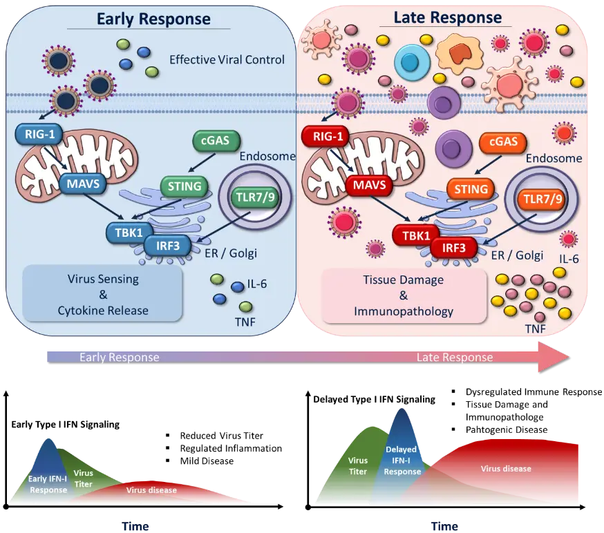
Figure 2. Conceptual model of temporal regulation of innate immune responses during viral infection. Early PRR activation promotes interferon production and viral control, whereas delayed or dysregulated responses may permit viral expansion and contribute to excessive inflammatory signaling, tissue damage, and immunopathology.
6. Viral Evasion of Innate Immune Sensing
A defining feature of successful viral pathogens is their ability to evade or actively subvert host innate immune sensing pathways. Given the central role of pattern recognition receptors (PRRs) in initiating antiviral responses, viruses have evolved diverse and often highly specific strategies to interfere with both the detection of viral components and the downstream signaling cascades that lead to interferon production. These immune evasion mechanisms are critical determinants of viral replication efficiency, transmission, and ultimately disease severity.
One common strategy employed by RNA viruses is the modification or concealment of viral nucleic acids to avoid recognition by cytosolic sensors such as RIG-I and MDA5. For instance, many viruses cap their RNA or remove 5′-triphosphate groups to mimic host mRNA structures, thereby escaping detection by RIG-I [18]. Coronaviruses, including severe acute respiratory syndrome coronavirus 2 (SARS-CoV-2), encode nonstructural proteins that mediate RNA capping and methylation, effectively masking viral RNA from host sensors [71]. In addition, replication of viral genomes within membrane-bound compartments can physically shield viral RNA intermediates from cytosolic recognition, as observed in flaviviruses and coronaviruses [72,73].
Beyond passive evasion, many viruses actively antagonize PRR signaling pathways by targeting key host proteins. Influenza A virus provides a well-characterized example through its nonstructural protein 1 (NS1), which inhibits RIG-I signaling by binding to viral RNA and interfering with downstream activation of interferon regulatory factor 3 (IRF3) [74]. Similarly, hepatitis C virus (HCV) disrupts RIG-I-mediated signaling by cleaving the adaptor protein MAVS via its NS3/4A protease, thereby preventing the induction of interferons [75]. These targeted disruptions highlight the evolutionary pressure on viruses to disable critical nodes within innate immune pathways.
The central role of the cGAS–STING pathway in antiviral immunity is further underscored by the extensive range of viral strategies that have evolved to antagonize its activation. DNA viruses, as well as several RNA viruses, actively target components of this pathway to suppress innate immune recognition and facilitate viral replication. Herpes simplex virus type 1 (HSV-1) represents a prominent example of such immune evasion, employing multiple viral proteins to disrupt cGAS–STING signaling and thereby attenuate type I interferon (IFN-I) responses [76,77]. Similarly, recent studies have demonstrated that SARS-CoV-2 encodes proteins, including ORF9b and NSP13, that interfere with interferon induction and signaling pathways, highlighting a remarkable convergence of immune evasion mechanisms across phylogenetically distinct virus families [78,79]. The recurrent targeting of the cGAS–STING axis by diverse viruses emphasizes its importance as a critical hub of host antiviral defense.
Beyond disrupting pathogen sensing itself, viruses frequently manipulate downstream signaling networks to broadly suppress antiviral immune programs. Inhibition of key transcription factors such as IRF3 and NF-κB prevents the induction of both interferons and pro-inflammatory cytokines, thereby limiting the establishment of effective innate immune responses. Furthermore, many viruses interfere with the Janus kinase–signal transducer and activator of transcription (JAK–STAT) pathway, which mediates the cellular response to secreted interferons. A well-characterized example is provided by paramyxoviruses, whose V proteins promote the degradation of STAT family members and consequently impair interferon-stimulated gene expression [80]. Through such mechanisms, viruses not only evade initial immune detection but also circumvent the antiviral state induced in surrounding, uninfected cells.
Collectively, these observations illustrate the dynamic co-evolution of host innate immune defenses and viral countermeasures. The continuous selective pressure exerted by immune surveillance has driven the emergence of highly specialized viral strategies that exploit regulatory vulnerabilities within host signaling pathways. Importantly, the efficiency, specificity, and breadth of these immune evasion mechanisms vary considerably among viral species and are often closely associated with differences in virulence, tissue tropism, and disease severity. Consequently, elucidating the molecular basis of viral immune evasion not only provides fundamental insights into virus–host interactions but also identifies promising targets for therapeutic intervention to restore innate immune function and enhance antiviral protection.
7. From Protection to Pathology
Innate immune sensing pathways are essential for the rapid control of viral infections. However, their activation must be tightly regulated to prevent collateral tissue damage. A growing body of evidence demonstrates that the same molecular mechanisms that mediate antiviral protection can, under conditions of dysregulation, drive immunopathology and contribute significantly to disease severity. This dual role underscores the concept that innate immune responses operate within a narrow therapeutic window, where both insufficient and excessive activation can be detrimental to the host.
Protective antiviral immunity is typically characterized by rapid and transient activation of pattern recognition receptor (PRR) signaling, leading to timely production of type I and III interferons and the induction of interferon-stimulated genes (ISGs) that restrict viral replication (Figure 3). Early interferon (IFN) responses are critical in limiting viral replication and improving clinical outcomes by initiating innate antiviral defenses, reducing lung inflammation, and controlling viral loads in respiratory and other infections [81,82,83,84]. Thus, delayed or impaired IFN responses often lead to increased viral persistence, excessive inflammation, and higher disease severity [85]. However, when these responses are delayed, prolonged, or excessive, they can contribute to tissue damage and impaired organ function. Importantly, the consequences of innate immune activation depend not only on its timing but also on its duration. Sustained type I interferon signaling can contribute to lung pathology by promoting inflammatory cell infiltration and epithelial damage [6].
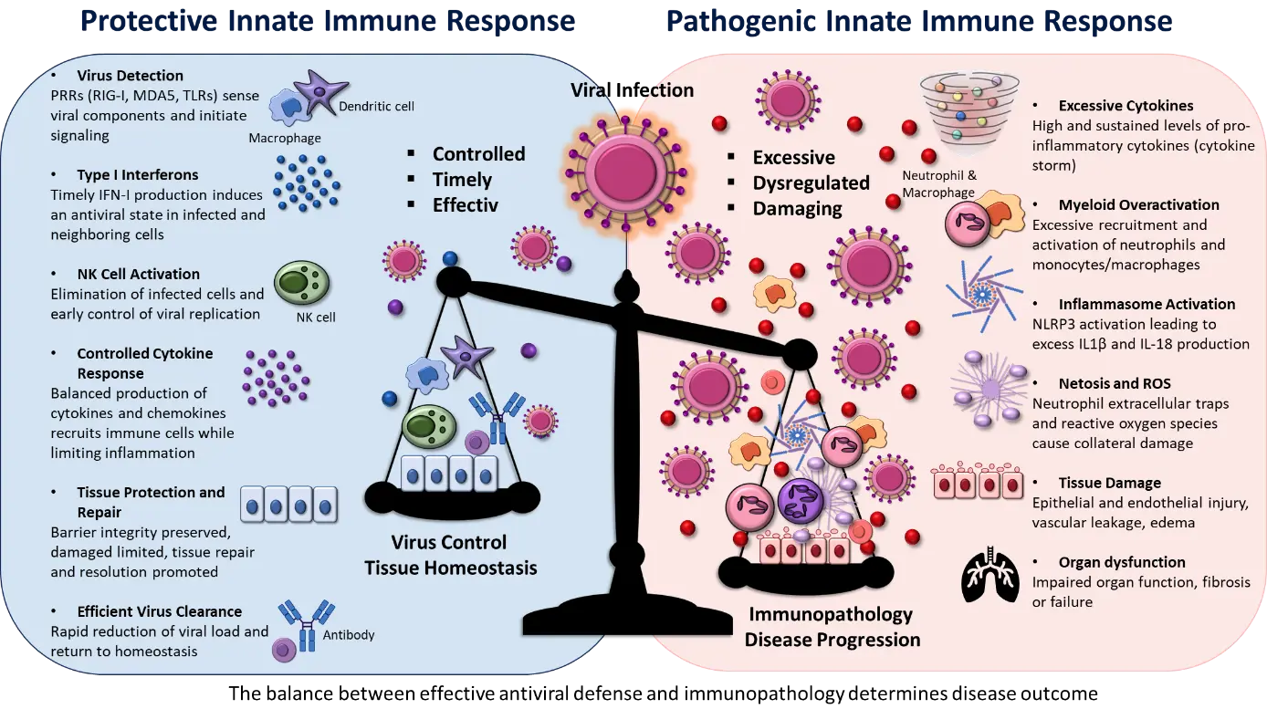
Figure 3. Protective versus pathogenic innate immune responses to viral infection. This figure illustrates how the balance between early protective responses and late inflammatory processes determines disease severity. Innate immune sensing pathways can mediate both protective antiviral immunity and pathological inflammation, depending on how the response is regulated. In the protective scenario, early and controlled activation of PRRs leads to the timely production of interferons and interferon-stimulated genes (ISGs), as well as immune cell activation, resulting in efficient viral clearance and limited inflammation. In contrast, the pathogenic response is characterized by delayed or dysregulated sensing, excessive cytokine production (cytokine storm), and uncontrolled immune activation. Elevated levels of pro-inflammatory cytokines drive the recruitment and overactivation of immune cells, contributing to tissue damage and immunopathology. This dichotomy highlights the importance of tightly regulated innate immune responses in determining the balance between host protection and disease progression.
A prominent example of the pathological consequences of dysregulated innate immunity is the development of hyperinflammatory responses, often referred to as “cytokine storms”. In severe cases of coronavirus disease 2019 (COVID-19), patients exhibit elevated levels of pro-inflammatory cytokines, including interleukin-6 (IL-6), tumor necrosis factor (TNF), and chemokines that drive the recruitment of inflammatory monocytes and neutrophils to the lung [60]. Notably, this hyperinflammatory state frequently occurs in the context of impaired early interferon responses, suggesting a temporal disconnect between antiviral defense and inflammatory signaling. Similar patterns have been observed in infections with highly pathogenic influenza strains (e.g., H5N1, H7N9, 1918 H1N1), where excessive cytokine production (cytokine storm) contributes to acute respiratory distress syndrome (ARDS) and increased mortality [86,87,88]. This uncontrolled inflammatory reaction leads to severe damage of the lung epithelial–endothelial barrier, resulting in massive lung edema, reduced gas exchange, and eventual multi-organ failure [89].
Beyond driving systemic inflammation, innate immune sensing pathways can directly contribute to virus-specific pathogenic processes, highlighting the complex relationship between host defense and disease progression.
In this context, innate immune activation represents a double-edged sword, as signaling pathways that normally promote antiviral defense can simultaneously contribute to vascular leakage, tissue injury, and clinical deterioration.
The cellular and spatial organization of host responses further influences the pathological consequences of innate immune activation. Within the respiratory tract and other infected tissues, epithelial cells, resident macrophages, and recruited leukocyte populations collectively generate a highly dynamic inflammatory microenvironment. While localized activation of these cells is essential for effective viral clearance, excessive or widespread immune activation can compromise tissue architecture and impair organ function. In respiratory infections, for example, uncontrolled inflammation may disrupt epithelial barrier integrity and impair gas exchange, thereby exacerbating disease severity. Furthermore, sustained activation of innate immune pathways can interfere with tissue regeneration and repair processes, prolonging recovery and increasing susceptibility to secondary complications.
To counterbalance these potentially detrimental effects, multiple regulatory mechanisms have evolved to constrain inflammatory signaling and restore tissue homeostasis. Negative feedback networks, including suppressor of cytokine signaling (SOCS) proteins and other inhibitory regulators, function to attenuate interferon- and cytokine-mediated signaling pathways once an effective immune response has been established [61]. Disruption of these regulatory circuits, whether resulting from genetic susceptibility, age-related factors, or overwhelming viral burden, can promote persistent immune activation and drive the transition from protective immunity to pathological inflammation.
Collectively, these observations support the concept that innate immune sensing pathways are neither inherently protective nor intrinsically harmful. Rather, their impact on disease outcome is highly context dependent. The precise regulation of the timing, magnitude, duration, and cellular distribution of innate immune responses determines the balance between viral control and immunopathology. Consequently, elucidating the mechanisms governing this balance remains a central challenge in the study of viral pathogenesis and is essential for developing therapeutic strategies that maximize antiviral efficacy while minimizing immune-mediated tissue damage.
8. Therapeutic Implications and Future Directions
The central role of innate immune sensing pathways in determining viral pathogenesis has important implications for therapeutic intervention. Rather than targeting viral components directly, host-directed approaches that modulate innate immune signaling may provide complementary strategies with the potential for broad-spectrum antiviral activity. However, because innate immune pathways contribute to both antiviral protection and immunopathology, therapeutic modulation requires careful consideration of the timing, magnitude, and cellular context of immune activation. Strategies that enhance innate responses may be beneficial during the early stages of infection, whereas inhibition of excessive downstream inflammation may be more appropriate once viral replication is controlled but immune-mediated tissue damage persists.
One of the most extensively investigated approaches involves therapeutic modulation of interferon responses. Type I interferons have demonstrated antiviral activity against a broad range of viral infections by inducing interferon-stimulated genes (ISGs) and establishing an antiviral state [59]. Importantly, clinical and experimental studies indicate that the therapeutic efficacy of interferon-based interventions is strongly dependent on timing. Early administration may enhance viral control, whereas delayed treatment can provide limited benefit or, in some settings, contribute to inflammatory pathology, as observed in influenza and SARS-CoV-2 infections [4,47,81,83]. These findings emphasize that therapeutic interferon administration should be considered in relation to the temporal dynamics of infection rather than as a uniformly beneficial intervention.
Type III interferons may represent an additional therapeutic avenue, particularly at epithelial barriers. Their more restricted receptor distribution compared with type I interferons enables potent antiviral activity at mucosal surfaces while potentially limiting systemic inflammatory effects. This property may be particularly relevant for respiratory viral infections, although the therapeutic window and effects on epithelial repair require further investigation. Together, these observations highlight the importance of temporally and spatially optimized interferon-based strategies.
Beyond interferon-based approaches, direct pharmacological modulation of upstream innate immune sensors represents a promising strategy for antiviral therapy. Agonists targeting cGAS–STING, RIG-I-like receptors, and TLRs have been developed to enhance innate immune activation and interferon production. However, the current translational landscape differs substantially between these pathways. While several innate immune agonists have entered clinical development, their clinical evaluation has largely occurred in oncology rather than in patients with viral infections. The underlying pharmacological principles may also be relevant to antiviral therapy. This translational gap highlights an important area for future investigation, particularly given the context-dependent effects of innate immune activation on viral control and immunopathology. Consequently, these studies provide important evidence that innate immune pathways can be pharmacologically targeted in humans, thereby establishing their clinical tractability.
For the cGAS–STING pathway, several agonists, including ADU-S100, MK-1454, IMSA101, and TAK-676, have undergone early-phase clinical evaluation, predominantly in patients with solid tumors (Table 1). These studies have primarily addressed safety, tolerability, and pharmacodynamic activity rather than antiviral efficacy. Accordingly, their relevance to viral disease lies mainly in demonstrating the feasibility of pharmacologically activating STING in humans. In contrast, antiviral applications of STING agonists remain predominantly preclinical [90]. For example, ADU-S100 binds directly to the STING protein and activates it. Although it demonstrated strong local immune activation, clinical efficacy (tumor shrinkage) in advanced solid tumors fell short of expectations, leading to a temporary pause in its development as a monotherapy. However, current research continues to investigate modified combination therapies (NCT03937141, NCT02675439, or with bispecific antibodies such as CLDN18.2) [91]. Initial data from a Phase I study (NCT03010176) showed that MK-1454 yielded few objective clinical rates of tumor reduction as a monotherapy. However, response rates increased when combined with the anti-PD-1 antibody pembrolizumab [92]. In a Phase I/II clinical trial (NCT04020185) involving patients with advanced solid tumors, IMSA101 (both as a monotherapy and in combination with a checkpoint inhibitor) was found to be well tolerated [36]. However, similar to ADU-S100 or MK-1454, as a monotherapy, it demonstrated only minimal intrinsic antitumor activity.
TAK-676 has been investigated in combination with pembrolizumab in several clinical trials (including Phase I/II NCT04420884 and Phase I NCT04879849). Unlike the previously described agonists, TAK-676 acts systemically. GSK3745417 is a synthetic STING agonist developed by GlaxoSmithKline (GSK). Unlike classical agents such as ADU-S100 or IMSA101, it is a non-CDN molecule based on an amidobenzimidazole (diABZI) scaffold. A first-in-human Phase I study (NCT03843359) is investigating the safety and dosing of GSK3745417, both as a monotherapy and in combination with the immune checkpoint inhibitor dostarlimab, in patients with advanced, treatment-refractory solid tumors.
Beyond immuno-oncology, diABZI has proven to be a potent antiviral agent in the field of infectious diseases. By rapidly activating cellular interferon defenses, diABZI effectively blocks the replication of a wide range of viruses. Studies demonstrate strong inhibition of SARS-CoV-2 variants in human lung cells, as well as complete protection against herpes simplex encephalitis (HSE) in animal models [93]. Like diABZI, SR-717 is also being investigated outside the field of oncology due to its potent ability to upregulate the innate interferon response. Studies show that prophylactic or early therapeutic administration of SR-717 can effectively protect cells against replication by RNA viruses (such as SARS-CoV-2) [94,95].
Conversely, inhibition of STING signaling may be beneficial in contexts where excessive activation contributes to inflammatory pathology. H-151, C-176, C-178, and SN-011 are small, synthetic molecules and represent highly specific STING inhibitors. They were developed to treat life-threatening autoimmune diseases, chronic inflammation, and excessive immune responses (such as cytokine storms) [96].
Table 1. Selected cGAS–STING modulators and their translational relevance to viral infections.
|
Compound |
Target/Pathway |
Development Stage |
Main Evidence/Context |
Relevance to Viral Infections |
|---|---|---|---|---|
|
ADU-S100 (MIW815) |
STING agonist |
Clinical development |
Primarily Solid tumors |
Demonstrates clinical tractability of pharmacological STING activation; antiviral applications remain preclinical |
|
MK-1454 |
STING agonist |
Clinical development |
Primarily Solid tumors, lymphoma |
Provides evidence for translational development of STING agonism |
|
TAK-676 |
STING agonist |
Clinical development |
Primarily Solid tumors |
Supports the feasibility of pharmacological STING modulation; antiviral potential remains investigational |
|
IMSA101 |
STING agonist |
Clinical development |
Primarily Solid tumors |
Demonstrates clinical development of STING-directed therapies |
|
GSK3745417 |
STING agonist |
Clinical development |
Primarily Solid tumors, AML, MDS |
Supports target tractability; no established clinical antiviral indication |
|
diABZI |
STING agonist |
Preclinical |
Antiviral activity reported in experimental infection models, Immuno-oncology |
Directly relevant to antiviral applications; investigated against multiple viral pathogens |
|
SR-717 |
STING agonist |
Preclinical |
Immunomodulatory and antiviral activity |
Supports the potential application of STING activation in viral infection |
|
STING inhibition |
STING |
Preclinical |
Investigated primarily in inflammatory and Autoimmune settings |
Potential strategy for limiting immunopathology during infections characterized by excessive innate immune activation |
Importantly, although STING agonists have been extensively investigated in oncology, their clinical translation as antiviral therapies remains limited, with current evidence for viral infections largely restricted to preclinical studies.
A similar distinction applies to RIG-I-directed approaches. RIG-I agonists such as MK-4621 and CV8102 have entered early clinical development, primarily in oncology, providing evidence that pharmacological activation of RIG-I is feasible in humans. However, antiviral applications remain largely preclinical. Experimental RNA ligands, including SLR14, have shown protective effects in models of SARS-CoV-2 infection, supporting the potential of RIG-I activation as an antiviral strategy [97,98,99].
TLR-directed approaches are comparatively more advanced clinically, with several agonists already approved for use as vaccine adjuvants and in topical treatments. Nevertheless, their therapeutic use in viral infections remains highly context dependent because TLR activation can promote antiviral defense but may also amplify inflammatory responses. Consequently, modulation of TLR signaling may require careful adjustment of receptor specificity, dose, and timing rather than indiscriminate activation of innate immunity [100,101].
In addition to enhancing antiviral sensing, therapeutic strategies may also aim to attenuate excessive downstream inflammation. This approach is particularly relevant in severe viral infections in which tissue injury is driven not only by viral replication but also by dysregulated host immune responses. Clinical experience from COVID-19 has demonstrated that inhibition of inflammatory signaling can improve outcomes in selected patient populations. In particular, blockade of interleukin-6 (IL-6) signaling with monoclonal antibodies (tocilizumab, sarilumab, and siltuximab) has been associated with reduced disease progression and mortality in severe disease, supporting a pathogenic role for excessive cytokine production [102].
Similarly, Janus kinase (JAK) inhibitors have been used to attenuate cytokine signaling downstream of multiple inflammatory receptors. Clinical studies with agents including baricitinib and other JAK inhibitors have provided further evidence that modulation of host inflammatory pathways can be beneficial when excessive immune activation contributes to disease severity [103]. These findings illustrate an important therapeutic complement to strategies aimed at enhancing innate antiviral responses—whereas early intervention may benefit from strengthening antiviral signaling, later stages of severe disease may require selective suppression of excessive inflammation.
Looking ahead, advances in systems immunology and high-resolution profiling technologies are providing new opportunities to define the heterogeneity of antiviral responses and identify context-specific therapeutic targets. Single-cell transcriptomics, spatial profiling, and genome-wide CRISPR-based functional screens have revealed substantial heterogeneity in innate immune responses across cell types, tissues, and disease states [104,105,106,107,108,109]. These approaches challenge the concept of a uniform antiviral response and instead emphasize that the consequences of innate immune activation depend strongly on cellular and tissue context. Increasing evidence also indicates that host genetic variation contributes to differences in susceptibility to severe viral disease. In the future, the integration of functional genomics, single-cell profiling, and clinical data may facilitate the identification of patient subgroups that are most likely to benefit from either enhancement or suppression of specific innate immune pathways. Despite these advances, several challenges remain. A major objective is to develop approaches that selectively modulate innate immune pathways while preserving their essential protective functions. The dynamic interaction between innate sensing, viral adaptation, and host-specific factors such as age, comorbidities, genetic variation, and the microbiome complicates this challenge. Furthermore, the optimal therapeutic strategy may differ depending on the stage of infection, the predominant infected cell type, and the balance between viral replication and immune-mediated tissue damage. Addressing these challenges will require integrative approaches that combine mechanistic studies with clinical, genetic, and systems-level analyses.
Collectively, current evidence supports innate immune sensing pathways as promising therapeutic targets in viral disease. However, the available evidence also indicates that therapeutic modulation cannot be reduced to simple activation or inhibition of innate immunity. Instead, successful intervention will likely depend on matching the direction, magnitude, and timing of pathway modulation to the stage and biological context of infection. Clinical experience with interferons and anti-inflammatory therapies, together with preclinical studies targeting upstream sensors such as STING and RIG-I, provides a foundation for this approach. Continued integration of molecular immunology, functional genomics, and clinical research will be important for determining how innate immune pathways can be therapeutically manipulated to enhance antiviral protection while limiting immunopathological consequences.
9. Conclusions
Viral pathogens are a common cause of illness and mortality worldwide. Because viruses are obligate intracellular parasites, they must use the cellular metabolic pathways of infected host cells to reproduce [110]. Innate immune sensing pathways have emerged as central determinants of viral pathogenesis, extending far beyond their classical role as the first line of host defense. Collectively, current evidence supports a central role for innate immune sensing in shaping viral disease trajectories. The integration of molecular, cellular, and clinical findings further emphasizes that antiviral protection and immunopathology are determined by the context in which innate pathways are activated and regulated.
A further layer of complexity arises from the dynamic interplay between host innate immune sensing mechanisms and viral immune evasion strategies, which together represent critical determinants of disease outcome. Successful viral pathogens have evolved sophisticated mechanisms to suppress, delay, or redirect innate immune detection, thereby creating a temporal window that facilitates viral replication and dissemination. However, this early immune evasion often comes at the cost of dysregulated host responses during later stages of infection, contributing to excessive inflammation and immunopathology. This phenomenon is observed across diverse viral families, including coronaviruses and flaviviruses, which encode multiple factors that target PRR-mediated signaling pathways and interferon induction cascades [14]. At the same time, accumulating evidence indicates that host-intrinsic factors substantially influence susceptibility to severe disease. In particular, genetic variation within key innate immune sensing and signaling pathways can profoundly alter antiviral responsiveness, underscoring the importance of interindividual heterogeneity and highlighting the growing relevance of personalized approaches to innate immunity.
The inherently dual nature of innate immune sensing, as both a cornerstone of antiviral defense and a potential driver of tissue damage, emphasizes the necessity for tight spatial, temporal, and quantitative regulation of these pathways. Protective immune responses depend not only on the magnitude of activation but also on the precise timing and duration of signaling events. Consequently, therapeutic strategies aimed at modulating innate immune pathways must account for the dynamic nature of host–pathogen interactions. Clinical experience with interferon-based therapies and anti-inflammatory interventions targeting excessive immune activation illustrates that therapeutic efficacy is highly dependent on the stage of infection and the underlying immunological context. These observations reinforce the concept that successful immune modulation requires a nuanced balance between enhancing antiviral protection and limiting collateral tissue injury.
Looking ahead, emerging technologies in systems immunology, single-cell biology, and functional genomics are transforming our understanding of the complexity and heterogeneity that characterize innate immune responses. These approaches provide unprecedented resolution for dissecting cellular and molecular networks involved in pathogen recognition and immune regulation, revealing previously unrecognized cell type-specific programs and regulatory circuits. Such advances are beginning to illuminate how early sensing events shape downstream immune responses and ultimately influence disease trajectories. Integrating these mechanistic insights with clinical and genetic data will be essential for establishing predictive models of disease progression and guiding the development of next-generation therapeutic strategies that enhance antiviral immunity while minimizing the risk of immunopathology.
In summary, innate immune sensing pathways occupy a central position at the nexus of viral recognition, immune regulation, and disease outcome. Far from serving merely as passive detectors of infection, these pathways function as dynamic integrators of host and pathogen-derived signals that shape the balance between protective immunity and pathological inflammation. Continued efforts to elucidate the molecular mechanisms governing their activation, regulation, and dysregulation will not only deepen our understanding of viral pathogenesis but also provide a conceptual framework for the development of innovative therapeutic approaches designed to harness the full potential of the innate immune system while preserving immune homeostasis.
Scope and Literature Search
This review focuses on recent advances in innate immune sensing during viral infections and their implications for antiviral immunity, immunopathology, and therapeutic development. Relevant literature was identified primarily through searches of PubMed and ClinicalTrials.gov, with the literature search conducted from February 2026 to August 2026 and with particular emphasis on recent mechanistic, translational and clinical studies. Search terms included combinations of innate immune sensing, pattern recognition receptors, RIG-I, MDA5, cGAS–STING, Toll-like receptors, interferons, inflammasomes, viral infection, immune evasion, immunopathology, and therapeutic targeting. Studies were considered eligible when they provided mechanistic, functional, or clinical insights into innate immune responses to viral infection, their regulation, or contribution to disease outcome, or the therapeutic modulation of these pathways. Primary research articles, clinical studies and relevant recent reviews were prioritized, whereas studies lacking a direct connection to viral innate immunity were not considered. Reference lists of key publications were additionally screened to identify relevant earlier studies and complementary literature. ClinicalTrials.gov was searched separately to identify ongoing and completed clinical studies targeting innate immune pathways.
Ethics Statement
Not applicable.
Informed Consent Statement
Not applicable.
Data Availability Statement
No new data were created or analyzed in this study. Data sharing is not applicable to this article.
Funding
This review received no external funding.
Declaration of Competing Interest
The author declares no conflicts of interest.
References
-
Kawai T, Akira S. The role of pattern-recognition receptors in innate immunity: Update on Toll-like receptors. Nat. Immunol. 2010, 11, 373–384. DOI:10.1038/ni.1863 [Google Scholar]
-
Wu J, Chen ZJ. Innate Immune Sensing and Signaling of Cytosolic Nucleic Acids. Annu. Rev. Immunol. 2014, 32, 461–488. DOI:10.1146/annurev-immunol-032713-120156 [Google Scholar]
-
Wu H, Campillo Prados M, Vaeth M. Metabolic Regulation of T Cell Exhaustion. Immune Discov. 2025, 1, 10005. DOI:10.70322/immune.2025.10005 [Google Scholar]
-
Hadjadj J, Yatim N, Barnabei L, Corneau A, Boussier J, Smith N, et al. Impaired type I interferon activity and inflammatory responses in severe COVID-19 patients. Science 2020, 369, 718–724. DOI:10.1126/science.abc6027 [Google Scholar]
-
Bastard P, Rosen LB, Zhang Q, Michailidis E, Hoffmann HH, Zhang Y, et al. Autoantibodies against type I IFNs in patients with life-threatening COVID-19. Science 2020, 370, eabd4585. DOI:10.1126/science.abd4585 [Google Scholar]
-
Davidson S, Crotta S, McCabe TM, Wack A. Pathogenic potential of interferon αβ in acute influenza infection. Nat. Commun. 2014, 5, 3864. DOI:10.1038/ncomms4864 [Google Scholar]
-
Guzman MG, Harris E. Dengue. Lancet 2015, 385, 453–465. DOI:10.1016/S0140-6736(14)60572-9 [Google Scholar]
-
Reinert LS, Lopušná K, Winther H, Sun C, Thomsen MK, Nandakumar R, et al. Sensing of HSV-1 by the cGAS–STING pathway in microglia orchestrates antiviral defence in the CNS. Nat. Commun. 2016, 7, 13348. DOI:10.1038/ncomms13348 [Google Scholar]
-
Shmuel-Galia L, Jiang Z, Stine L, Cahill S, Ng SL, Wilson R, et al. Alternative cGAS signaling promotes herpes simplex encephalitis. Proc. Natl. Acad. Sci. USA 2025, 122, e2423873122. DOI:10.1073/pnas.2423873122 [Google Scholar]
-
Lio CWJ, McDonald B, Takahashi M, Dhanwani R, Sharma N, Huang J, et al. cGAS-STING Signaling Regulates Initial Innate Control of Cytomegalovirus Infection. J. Virol. 2016, 90, 7789–7797. DOI:10.1128/JVI.01040-16 [Google Scholar]
-
Riou R, Bressollette-Bodin C, Boutoille D, Gagne K, Rodallec A, Lefebvre M, et al. Severe Symptomatic Primary Human Cytomegalovirus Infection despite Effective Innate and Adaptive Immune Responses. J. Virol. 2017, 91, e02245-16. DOI:10.1128/JVI.02245-16 [Google Scholar]
-
Lam E, Stein S, Falck-Pedersen E. Adenovirus Detection by the cGAS/STING/TBK1 DNA Sensing Cascade. J. Virol. 2014, 88, 974–981. DOI:10.1128/JVI.02702-13 [Google Scholar]
-
Anghelina D, Lam E, Falck-Pedersen E. Diminished Innate Antiviral Response to Adenovirus Vectors in cGAS/STING-Deficient Mice Minimally Impacts Adaptive Immunity. J. Virol. 2016, 90, 5915–5927. DOI:10.1128/JVI.00500-16 [Google Scholar]
-
Beachboard DC, Horner SM. Innate immune evasion strategies of DNA and RNA viruses. Curr. Opin. Microbiol. 2016, 32, 113–119. DOI:10.1016/j.mib.2016.05.015 [Google Scholar]
-
Xiao R, Zhang A. Involvement of the STING signaling in COVID-19. Front. Immunol. 2022, 13, 1006395. DOI:10.3389/fimmu.2022.1006395 [Google Scholar]
-
Chen K, Xiao F, Hu D, Ge W, Tian M, Wang W, et al. SARS-CoV-2 Nucleocapsid Protein Interacts with RIG-I and Represses RIG-Mediated IFN-β Production. Viruses 2020, 13, 47. DOI:10.3390/v13010047 [Google Scholar]
-
Hornung V, Ellegast J, Kim S, Brzózka K, Jung A, Kato H, et al. 5’-Triphosphate RNA Is the Ligand for RIG-I. Science 2006, 314, 994–997. DOI:10.1126/science.1132505 [Google Scholar]
-
Rehwinkel J, Gack MU. RIG-I-like receptors: Their regulation and roles in RNA sensing. Nat. Rev. Immunol. 2020, 20, 537–551. DOI:10.1038/s41577-020-0288-3 [Google Scholar]
-
Kato H, Takeuchi O, Sato S, Yoneyama M, Yamamoto M, Matsui K, et al. Differential roles of MDA5 and RIG-I helicases in the recognition of RNA viruses. Nature 2006, 441, 101–105. DOI:10.1038/nature04734 [Google Scholar]
-
Sun L, Wu J, Du F, Chen X, Chen ZJ. Cyclic GMP-AMP synthase is a cytosolic DNA sensor that activates the type I interferon pathway. Science 2013, 339, 786–791. DOI:10.1126/science.1232458 [Google Scholar]
-
West AP, Khoury-Hanold W, Staron M, Tal MC, Pineda CM, Lang SM, et al. Mitochondrial DNA stress primes the antiviral innate immune response. Nature 2015, 520, 553–557. DOI:10.1038/nature14156 [Google Scholar]
-
Wobma H, Shin DS, Chou J, Dedeoğlu F. Dysregulation of the cGAS-STING Pathway in Monogenic Autoinflammation and Lupus. Front. Immunol. 2022, 13, 905109. DOI:10.3389/fimmu.2022.905109 [Google Scholar]
-
Kawai T, Akira S. Innate immune recognition of viral infection. Nat. Immunol. 2006, 7, 131–137. DOI:10.1038/ni1303 [Google Scholar]
-
van der Made CI, Simons A, Schuurs-Hoeijmakers J, van den Heuvel G, Mantere T, Kersten S, et al. Presence of Genetic Variants Among Young Men With Severe COVID-19. JAMA 2020, 324, 663. DOI:10.1001/jama.2020.13719 [Google Scholar]
-
He X, Wedn A, Wang J, Gu Y, Liu H, Zhang J, et al. IUPHAR ECR review: The cGAS-STING pathway: Novel functions beyond innate immune and emerging therapeutic opportunities. Pharmacol. Res. 2024, 201, 107063. DOI:10.1016/j.phrs.2024.107063 [Google Scholar]
-
Zhang Z, Zhang C. Regulation of cGAS–STING signalling and its diversity of cellular outcomes. Nat. Rev. Immunol. 2025, 25, 425–444. DOI:10.1038/s41577-024-01112-7 [Google Scholar]
-
Chen ZJ. Discovery of cGAS as a DNA-Sensing Enzyme That Triggers Inflammation. JAMA 2024, 332, 1693. DOI:10.1001/jama.2024.17873 [Google Scholar]
-
Patel DJ, Yu Y, Xie W. cGAMP-activated cGAS–STING signaling: its bacterial origins and evolutionary adaptation by metazoans. Nat. Struct. Mol. Biol. 2023, 30, 245–260. DOI:10.1038/s41594-023-00933-9 [Google Scholar]
-
Hu MM, Shu HB. Mitochondrial DNA-triggered innate immune response: Mechanisms and diseases. Cell. Mol. Immunol. 2023, 20, 1403–1412. DOI:10.1038/s41423-023-01086-x [Google Scholar]
-
Liu C, Choi Y. ADAR1: A central regulator of dsRNA sensing in host-virus interactions. Front. Immunol. 2026, 17, 1756517. DOI:10.3389/fimmu.2026.1756517 [Google Scholar]
-
de Reuver R, Dierick E, Wiernicki B, Staes K, Seys L, De Meester E, et al. ADAR1 interaction with Z-RNA promotes editing of endogenous double-stranded RNA and prevents MDA5-dependent immune activation. Cell Rep. 2021, 36, 109500. DOI:10.1016/j.celrep.2021.109500 [Google Scholar]
-
Pfaller CK, George CX, Samuel CE. Adenosine Deaminases Acting on RNA (ADARs) and Viral Infections. Annu. Rev. Virol. 2021, 8, 239–264. DOI:10.1146/annurev-virology-091919-065320 [Google Scholar]
-
Taguchi T, Mukai K, Takaya E, Shindo R. STING Operation at the ER/Golgi Interface. Front. Immunol. 2021, 12, 646304. DOI:10.3389/fimmu.2021.646304 [Google Scholar]
-
Islam S, Islam MM, Akhand MRN, Park BY, Akanda MR. Recent advancements in cGAS-STING activation, tumor immune evasion, and therapeutic implications. Med. Oncol. 2024, 41, 291. DOI:10.1007/s12032-024-02539-7 [Google Scholar]
-
Nerdinger YG, Binder AK, Bremm F, Feuchter N, Schaft N, Dörrie J. STINGing Cancer: Development, Clinical Application, and Targeted Delivery of STING Agonists. Int. J. Mol. Sci. 2025, 26, 9008. DOI:10.3390/ijms26189008 [Google Scholar]
-
Shi J, Zhang Y, Zhao N, Seki E, Ma L, Kocic G, et al. Precision targeting of STING: Challenges, innovations, and clinical outlook for cancer therapy. Innovation 2026, 7, 101074. DOI:10.1016/j.xinn.2025.101074 [Google Scholar]
-
Fahey CG, Cordova AF, Gedeon PC, Barbie DA. Targeting STING to generate therapeutic anti-tumor immunity. Cancer Cell 2026, 44, 260–280. DOI:10.1016/j.ccell.2025.12.002 [Google Scholar]
-
Blest HTW, Chauveau L. cGAMP the travelling messenger. Front. Immunol. 2023, 14, 1150705. DOI:10.3389/fimmu.2023.1150705 [Google Scholar]
-
Gentili M, Kowal J, Tkach M, Satoh T, Lahaye X, Conrad C, et al. Transmission of innate immune signaling by packaging of cGAMP in viral particles. Science 2015, 349, 1232–1236. DOI:10.1126/science.aab3628 [Google Scholar]
-
Honda K, Takaoka A, Taniguchi T. Type I Inteferon Gene Induction by the Interferon Regulatory Factor Family of Transcription Factors. Immunity 2006, 25, 349–360. DOI:10.1016/j.immuni.2006.08.009 [Google Scholar]
-
Fitzgerald KA, McWhirter SM, Faia KL, Rowe DC, Latz E, Golenbock DT, et al. IKKε and TBK1 are essential components of the IRF3 signaling pathway. Nat. Immunol. 2003, 4, 491–496. DOI:10.1038/ni921 [Google Scholar]
-
Mao H, Zhao X, Sun SC. NF-κB in inflammation and cancer. Cell. Mol. Immunol. 2025, 22, 811–839. DOI:10.1038/s41423-025-01310-w [Google Scholar]
-
Broggi A, Ghosh S, Sposito B, Spreafico R, Balzarini F, Lo Cascio A, et al. Type III interferons disrupt the lung epithelial barrier upon viral recognition. Science 2020, 369, 706–712. DOI:10.1126/science.abc3545 [Google Scholar]
-
Lozhkov A, Dobrovolskaya O, Romanovskaya-Romanko E, Shishlyannikov S, Elpaeva E, Garshinina A, et al. Exploring the protective role of recombinant type III interferons in respiratory infections: Insights from an Mx1-deficient mouse model. Int. J. Antimicrob. Agents 2025, 66, 107491. DOI:10.1016/j.ijantimicag.2025.107491 [Google Scholar]
-
Hendricks MR, Savan R. Interferon-λ at the Center of the Storm. Immunity 2020, 53, 245–247. DOI:10.1016/j.immuni.2020.07.024 [Google Scholar]
-
Andreakos E, Salagianni M, Galani IE, Koltsida O. Interferon-λs: Front-Line Guardians of Immunity and Homeostasis in the Respiratory Tract. Front. Immunol. 2017, 8, 1232. DOI:10.3389/fimmu.2017.01232 [Google Scholar]
-
Park A, Iwasaki A. Type I and type III Interferons—Induction, Signaling, Evasion and Application to Combat COVID-19. Cell Host Microbe 2020, 27, 870–878. DOI:10.1016/j.chom.2020.05.008 [Google Scholar]
-
Haller O, Staeheli P, Schwemmle M, Kochs G. Mx GTPases: Dynamin-like antiviral machines of innate immunity. Trends Microbiol. 2015, 23, 154–163. DOI:10.1016/j.tim.2014.12.003 [Google Scholar]
-
García MA, Meurs EF, Esteban M. The dsRNA protein kinase PKR: Virus and cell control. Biochimie 2007, 89, 799–811. DOI:10.1016/j.biochi.2007.03.001 [Google Scholar]
-
Lee D, Malathi K, Okano T, Nakajima K, Cobat A, Morio T, et al. The OAS–RNase L pathway: Insights from experiments of nature. Sci. Immunol. 2026, 11, eads9407. DOI:10.1126/sciimmunol.ads9407 [Google Scholar]
-
Silverman RH. Viral Encounters with 2’,5’-Oligoadenylate Synthetase and RNase L during the Interferon Antiviral Response. J. Virol. 2007, 81, 12720–12729. DOI:10.1128/JVI.01471-07 [Google Scholar]
-
Shi W, Jin M, Chen H, Wu Z, Yuan L, Liang S, et al. Inflammasome activation by viral infection: Mechanisms of activation and regulation. Front. Microbiol. 2023, 14, 1247377. DOI:10.3389/fmicb.2023.1247377 [Google Scholar]
-
Wu N, Zheng C, Xu J, Ma S, Jia H, Yan M, et al. Race between virus and inflammasomes: Inhibition or escape, intervention and therapy. Front. Cell. Infect. Microbiol. 2023, 13, 1173505. DOI:10.3389/fcimb.2023.1173505 [Google Scholar]
-
Oh S, Lee S. Recent advances in ZBP1-derived PANoptosis against viral infections. Front. Immunol. 2023, 14, 1148727. DOI:10.3389/fimmu.2023.1148727 [Google Scholar]
-
Boyd DF, Jordan SV, Balachandran S. ZBP1-driven cell death in severe influenza. Trends Microbiol. 2025, 33, 521–532. DOI:10.1016/j.tim.2024.12.008 [Google Scholar]
-
Yin C, Balachandran S. ZBP1 inflames the SARS-CoV-2-infected lung. Cell Res. 2023, 33, 333–334. DOI:10.1038/s41422-023-00784-5 [Google Scholar]
-
Elsharkawy A, Jahantigh HR, Guglani A, Stone S, Arora K, Kumar M. Virus-specific host responses and gene signatures following infection with major SARS-CoV-2 variants of concern: Role of ZBP1 in viral clearance and lung inflammation. Front. Immunol. 2025, 16, 1557535. DOI:10.3389/fimmu.2025.1557535 [Google Scholar]
-
Yang B, Hu A, Wang T, Chen X, Ma C, Yang X, et al. SARS-CoV-2 infection induces ZBP1-dependent PANoptosis in bystander cells. Proc. Natl. Acad. Sci. USA 2025, 122, e2500208122. DOI:10.1073/pnas.2500208122 [Google Scholar]
-
Ivashkiv LB, Donlin LT. Regulation of type I interferon responses. Nat. Rev. Immunol. 2014, 14, 36–49. DOI:10.1038/nri3581 [Google Scholar]
-
Lucas C, Wong P, Klein J, Castro TBR, Silva J, Sundaram M, et al. Longitudinal analyses reveal immunological misfiring in severe COVID-19. Nature 2020, 584, 463–469. DOI:10.1038/s41586-020-2588-y [Google Scholar]
-
Yoshimura A, Naka T, Kubo M. SOCS proteins, cytokine signalling and immune regulation. Nat. Rev. Immunol. 2007, 7, 454–465. DOI:10.1038/nri2093 [Google Scholar]
-
Monk PD, Marsden RJ, Tear VJ, Brookes J, Batten TN, Mankowski M, et al. Safety and efficacy of inhaled nebulised interferon beta-1a (SNG001) for treatment of SARS-CoV-2 infection: A randomised, double-blind, placebo-controlled, phase 2 trial. Lancet Respir. Med. 2021, 9, 196–206. DOI:10.1016/S2213-2600(20)30511-7 [Google Scholar]
-
Sardiah S. The Role of Interferon Beta Administration in COVID-19 Treatment. Am. J. Biomed. Sci. Res. 2021, 14, 310–311. DOI:10.34297/AJBSR.2021.14.002001 [Google Scholar]
-
Wang N, Zhan Y, Zhu L, Hou Z, Liu F, Song P, et al. Retrospective Multicenter Cohort Study Shows Early Interferon Therapy Is Associated with Favorable Clinical Responses in COVID-19 Patients. Cell Host Microbe 2020, 28, 455–464.e2. DOI:10.1016/j.chom.2020.07.005 [Google Scholar]
-
Liu Q, Zhou YH, Yang ZQ. The cytokine storm of severe influenza and development of immunomodulatory therapy. Cell. Mol. Immunol. 2016, 13, 3–10. DOI:10.1038/cmi.2015.74 [Google Scholar]
-
Chang D, Dela Cruz C, Sharma L. Beneficial and Detrimental Effects of Cytokines during Influenza and COVID-19. Viruses 2024, 16, 308. DOI:10.3390/v16020308 [Google Scholar]
-
de la Fuente A, López-Sánchez J, Vaquero-Roncero LM, Merino García M, Sánchez Barrado ME, Sánchez-Hernández MV, et al. Synergistic impact of innate immunity hyper-activation and endothelial dysfunction on the magnitude of organ failure in the infection-sepsis continuum. Int. J. Infect. Dis. 2024, 146, 107142. DOI:10.1016/j.ijid.2024.107142 [Google Scholar]
-
Chen L, Deng H, Cui H, Fang J, Zuo Z, Deng J, et al. Inflammatory responses and inflammation-associated diseases in organs. Oncotarget 2018, 9, 7204–7218. DOI:10.18632/oncotarget.23208 [Google Scholar]
-
Zhang Q, Bastard P, Liu Z, Le Pen J, Moncada-Velez M, Chen J, et al. Inborn errors of type I IFN immunity in patients with life-threatening COVID-19. Science 2020, 370, eabd4570. DOI:10.1126/science.abd4570 [Google Scholar]
-
Hernandez N, Melki I, Jing H, Habib T, Huang SSY, Danielson J, et al. Life-threatening influenza pneumonitis in a child with inherited IRF9 deficiency. J. Exp. Med. 2018, 215, 2567–2585. DOI:10.1084/jem.20180628 [Google Scholar]
-
Züst R, Cervantes-Barragan L, Habjan M, Maier R, Neuman BW, Ziebuhr J, et al. Ribose 2′-O-methylation provides a molecular signature for the distinction of self and non-self mRNA dependent on the RNA sensor Mda5. Nat. Immunol. 2011, 12, 137–143. DOI:10.1038/ni.1979 [Google Scholar]
-
van den Elsen K, Quek JP, Luo D. Molecular Insights into the Flavivirus Replication Complex. Viruses 2021, 13, 956. DOI:10.3390/v13060956 [Google Scholar]
-
Doyle N, Simpson J, Hawes PC, Maier HJ. Coronavirus RNA Synthesis Takes Place within Membrane-Bound Sites. Viruses 2021, 13, 2540. DOI:10.3390/v13122540 [Google Scholar]
-
García-Sastre A, Egorov A, Matassov D, Brandt S, Levy DE, Durbin JE, et al. Influenza A Virus Lacking the NS1 Gene Replicates in Interferon-Deficient Systems. Virology 1998, 252, 324–330. DOI:10.1006/viro.1998.9508 [Google Scholar]
-
Li XD, Sun L, Seth RB, Pineda G, Chen ZJ. Hepatitis C virus protease NS3/4A cleaves mitochondrial antiviral signaling protein off the mitochondria to evade innate immunity. Proc. Natl. Acad. Sci. USA 2005, 102, 17717–17722. DOI:10.1073/pnas.0508531102 [Google Scholar]
-
Deng L, Xu Z, Li F, Zhao J, Jian Z, Deng H, et al. Insights on the cGAS-STING Signaling Pathway During Herpesvirus Infections. Front. Immunol. 2022, 13, 931885. DOI:10.3389/fimmu.2022.931885 [Google Scholar]
-
Wang A, Peng Q, Fan H, Ji W, Lou J, Zhou X, et al. Herpes simplex virus 1 encodes a STING antagonist that can be therapeutically targeted. Cell Rep. Med. 2025, 6, 102051. DOI:10.1016/j.xcrm.2025.102051 [Google Scholar]
-
Han L, Zhuang M, Deng J, Zheng Y, Zhang J, Nan M, et al. SARS‐CoV‐2 ORF9b antagonizes type I and III interferons by targeting multiple components of the RIG‐I/MDA‐5–MAVS, TLR3–TRIF, and cGAS–STING signaling pathways. J. Med. Virol. 2021, 93, 5376–5389. DOI:10.1002/jmv.27050 [Google Scholar]
-
Xia H, Cao Z, Xie X, Zhang X, Chen JYC, Wang H, et al. Evasion of Type I Interferon by SARS-CoV-2. Cell Rep. 2020, 33, 108234. DOI:10.1016/j.celrep.2020.108234 [Google Scholar]
-
Didcock L, Young DF, Goodbourn S, Randall RE. The V Protein of Simian Virus 5 Inhibits Interferon Signalling by Targeting STAT1 for Proteasome-Mediated Degradation. J. Virol. 1999, 73, 9928–9933. DOI:10.1128/JVI.73.12.9928-9933.1999 [Google Scholar]
-
Iwasaki A, Pillai PS. Innate immunity to influenza virus infection. Nat. Rev. Immunol. 2014, 14, 315–328. DOI:10.1038/nri3665 [Google Scholar]
-
Li K, McCaw JM, Cao P. Enhanced viral infectivity and reduced interferon production are associated with high pathogenicity for influenza viruses. PLoS Comput. Biol. 2023, 19, e1010886. DOI:10.1371/journal.pcbi.1010886 [Google Scholar]
-
Feng E, Balint E, Poznanski SM, Ashkar AA, Loeb M. Aging and Interferons: Impacts on Inflammation and Viral Disease Outcomes. Cells 2021, 10, 708. DOI:10.3390/cells10030708 [Google Scholar]
-
Bencze D, Fekete T, Pázmándi K. Correlation between Type I Interferon Associated Factors and COVID-19 Severity. Int. J. Mol. Sci. 2022, 23, 10968. DOI:10.3390/ijms231810968 [Google Scholar]
-
Metcalf J, Alexander J, Booth JL, Miller C, Drevets D, Wu W. Impaired Early Interferon Responses to Influenza Virus Infection in Aged Mice are Associated With Increased Pulmonary Inflammation. J. Immunol. 2024, 212, 0940_5652–0940_5652. DOI:10.4049/jimmunol.212.supp.0940.5652 [Google Scholar]
-
Gu Y, Zuo X, Zhang S, Ouyang Z, Jiang S, Wang F, et al. The Mechanism behind Influenza Virus Cytokine Storm. Viruses 2021, 13, 1362. DOI:10.3390/v13071362 [Google Scholar]
-
Niles MA, Kronhart S, Decker KE, Gogesch P, Sawatsky B, Stock S, et al. Influenza A virus-induced interferon-alpha production by myeloid dendritic cells and macrophages requires productive infection. Emerg. Microbes Infect. 2025, 14, 2556718. DOI:10.1080/22221751.2025.2556718 [Google Scholar]
-
Oldstone MBA, Rosen H. Cytokine Storm Plays a Direct Role in the Morbidity and Mortality from Influenza Virus Infection and is Chemically Treatable with a Single Sphingosine-1-Phosphate Agonist Molecule. Curr. Top. Microbiol. Immunol. 2014, 378, 129–147. DOI:10.1007/978-3-319-05879-5_6 [Google Scholar]
-
Short KR, Veldhuis Kroeze EJB, Reperant LA, Richard M, Kuiken T. Influenza virus and endothelial cells: A species specific relationship. Front. Microbiol. 2014, 5, 653. DOI:10.3389/fmicb.2014.00653 [Google Scholar]
-
Decout A, Katz JD, Venkatraman S, Ablasser A. The cGAS–STING pathway as a therapeutic target in inflammatory diseases. Nat. Rev. Immunol. 2021, 21, 548–569. DOI:10.1038/s41577-021-00524-z [Google Scholar]
-
Zhou T. A phase 2 study of STING agonist ADU-S100 combined with a CLDN18.2 bispecific antibody in patients with advanced pancreatic cancer. J. Clin. Oncol. 2026, 44, 749–749. DOI:10.1200/JCO.2026.44.2_suppl.749 [Google Scholar]
-
Harrington KJ, Champiat S, Brody JD, Cho BC, Romano E, Golan T, et al. Phase I and II Clinical Studies of the STING Agonist Ulevostinag with and without Pembrolizumab in Participants with Advanced or Metastatic Solid Tumors or Lymphomas. Clin. Cancer Res. 2025, 31, 3400–3411. DOI:10.1158/1078-0432.CCR-24-3630 [Google Scholar]
-
Humphries F, Shmuel-Galia L, Jiang Z, Wilson R, Landis P, Ng SL, et al. A diamidobenzimidazole STING agonist protects against SARS-CoV-2 infection. Sci. Immunol. 2021, 6, eabi9002. DOI:10.1126/sciimmunol.abi9002 [Google Scholar]
-
Si X, Wang X, Wu H, Yan Z, You L, Liu G, et al. Inhibition Effect of STING Agonist SR717 on PRRSV Replication. Viruses 2024, 16, 1373. DOI:10.3390/v16091373 [Google Scholar]
-
Zhou Z, Zhang X, Lei X, Xiao X, Jiao T, Ma R, et al. Sensing of cytoplasmic chromatin by cGAS activates innate immune response in SARS-CoV-2 infection. Signal Transduct. Target. Ther. 2021, 6, 382. DOI:10.1038/s41392-021-00800-3 [Google Scholar]
-
Tian M, Li F, Pei H. RETRACTED: Tian et al. The cGAS-STING Pathway: A New Therapeutic Target for Ischemia–Reperfusion Injury in Acute Myocardial Infarction? Biomedicines 2025, 13, 2818. DOI:10.3390/biomedicines13112818 [Google Scholar]
-
Moreno V, Calvo E, Middleton MR, Barlesi F, Gaudy-Marqueste C, Italiano A, et al. Treatment with a retinoic acid-inducible gene I (RIG-I) agonist as monotherapy and in combination with pembrolizumab in patients with advanced solid tumors: Results from two phase 1 studies. Cancer Immunol. Immunother. 2022, 71, 2985–2998. DOI:10.1007/s00262-022-03191-8 [Google Scholar]
-
Eigentler T, Thomas I, Samoylenko I, Erdmann M, Heinzerling L, Ochsenreither S, et al. Phase I study of intratumoral administration of CV8102 in patients with advanced melanoma, squamous cell carcinoma of the skin, squamous cell carcinoma of the head and neck, or adenoid cystic carcinoma. J. Immunother. Cancer 2025, 13, e009352. DOI:10.1136/jitc-2024-009352 [Google Scholar]
-
Mao T, Israelow B, Lucas C, Vogels CBF, Gomez-Calvo ML, Fedorova O, et al. A stem-loop RNA RIG-I agonist protects against acute and chronic SARS-CoV-2 infection in mice. J. Exp. Med. 2022, 219, e20211818. DOI:10.1084/jem.20211818 [Google Scholar]
-
Chakraborty S, Ye J, Wang H, Sun M, Zhang Y, Sang X, et al. Application of toll-like receptors (TLRs) and their agonists in cancer vaccines and immunotherapy. Front. Immunol. 2023, 14, 1227833. DOI:10.3389/fimmu.2023.1227833 [Google Scholar]
-
Kawai T, Ikegawa M, Ori D, Akira S. Decoding Toll-like receptors: Recent insights and perspectives in innate immunity. Immunity 2024, 57, 649–673. DOI:10.1016/j.immuni.2024.03.004 [Google Scholar]
-
Parums DV. Editorial: Tocilizumab, a Humanized Therapeutic IL-6 Receptor (IL-6R) Monoclonal Antibody, and Future Combination Therapies for Severe COVID-19. Med. Sci. Monit. 2021, 27, e933973. DOI:10.12659/MSM.933973 [Google Scholar]
-
Karunakar KK, Cheriyan BV, Philiph S, Shanmugam RK, Sree J. Next-generation Janus kinase inhibitors: Integrating synthetic innovation, structural biology, and computational design for precision drug discovery. Pharm. Sci. Adv. 2026, 4, 100116. DOI:10.1016/j.pscia.2026.100116 [Google Scholar]
-
Schoggins JW, Wilson SJ, Panis M, Murphy MY, Jones CT, Bieniasz P, et al. Erratum: Corrigendum: A diverse range of gene products are effectors of the type I interferon antiviral response. Nature 2015, 525, 144. DOI:10.1038/nature14554 [Google Scholar]
-
Park RJ, Wang T, Koundakjian D, Hultquist JF, Lamothe-Molina P, Monel B, et al. A genome-wide CRISPR screen identifies a restricted set of HIV host dependency factors. Nat. Genet. 2017, 49, 193–203. DOI:10.1038/ng.3741 [Google Scholar]
-
Wei J, Alfajaro MM, DeWeirdt PC, Hanna RE, Lu-Culligan WJ, Cai WL, et al. Genome-wide CRISPR Screens Reveal Host Factors Critical for SARS-CoV-2 Infection. Cell 2021, 184, 76–91.e13. DOI:10.1016/j.cell.2020.10.028 [Google Scholar]
-
Wilk AJ, Rustagi A, Zhao NQ, Roque J, Martínez-Colón GJ, McKechnie JL, et al. A single-cell atlas of the peripheral immune response in patients with severe COVID-19. Nat. Med. 2020, 26, 1070–1076. DOI:10.1038/s41591-020-0944-y [Google Scholar]
-
Chua RL, Lukassen S, Trump S, Hennig BP, Wendisch D, Pott F, et al. COVID-19 severity correlates with airway epithelium–immune cell interactions identified by single-cell analysis. Nat. Biotechnol. 2020, 38, 970–979. DOI:10.1038/s41587-020-0602-4 [Google Scholar]
-
Liao M, Liu Y, Yuan J, Wen Y, Xu G, Zhao J, et al. Single-cell landscape of bronchoalveolar immune cells in patients with COVID-19. Nat. Med. 2020, 26, 842–844. DOI:10.1038/s41591-020-0901-9 [Google Scholar]
-
Luan H. Cell-Autonomous and Non-Cell-Autonomous Antiviral Immunity via siRNA-Directed RNAi in Drosophila melanogaster. Immune Discov. 2025, 1, 10002. DOI:10.70322/immune.2025.10002 [Google Scholar]