
Applications of Single-Cell Transcriptomics in Horticultural Plants

Applications of Single-Cell Transcriptomics in Horticultural Plants
Ting Zhang 1,2,3 Shuwei Xiao 1,2,3 Ran Kui 1,2,3 Tao Wu 1,2,3,* Ning Hao 1,2,3,*
Received: 03 July 2026 Revised: 06 August 2026 Accepted: 08 October 2026 Published: 22 October 2026

© 2026 The authors. This is an open access article under the Creative Commons Attribution 4.0 International License (https://creativecommons.org/licenses/by/4.0/).
1. Introduction
Plants have complex organic systems, hierarchically composed of different cell types, tissues, and organs [1]. Cellular heterogeneity is a prevalent feature of multi-cellular organisms, and cell-specific regulatory mechanisms play a key role in controlling plant cell differentiation, metabolic compartmentalization, and environmental response [2,3,4]. However, conventional transcriptome sequencing averages gene expression across all cells within an organ or tissue, making it difficult to identify the specificity and heterogeneity of cellular expression. Recently, scRNA-seq has been used to partially resolve this problem by effectively identifying the transcriptional characteristics of different cell types in a tissue, thereby providing a valuable tool for comprehensively elucidating the heterogeneity of gene expression among cells [5].
The scRNA-seq method was initially developed based on improvements in mRNA sequencing technology. It was first applied to mouse blastomeres in 2009 [6]. This study identified multiple previously unknown splice junctions, revealing the complexity of transcript variants in individual cells [6]. Subsequently, scRNA-seq has been extensively employed to study various biological processes in mammals, including the development of tissues and organs, cellular immune responses, and pathogenesis of tumor diseases [7,8,9,10]. However, the application of scRNA-seq in plant research has been relatively delayed, primarily because of the challenges posed by the plant cell wall and the larger size of plant cells compared to animal cells [11]. In 2015, scRNA-seq analysis was successfully applied to the meiotic process in maize [12], in which researchers used a thin glass pipette system to manually separate four microspores from the same developing tetrad and destroy the cell wall, marking a significant breakthrough in plant single-cell genomics.
Currently, analysis of scRNA-seq data provides unprecedented insights into the mechanisms underlying plant growth, development, and responses to environmental stress at the single-cell level [13]. This technological advancement has significantly advanced the research on cellular heterogeneity and developmental diversity in plants, particularly in horticultural species. Horticultural plants, as a large and economically valuable group within the plant kingdom, exhibit developmental diversity and heterogeneity at the level of whole plant, organ, tissue, and cell levels. These characteristics make horticultural plants, such as tomato, ideal materials for scRNA-seq studies [14].
Here, we provide a comprehensive overview of the technological developments in scRNA-seq and highlight the key progress in its application to horticultural plants. We not only outlined the general workflow of the scRNA-seq method, but also emphasized key considerations specific to plant research. Furthermore, we discuss the opportunities and challenges associated with this rapidly evolving technology and offer insights into its potential for identifying valuable targets for breeding and genetic engineering. To ensure a balanced and comprehensive literature base, we searched the Web of Science, PubMed, and Google Scholar for publications up to July 2026, using keywords covering single-cell technologies and horticultural plants. Additional papers were identified by cross-checking reference lists of the retrieved articles and relevant reviews. As this review is narrative in scope which focused on a critical appraisal of selected representative studies to offer a broad yet integrative perspective on the field.
2. Technical Workflow and Methodological Advances of scRNA-Seq in Plants
Generation and analysis of single-cell transcriptome datasets from plant samples involve a multi-step process. While the specific steps of scRNA-seq may vary depending on the biological question under investigation, the fundamental processes generally remain consistent, including sample preparation, library construction and sequencing, and data analysis [2].
Originally developed for animal cells, scRNA-seq protocols required significant adaptation to plant systems because of their cellular differences, particularly in protoplast extraction and cell isolation [6]. Recent technological advances in microfluidics, bioinformatics, single-cell isolation techniques, and next-generation sequencing (NGS) have revolutionised high-throughput single-cell transcriptomics approaches in plants. To date, droplet-based and microporous scRNA-seq methods have been widely used in diverse plant tissues, including Arabidopsis roots [15,16,17], cotyledons [18], and leaves [19], as well as rice florets and inflorescence meristems [20]. The continuous updating of these methodologies has facilitated the widespread adoption of scRNA-seq in plant research, with applications now extending to horticultural plant species (Figure 1), including tomatoes and Chinese cabbage.
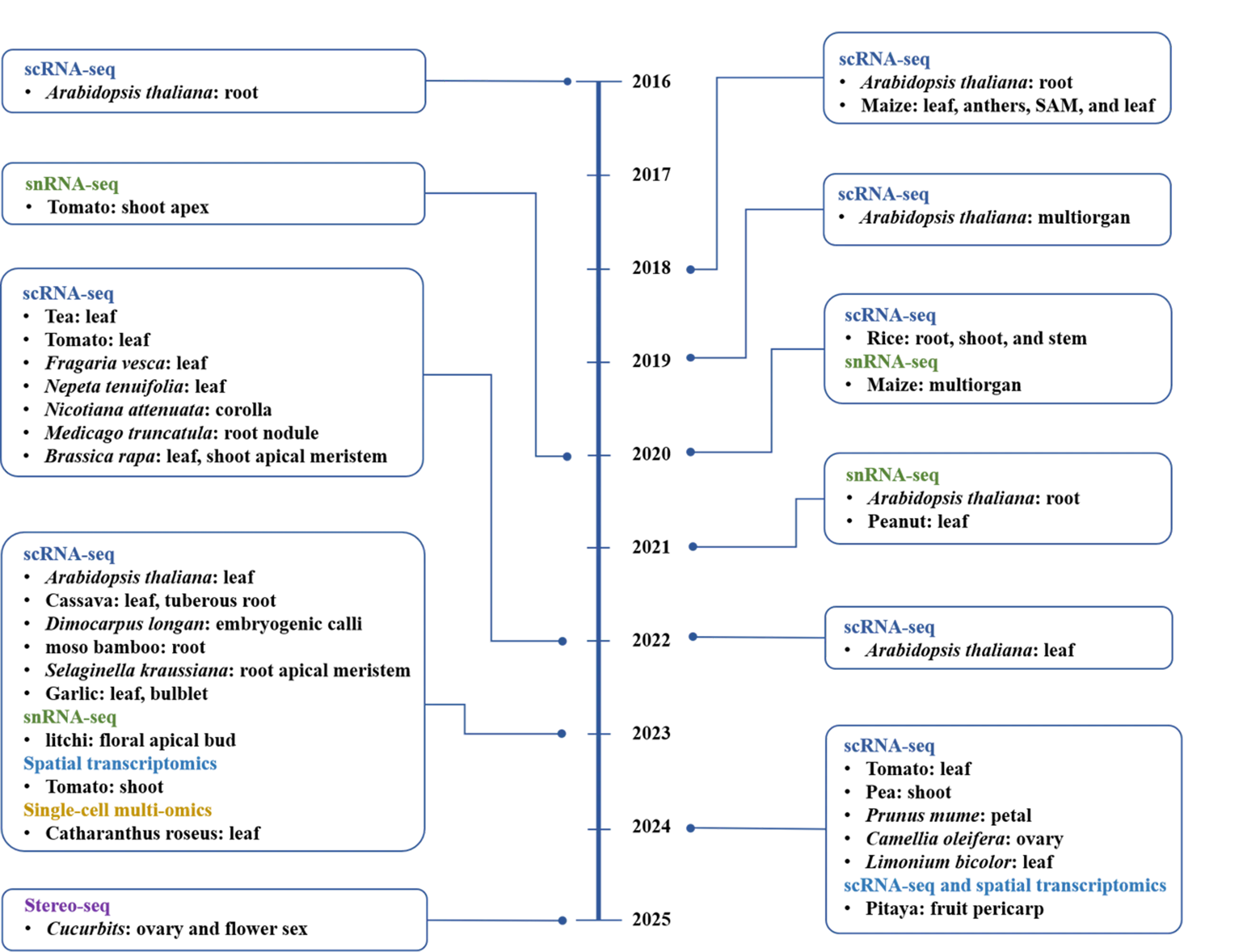
Figure 1. Timeline of major single-cell transcriptomics studies in model plants and horticultural plants.
2.1. Sample Preparation Methods in Plants
The success of scRNA-seq depends on obtaining high-quality single-cell suspensions, and proper sample preparation is the most critical initial step. This process requires careful optimisation of three key parameters: the plant developmental stage, tissue selection, and cell isolation method [2]. In the study of plant development, root-tip tissues, leaves, and floral organs serve as model systems because of their well-characterized cellular differentiation patterns. For instance, to trace the trajectories of phloem parenchyma cells during root meristem formation in tomato (Solanum lycopersicum), fluorescence-activated cell sorting (FACS) was used to isolate individual cells from tomato internode I sections after enzymatic dissociation, ensuring high-quality samples for subsequent scRNA-seq analysis [21]. Parallel studies on specialised plant structures have revealed that the salt gland differentiation process in the recretohalophyte Limonium bicolor exhibits distinct developmental stages [22]. Notably, the first true leaf undergoes critical transitions in salt gland maturity between days 4 and 8 post-emergence, prompting researchers to focus on protoplast isolation from day 4 to 7 leaves for developmental trajectory analysis [22].
The selection of optimal temporal sampling points remains crucial in plant stress response studies, because plants rapidly activate a series of physiological and molecular responses to both abiotic and biotic stresses. For instance, Arabidopsis thaliana exhibits significant heterogeneity in the responses of different cell types to heat stress [23]. This temporal and spatial complexity was further demonstrated in comparative analyses of rice (Oryza sativa) varieties, in which scRNA-seq profiling of leaf sheath tissues from the brown planthopper (BPH)-susceptible TN1 and BPH-resistant YHY15 cultivars at 48 h post-infection revealed distinct transcriptional landscapes [24].
Advancements in plant cell isolation methodologies have led to significant progress in single-cell transcriptomics. Protoplast-based approaches have particular technical merits in their capacity to simultaneously capture nuclear and cytoplasmic RNA, typically yielding 3–5 times more detectable than single-nucleus RNA sequencing (snRNA-seq) under equivalent experimental conditions, thus providing a more comprehensive view of gene expression patterns and regulatory processes within a single cell [25,26,27,28]. However, due to cell walls and the effects of enzymatic digestion, isolating individual plant cells may introduce significant bias in gene expression [29]. snRNA-seq has a simpler experimental workflow that avoids the dissociation biases introduced by enzymatic digestion, thereby improving the accuracy of sequencing results. This technique has been successfully applied to various plant species and tissues, including Arabidopsis roots, tomato stem tips, and litchi flower organs [25,29,30,31,32,33]. These studies indicated that isolated nuclei can serve as an alternative to protoplasts for obtaining high-quality transcriptomic information from plant cells. However, snRNA-seq may miss valuable information regarding cytoplasmic RNA [32].
2.2. Library Construction and Sequencing
Currently, FACS techniques are widely used in plant research to isolate and purify cells, thereby enriching specific target cell populations or reducing unwanted cellular debris [21,30,33,34,35]. For instance, in tomato, the FACS-mediated isolation of phloem parenchyma cells identified a transitional state indicative of incipient root meristem formation [21]. Similar applications in maize ears combined filtration with FACS to eliminate cellular debris and generate a comprehensive single-cell atlas for developing inflorescences [36]. Notwithstanding its utility, cell sorting introduces additional procedural complexity and extends the processing time, with efficiency markedly influenced by tissue characteristics, species-specific factors, and instrumentation platforms [2].
FACS-based approaches typically require integration with high-throughput sequencing platforms, such as the 10x Genomics system (Pleasanton, CA, USA), which is predominantly used for large-scale scRNA-seq library generation across plant species [37]. Among single-cell platforms, 10x Genomics has been widely adopted for its high sensitivity and mature ecosystem, whereas BD Rhapsody (BD Biosciences, Franklin Lakes, NJ, USA) uses a microwell design that is gentler on cells and more effective at capturing low-mRNA cell types such as epidermal cells and meristem initials [38]. Rhapsody has been successfully applied to capture Arabidopsis callus cells for regeneration trajectory analysis [39] and rice inflorescence cells for studies of floret and meristem development [20]. For spatial transcriptomics, stereo-seq provides subcellular resolution and large capture areas suitable for whole-organ mapping, whereas 10x Visium offers a streamlined commercial workflow, with the upgraded Visium HD delivering substantially higher resolution [40].
2.3. scRNA-Seq Data Processing and Analysis
scRNA-seq data processing and analysis constitute a multi-step process, typically involving data pre-processing, dimensionality and clustering, and downstream analysis [41,42]. The initial step generally involves aligning reads to a reference genome and collapsing them using a unique molecular identifier (UMI) to generate a gene-cell matrix of integer feature counts. Due to the impact of non-barcoded cellular debris, mRNA multiplets, and batch effects, rigorous quality control and normalisation are essential to ensure data reliability. In plants, doublets and multiplets can be detected by SoCube [43]. Because each count in the matrix results from captured and reverse-transcribed mRNA molecules, technical variations may lead to differences in sequencing depth among cells. Normalisation corrects these discrepancies by scaling the counts to accurately reflect relative gene expression abundance, thereby serving as a critical step in downstream analyses [42]. The high dimensionality and complexity of scRNA-seq data necessitate their projection into a lower-dimensional space for optimal representation. The DREAM technique was recently employed for dimensionality reduction in data analysis [44]. Downstream analysis of scRNA-seq data typically involves three major steps: clustering, trajectory inference, and cell-type annotation [45]. Clustering serves as the initial step in inferring cell identity and extracting biological information. It grouped cells based on similarities in their gene expression profiles [42]. However, variations in the clustering parameters can substantially affect the output and identification of sub-cell types, necessitating careful consideration [2]. However, pseudo-time analysis overcomes the intrinsic limitation of discrete clustering in capturing progressive cellular dynamics by reconstructing developmental trajectories through cell ordering based on expression pattern resemblance [46].
Although unsupervised clustering groups cells into distinct clusters, it typically lacks intrinsic biological interpretability. Cellular identities within each cluster were determined based on the distinctive expression of specific marker genes. Currently, manual annotation using canonical cell-type-specific markers remains the prevailing method for defining cell types in Arabidopsis thaliana roots [16,47,48]. scRNA-seq has been successfully employed to construct single-cell atlases and identify cell populations in various horticultural plants. However, the scarcity of well-characterised marker genes for diverse cell types and states in non-model crops complicates manual annotation efforts. No single analytical pipeline performed best for every single-cell dataset. The choice of the model and parameters depends on the experimental design, sequencing platform, tissue type, sample size, sequencing depth, and specific biological questions. An important issue is the risk of over-correction during batch integration. These methods help remove technical noise from replicates, batches, genotypes, stages, or platforms. However, when applied too strongly, they may eliminate the genuine biological differences [49]. This is particularly concerning when the cell states of interest are defined by specific genotypes, treatments, developmental stages, or stress responses.
Recently, spatial transcriptomics has emerged as a powerful complement to scRNA-seq. It enables marker-free inference of cell identity and spatial organisation and thus helps elucidate tissue architecture and transcriptional context. This technology is particularly relevant for plants. Unlike many mobile animal cell populations, plant cells are constrained by rigid cell walls, generally maintain stable spatial positions within tissues. Consequently, cell identity, differentiation trajectory, intercellular signaling, and developmental fate are all closely linked to the tissue position. For instance, its application to the pitaya (Hylocereus undatus) pericarp has identified the central role of the early response transcription factor HuCMB1 in the senescence regulatory network [50]. Moreover, an integrated analysis combining snRNA-seq and spatial transcriptomics identified rare cell subtypes and elucidated their critical functions in soybean nodule maturation and physiological processes [51]. The stereo-seq method has been employed to analyse the functions of key genes, including novel KNOX1 transcription factors that play roles in ovary development and floral sex determination [52]. Nevertheless, the accuracy of deconvolution for rare cell types is constrained not only by the low abundance of their transcriptional signals, but also by the mixed cellular composition inherent in each spatial spot. Therefore, any inference regarding the colocalisation of rare populations needs to be treated with care. Where feasible, orthogonal lines of evidence, such as high-resolution spatial technologies, fluorescence imaging, RNA in situ hybridisation, or immunostaining, can provide valuable validation. Together, these complementary approaches offer a solid basis for understanding how rare cell types are spatially organised within plant tissues. Spatial transcriptomics revealed that light-induced chlorenchyma cells drive shoot regeneration via sugar driving target-of-rapamycin (TOR) signaling in tomato [53]. This study revealed the specific expression of several genes in the lip and tepals, including AGL6-like genes, thereby systematically elucidating the molecular mechanisms underlying the complex floral organ development in orchids [54].
3. Single-Cell Transcriptomics Studies in Horticultural Plants
The exceptional diversity of horticultural plants—spanning vegetable-producing, fruit-bearing, ornamental, and beverage-producing plants and herbal medicinal plants—presents both a challenge and a unique opportunity for biological research [55]. This heterogeneity makes scRNA-seq a particularly powerful tool as it can precisely resolve the cellular composition and dynamic gene expression programs that govern the formation and function of diverse organs. By deconstructing complex tissues into their constituent cell types, scRNA-seq offers an unparalleled opportunity to elucidate the mechanisms underlying organ development, quality traits, metabolite biosynthesis, adaptive responses, and evolution in economically important species. This section discusses the applications and future directions of scRNA-seq research in horticultural plants, with the plant species and corresponding tissues/organs investigated using each approach summarised in Table 1.
Table 1. Overview of scRNA-seq studies in horticultural plants.
|
Horticultural Category |
Species |
Organ/Tissue |
Method |
Library Construction |
Key Finding |
References |
|---|---|---|---|---|---|---|
|
Vegetable-producing |
Allium sativum |
bulbs |
scRNA-seq |
10x Genomics |
Clove organogenesis and garlic–virus interaction |
[56] |
|
Brassica campestris |
root tips |
snRNA-seq; snATAC-seq; spatial RNA-seq |
DNBelab C; Stereo-seq |
Salt stress; BcRAP2.11–BcIRT2 in root hairs |
[57] |
|
|
Brassica rapa |
shoot apices and leaves |
scRNA-seq |
10x Genomics |
Heat stress; subgenome dominance in mesophyll cells |
[58] |
|
|
Brassica rapa |
young leaves |
scRNA-seq |
Not specified |
Distinct palisade vs. spongy mesophyll cell functions |
[59] |
|
|
Brassica rapa |
leaf vasculature |
scRNA-seq |
10x Genomics |
Xylem/phloem differentiation; BrNAC45.1/2 |
[60] |
|
|
Brassica rapa |
young leaves |
scRNA-seq |
10x Genomics |
Vernalization response at single-cell resolution |
[61] |
|
|
Cucumis sativus |
floral buds |
spatial RNA-seq |
Stereo-seq |
Floral bud development and sex determination |
[52] |
|
|
Ipomoea aquatica |
root tips |
scRNA-seq |
10x Genomics |
Cd uptake between low-/high-Cd cultivars |
[62] |
|
|
Solanum lycopersicum |
shoot-borne root primordia |
scRNA-seq |
mcSCRB-seq |
Shoot-borne root origin from phloem parenchyma |
[21] |
|
|
Solanum lycopersicum |
calli |
spatial RNA-seq; snRNA-seq |
Stereo-seq; BMKMANU S1000; 10x Visium; 10x Genomics |
Shoot regeneration via chlorenchyma cells |
[53] |
|
|
Solanum lycopersicum |
leaves |
scRNA-seq |
10x Genomics |
ToCV infection; mesophyll transition and SKIP2–chlorophyll |
[63] |
|
|
Solanum lycopersicum |
apical hooks and hypocotyls |
snRNA-seq |
DNBelab C |
Cell-type-specific ethylene/TOR signaling; JA1 |
[64] |
|
|
Solanum lycopersicum |
leaf, stem, root, flower, fruit |
scRNA-seq |
10x Genomics |
PGPR-mediated metabolic rewiring |
[65] |
|
|
Spinacia oleracea |
inflorescences |
spatial RNA-seq |
BMKMANU S1000 |
Sex differentiation via MSI1/HDT2 |
[66] |
|
|
Pisum sativum |
shoot apices |
scRNA-seq |
10x Genomics |
B deficiency; mesophyll, guard, and SAM cell responses |
[67] |
|
|
Euryale ferox |
leaves |
scRNA-seq |
10x Genomics |
Aquatic adaptation; epidermal flavonoid C-glycosides |
[68] |
|
|
Fruit-bearing |
Prunus persica |
flower buds |
spatial RNA-seq |
10x Visium |
Flower bud development and dormancy |
[69] |
|
Fragaria vesca |
leaves |
scRNA-seq |
10x Genomics |
Botrytis cinerea; early-responding epidermal and mesophyll cells |
[70] |
|
|
Hylocereus undatus |
pericarps |
scRNA-seq; spatial RNA-seq |
10x Genomics; 10x Visium |
Fruit senescence via HuCMB1 |
[50] |
|
|
Litchi chinensis |
resting buds; vegetative buds; floral buds |
snRNA-seq |
BD Rhapsody |
Floral transition; LcFT1/LcTFL1-2 transport |
[31] |
|
|
Citrus jambhiri |
root apexes |
snRNA-seq |
10x Genomics |
DOF2.4 in vascular development and immunity |
[71] |
|
|
Crataegus spp. |
leaves |
scRNA-seq; snRNA-seq |
BD Rhapsody; 10x Genomics |
Leaf cell atlas; flavonoid biosynthesis compartmentalization |
[72] |
|
|
Prunus spp. |
young leaves |
scRNA-seq |
10x Genomics |
Leaf development; PpAPL/PpCER1 |
[73] |
|
|
Vitis vinifera |
lateral buds |
snRNA-seq |
10x Genomics |
Autophagy/hormone signaling in tendril determination |
[74] |
|
|
Vitis vinifera |
leaves |
scRNA-seq; spatial RNA-seq |
10x Genomics; Stereo-seq |
Downy mildew defense; guard cell reprogramming |
[75] |
|
|
Malus domestica |
seedlings |
snRNA-seq |
DNBelab C |
Reference genome and cell-type annotation evaluation |
[76] |
|
|
Prunus mume |
petals |
scRNA-seq |
10x Genomics |
Petal cell atlas; floral volatile synthesis |
[77] |
|
|
Phalaenopsis |
inflorescences |
spatial RNA-seq |
10x Visium |
Flower organogenesis; anther development |
[54] |
|
|
Rosa hybrida |
petals |
scRNA-seq |
10x Genomics |
Gray mold response in epidermal/Fra cells |
[78] |
|
|
Tillandsia duratii |
roots and leaves |
spatial RNA-seq |
BMKMANU S1000; Stereo-seq |
Land-to-air adaptive evolution |
[79] |
|
|
Phyllostachys edulis |
basal root tips |
scRNA-seq |
Not specified |
Root atlas; PheWOX13a/b |
[80] |
|
|
Dendrobium devonianum |
flower buds |
scRNA-seq |
10x Genomics |
Flower color differentiation (carotenoid/anthocyanin) |
[81] |
|
|
Dendrobium hybrid |
flower buds |
scRNA-seq |
Singleron GEXSCOPE |
Floral specialization; tepal/pigment cell programs |
[82] |
|
|
Rosa sericea |
leaves |
snRNA-seq |
10x Genomics |
Alpine adaptation; trichome development |
[83] |
|
|
Beverage-producing |
Camellia sinensis |
leaves |
scRNA-seq |
10x Genomics |
Catechin (ECG) biosynthesis |
[84] |
|
Camellia sinensis |
root tips |
scRNA-seq |
10x Genomics |
Theanine biosynthesis in pericycle/xylem cells |
[85] |
|
|
Camellia sinensis |
buds and leaves |
snRNA-seq |
Not specified |
Leaf development and flavor; CsmiRNA396b-CsGRF |
[86] |
|
|
Camellia sinensis |
leaves |
snRNA-seq |
10x Genomics |
Trichome development; CsCUT1/CsMYB4 |
[87] |
|
|
Herbal medicinal |
Catharanthus roseus |
leaves |
scRNA-seq |
10x Genomics |
MIA biosynthesis compartmentalization |
[88] |
|
Catharanthus roseus |
young leaves |
scRNA-seq |
10x Genomics |
MIA biosynthesis |
[89] |
|
|
Limonium bicolor |
young leaves |
scRNA-seq |
10x Genomics |
Salt gland cell fate determination |
[22] |
3.1. Exploring the Developmental Molecular Mechanism at the Cellular Level
ScRNA-seq has been widely employed to dissect the cellular composition and developmental trajectories of roots during root development and regeneration. scRNA-seq was performed on tomato shoot-borne at different developmental stages, depicting the developmental trajectories of phloem parenchyma cells as they form the root meristem, and addressed the question of the origin of shoot-borne roots [21] (Figure 2). Similarly, scRNA-seq analysis of moso bamboo (Phyllostachys edulis) basal roots constructed a single-cell transcriptome atlas that captured spatiotemporal developmental information, revealed the heterogeneity of bamboo root development, and identified the functions of the key regulatory factors PheWOX13a and PheWOX13b in plant root development [80]. In addition, the expression patterns of the SHR-SCR and euANT-WOX modules in S. kraussiana root tips were similar to those in A. thaliana root tips. However, differences in the molecular mechanisms regulating stem cells between the two species, including those involving the WOX and SHR-SCR modules, provide new insights into the evolutionary conservation of root tip stem cells [90].
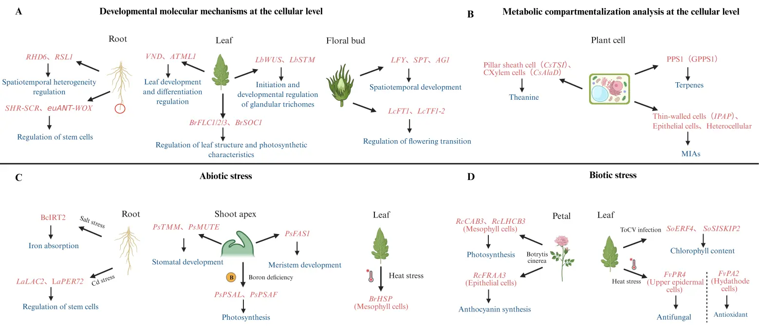
Figure 2. Applications of scRNA-seq in dissecting plant development, specialized metabolism, and stress response at cellular resolution. (A) Cell-type-specific gene regulatory modules governing developmental programs of roots, leaves, and floral buds are revealed to dissect tissue and organ differentiation. (B) Cell-specific metabolic gene expression patterns clarify the compartmentalized biosynthetic sites of diverse specialized metabolites. (C) Distinct response characteristics of cell populations from roots, leaves, and shoot apices interpret the tissue-specific adaptation mechanisms to multiple abiotic stresses. (D) Tissue-specific cell defense cascades illustrate divergent immune regulatory modes under pathogen infection and heat-biotic combined stress. Created with BioRender. Agreement number: ZE2A2Q88DW [91].
For shoot apical meristem development and floral bud differentiation, an snRNA-seq study of the tomato shoot apex established a high-resolution expression atlas that revealed the spatiotemporal developmental trajectory of tomato shoot apex cells and identified key regulators during the developmental process [92]. The first snRNA-seq atlas of apical bud cells was established to identify genes related to floral transitions. Integrating snRNA-seq with RNA in situ hybridization revealed that LcFT1 and LcTFL1-2 mRNAs were transported from the leaves to the shoot apical meristem (SAM), thus revealing a key molecular mechanism governing floral transition [31].
During leaf development and specialised cell differentiation, the cellular heterogeneity and transcriptional features of cassava leaves were revealed by scRNA-seq, which identified six genes associated with cell differentiation fate and analysed the structural and photosynthetic properties of cassava leaves, providing a paradigm for generating cellular atlases in non-model plants [93]. In the medicinal plant Nepeta tenuifolia, scRNA-seq analysis of leaves revealed the differentiation trajectories of specific glandular trichome cells, providing a basis for understanding the molecular regulatory network involved in glandular trichome initiation and development [94]. Furthermore, a study of Chinese cabbage leaf vasculature has revealed the developmental trajectories of the xylem and phloem through pseudo-time analysis, suggesting that auxin response elements (such as MP and LAX2) and ethylene response elements (such as ERF1 and ERF13) may be involved in early xylem development. Additionally, several NAC transcription factors (e.g., BrNAC45.1 and BrNAC45.2) have been identified as key candidate regulators of sieve elements and companion cell differentiation [60]. scRNA-seq analysis was employed to identify AG2 (AGAMOUS 2), which served as a molecular marker for the ovule in Dendrobium devonianum [81].
3.2. Exploring Quality Trait Formation at the Cellular Level
Quality traits, such as flower colour luster, flavour, and shape, are of significant economic importance in horticultural plants. With advances in scRNA-seq technology, elucidating the key metabolic processes underlying the formation of these quality traits and identifying their specific marker genes has become a major research focus in this field. In floral quality research, a pioneering single-cell atlas of mei (Prunus mume) petals was constructed, which identified six major cell types in the petals and revealed the expression dynamics of differentially expressed genes (DEGs) among different cell types at different floral developmental stages, shedding light on the molecular mechanisms of floral volatile synthesis [77] (Figure 2). The combined application of scRNA-seq and spatial transcriptome sequencing revealed the function of the early response factor HuCMB1 in regulating fruit colour, water loss, lesion formation, and the senescence process, demonstrating that this gene plays a key role in the comprehensive regulation of post harvest fruit quality [50]. Furthermore, scRNA-seq has revolutionised the understanding of complex metabolic pathways. The unexpected discovery of catechin ester glucosyltransferases involved in ECG glucoside formation has enhanced understanding of the complex metabolic pathways of catechin esters in tea [84]. snRNA-seq combined with metabolomics revealed that key regulatory networks, including CsmiRNA396b-CsGRF and CsTCP3/CsTCP14, play a coordinating role in regulating leaf development and flavour compound biosynthesis, providing an important theoretical basis for quality improvement in tea plants [86].
3.3. Elucidating Metabolic Compartmentalization at the Cellular Level
The biosynthesis and storage of secondary metabolites in horticultural plants are typically confined to specific cell types and exhibit distinct metabolic compartmentalisation. Traditional methods only capture the average content at the tissue level, making it difficult to identify the cellular sources of these metabolites. In contrast, scRNA-seq allows for the precise localisation of metabolic pathway gene expression in specific cell subpopulations, thereby identifying the cells responsible for synthesising secondary metabolites. For example, scRNA-seq analysis has revealed that the biosynthesis of theanine exhibits a multi-cellular compartmentalisation, with its precursors and final products synthesised in different cell types. Specifically, CsTSI is primarily concentrated in pericycle cells, whereas CsAlaDC is highly expressed in xylem cells [85]. Similarly, by integrating scRNA-seq with multi-omics technologies, the first single-cell atlas of C. roseus leaves was constructed, revealing the compartmentalisation of the biosynthetic pathways of monoterpenoid indole alkaloids (MIAs). Specifically, MIA biosynthesis was found to be initialised in internal phloem-associated parenchyma (IPAP) cells, with intermediate enzymatic steps occurring in epidermal cells (ECs) and late steps preferentially localised in idioblast cells (ICs) [88,89] (Figure 2). These findings indicated that scRNA-seq technologies drive research on secondary metabolism at the single-cell level and offer new insights into the regulation of compartmenalised metabolic networks.
3.4. Understanding Stress Response Mechanisms at the Cellular Level
Horticultural plants face a variety of environmental stresses, both abiotic and biotic, during their growth and development. Different plant cell types exhibit marked heterogeneity in their responses to stress conditions, and scRNA-seq technology is an effective method for analysing cellular heterogeneity and identifying key stress-responsive genes.
In abiotic stress studies, scRNA-seq has been successfully applied to various horticultural plants and non-model crops. scRNA-seq analysis of palisade mesophyll cells (PMCs) and spongy mesophyll cells (SMCs) in Brassica rapa showed significant functional differences. PMCs were related to photosynthesis, whereas SMCs responded to external environmental stress, providing important information for understanding the mechanism of leaf development under environmental stress in leafy vegetables [59] (Figure 2). Furthermore, an scRNA-seq map of Chinese cabbage leaves under high-temperature stress was constructed, revealing dominant subgenome expression and transcriptional regulatory networks at the cellular level in response to heat stress [58]. Additionally, a study on boron-stressed pea shoots revealed cellular heterogeneity among distinct cell types and identified key genes and regulatory factors that govern shoot development under boron stress, thereby shedding light on adaptation strategies to boron deficiency in plants [67]. scRNA-seq analysis of young leaves in the recretohalophyte (Limonium bicolor) revealed the expression characteristics of genes related to salt gland cell fate determination and the involvement of cytokinin, providing a basis for exploring the molecular mechanisms of plant salinity tolerance [22]. In addition, the integration of scRNA-seq and snATAC-seq data makes it possible to link cell type-specific open chromatin regions to the genes they may regulate, and to reconstruct transcription factor centered networks. This shifts the analysis from descriptive observations towards testable regulatory models. Salt-stress responses in root hairs are used as a concrete example, tracing a complete analytical path from the identification of the transcription factor BcRAP2.11 to the coordinated changes in chromatin accessibility and in the expression of its downstream target BcIRT2 [57]. scRNA-seq data indicated that chilling triggered a biphasic transcriptional program in mesophyll cells and led to preferential enrichment of flowering transcripts in vascular tissues, with BrFLC identified as a key gene in the vernalization response [61]. Collectively, these studies revealed cell type-specific response mechanisms to abiotic stress.
scRNA-seq has also demonstrated tremendous potential for biotic stress analysis. snRNA-seq analysis has revealed the immune response mechanism of rough lemon to Candidatus Liberibacter asiaticus and identified the dual core function of the zinc finger transcription factor DOF2.4 in regulating vascular cell development and activating immune responses [71]. In strawberries, a single-cell atlas was constructed to analyse the distinct gene expression profiles of leaf cells infested with Botrytis cinerea at different pathogenesis stages. This study discovered dynamic changes in the expression of disease-related genes PR4 and the peroxidase PA2 during lesion development, providing a broader perspective on the cellular heterogeneity and holistic dynamics of plant-pathogen interactions [70]. Recently, scRNA-seq analysis of healthy and chlorosis virus-infected tomato leaves depicted a single-cell transcriptome landscape and identified the F-box protein SKIP2, which plays a key role in chlorophyll maintenance during viral infection in tomatoes, providing new insights into cellular development and physiological functions under biotic stress [63]. Furthermore, another study revealed the cellular heterogeneity and molecular mechanisms underlying the response of rose petals to gray mold infection, identifying key cell types (e.g., epidermal/Fra cells) and genes (e.g., Fra, TIP2, EP3) [78]. Viral infection up-regulated glutathione synthase/reductase genes and altered glutathione metabolism, implicating this pathway in garlic antiviral immunity [56].
3.5. Resolving Evolutionary Conservation and Divergence at the Cellular Level
Analyses of scRNA-seq data have revealed both conserved and divergent features of regulatory networks between horticultural plants and model species. A comparative single-cell atlas of guard cells across Arabidopsis, tomato, soybean, rice, and sorghum revealed that, despite substantial evolutionary distance, these species share a core guard-cell transcriptome enriched for genes involved in stomatal movement, ABA signaling, reactive oxygen species metabolism, and ion transport, including the transcription factors MYB60 and FAMA, and transporters KAT1 and SLAC1. This indicated a high degree of functional conservation at the key cell type level [95]. At the same time, the study uncovered species-specific regulatory modules, indicating that lineage-specific divergence has occurred on top of the conserved core program. These findings suggest that conserved orthologous marker genes and regulatory modules can serve as useful references for identifying major cell types and developmental programs across species. However, the direct transfer of cell identities or regulatory relationships from model plants to horticultural species should be undertaken with caution. Even when major cell types are evolutionarily conserved, their underlying transcriptional programs and regulatory logic may show species- or lineage-specific features, making experimental validation of target species indispensable.
4. Challenges and Perspectives
Horticultural plants are domesticated to meet various human needs and have substantial economic value. Therefore, understanding single-cell-level heterogeneity is crucial for studying growth, development, and differentiation. Future research in this field will focus on several aspects, including the developmental trajectories of plant cell lineages in key tissues (e.g., tuberous roots and tubers); the fine-scale gene regulatory networks and epigenetic modifications of fruits and seeds; and the molecular mechanisms of trichome formation and development.
Beyond these fundamental questions, single-cell technologies can support breeding in multiple ways: identifying cell type-specific genes for marker development, providing tissue-specific promoters for targeted expression, and providing precise genome editing. In kiwifruit, scRNA-seq of cold-tolerant versus cold-sensitive leaves revealed a striking difference in the guard cell proportion (2% vs. 40%), identified AaTIFY as a negative regulator of cold tolerance, and enabled its use as a CRISPR/Cas9 target for breeding cold-hardy cultivars [96]. Spatial transcriptomics of floral buds identified KNAT2-like1 as the master switch for receptacle expansion and inferior ovary formation in cucumbers, offering both a promoter resource for fruit shape engineering and an entry point for dissecting complex developmental traits [52].
In addition to identifying candidate genes and regulatory elements, single-cell technologies allow functional validation through transgenic and gene editing approaches. For example, scRNA-seq has been used to identify the key regulators of regeneration and stress responses in horticultural species. The functions of these regulators were verified using CRISPR/Cas9 knockout and transgenic overexpression. This workflow moves from cell type-specific discovery to targeted mutagenesis or ectopic expression. This helps establish causal links between regulatory networks and important agronomic traits. Although this integration of single-cell profiling and reverse genetics is still emerging in horticultural plants, it provides a promising method to convert descriptive cell atlases into practical targets for breeding and biotechnology.
Continuous advancements in high-throughput sequencing technology will accelerate research into the principles, patterns, and mechanisms of horticultural plant growth and development. scRNA-seq offers high-resolution transcriptional profiling at the single-cell level, yet its success hinges on efficient protoplast isolation, a step that can introduce cell-type bias and trigger stress-related transcriptional changes. snRNA-seq, in contrast, addresses many of these issues and is better suited for recalcitrant tissues, including lignified, mature, frozen, or metabolite-rich horticultural samples, although it captures primarily nuclear transcripts and may not fully reflect cytoplasmic mRNA pools [14]. However, tissue dissociation methods can lead to loss of spatial information and alterations in cell type proportions. Furthermore, spatial transcriptomics retains the anatomical context, which is critical for studying plant development and tissue patterning, but its practical utility is still constrained by limited resolution, moderate sensitivity, and relatively high costs. Single-cell multi-omics offers a route to uncover regulatory relationships that transcriptome-only approaches miss, but it demands specialised computational skills, complex data integration, and substantial financial resources. Combining multi-disciplinary knowledge from genomes, transcriptomes, epigenomes, proteomes, and metabolomes will provide an unprecedented perspective for understanding cellular heterogeneity in horticultural plants. Despite facing many challenges, such as low resolution and low sequencing depth, single-cell multi-omics analysis will increasingly become a vital tool in horticultural plant research as the technology continues to develop and mature.
Statement of the Use of Generative AI and AI-Assisted Technologies in the Writing Process
During the preparation of this manuscript, the authors used ChatGPT and DeepSeek to correct grammar errors. After using this tool, the authors reviewed and edited the content as needed and take full responsibility for the content of the published article.
Acknowledgments
The authors gratefully acknowledge the financial support received for this work.
Author Contributions
T.Z., N.H. and T.W. drafted the manuscript. S.X. and R.K. prepared the tables and figures. All authors reviewed and approved the final version.
Ethics Statement
Not applicable.
Informed Consent Statement
Not applicable.
Data Availability Statement
All supporting data are within the article.
Funding
This research was supported by National Natural Science Foundation of China (32402570), Yuelushan Laboratory Breeding Program (2024RC2009), 2025 Furong Program Science and Technology Leading Talents Project Innovation and Entrepreneurship Team (University Sector), Innovative Research Groups of the National Natural Science Foundation of Hunan Province (2025JJ10003).
Declaration of Competing Interest
The authors declare that they have no conflict of interest.
References
-
Barlow PW. Differential growth in plants—A phenomenon that occurs at all levels of organization. Environ. Exp. Bot. 1989, 29, 1–5. DOI:10.1016/0098-8472(89)90034-8 [Google Scholar]
-
Seyfferth C, Renema J, Wendrich JR, Eekhout T, Seurinck R, Vandamme N, et al. Advances and Opportunities in Single-Cell Transcriptomics for Plant Research. Annu. Rev. Plant Biol. 2021, 72, 847–866. DOI:10.1146/annurev-arplant-081720-010120 [Google Scholar]
-
Kalisky T, Blainey P, Quake SR. Genomic Analysis at the Single-Cell Level. Annu. Rev. Genet. 2011, 45, 431–445. DOI:10.1146/annurev-genet-102209-163607 [Google Scholar]
-
Ryu KH, Zhu Y, Schiefelbein J. Plant Cell Identity in the Era of Single-Cell Transcriptomics. Annu. Rev. Genet. 2021, 55, 479–496. DOI:10.1146/annurev-genet-071719-020453 [Google Scholar]
-
Tang F, Lao K, Surani MA. Development and applications of single-cell transcriptome analysis. Nat. Methods 2011, 8, S6–S11. DOI:10.1038/nmeth.1557 [Google Scholar]
-
Tang F, Barbacioru C, Wang Y, Nordman E, Lee C, Xu N, et al. mRNA-Seq whole-transcriptome analysis of a single cell. Nat. Methods 2009, 6, 377–382. DOI:10.1038/nmeth.1315 [Google Scholar]
-
Shalek AK, Satija R, Adiconis X, Gertner RS, Gaublomme JT, Raychowdhury R, et al. Single-cell transcriptomics reveals bimodality in expression and splicing in immune cells. Nature 2013, 498, 236–240. DOI:10.1038/nature12172 [Google Scholar]
-
Yan L, Yang M, Guo H, Yang L, Wu J, Li R, et al. Single-cell RNA-Seq profiling of human preimplantation embryos and embryonic stem cells. Nat. Struct. Mol. Biol. 2013, 20, 1131–1139. DOI:10.1038/nsmb.2660 [Google Scholar]
-
Treutlein B, Brownfield DG, Wu AR, Neff NF, Mantalas GL, Espinoza FH, et al. Reconstructing lineage hierarchies of the distal lung epithelium using single-cell RNA-seq. Nature 2014, 509, 371–375. DOI:10.1038/nature13173 [Google Scholar]
-
Patel AP, Tirosh I, Trombetta JJ, Shalek AK, Gillespie SM, Wakimoto H, et al. Single-cell RNA-seq highlights intratumoral heterogeneity in primary glioblastoma. Science 2014, 344, 1396–1401. DOI:10.1126/science.1254257 [Google Scholar]
-
Ivakov A, Persson S. Plant cell shape: Modulators and measurements. Front. Plant Sci. 2013, 4, 439. DOI:10.3389/fpls.2013.00439 [Google Scholar]
-
Li X, Li L, Yan J. Dissecting meiotic recombination based on tetrad analysis by single-microspore sequencing in maize. Nat. Commun. 2015, 6, 6648. DOI:10.1038/ncomms7648 [Google Scholar]
-
McFaline-Figueroa JL, Trapnell C, Cuperus JT. The promise of single-cell genomics in plants. Curr. Opin. Plant Biol. 2020, 54, 114–121. DOI:10.1016/j.pbi.2020.04.002 [Google Scholar]
-
Zhang J, Ahmad M, Gao H. Application of single-cell multi-omics approaches in horticulture research. Mol. Hortic. 2023, 3, 18. DOI:10.1186/s43897-023-00067-y [Google Scholar]
-
Shahan R, Hsu CW, Nolan TM, Cole BJ, Taylor IW, Greenstreet L, et al. A single-cell Arabidopsis root atlas reveals developmental trajectories in wild-type and cell identity mutants. Dev. Cell 2022, 57, 543–560.e9. DOI:10.1016/j.devcel.2022.01.008 [Google Scholar]
-
Denyer T, Ma X, Klesen S, Scacchi E, Nieselt K, Timmermans MCP. Spatiotemporal Developmental Trajectories in the Arabidopsis Root Revealed Using High-Throughput Single-Cell RNA Sequencing. Dev. Cell 2019, 48, 840–852.e5. DOI:10.1016/j.devcel.2019.02.022 [Google Scholar]
-
Dorrity MW, Alexandre CM, Hamm MO, Vigil AL, Fields S, Queitsch C, et al. The regulatory landscape of Arabidopsis thaliana roots at single-cell resolution. Nat. Commun. 2021, 12, 3334. DOI:10.1038/s41467-021-23675-y [Google Scholar]
-
Liu Z, Zhou Y, Guo J, Li J, Tian Z, Zhu Z, et al. Global Dynamic Molecular Profiling of Stomatal Lineage Cell Development by Single-Cell RNA Sequencing. Mol. Plant 2020, 13, 1178–1193. DOI:10.1016/j.molp.2020.06.010 [Google Scholar]
-
Liu W, Zhang Y, Fang X, Tran S, Zhai N, Yang Z, et al. Transcriptional landscapes of de novo root regeneration from detached Arabidopsis leaves revealed by time-lapse and single-cell RNA sequencing analyses. Plant Commun. 2022, 3, 100306. DOI:10.1016/j.xplc.2022.100306 [Google Scholar]
-
Zong J, Wang L, Zhu L, Bian L, Zhang B, Chen X, et al. A rice single cell transcriptomic atlas defines the developmental trajectories of rice floret and inflorescence meristems. New Phytol. 2022, 234, 494–512. DOI:10.1111/nph.18008 [Google Scholar]
-
Omary M, Gil-Yarom N, Yahav C, Steiner E, Hendelman A, Efroni I. A conserved superlocus regulates above- and belowground root initiation. Science 2022, 375, eabf4368. DOI:10.1126/science.abf4368 [Google Scholar]
-
Zhao B, Gao Y, Ma Q, Wang X, Zhu JK, Li W, et al. Global dynamics and cytokinin participation of salt gland development trajectory in recretohalophyte Limonium bicolor. Plant Physiol. 2024, 195, 2094–2110. DOI:10.1093/plphys/kiae199 [Google Scholar]
-
Jean-Baptiste K, McFaline-Figueroa JL, Alexandre CM, Dorrity MW, Saunders L, Bubb KL, et al. Dynamics of Gene Expression in Single Root Cells of Arabidopsis thaliana. Plant Cell 2019, 31, 993–1011. DOI:10.1105/tpc.18.00785 [Google Scholar]
-
Zha W, Li C, Wu Y, Chen J, Li S, Sun M, et al. Single-Cell RNA sequencing of leaf sheath cells reveals the mechanism of rice resistance to brown planthopper (Nilaparvata lugens). Front. Plant Sci. 2023, 14, 1200014. DOI:10.3389/fpls.2023.1200014 [Google Scholar]
-
Minne M, Ke Y, Saura-Sanchez M, De Rybel B. Advancing root developmental research through single-cell technologies. Curr. Opin. Plant Biol. 2022, 65, 102113. DOI:10.1016/j.pbi.2021.102113 [Google Scholar]
-
Farmer A, Thibivilliers S, Ryu KH, Schiefelbein J, Libault M. Single-nucleus RNA and ATAC sequencing reveals the impact of chromatin accessibility on gene expression in Arabidopsis roots at the single-cell level. Mol. Plant 2021, 14, 372–383. DOI:10.1016/j.molp.2021.01.001 [Google Scholar]
-
Long Y, Liu Z, Jia J, Mo W, Fang L, Lu D, et al. FlsnRNA-seq: Protoplasting-free full-length single-nucleus RNA profiling in plants. Genome Biol. 2021, 22, 66. DOI:10.1186/s13059-021-02288-0 [Google Scholar]
-
Sunaga‐Franze DY, Muino JM, Braeuning C, Xu X, Zong M, Smaczniak C, et al. Single‐nucleus RNA sequencing of plant tissues using a nanowell‐based system. Plant J. 2021, 108, 859–869. DOI:10.1111/tpj.15458 [Google Scholar]
-
Wang K, Zhao C, Xiang S, Duan K, Chen X, Guo X, et al. An optimized FACS-free single-nucleus RNA sequencing (snRNA-seq) method for plant science research. Plant Sci. 2023, 326, 111535. DOI:10.1016/j.plantsci.2022.111535 [Google Scholar]
-
Efroni I, Mello A, Nawy T, Ip PL, Rahni R, DelRose N, et al. Root Regeneration Triggers an Embryo-like Sequence Guided by Hormonal Interactions. Cell 2016, 165, 1721–1733. DOI:10.1016/j.cell.2016.04.046 [Google Scholar]
-
Yang MC, Wu ZC, Chen RY, Abbas F, Hu GB, Huang XM, et al. Single-nucleus RNA sequencing and mRNA hybridization indicate key bud events and LcFT1 and LcTFL1-2 mRNA transportability during floral transition in litchi. J. Exp. Bot. 2023, 74, 3613–3629. DOI:10.1093/jxb/erad103 [Google Scholar]
-
Islam MT, Liu Y, Hassan MM, Abraham PE, Merlet J, Townsend A, et al. Advances in the Application of Single-Cell Transcriptomics in Plant Systems and Synthetic Biology. BioDesign Res. 2024, 6, 0029. DOI:10.34133/bdr.0029 [Google Scholar]
-
Ryu KH, Huang L, Kang HM, Schiefelbein J. Single-Cell RNA Sequencing Resolves Molecular Relationships Among Individual Plant Cells. Plant Physiol. 2019, 179, 1444–1456. DOI:10.1104/pp.18.01482 [Google Scholar]
-
Roszak P, Heo JO, Blob B, Toyokura K, Sugiyama Y, de Luis Balaguer MA, et al. Cell-by-cell dissection of phloem development links a maturation gradient to cell specialization. Science 2021, 374, eaba5531. DOI:10.1126/science.aba5531 [Google Scholar]
-
Lopez-Anido CB, Vatén A, Smoot NK, Sharma N, Guo V, Gong Y, et al. Single-cell resolution of lineage trajectories in the Arabidopsis stomatal lineage and developing leaf. Dev. Cell 2021, 56, 1043–1055.e4. DOI:10.1016/j.devcel.2021.03.014 [Google Scholar]
-
Xu X, Crow M, Rice BR, Li F, Harris B, Liu L, et al. Single-cell RNA sequencing of developing maize ears facilitates functional analysis and trait candidate gene discovery. Dev. Cell 2021, 56, 557–568.e6. DOI:10.1016/j.devcel.2020.12.015 [Google Scholar]
-
Zheng D, Xu J, Lu Y, Chen H, Chu Q, Fan L. Recent progresses in plant single-cell transcriptomics. Crop Des. 2023, 2, 100041. DOI:10.1016/j.cropd.2023.100041 [Google Scholar]
-
Eekhout T, Veirman LD, Block JD, Persyn F, Goossens V, Audenaert D, et al. Benchmarking plant single cell RNA-sequencing sample processing strategies. EMBO J. 2026, 45, 4337–4359. DOI:10.1038/s44318-026-00800-5 [Google Scholar]
-
Zhai N, Xu L. Pluripotency acquisition in the middle cell layer of callus is required for organ regeneration. Nat. Plants 2021, 7, 1453–1460. DOI:10.1038/s41477-021-01015-8 [Google Scholar]
-
Moskal K, Puchta-Jasińska M, Bolc P, Motor A, Frankowski R, Pietrusińska-Radzio A, et al. Why “Where” Matters as Much as “How Much”: Single-Cell and Spatial Transcriptomics in Plants. Int. J. Mol. Sci. 2025, 26, 11819. DOI:10.3390/ijms262411819 [Google Scholar]
-
Wu Y, Zhang K. Tools for the analysis of high-dimensional single-cell RNA sequencing data. Nat. Rev. Nephrol. 2020, 16, 408–421. DOI:10.1038/s41581-020-0262-0 [Google Scholar]
-
Luecken MD, Theis FJ. Current best practices in single‐cell RNA‐seq analysis: A tutorial. Mol. Syst. Biol. 2019, 15, MSB188746. DOI:10.15252/msb.20188746 [Google Scholar]
-
Zhang H, Lu M, Lin G, Zheng L, Zhang W, Xu Z, et al. SoCube: An innovative end-to-end doublet detection algorithm for analyzing scRNA-seq data. Brief. Bioinform. 2023, 24, bbad104. DOI:10.1093/bib/bbad104 [Google Scholar]
-
Jiang J, Xu J, Liu Y, Song B, Guo X, Zeng X, et al. Dimensionality reduction and visualization of single-cell RNA-seq data with an improved deep variational autoencoder. Brief. Bioinform. 2023, 24, bbad152. DOI:10.1093/bib/bbad152 [Google Scholar]
-
Zhang Z, Cui F, Lin C, Zhao L, Wang C, Zou Q. Critical downstream analysis steps for single-cell RNA sequencing data. Brief. Bioinform. 2021, 22, bbab105. DOI:10.1093/bib/bbab105 [Google Scholar]
-
Haghverdi L, Büttner M, Wolf FA, Buettner F, Theis FJ. Diffusion pseudotime robustly reconstructs lineage branching. Nat. Methods 2016, 13, 845–848. DOI:10.1038/nmeth.3971 [Google Scholar]
-
Wendrich JR, Yang B, Vandamme N, Verstaen K, Smet W, Van de Velde C, et al. Vascular transcription factors guide plant epidermal responses to limiting phosphate conditions. Science 2020, 370, eaay4970. DOI:10.1126/science.aay4970 [Google Scholar]
-
Shulse CN, Cole BJ, Ciobanu D, Lin J, Yoshinaga Y, Gouran M, et al. High-Throughput Single-Cell Transcriptome Profiling of Plant Cell Types. Cell Rep. 2019, 27, 2241–2247.e4. DOI:10.1016/j.celrep.2019.04.054 [Google Scholar]
-
Li S, Lücken M, Marioni JC, Teichmann SA, He P. Toward informed batch correction for single-cell transcriptome integration. Nat. Comput. Sci. 2026, 6, 123–133. DOI:10.1038/s43588-025-00943-1 [Google Scholar]
-
Li X, Li B, Gu S, Pang X, Mason P, Yuan J, et al. Single-cell and spatial RNA sequencing reveal the spatiotemporal trajectories of fruit senescence. Nat. Commun. 2024, 15, 3108. DOI:10.1038/s41467-024-47329-x [Google Scholar]
-
Liu Z, Kong X, Long Y, Liu S, Zhang H, Jia J, et al. Integrated single-nucleus and spatial transcriptomics captures transitional states in soybean nodule maturation. Nat. Plants 2023, 9, 515–524. DOI:10.1038/s41477-023-01387-z [Google Scholar]
-
Dong Z, Liu X, Guo X, Liu X, Wang B, Shao W, et al. Developmental innovation of inferior ovaries and flower sex orchestrated by KNOX1 in cucurbits. Nat. Plants 2025, 11, 861–877. DOI:10.1038/s41477-025-01950-w [Google Scholar]
-
Song X, Guo P, Xia K, Wang M, Liu Y, Chen L, et al. Spatial transcriptomics reveals light-induced chlorenchyma cells involved in promoting shoot regeneration in tomato callus. Proc. Natl. Acad. Sci. USA 2023, 120, e2310163120. DOI:10.1073/pnas.2310163120 [Google Scholar]
-
Liu C, Leng J, Li Y, Ge T, Li J, Chen Y, et al. A spatiotemporal atlas of organogenesis in the development of orchid flowers. Nucleic Acids Res. 2022, 50, 9724–9737. DOI:10.1093/nar/gkac773 [Google Scholar]
-
Chen F, Song Y, Li X, Chen J, Mo L, Zhang X, et al. Genome sequences of horticultural plants: past, present, and future. Hortic. Res. 2019, 6, 112. DOI:10.1038/s41438-019-0195-6 [Google Scholar]
-
Gao S, Li F, Zeng Z, He Q, Mostafa HHA, Zhang S, et al. A single-cell transcriptomic atlas reveals the cell differentiation trajectory and the response to virus invasion in swelling clove of garlic. Hortic. Res. 2025, 12, uhae365. DOI:10.1093/hr/uhae365 [Google Scholar]
-
Liu Q, Kang J, Du L, Liu Z, Liang H, Wang K, et al. Single‐cell multiome reveals root hair‐specific responses to salt stress. New Phytol. 2025, 246, 2634–2651. DOI:10.1111/nph.70160 [Google Scholar]
-
Sun X, Feng D, Liu M, Qin R, Li Y, Lu Y, et al. Single-cell transcriptome reveals dominant subgenome expression and transcriptional response to heat stress in Chinese cabbage. Genome Biol. 2022, 23, 262. DOI:10.1186/s13059-022-02834-4 [Google Scholar]
-
Guo X, Liang J, Lin R, Zhang L, Zhang Z, Wu J, et al. Single‐cell transcriptome reveals differentiation between adaxial and abaxial mesophyll cells in Brassica rapa. Plant Biotechnol. J. 2022, 20, 2233–2235. DOI:10.1111/pbi.13919 [Google Scholar]
-
Guo X, Yuan J, Zhang Y, Wu J, Wang X. Developmental landscape and asymmetric gene expression in the leaf vasculature of Brassica rapa revealed by single-cell transcriptome. Hortic. Res. 2025, 12, uhaf060. DOI:10.1093/hr/uhaf060 [Google Scholar]
-
Zhang Z, Cai X, Liang J, Liu J, Guo J, Yang W, et al. Time-resolved single-cell atlas identifies the spatiotemporal transcription dynamics in vernalization response in Brassica rapa. Cell Rep. 2025, 44, 115725. DOI:10.1016/j.celrep.2025.115725 [Google Scholar]
-
Shen C, Huang BF, Liao Q, Chen KF, Xin JL, Huang YY. Uncovering differences in cadmium accumulation capacity of different Ipomoea aquatica cultivars at the level of root cell types. Hortic. Res. 2025, 12, uhaf077. DOI:10.1093/hr/uhaf077 [Google Scholar]
-
Yue H, Chen G, Zhang Z, Guo Z, Zhang Z, Zhang S, et al. Single‐cell transcriptome landscape elucidates the cellular and developmental responses to tomato chlorosis virus infection in tomato leaf. Plant Cell Environ. 2024, 47, 2658–2672. DOI:10.1111/pce.14906 [Google Scholar]
-
Huang W, Yang L, Hu N, Jiang Q, Zhou P, He J, et al. Single-nucleus transcriptome profiling unveils cell-type-specific ethylene and TOR signaling in tomato. Hortic. Res. 2026, 13, uhag044. DOI:10.1093/hr/uhag044 [Google Scholar]
-
Fu W, Sun C, Sun B, Xue Q, Guo Q, Lai H. Single-cell RNA-seq uncovers landscape of tomato metabolic rewiring mediated by plant growth-promoting rhizobacteria. Plant Physiol. Biochem. 2026, 237, 111521. DOI:10.1016/j.plaphy.2026.111521 [Google Scholar]
-
You C, Yang H, Zhao Y, Wang X, Wei S, Chen N, et al. Spatiotemporal Transcriptomic Atlas Reveals the Regulatory Mechanisms Underlying Early Inflorescence Development and Sex Differentiation in Spinach. Adv. Sci. 2025, 12, e07818. DOI:10.1002/advs.202507818 [Google Scholar]
-
Chen X, Ru Y, Takahashi H, Nakazono M, Shabala S, Smith SM, et al. Single‐cell transcriptomic analysis of pea shoot development and cell‐type‐specific responses to boron deficiency. Plant J. 2024, 117, 302–322. DOI:10.1111/tpj.16487 [Google Scholar]
-
Liu A, Qu C, Ling W, Wang T, Zhang Y, Fang Y, et al. A model for the adaptation of Euryale ferox leaves to aquatic environments through EfCGT1‐controlled flavonoid C‐glycoside‐specific accumulation in epidermis cells. Plant Biotechnol. J. 2025, 23, 3333–3348. DOI:10.1111/pbi.70155 [Google Scholar]
-
Zhao YL, Li Y, Guo DD, Chen XJ, Cao K, Wu JL, et al. Spatiotemporally transcriptomic analyses of floral buds reveal the high-resolution landscape of flower development and dormancy regulation in peach. Hortic. Res. 2025, 12, uhaf029. DOI:10.1093/hr/uhaf029 [Google Scholar]
-
Bai Y, Liu H, Lyu H, Su L, Xiong J, Cheng ZM. Development of a single-cell atlas for woodland strawberry (Fragaria vesca) leaves during early Botrytis cinerea infection using single-cell RNA-seq. Hortic. Res. 2022, 9, uhab055. DOI:10.1093/hr/uhab055 [Google Scholar]
-
Tian XB, Zhou J, Li J, Li Y, Zhou C, Song Z. Single-nucleus transcriptomics reveals the cellular immune responses to Candidatus Liberibacter asiaticus in rough lemon. Hortic. Res. 2026, 13, uhaf265. DOI:10.1093/hr/uhaf265 [Google Scholar]
-
Zhao G, Wu X, Wang B, Li X, Zheng J, Ding Z, et al. Single-cell RNA-sequencing profiles reveal the developmental landscape of hawthorn leaves. BMC Plant Biol. 2026, 26, 1296. DOI:10.1186/s12870-026-09127-z [Google Scholar]
-
Li M, Gao X, Nie P, Li G, Gong Q, Dong X, et al. Identification of key regulators for the development of Prunus leaves using single-cell RNA sequencing. BMC Plant Biol. 2026, 26, 521. DOI:10.1186/s12870-026-08348-6 [Google Scholar]
-
Li Y, Bian Y, Ni Y, Xie Z. Single‐nucleus transcriptomic atlas reveals autophagy and hormone signaling in early grapevine (Vitis vinifera L.) tendril versus inflorescence determination. Plant J. 2026, 125, e70804. DOI:10.1111/tpj.70804 [Google Scholar]
-
Yao X, Xu Z, Xi Y, He X, Gao Q, Lu J, et al. Unveiling the early defense response dynamics in grapevines against Plasmopara viticola by single-cell transcriptomics. Genome Biol. 2026, 27, 43. DOI:10.1186/s13059-025-03904-z [Google Scholar]
-
Hu S, Liu Y, Zhao Y, Li B, Shen Y, Mimata Y, et al. Comparative evaluation of reference genomes and cell-type annotation frameworks for single-nucleus transcriptomic analysis in apple. Agric. Commun. 2026, 4, 100154. DOI:10.1016/j.agrcom.2026.100154 [Google Scholar]
-
Guo Y, Chen X, Li J, Wang Q, Zhang S, Liu N, et al. Single-cell RNA sequencing reveals a high-resolution cell atlas of petals in Prunus mume at different flowering development stages. Hortic. Res. 2024, 11, uhae189. DOI:10.1093/hr/uhae189 [Google Scholar]
-
Li X, Siman Y, Zhao Y, Peng L, Wu H, Guan W, et al. Single-cell transcriptomic analyses reveal cellular and molecular patterns of rose petal responses to gray mold infection. Hortic. Res. 2025, 12, uhaf152. DOI:10.1093/hr/uhaf152 [Google Scholar]
-
Lyu X, Li P, Jin L, Yang F, Pucker B, Wang C, et al. Tracing the evolutionary and genetic footprints of atmospheric tillandsioids transition from land to air. Nat. Commun. 2024, 15, 9599. DOI:10.1038/s41467-024-53756-7 [Google Scholar]
-
Cheng Z, Mu C, Li X, Cheng W, Cai M, Wu C, et al. Single-cell transcriptome atlas reveals spatiotemporal developmental trajectories in the basal roots of moso bamboo (Phyllostachys edulis). Hortic. Res. 2023, 10, uhad122. DOI:10.1093/hr/uhad122 [Google Scholar]
-
Wang J, Zhou Y, Zhang M, Li X, Liu T, Liu Y, et al. Resolving floral development dynamics using genome and single‐cell temporal transcriptome of Dendrobium devonianum. Plant Biotechnol. J. 2025, 23, 2997–3011. DOI:10.1111/pbi.70094 [Google Scholar]
-
Li Y, Zhang S, Zhang B, Yin Y, Yu H. Single-cell and haplotype-resolved transcriptomics reveal molecular signatures of orchid floral structures. Mol. Plant 2026, 19, 1239–1254. DOI:10.1016/j.molp.2026.03.016 [Google Scholar]
-
Deng H, Ru J, Liang Z, Tang Z, Wang Y, Yuan W, et al. Single-cell transcriptomics reveals cellular and genetic mechanisms of alpine adaptation in Rosa sericea. Front. Plant Sci. 2026, 17, 1733247. DOI:10.3389/fpls.2026.1733247 [Google Scholar]
-
Wang Q, Wu Y, Peng A, Cui J, Zhao M, Pan Y, et al. Single‐cell transcriptome atlas reveals developmental trajectories and a novel metabolic pathway of catechin esters in tea leaves. Plant Biotechnol. J. 2022, 20, 2089–2106. DOI:10.1111/pbi.13891 [Google Scholar]
-
Lin S, Zhang Y, Zhang S, Wei Y, Han M, Deng Y, et al. Root-specific theanine metabolism and regulation at the single-cell level in tea plants (Camellia sinensis). eLife 2024, 13, RP95891. DOI:10.7554/eLife.95891 [Google Scholar]
-
Zhao X, Xu X, Chi N, Liu Y, Zhou X, Jin J, et al. Integrated single-nucleus transcriptomic and metabolomic insights into bud-to-leaf development and metabolite synthesis in tea plant. Hortic. Res. 2026, 13, uhaf281. DOI:10.1093/hr/uhaf281 [Google Scholar]
-
Deng X, Tang Y, Zhang Q, Kong W, Lin X, Chen X, et al. Exploring the developmental mechanisms of tea plant trichomes using genomics and single-cell transcriptome sequencing. Hortic. Res. 2026, 13, uhaf352. DOI:10.1093/hr/uhaf352 [Google Scholar]
-
Sun S, Shen X, Li Y, Li Y, Wang S, Li R, et al. Single-cell RNA sequencing provides a high-resolution roadmap for understanding the multicellular compartmentation of specialized metabolism. Nat. Plants 2022, 9, 179–190. DOI:10.1038/s41477-022-01291-y [Google Scholar]
-
Li C, Wood JC, Vu AH, Hamilton JP, Rodriguez Lopez CE, Payne RME, et al. Single-cell multi-omics in the medicinal plant Catharanthus roseus. Nat. Chem. Biol. 2023, 19, 1031–1041. DOI:10.1038/s41589-023-01327-0 [Google Scholar]
-
Liu W, Cai G, Zhai N, Wang H, Tang T, Zhang Y, et al. Genome and transcriptome of Selaginella kraussiana reveal evolution of root apical meristems in vascular plants. Curr. Biol. 2023, 33, 4085–4097.e5. DOI:10.1016/j.cub.2023.08.061 [Google Scholar]
-
Xiao S, Kui R. Applications of scRNA-seq in Dissecting Plant Development, Specialized Metabolism, and Stress Response at Cellular Resolution. Created in BioRender. 2026. Available online: https://BioRender.com/7nk7ypa (accessed on 13 August 2026).
-
Tian C, Du Q, Xu M, Du F, Jiao Y. Single-nucleus RNA-seq resolves spatiotemporal developmental trajectories in the tomato shoot apex. bioRxiv. 2020. DOI:10.1101/2020.09.20.305029 [Google Scholar]
-
Zang Y, Pei Y, Cong X, Ran F, Liu L, Wang C, et al. Single-cell RNA-sequencing profiles reveal the developmental landscape of the Manihot esculenta Crantz leaves. Plant Physiol. 2023, 194, 456–474. DOI:10.1093/plphys/kiad500 [Google Scholar]
-
Zhou P, Chen H, Dang J, Shi Z, Shao Y, Liu C, et al. Single-cell transcriptome of Nepeta tenuifolia leaves reveal differentiation trajectories in glandular trichomes. Front. Plant Sci. 2022, 13, 988594. DOI:10.3389/fpls.2022.988594 [Google Scholar]
-
Nguyen CC, Thibivilliers S, Li Y, Fazekas CT, Yang EJY, Asiamah JY, et al. Uncovering the core genetic programs governing plant guard cell biology. New Phytol. 2026, 249, 198–217. DOI:10.1111/nph.70678 [Google Scholar]
-
Lin M, Zhang Z, Zhang Q, Sun S, Li Y, Liu M, et al. Single-cell sequencing reveals dynamic cell development trajectories in two kiwifruit (Actinidia chinensis) genotypes with contrasting cold resistance. Hortic Adv. 2025, 3, 1–16. DOI:10.1007/s44281-025-00077-4 [Google Scholar]