
Unlocking the Immunomodulatory Potential of Lactic Acid Bacteria in Livestock and Poultry: Mechanisms and Implications
Unlocking the Immunomodulatory Potential of Lactic Acid Bacteria in Livestock and Poultry: Mechanisms and Implications
Wenjie Li 1,2 Jiabing Mao 1,2 Sha Wu 1,2 Jie Yin 1,2 Cong Lan 1,2,*
Received: 03 July 2026 Revised: 01 September 2026 Accepted: 12 October 2026 Published: 23 October 2026

© 2026 The authors. This is an open access article under the Creative Commons Attribution 4.0 International License (https://creativecommons.org/licenses/by/4.0/).
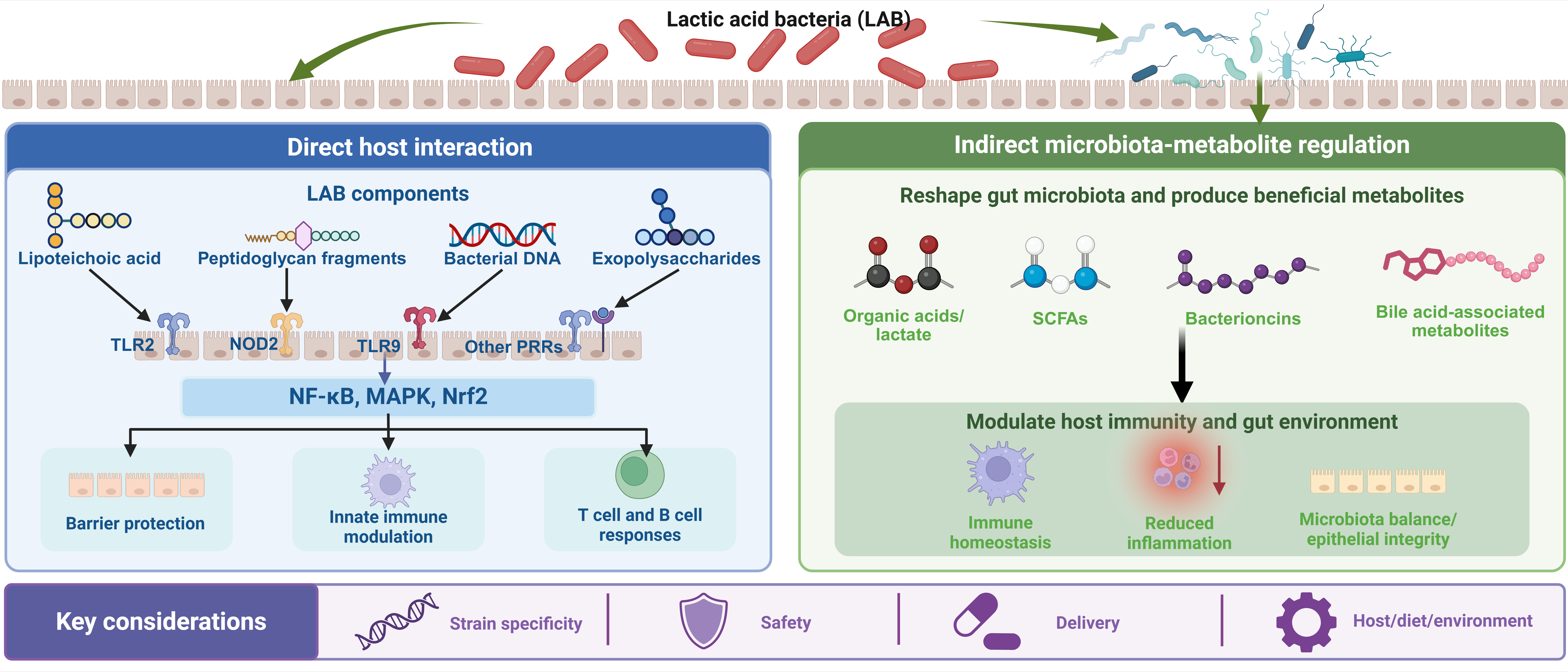
Graphical Abstract
1. Introduction
Modern intensive livestock production, while meeting the growing global demand for meat, eggs, and milk, is increasingly challenged by the pressures of disease prevention and the rising threat of antimicrobial resistance (AMR). The extensive and inappropriate application of antibiotics imposes strong selective pressure on microbial populations, causing the spread of resistance within high-density animal production systems [1]. Antibiotic exposure can also disrupt the gut microbiota and compromise intestinal homeostasis, thereby adversely affecting animal health and production efficiency [2]. Consequently, there is an increasing need for sustainable strategies that enhance disease resilience while maintaining immune and intestinal homeostasis without relying on routine antibiotic use. Among these strategies, lactic acid bacteria (LAB) have attracted considerable attention because of their favorable safety profiles and their capacity to interact with the intestinal microbiota and host immune system [3,4]. Evidence from swine, poultry, and cattle indicates that LAB supplementation can improve intestinal health, reduce pathogen burden, and enhance production performance [4,5,6], supporting their potential application in low-antibiotic or antibiotic-free production systems.
Importantly, the beneficial effects of LAB cannot be explained solely by competitive exclusion of pathogens or changes in gut microbial composition. The intestinal mucosa represents a major interface through which LAB communicate with the host immune system. LAB-associated molecular structures, including lipoteichoic acids, peptidoglycan-derived fragments, surface proteins, exopolysaccharides, and bacterial DNA, can be sensed by intestinal epithelial and immune cells through pattern-recognition receptors such as TLR2, NOD2, and TLR9 [7,8,9,10,11]. These interactions can subsequently modulate intracellular inflammatory and stress-response pathways, influence macrophage and dendritic cell functions, shape T- and B-cell responses, and contribute to the maintenance of intestinal barrier integrity and immune homeostasis. In parallel, LAB-derived products and LAB-associated microbial metabolites, including bacteriocins, short-chain fatty acids (SCFAs), and bile acid derivatives, provide an additional layer of immune regulation by linking microbial metabolic activity with epithelial, innate, and adaptive immune responses [12,13,14,15,16,17,18,19]. Despite increasing evidence for these immunomodulatory effects, important mechanistic questions remain unresolved.
Existing reviews have extensively discussed probiotics and postbiotics as alternatives to antibiotics, their effects on gut microbiota and productive performance, and their applications in individual livestock species [3,4,6,20]. However, the mechanisms linking the interaction of LAB with the intestinal mucosa of livestock and poultry to host immune receptor-mediated recognition, downstream cellular signaling, metabolite-mediated regulation, and adaptive immune responses have not been sufficiently summarized and analyzed. In addition, strain-specific characteristics of different LAB strains contribute to variation in their functional properties and application outcomes, but these differences remain insufficiently understood.
Because LAB-mediated immunomodulation is strongly influenced by host physiology, intestinal architecture, immune organization, and microbial ecology, this review prioritizes evidence obtained directly from target livestock species, particularly pigs, chickens, and cattle. These species represent major livestock and poultry production systems and therefore provide the most relevant evidence for practical application in animal production [4,5,6]. Evidence from murine, human, or in vitro models is included primarily when it provides mechanistic insight that has not yet been directly resolved in livestock and poultry, and such evidence is clearly distinguished from findings obtained in target animal species.
Accordingly, this review will comprehensively discuss the mechanisms, application scenarios, and challenges of LAB-mediated immune regulation in livestock and poultry, with the aim of providing new insights and directions for the standardized application of LAB in animal production.
2. Lactic Acid Bacteria–Host Immune Interactions in Livestock and Poultry
2.1. Overview of Lactic Acid Bacteria
LAB comprise a physiologically defined group of bacteria characterized primarily by the production of lactic acid through carbohydrate fermentation, rather than a single formal taxonomic lineage. They are typically Gram-positive, non-spore-forming, fermentative bacteria and are widely distributed in the gastrointestinal tract of animals, fermented feeds, foods, and other nutrient-rich environments. Most classical LAB belong to the phylum Bacillota (traditionally Firmicutes), class Bacilli, and order Lactobacillales. Taxonomically diverse genera within this group include Lactobacillus sensu stricto, Lactiplantibacillus, Lacticaseibacillus, Limosilactobacillus, Pediococcus, Lactococcus, Enterococcus, Leuconostoc, and Weissella, among others [21,22]. Importantly, membership in the LAB group does not, by itself, imply probiotic functionality, and the beneficial and safety characteristics of candidate strains require strain-level evaluation.
Phylogenetic and phylogenomic studies have substantially reshaped the taxonomy of LAB. Traditional classification based on cell morphology and fermentation characteristics does not always correspond to evolutionary relationships inferred from 16S rRNA sequences or whole-genome data [23]. A particularly important taxonomic revision involved the former genus Lactobacillus, which exhibited extensive phylogenetic, ecological, and phenotypic diversity. Based on core-genome phylogeny, average amino acid identity, clade-specific signature genes, physiological characteristics, and ecological associations, the former Lactobacillus genus was reclassified into 25 genera, comprising the emended genus Lactobacillus, Paralactobacillus, and 23 newly described genera [22]. This reclassification introduced several genera frequently encountered in livestock probiotic research, including Lactiplantibacillus, Lacticaseibacillus, and Limosilactobacillus, and more accurately reflects their evolutionary and ecological relationships.
At the genomic level, LAB exhibit substantial interspecific and intraspecific diversity. Comparative genomic analyses indicate that, in addition to conserved core functions required for cellular maintenance and fermentative metabolism, LAB possess variable accessory gene repertoires associated with carbohydrate utilization, membrane transport, environmental stress responses, adhesion, exopolysaccharide production, bacteriocin biosynthesis, and adaptation to specific ecological niches [23,24,25,26,27,28]. Gene gain and loss, horizontal gene transfer, and other genome-level changes have contributed to the evolutionary diversification and niche adaptation of LAB. Consequently, even closely related strains can differ markedly in their metabolic and host-interaction phenotypes.
The biological properties of probiotic LAB further determine their ability to survive gastrointestinal transit and interact with livestock and poultry hosts. Selected strains exhibit tolerance to gastric acidity and bile salts, while surface proteins, mucus-binding factors, peptidoglycan, lipoteichoic acids, and exopolysaccharides can facilitate adhesion or prolonged residence at the intestinal mucosal surface [7]. Such interactions increase opportunities for communication with intestinal epithelial and immune cells. LAB can also compete with pathogens for nutrients and adhesion sites and produce antimicrobial metabolites, including organic acids and bacteriocins [29,30]. Organic acids such as lactic acid and acetic acid can inhibit many susceptible microorganisms through extracellular acidification and disruption of intracellular pH homeostasis. However, this effect is not universal, because acid-tolerant or acid-adapted pathogens, including Salmonella enterica, Listeria monocytogenes, and enterohemorrhagic Escherichia coli, possess mechanisms that enhance their survival under acidic conditions [31,32]. In addition, many LAB produce bacteriocins, such as nisin and enterocin, which can directly inhibit susceptible bacteria [30]. Together, these taxonomic, genomic, and physiological characteristics provide the biological basis for strain-specific interactions between LAB and the intestinal microbiota and the host immune system.
2.2. Immunomodulatory Properties of LAB in Livestock and Poultry
In livestock and poultry, immune regulation refers to the dynamic processes through which the immune system maintains physiological equilibrium while responding to environmental, microbial, and metabolic challenges. Beyond its classical role in pathogen defense, the immune system coordinates inflammatory responses, tissue repair, and host-microbiota interactions to preserve systemic stability and host integrity. Accordingly, immune activation represents a regulated response aimed at restoring disrupted physiological equilibrium rather than merely eliminating invading pathogens [33,34]. In production animals, maintaining immune homeostasis is particularly critical because immune activity is tightly linked to metabolic resource allocation. Excessive or chronic immune stimulation diverts nutrients and energy away from growth and productive functions toward immune defense, thereby reducing feed efficiency and growth performance [35]. Conversely, a balanced immune system promotes intestinal integrity, microbial stability, and effective responses to pathogens, ultimately supporting animal health and productive capacity. Disruption of immune homeostasis, often termed immune stress, has been shown to impair growth performance and alter gut microbial communities in poultry, illustrating the strong connection between immune balance and production outcomes [36]. Accordingly, the concept of immune regulation in livestock production extends beyond pathogen defense to encompass the maintenance of immune equilibrium that optimizes resilience, metabolic efficiency, and productivity under intensive farming conditions [37].
The immune system of livestock and poultry comprises two integrated arms—innate immunity and adaptive immunity—which together provide protection against pathogens while maintaining physiological homeostasis. Innate immunity constitutes the first line of defense, relying on physical barriers, antimicrobial peptides, and specialized immune cells such as macrophages, dendritic cells, heterophils, and natural killer cells to rapidly recognize invading microorganisms through pattern recognition receptors (PRRs) that detect conserved microbial structures known as microbe-associated molecular patterns (MAMPs) [38]. In contrast, adaptive immunity develops more slowly but provides highly specific and long-lasting responses mediated primarily by T lymphocytes and B lymphocytes, which generate antigen-specific cellular responses and immunological memory following antigen exposure [39]. In livestock species, these two immune branches are closely interconnected, with innate immune signaling directing antigen presentation and shaping downstream adaptive immune responses in mucosal tissues such as the gut-associated lymphoid tissue (GALT), a central site for host-microbiota immune interactions [40].
3. Mechanisms of Action of LAB in Modulating Host Immunity
LAB-mediated immunoregulation involves both direct recognition of bacterial structural components and signaling mediated by soluble bioactive products and microbiota-associated metabolites. These factors differ substantially in their origin, molecular targets, and level of experimental support in livestock and poultry. To distinguish direct LAB-derived molecules from metabolites generated through community-level microbial metabolism, the major LAB-derived and LAB-associated bioactive factors, their principal immune targets, and the available livestock- and poultry-related evidence are summarized in Table 1. In addition, the strain-specific characteristics of LAB determine the effects of LAB-mediated host immune regulation and will be further discussed in this section.
Table 1. Major LAB-derived and LAB-associated bioactive factors involved in immune regulation in livestock and poultry.
|
Category |
Bioactive Factor |
Relationship to LAB |
Major Host Targets or Mechanisms |
Major Biological/ Immunological Effects |
References |
|---|---|---|---|---|---|
|
Cell-associated structural component |
Lipoteichoic acids (LTAs) |
Direct component of the Gram-positive LAB cell envelope |
TLR2-associated recognition and downstream inflammatory signaling |
Modulation of innate immune sensing and cytokine responses |
[10] |
|
Cell-associated structural component |
Peptidoglycan-derived muropeptides |
Released from the LAB cell wall during growth or turnover |
Intracellular NOD2 recognition |
Regulation of inflammatory signaling and mucosal immune responses |
|
|
Cell-associated structural component |
Bacterial DNA/CpG motifs |
Direct bacterial cellular component |
TLR9-dependent sensing |
Regulation of innate immune signaling and cytokine production |
[8] |
|
Cell-associated structural component |
Surface proteins |
LAB surface-associated proteins and adhesins |
Mucus/epithelial binding and strain-specific host interactions |
Promotion of mucosal retention and prolonged host-microbe contact |
|
|
Surface-associated or secreted polymer |
EPS |
Directly synthesized by LAB and either surface-associated or released extracellularly |
RP105/MD1-, TLR- and MAPK-associated signaling; interference with pathogen adhesion |
Regulation of inflammatory responses, epithelial barrier protection, macrophage activity, and pathogen exclusion |
|
|
Direct fermentation product |
Lactic acid and other organic acids |
Direct products of LAB carbohydrate fermentation; production pattern is strain-and pathway-dependent |
Extracellular acidification and intracellular weak-acid stress in susceptible microorganisms |
Pathogen suppression and indirect reduction of pathogen-associated inflammatory pressure |
|
|
Ribosomally synthesized peptide |
Bacteriocins (e.g., nisin, plantaricins, enterocins) |
Directly synthesized by bacteriocin-producing LAB strains |
Primarily bacterial membrane/cell-wall targets; context-dependent epithelial and immune effects |
Pathogen inhibition, barrier protection, modulation of inflammatory responses, and possible direct immune-cell regulation |
|
|
LAB-associated microbial metabolite |
Short-chain fatty acids (acetate, propionate, and butyrate) |
Mainly generated through community-level intestinal fermentation; LAB may influence their abundance through metabolic interactions and cross-feeding |
FFAR3/GPR41, FFAR2/GPR43, HCAR2/GPR109A, and HDAC inhibition |
Maintenance of epithelial barrier integrity, regulation of macrophages and dendritic cells, promotion of Treg function, and modulation of IL-22 production |
|
|
Host-derived and microbiota-modified metabolite |
Bile acid-associated metabolites |
LAB can contribute to BA deconjugation through strain-specific BSH activity, whereas most secondary BA formation requires broader microbial metabolism |
FXR, TGR5, NF-κB/NLRP3-associated signaling, and BA-dependent T-cell regulation |
Regulation of epithelial integrity, innate inflammation, macrophage/DC activity, and Th17–Treg balance |
3.1. Direct Regulation Through Host-Microbe Interaction
3.1.1. Interaction with the Intestinal Mucosal Interface
The first step in direct immunoregulation is the encounter of LAB with the intestinal mucosal interface. This interface is not a passive barrier but a structured immunological landscape composed of the mucus layer, intestinal epithelial cells (IECs), intraepithelial lymphocytes, lamina propria leukocytes, and an organized GALT. In poultry and swine, the architecture of the mucosal immune system imposes species-specific constraints on how LAB are first sensed. Chickens lack conventional lymph nodes and instead rely heavily on a well-developed mucosa-associated lymphoid network, including Peyer’s patches, cecal tonsils, and diffuse lymphoid aggregates distributed along the intestine. Recent work further shows that organized lymphoid tissues in the chicken gut develop in a spatiotemporally regulated manner, creating region-specific sites where luminal antigens and commensals can be sampled [62]. In pigs, extensive ileal Peyer’s patches and developmentally distinct lymphoid structures in the large intestine provide additional sites for microbial sensing and immune activation [63]. These species-specific differences suggest that the location and persistence of LAB within the intestine may influence the nature and magnitude of subsequent immune responses.
For LAB, effective interaction with the host depends not only on bacterial abundance but also on the ability of individual strains to tolerate gastrointestinal conditions and remain in proximity to the mucosal surface. Adhesion-related surface proteins, mucus-binding domains, and physicochemical interactions with host glycoproteins can facilitate bacterial retention within the mucus layer or near epithelial cells [42]. Such retention may prolong the duration of host–microbe contact and increase the opportunity for repeated exposure of epithelial and immune cells to LAB-associated structural signals.
Experimental evidence in poultry and swine indicates that the earliest probiotic effects of LAB depend on sustained mucosal association rather than receptor engagement alone. In poultry, Lactobacillus crispatus and Lactobacillus gallinarum strains with stronger epithelial adhesion and persistence show improved gastrointestinal colonization, although this trait must act in concert with other fitness-related properties such as bile tolerance [64]. In swine, early mucosal contact with LAB is likewise functionally consequential: oral administration of Limosilactobacillus reuteri D8 in newborn piglets promoted epithelial development and enhanced mucosal immune indices [65], while porcine epithelial models have shown that selected immunobiotic LAB can modulate antiviral and inflammatory responses at the level of intestinal epithelial cells and in epithelial–immune cell co-culture systems [66]. Together, these findings suggest that the initial host response to LAB is shaped by bacterial survival, spatial persistence at the mucosal surface, and early epithelial–immune crosstalk, highlighting the intestinal epithelium as a primary site of probiotic signal initiation rather than a passive downstream target.
3.1.2. Recognition of LAB by Host Immune Receptors
After establishing an association with the mucosal surface, LAB are recognized by the host through specific cell-associated molecular structures (Figure 1). At the intestinal epithelial interface, intestinal epithelial cells, macrophages, and dendritic cells can recognize a variety of LAB-associated molecular patterns, including lipoteichoic acids, peptidoglycan-derived muropeptides, surface proteins, extracellular exopolysaccharides, and bacterial DNA [10]. Current research has mainly focused on three major receptors: TLR2 primarily participates in the sensing of peptidoglycan, an important component of the cell wall of Gram-positive bacteria [11], NOD2 is mainly responsible for the intracellular recognition of peptidoglycan fragments [9], and TLR9 participates in the recognition of bacterial DNA enriched in CpG sequences [8]. This recognition process is influenced by factors including ligand chemical structure, bacterial viability, exposure dose, and the cellular compartment in which signaling occurs, thereby affecting the intensity and nature of host sensing of LAB [20]. In addition, the anti-inflammatory activity of some lactobacilli depends on NOD2 recognition of specific peptidoglycan-derived muropeptides, further indicating that receptor activation depends on molecular composition rather than simply on the presence of lactobacilli [41]. Therefore, immune responses involving TLR2, NOD2, or other PRRs should be considered ligand- and strain-dependent responses rather than invariant immune effects following LAB exposure, warranting further investigation.
Compared with studies in mice or humans, direct evidence linking specific LAB molecules to defined host receptors in livestock and poultry remains relatively limited. Recent animal studies have shown that, in pigs infected with F18+ enterotoxigenic Escherichia coli, dietary supplementation with Lactobacillus-derived postbiotics reduced the expression of TLR4, NOD1, and CD14 in the jejunum [67], suggesting that lactobacilli may alleviate intestinal pathogen-associated responses through modulation of pattern-recognition receptor signaling.
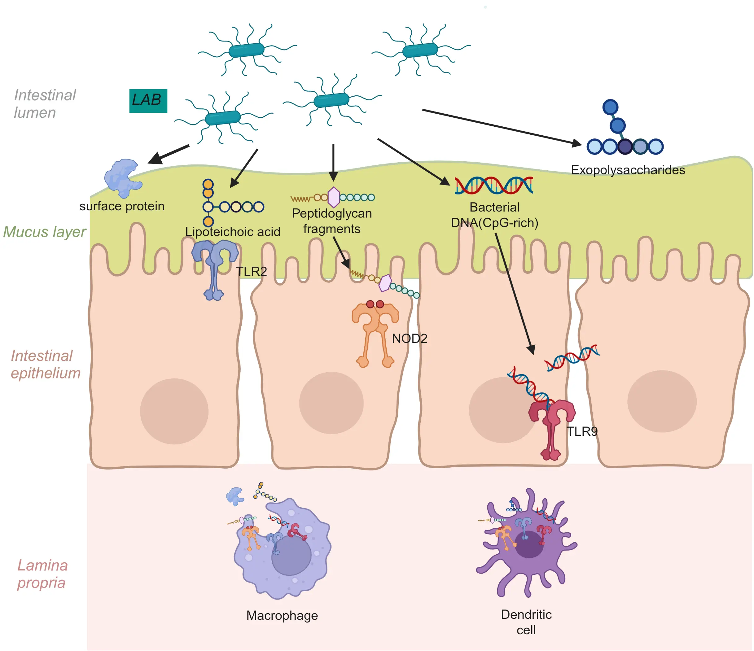
Figure 1. Pattern recognition of LAB at the intestinal mucosal interface. Notes: At the intestinal mucosal interface, lactic acid bacteria are detected by epithelial and immune cells through conserved microbial-associated molecular patterns—such as lipoteichoic acids, peptidoglycan fragments, surface proteins, exopolysaccharides, and DNA—engaging TLR2, NOD2, and TLR9 in a context-dependent manner shaped by ligand type, bacterial viability, dose, and cellular compartmentalization.
3.1.3. Cellular and Signaling Consequences of LAB Recognition
Following recognition of LAB by host pattern-recognition receptors, signal transduction induces changes in downstream cellular and immune effectors. In porcine intestinal epithelial cell models, Limosilactobacillus fermentum ZC529 activated the Keap1–Nrf2 pathway while suppressing NF-κB signaling, thereby maintaining epithelial barrier integrity under inflammatory stress [68]. In weaned piglets, a Limosilactobacillus reuteri strain isolated from Ningxiang pigs reduced intestinal IL-6 and TNF-α levels while increasing IL-10 levels [69]. In poultry macrophages, probiotic lactobacilli similarly influenced phagocytosis, nitric oxide production, cytokine expression, and TLR2/TLR4-related responses under heat stress [70]. In broiler chickens, dietary supplementation with Lacticaseibacillus paracasei NSMJ15 increased the proportion of jejunal CD3⁺CD4⁺CD8⁻ T cells and decreased CD3⁺CD8⁺TCRγδ⁺ T cells [71], while embryonic exposure to poultry-derived lactobacilli increased the expression of BAFF, BAFF-R, IL-10, TGF-β, and cytokines associated with multiple T-helper cell programs in the bursa of Fabricius [72]. These studies indicate that LAB can modulate host immune status by altering the expression of downstream inflammation-related signaling pathways and the distribution of T-cell subsets, potentially contributing to the attenuation of intestinal inflammation.
This may represent a key mechanistic link between LAB-mediated innate immune recognition and adaptive immunity, particularly through the functional reprogramming of antigen-presenting cells, such as dendritic cells (DCs). In porcine monocyte-derived DCs, Lactobacillus johnsonii increased the expression of MHC-II and the costimulatory molecule CD80, enhanced DC-mediated CD4+ T-cell proliferation, and altered cytokine production as well as TLR2/TLR6 expression. In DC–T-cell co-culture systems, these changes were associated with differential polarization toward Th1, Th2, and Treg-type responses, whereas cell-free bacterial supernatants preferentially promoted Th2/ and Treg-associated responses [73]. Similarly, oral administration of Lactobacillus delbrueckii to suckling piglets promoted DC activation and was accompanied by changes in NF-κB and MAPK signaling pathways, providing in vivo evidence supporting DCs as intermediary cells linking LAB sensing to downstream adaptive immune responses [74].
More direct mechanistic evidence linking LAB to B-cell and mucosal IgA responses has been established in pigs. Lacticaseibacillus rhamnosus GG promoted early B-cell lineage development in piglets and increased IgA production in the intestinal lamina propria [75]. Importantly, this effect was closely associated with the LAB-derived p40 protein. p40 activated EGFR/AKT and NF-κB signaling in porcine intestinal epithelial cells and induced the secretion of a proliferation-inducing ligand (APRIL), which subsequently promoted IgA production by B cells. Further studies identified candidate epithelial binding partners of p40, including ARF4 and DIF3, thereby establishing a molecular link among a defined LAB effector molecule, epithelial cell signaling, and downstream B-cell function [75]. This pathway illustrates how microbial signals can be translated into adaptive mucosal immunity through epithelial cell–B-cell communication rather than simply through changes in lymphocyte abundance. More broadly, BAFF, APRIL, TGF-β, and IL-10 collectively constitute a signaling network that regulates B-cell survival, IgA class switching, and plasma-cell differentiation, although the relative contributions of these pathways in livestock and poultry remain to be determined [76,77].
Overall, the available evidence supports a hierarchical mechanistic model in which LAB-associated molecules are first sensed by intestinal epithelial cells and innate immune cells, followed by intracellular signal transduction and functional reprogramming of antigen-presenting cells; these changes subsequently shape T-cell polarization and B-cell/IgA responses. However, the strength of evidence differs across immune pathways. The p40–epithelial signaling–APRIL–IgA axis established in pigs provides direct mechanistic evidence, whereas many current studies in poultry primarily provide evidence of immune phenotypes or transcriptional associations and require further mechanistic investigation.
3.2. Metabolite-Mediated Signaling
3.2.1. Short-Chain Fatty Acids
Organic acids represent an important class of bioactive products associated with LAB metabolism. A defining metabolic characteristic of LAB is the fermentation of carbohydrates with lactic acid as a major end product. Depending on the species, fermentation pathway, substrate availability, and environmental conditions, some LAB can also produce acetate and other organic acids [47]. Metabolic studies conducted under defined culture or fermentation conditions demonstrate that LAB can produce lactic acid and, in some strains, additional organic acids [47].
Importantly, SCFAs detected in the intestinal environment should not all be considered direct metabolic products of LAB. LAB primarily contribute lactate and, in some strains, acetate or other organic acids through their own fermentation pathways. In contrast, propionate and particularly butyrate are generally produced predominantly by other members of the gut microbiota. LAB can nevertheless contribute indirectly to their formation through microbial cross-feeding: lactate and acetate released by LAB can serve as substrates for specialized intestinal bacteria that subsequently generate butyrate and other fermentation products [12,48,78]. Thus, LAB supplementation may alter intestinal SCFA profiles through two distinct but interconnected routes—direct production of selected fermentation products and indirect modulation of community-level metabolism. This distinction is important when interpreting increases in intestinal or fecal SCFAs following LAB supplementation, because such changes should not automatically be attributed to direct production by the administered LAB strain.
SCFAs are increasingly recognized as signaling metabolites that link microbial metabolism with epithelial and immune functions. Acetate, propionate, and butyrate can signal through G protein-coupled receptors, including FFAR3 (GPR41), FFAR2 (GPR43), and HCAR2 (GPR109A), depending on the metabolite and host cell type, while propionate and butyrate also exert important epigenetic effects through inhibition of histone deacetylases (HDACs) [12,79]. Through these pathways, SCFAs can regulate epithelial gene expression, reinforce tight-junction integrity, influence mucus production, and modulate inflammatory signaling in epithelial cells, macrophages, and dendritic cells. These effects can restrain excessive NF-κB-associated inflammatory responses while preserving mucosal defense, thereby contributing to intestinal immune homeostasis rather than generalized immune suppression [12,79].
The immunological influence of SCFAs further extends to adaptive immunity. Butyrate and propionate can promote the differentiation and functional stability of regulatory T cells through metabolic and epigenetic mechanisms, while SCFA-dependent signaling can also influence T-cell polarization and mucosal cytokine production, including IL-22 [80]. These responses contribute to immune tolerance, epithelial repair, and antimicrobial defense.
Within the gastrointestinal tract, the relationship between LAB metabolism and SCFAs is further complicated by microbial cross-feeding. Acetate, propionate, and butyrate are generated by the collective metabolic activity of diverse intestinal microorganisms rather than by LAB alone [12,78]. Metabolites released by one microbial population can serve as substrates for another; in particular, lactate and acetate can be utilized by specialized intestinal bacteria to produce butyrate and other fermentation products [48]. In weaned piglets, supplementation with a complex probiotic preparation containing Lactobacillus acidophilus altered fecal SCFA composition and was accompanied by improved average daily gain [54]. However, because the intervention contained multiple probiotic organisms and SCFAs were measured at the intestinal community level, this study supports an association between probiotic-induced microbial metabolic changes and host performance rather than direct SCFA production by L. acidophilus. SCFAs are also increasingly recognized as modulators of porcine intestinal barrier function and mucosal immunity [81].
Therefore, LAB may influence the intestinal SCFA profile both through their own metabolic products and indirectly by modifying substrate availability, microbial community structure, and cross-feeding networks. These findings indicate that changes in fecal or intestinal SCFA concentrations following LAB supplementation may be mediated by interactions between LAB and the resident microbiota, as well as by LAB-induced effects on the host that subsequently alter the intestinal microbial community. These mechanisms warrant further investigation.
3.2.2. Bacteriocins
Bacteriocins are ribosomally synthesized antimicrobial peptides produced by diverse bacteria, including many LAB species. Among LAB-derived bacteriocins, nisin produced by Lactococcus lactis is the most extensively characterized, while other representative compounds include plantaricins produced by Lactiplantibacillus plantarum and enterocins produced by Enterococcus spp. Their primary biological function is antimicrobial activity, which may be narrow- or broad-spectrum depending on the bacteriocin [15,50]. For example, nisin binds to lipid II, an essential precursor of bacterial cell-wall synthesis, and subsequently promotes membrane permeabilization and pore formation, resulting in rapid inhibition of susceptible Gram-positive bacteria [82]. Therefore, the contribution of bacteriocins to host health should first be considered in the context of pathogen control and microbial ecological regulation rather than being interpreted solely as direct immune stimulation.
In addition to their antimicrobial activity, LAB-derived bacteriocins may directly or indirectly influence epithelial barrier function and inflammatory responses. In a recent in vitro study, nisin, enterocin Gr17, and plantaricin RX-8 reduced Listeria monocytogenes adhesion and invasion in Caco-2 intestinal epithelial cells, preserved tight junction proteins, including ZO-1, occludin, and claudin-1, and attenuated the expression of IL-6, IL-1β, and TNF-α by reducing MAPK and NF-κB activation [14]. In porcine peripheral blood leukocytes, nisin exerted concentration- and activation-state-dependent effects. At higher concentrations, nisin stimulated proliferation, and IL-1β and IL-6 production, and increased the proportion of CD4+CD8+ T cells in unstimulated leukocytes, whereas in Escherichia coli-stimulated cells it reduced proliferative activity, phagocytosis, and IL-6 production [51]. These bidirectional responses indicate that nisin does not uniformly enhance or suppress immunity; instead, its effects depend on the physiological and inflammatory state of immune cells. At the same time, in vitro studies cannot establish that bacteriocins directly regulate host immunity in vivo.
In vivo studies have shown that, in broilers challenged with Clostridium perfringens, dietary nisin supplementation alleviated intestinal injury, reduced serum TNF-α concentrations, increased jejunal claudin-1 expression, and decreased apoptosis-related signaling, while also modifying cecal microbial composition [52]. More recently, dietary nisin supplementation in yellow-feathered broilers increased serum IgY concentrations and reduced circulating IL-1β, IL-6, and TNF-α, together with changes in intestinal SCFA concentrations and microbial community structure [83]. In lactating cows with subclinical mastitis, intramammary administration of nisin Z increased bacteriological cure rates and reduced somatic cell counts and N-acetyl-β-d-glucosaminidase (NAGase) activity in milk [53]. These studies indicate that nisin can modulate inflammatory and immune-associated phenotypes in animal models. Nevertheless, because antimicrobial activity, microbiota remodeling, barrier protection, and immune responses occur simultaneously in vivo, the extent to which these outcomes reflect direct signaling by nisin versus secondary consequences of altered pathogen burden or microbiota composition remains unresolved. Future studies using microbiota-depleted or microbiota-controlled models could help determine whether bacteriocins exert direct effects on host immune responses independent of their antimicrobial and microbiota-mediated effects.
3.2.3. Exopolysaccharides
Exopolysaccharides (EPSs) are high-molecular-weight carbohydrate polymers synthesized by many LAB and either retained in close association with the bacterial surface or released into the extracellular environment. LAB-derived EPSs comprise structurally diverse homopolysaccharides and heteropolysaccharides that differ substantially in molecular weight, monosaccharide composition, glycosidic linkage, degree of branching, charge, and non-carbohydrate substituents. These physicochemical characteristics are important determinants of their biological activity; therefore, EPSs should not be considered a functionally uniform class of molecules [43]. In the context of LAB-mediated immunoregulation, this structural heterogeneity provides a plausible molecular basis for the marked strain-specificity of EPS-associated host responses.
Purified LAB-derived EPSs can directly interact with intestinal epithelial and immune cells and modulate inflammatory signaling. In porcine intestinal epithelial cells, EPS fractions produced by Lactiplantibacillus plantarum N14 were shown to attenuate enterotoxigenic Escherichia coli-induced inflammatory responses. In particular, acidic EPS reduced the expression of pro-inflammatory mediators, including IL-6, IL-8, and MCP-1, and enhanced the expression of negative regulators of Toll-like receptor signaling, such as IRAK-M and MKP-1. Mechanistic analyses implicated the RP105/MD1 complex, together with TLR-associated signaling, in these regulatory effects [44]. These observations indicate that EPS-mediated immunoregulation can involve active modulation of pattern-recognition signaling rather than merely forming a physical barrier around bacterial cells.
Comparable effects have been reported for EPSs from other LAB strains in porcine intestinal epithelial models. EPS isolated from Limosilactobacillus reuteri L26 attenuated the upregulation of pro-inflammatory mediators induced by Salmonella Typhimurium in IPEC-J2 cells, including changes in IL-6, IL-8, TNF-α, and TLR-associated responses [45]. Similarly, EPS production by Lactiplantibacillus plantarum T10 contributed substantially to its barrier-protective and anti-inflammatory effects in ETEC-challenged IPEC-J2 cells. The EPS fraction enhanced epithelial barrier function and modulated inflammatory responses through TLR-associated p38 MAPK signaling; however, the corresponding in vivo validation in that study was performed in a murine colitis model rather than in livestock [84]. Together, these findings support a direct capacity of defined LAB-derived EPSs to influence porcine epithelial immune signaling, while also illustrating that mechanistic validation at the whole-animal livestock level remains limited.
EPSs may additionally protect the intestinal mucosa through mechanisms that are not necessarily dependent on direct immune-receptor signaling. In a porcine small-intestinal segment perfusion model, reuteran and levan produced by Limosilactobacillus reuteri strains were evaluated during challenge with enterotoxigenic E. coli K88. Reuteran significantly reduced adherent ETEC, and the tested glycans reduced ETEC-associated intestinal fluid loss [46]. These findings demonstrate that LAB-derived EPSs can interfere with pathogen adhesion, thereby reducing epithelial exposure to microbial virulence factors. However, this anti-adhesive effect should be distinguished from direct immunomodulation, because reduced pathogen–epithelium contact may secondarily alleviate inflammatory pressure without requiring direct activation or inhibition of host immune receptors.
The immunological activity of EPSs is also strongly strain- and structure-dependent. EPSs purified from two Lacticaseibacillus casei strains, AP and AG, differed in carbohydrate content, elemental composition, and immunological activity. Both EPS preparations enhanced phagocytic activity and nitric oxide production in RAW264.7 macrophages, but the EPS from strain AP showed stronger activity; notably, phosphorus was detected in AP-derived EPS but not in AG-derived EPS [85]. These results reinforce the concept that apparently closely related LAB strains can produce chemically distinct EPSs with different biological activities. Nevertheless, because these experiments used a murine macrophage cell line and strains of human origin, they provide mechanistic evidence for structure-dependent EPS activity rather than direct livestock-specific proof.
Overall, LAB-derived EPSs can contribute to host protection through several complementary mechanisms, including modulation of epithelial pattern-recognition signaling, attenuation of excessive inflammatory responses, preservation of epithelial barrier integrity, and interference with pathogen adhesion. Importantly, these effects are determined by the physicochemical properties of individual EPSs and cannot be generalized across LAB species or strains. Current evidence is particularly informative in porcine intestinal epithelial systems, whereas direct in vivo evidence defining EPS-specific immune mechanisms in livestock—especially poultry—remains comparatively limited. Future studies integrating detailed structural characterization of purified EPSs with receptor-level analyses and controlled livestock and poultry intervention models will therefore be essential to establish structure–function relationships and determine whether specific EPS signatures can serve as reliable criteria for selecting immunomodulatory LAB strains.
3.2.4. Bile Acid-Associated Metabolites
Bile acids (BAs) represent another class of bioactive metabolites through which LAB may indirectly or directly influence host physiology and immune homeostasis (Figure 2). Unlike microbial fermentation products, primary BAs are synthesized from cholesterol by the host liver, subsequently conjugated predominantly with glycine or taurine, secreted into the intestine, and extensively recycled through the enterohepatic circulation. Once they enter the intestinal lumen, however, their chemical composition can be substantially modified by the gut microbiota, generating a structurally diverse BA pool with distinct signaling properties [19,56]. Therefore, the contribution of LAB to BA-mediated immunoregulation is more accurately described as LAB-associated modulation of the intestinal BA pool rather than direct production of the entire spectrum of secondary BAs.
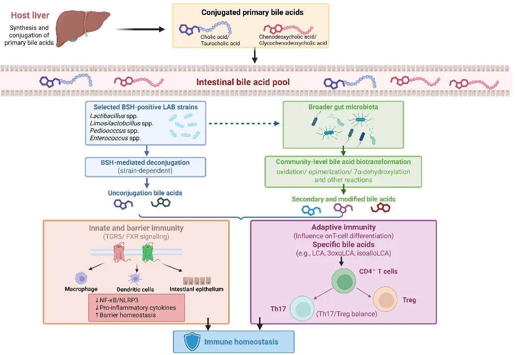
Figure 2. LAB-associated modulation of the bile acid pool and its potential impact on host immune homeostasis. Notes: LAB can reshape the intestinal bile acid pool through BSH-mediated deconjugation and microbiota remodeling. Microbe-derived bile acid metabolites subsequently regulate epithelial and immune responses through TGR5, FXR, and T-cell-associated pathways.
One of the best-characterized mechanisms by which LAB can directly modify BAs is bile salt hydrolase (BSH)-mediated deconjugation. BSH removes the amino acid moiety from conjugated BAs, producing unconjugated BA substrates that can subsequently undergo further microbial transformations. Importantly, BSH activity is not universal among LAB. Screening studies have shown that it is common in selected intestinal Lactobacillus species and strains but absent or substantially weaker in several other LAB taxa, indicating marked species- and strain-level heterogeneity [86]. Recent reviews likewise emphasize substantial variation in substrate specificity and catalytic activity among BSH enzymes from Lactobacillus, Bifidobacterium, and Enterococcus [58]. Thus, the ability of a LAB strain to modify the BA pool cannot be inferred solely from its taxonomic identity.
Specific LAB with experimentally characterized BSH activity have been identified. For example, the chicken-derived Ligilactobacillus salivarius NRRL B-30514 possesses a functionally characterized BSH with activity toward both glycine- and taurine-conjugated bile salts, providing direct evidence that certain poultry-associated LAB can enzymatically deconjugate host BAs [87]. In pigs, the Lactobacillus delbrueckii strain used by Hou et al. was shown to harbor the BSH2 gene and exhibit BSH activity in vitro; dietary supplementation enhanced ileal BA deconjugation and altered the enterohepatic FXR–FGF19 axis in vivo [59]. Broader phylogenomic analyses have also identified BSH-encoding members among Limosilactobacillus reuteri, Lactiplantibacillus plantarum, Lacticaseibacillus rhamnosus, Lacticaseibacillus casei, and several other intestinal lactobacilli, although BSH activity and substrate specificity remain strongly strain-dependent [88]. Importantly, these findings demonstrate LAB-mediated BA deconjugation, but they do not establish that these LAB strains directly generate the downstream secondary BAs responsible for immune signaling. Direct strain–enzyme–BA–immune causal evidence remains limited in livestock and poultry.
Following deconjugation, further BA transformations are carried out by the broader intestinal microbial community. Oxidation, epimerization, and, in particular, 7α-dehydroxylation generate a range of secondary and modified BAs. The latter pathway is restricted to a relatively small group of anaerobic intestinal bacteria carrying bile acid-inducible (bai) genes, including certain Clostridium species, rather than representing a general metabolic property of LAB [56]. Consequently, LAB can influence secondary BA profiles indirectly by providing deconjugated substrates, changing the abundance of BA-transforming microorganisms, or modifying the intestinal ecological environment. This community-level perspective is essential for avoiding over-attribution of secondary BA formation to LAB themselves.
Evidence from pigs directly supports this distinction. In growing-finishing pigs, dietary supplementation with Lactobacillus delbrueckii enhanced ileal BA deconjugation, increased fecal BA excretion, altered serum and hepatic BA composition, and modified the intestinal FXR–FGF19 axis, providing in vivo evidence that a LAB strain can reshape BA metabolism through BSH-related activity [59]. In weaned piglets, oral Lactiplantibacillus plantarum supplementation reduced ileal lithocholic acid concentrations and altered the abundance of several bacterial genera involved in BA biotransformation, including Clostridium, Parabacteroides, and Ruminococcus [60]. These findings suggest that LAB can modify the host BA pool through both direct enzymatic activity and indirect restructuring of BA-transforming microbial communities. However, neither study demonstrates that the administered LAB strain itself synthesized all of the secondary BAs detected in vivo.
Changes in BA composition are immunologically relevant because different BA species exhibit distinct affinities for host receptors and immune-regulatory targets. Two major signaling nodes are the nuclear receptor farnesoid X receptor (FXR) and the membrane G protein-coupled bile acid receptor TGR5. Through these pathways, BAs can regulate epithelial barrier integrity, inflammatory signaling, macrophage and dendritic-cell activity, and systemic metabolic responses [18,19]. In innate immune cells, TGR5 activation can increase intracellular cAMP and activate protein kinase A-dependent signaling, thereby attenuating excessive NF-κB activity and NLRP3 inflammasome activation. For example, microbiota-derived secondary BAs alleviated Staphylococcus aureus-induced mastitis in a mouse model through the TGR5–cAMP–PKA–NF-κB/NLRP3 axis [17]. Although this study provides useful mechanistic evidence, it does not demonstrate that the responsible secondary BAs were directly generated by LAB, nor does it establish an equivalent pathway in livestock.
BA-associated immune regulation also extends to adaptive immunity. Specific microbiota-derived LCA derivatives can directly influence CD4+ T-cell differentiation. In mechanistic mouse studies, 3-oxoLCA inhibited Th17 differentiation through direct interaction with RORγt, whereas isoalloLCA promoted regulatory T-cell differentiation through mitochondrial reactive oxygen species-dependent enhancement of FoxP3 expression [61]. These findings establish that microbial BA metabolites can shape the Th17–Treg balance, but they should be interpreted as evidence for the broader microbiota–BA–immune axis rather than as LAB-specific effects. Because BA pools and microbial BA-transforming capacities vary considerably among host species, extrapolation from murine and human systems to pigs and poultry requires caution.
Overall, current evidence supports a model in which LAB influence BA-associated immune signaling primarily by reshaping the intestinal BA pool, rather than by directly synthesizing the full range of secondary BAs. This modulation can occur through strain-specific BSH activity, changes in the abundance or function of other BA-transforming microorganisms, and downstream alterations in FXR-, TGR5-, and BA-responsive immune pathways. Direct evidence is currently strongest in swine for LAB-mediated changes in BA metabolism, whereas livestock- and poultry-specific studies linking defined LAB strains, individual BA metabolites, and immune outcomes remain limited. Future studies that combine strain-level BSH characterization, targeted BA metabolomics, microbiome functional analysis, and immune phenotyping will be required to resolve the causal contribution of LAB to the BA–immune axis in animal production.
3.3. Strain-Specific Effects on Immune Responses
Accumulating evidence demonstrates that the immunomodulatory effects of LAB are highly strain-specific and context-dependent. Even at the species level, different strains of lactic acid bacteria (LAB) exhibit marked heterogeneity in functional properties, including adhesion capacity, metabolite production, and immunomodulatory activity. This strain-specific functional diversity is fundamentally associated with genomic variation. Comparative genomic analyses have revealed extensive intra-species genetic diversity among LAB, with substantial differences in gene content even among strains of the same species. For instance, pan-genome analysis of 34 Lacticaseibacillus paracasei strains demonstrated an open pan-genome characterized by extensive gene gain and loss [24], and similar genomic diversity has been reported across multiple LAB species [25]. Importantly, higher genome sequence identity between strains does not reliably predict similarity in functional phenotypes, including immunomodulatory activity [26], underscoring that subtle genomic and regulatory differences, rather than species-level taxonomy alone, shape strain-specific host interactions [27,28]. As genomic characterization of LAB continues to advance, research focus has increasingly shifted from species-level classification toward strain-level resolution, reflecting the recognition that probiotic functionality is a strain-dependent trait driven by both genomic content and regulatory architecture. Importantly, strain-specific functional variation is also evident in vivo. Studies in pigs, poultry, and calves demonstrate that individual LAB strains differ in their capacity to reshape the intestinal microbiota and in the magnitude of their effects on growth, intestinal health, immune status, and disease resistance. Even closely related strains can generate distinct microbial and host responses.
Comparative characterization of broiler-derived Limosilactobacillus reuteri isolates demonstrated substantial within-species functional heterogeneity. Among the eight L. reuteri isolates examined, LR21, LR29, and LR61 exhibited relatively high surface hydrophobicity, whereas LR42 showed the strongest adhesion to Caco-2 cells. Moreover, reuterin production was detected only in LR21, LR42, LR61, and LR77, further demonstrating that closely related strains of the same species can differ markedly in probiotic-associated functional traits [89].
Strain-specific functional differences have also been demonstrated in porcine systems. In pigs, two strains of the same species, Lactiplantibacillus plantarum 22F and 25F, produced distinct effects on intestinal microbial parameters despite both showing probiotic potential, supporting functional variation at the strain level [90]. More recent mechanistic evidence further supports this concept. Seven L. plantarum strains isolated from porcine colostrum and milk induced markedly different immune responses in porcine intestinal epithelial cells. In particular, strain 4M4-417 strongly modulated the expression of PGRP1, PGRP3, NOD1, NOD2, and pBD1, whereas strain 4M4-326 showed a particularly strong capacity to enhance IFN-α and IFN-β expression following poly(I:C) challenge [91]. These findings demonstrate that even strains belonging to the same species can differ substantially in their interactions with porcine intestinal innate immune pathways. However, the latter evidence was obtained in a porcine epithelial cell model and therefore requires further in vivo validation in pigs. Additional L. plantarum strains, including P-8 in piglets, HJLP-1, HJZW08, and LPJZ-658 in broilers, and 299v in calves, have also been associated with distinct changes in gut microbiota, growth performance, intestinal health, and immune-related outcomes (Table 2).
Table 2. Strain-specific effects of LAB on gut microbiota, growth, and health in livestock and poultry: insights from Lactiplantibacillus plantarum.
|
LAB Species |
Strain ID |
Effects on the Gut Microbiota of the Animal |
Effects on the Growth and Health of the Animal |
References |
|---|---|---|---|---|
|
L. plantarum |
22F |
Pigs: Increased viable fecal lactobacilli and reduced Enterobacteriaceae compared with the control. |
Improved ADG and feed conversion relative to controls and increased intestinal villus height and VH:CD ratio. |
[90] |
|
L. plantarum |
25F |
Pigs: Increased fecal lactobacilli and reduced Enterobacteriaceae, although viable lactobacilli counts were lower than those observed with the other probiotic treatments. |
Improved growth efficiency and intestinal morphology compared with the untreated control. |
[90] |
|
L. plantarum |
P-8 |
Weaned piglets: Altered ileal and colonic microbiota, increased recovery of L. plantarum, and changed microbial functional pathways related to carbohydrate degradation and nutrient utilization. |
Improved feed conversion, intestinal morphology and antioxidant capacity; overall body weight and early-stage ADG were improved relative to controls. |
[92] |
|
L. plantarum |
HJLP-1 |
Broilers: Increased cecal microbial richness and enriched SCFA-associated bacteria including Ruminococcus and Lachnospiraceae; cecal acetate and butyrate concentrations were increased. |
Increased average daily weight gain and reduced feed-to-gain ratio; increased IgM, IgY and IL-10, decreased IL-1β, and enhanced antioxidant capacity. |
[93] |
|
L. plantarum |
HJZW08 |
Broilers: Altered α-diversity and cecal microbial structure and enriched taxa including Bacteroides, Lactimicrobium massiliense, Intestinimonas butyriciproducens, and Barnesiella viscericola. |
Increased BW and ADG; enhanced IgA, IgY, IL-10 and antioxidant status while decreasing IL-1β, IL-6 and TNF-α. |
[94] |
|
L. plantarum |
LPJZ-658 |
Broilers: Altered cecal microbial diversity and composition, reduced Streptococcus, Veillonella, Neisseria, and Haemophilus, and increased several potentially beneficial taxa including Bacteroides, Phascolarctobacterium, Bacillus, and Akkermansia. |
Increased ADG and ADFI and reduced FCR; improved intestinal morphology, carcass traits and meat-quality-related indices. |
[95] |
|
L. plantarum |
299v |
Preweaning calves: Increased fecal Bacteroidetes and bacterial diversity indices while reducing Firmicutes and Proteobacteria. |
Increased starter intake and ADG, reduced diarrhea incidence, increased IgA, IFN-γ and soluble CD4, and reduced IL-6. |
[96] |
Note: ADG, average daily gain; BW, body weight; FCR, feed conversion ratio; ADFI, average daily feed intake; VH:CD, villus height-to-crypt depth ratio
Strain-specific differences are also evident in LAB-derived metabolites. In Staphylococcus haemolyticus-challenged RAW264.7 murine macrophages, metabolites derived from two Lactiplantibacillus plantarum strains induced distinct transcriptional responses. qRT-PCR showed that metabolites from strain AD73 increased iNOS and IL-6 mRNA expression by 22.59- and 16.82-fold, respectively, whereas metabolites from strain TISTR2070 preferentially increased COX-2 mRNA expression by approximately 18.43-fold [57]. These differential responses, together with strain-resolved metabolomic profiles, support an association between strain-dependent metabolic output and immune activity. Because this study used a murine macrophage model developed in the context of bovine mastitis, it should be interpreted as mechanistic evidence rather than direct evidence in cattle. Collectively, these findings demonstrate that LAB-mediated immunomodulation cannot be generalized at the species level and support the need for strain-level functional validation. These biological differences provide the rationale for the mechanism-guided strain-selection framework discussed in Section 5.1.
4. Practical Applications in Livestock and Poultry Production
4.1. Modulation of the Gut Microbiota
LAB can reshape the intestinal microbial ecosystem through competition for nutrients and adhesion sites, antimicrobial activity, and modification of the intestinal physicochemical and metabolic environment [3,29]. However, available studies do not indicate a uniform shift toward higher microbial diversity. Instead, changes in community richness, structure, specific taxa, and metabolic functions vary according to LAB strain, dose, host species, and physiological context.
In pigs, supplementation with Limosilactobacillus reuteri ZLR003 increased microbial species richness and the abundance of Bacteroidetes, while increasing Bifidobacterium, Lactobacillus, Christensenellaceae R-7 group, and Rikenellaceae RC9 gut group and decreasing Clostridium sensu stricto 1, Terrisporobacter, Blautia, Corynebacterium, and Streptococcus [97]. The two doses also produced distinct microbial and metabolic responses; notably, acetate and butyrate concentrations were higher at the lower dose than at the higher dose, indicating a nonlinear dose–microbiota relationship [97]. By contrast, Lactiplantibacillus plantarum P-8 did not significantly alter ileal α-diversity but significantly reduced the Shannon and Simpson indices of the colonic microbiota, while enriching L. plantarum itself [92]. Functional profiling further showed enrichment of microbial pathways involved in galactose and stachyose degradation and other nutrient-utilization processes. Thus, beneficial host responses following LAB supplementation are not necessarily accompanied by increased microbial α-diversity [92].
A mixed L. plantarum–Bacillus subtilis fermented feed produced a different pattern in Bamei pigs. After 50 days, microbial community structure differed among dietary groups, and fermented-feed treatments generally maintained higher diversity than the control or antibiotic treatment. The mixed treatment increased Lactobacillus abundance and suppressed Clostridium and Streptococcus; at day 50, Lactobacillus accounted for approximately 9.5% of the microbiota in the mixed-fermented group compared with 1.3% in the basal-diet control [98]. Because this intervention also contained B. subtilis, however, these microbial changes cannot be attributed specifically to LAB.
Comparable strain- and context-dependent changes have been reported in poultry. In broilers, L. plantarum HJZW08 altered cecal α-diversity and community structure, increased Actinobacteriota at the higher dose, and enriched taxa including Lactimicrobium massiliense, Intestinimonas butyriciproducens, and Barnesiella viscericola [94]. These changes were accompanied by increased isobutyrate and isovalerate concentrations and improved immune indices, although the study does not establish that the microbial changes directly mediated the immune effects.
Under pathogen challenge, LAB appear to act partly by limiting infection-associated dysbiosis rather than simply increasing overall diversity. In Salmonella Enteritidis-infected broilers, supplementation with L. plantarum Lp71 and Enterococcus faecium Ef72 prevented the infection-associated decline in cecal microbial diversity, reduced microbiota dysbiosis, and restored taxa including Oscillospiraceae and Lachnospiraceae; it also counteracted the abnormal expansion of Akkermansia muciniphila induced by infection [99]. Functional prediction indicated recovery of pathways related to transcription and signal transduction [99]. Similarly, in Salmonella Typhimurium-challenged yellow-feathered broilers, L. plantarum GX17 did not significantly alter Chao1 or Shannon α-diversity, but β-diversity differed among treatment groups (Adonis, p = 0.021); GX17 increased Lactobacillus abundance and counteracted the infection-associated increase in Anaerotruncus [100].
Evidence from cattle further indicates that LAB-associated microbial responses depend strongly on production stage and dietary context. During a 163-day feedlot study, probiotic LAB altered the relative abundance of major fecal bacterial groups, with Clostridiaceae, Lachnospiraceae, Ruminococcaceae, and Bifidobacteriaceae forming prominent core communities. Interestingly, microbial diversity was influenced more strongly by the administration period than by probiotic supplementation itself, while predicted metabolic functions during the first 40 days were enriched for complex plant-polysaccharide degradation and amino-acid-related metabolism [5].
Collectively, these studies show that LAB-mediated microbiota modulation cannot be defined simply as an increase in microbial diversity or enrichment of a universal set of “beneficial bacteria”. Depending on the strain and host context, LAB may increase, decrease, or leave α-diversity unchanged while still producing marked changes in β-diversity, specific microbial taxa, and metabolic functions. A more consistent pattern is the restructuring of particular ecological and functional features, including suppression of pathogen-associated taxa, enrichment of fermentative or SCFA-associated microorganisms, and modification of carbohydrate and nutrient metabolism. However, because microbiota and immune outcomes are usually measured concurrently, these associations should not be interpreted as proof that microbial restructuring directly causes the observed immune benefits.
4.2. Promoting Growth in Livestock and Poultry
Improvement of productive performance is one of the major practical outcomes associated with the use of selected LAB strains in animal production. LAB supplementation has been reported to influence average daily gain (ADG), feed conversion efficiency, nutrient digestibility, and intestinal development, although the magnitude of these responses varies according to strain, dosage, animal age, diet, and production conditions. Representative applications of LAB supplementation in livestock and poultry are summarized in Table 3.
Table 3. Effects of LAB in livestock and poultry production.
|
LAB Strain |
Host Species |
Supplementation Methods and Dosages |
Primary Effects |
References |
|---|---|---|---|---|
|
Lactobacillus lactis (LL) |
Growing-finishing pigs |
Basal diet supplemented with 0.01% and 0.03% LL (5.0 × 10¹² CFU/g) |
Improves growth performance and meat quality |
[101] |
|
Lactiplantibacillus plantarum BG0001 |
Weaned piglets |
Basal diet supplemented with 0.1% and 0.2% BG0001 |
Improves growth performance and modulates fecal microbiota |
[102] |
|
Lactiplantibacillus plantarum HW1 |
Broiler chickens (necrotic enteritis challenge model) |
Basal diet supplemented with HW1 (4 × 10⁶ CFU/g) |
Alleviates intestinal damage and enhances growth performance |
[103] |
|
Lacticaseibacillus rhamnosus SN211 & Lactiplantibacillus plantarum SN212 |
Broiler chickens (Salmonella Typhimurium infection) |
0.2 mL of bacterial (1 × 10⁹ CFU/mL) suspension via intragastric gavage |
Exhibits anti-Salmonella activity and improves safety outcomes |
[104] |
|
L. plantarum NC8 |
Poultry (Salmonella Gallinarum infection) |
Bacterial (1 × 10⁸ CFU/mL) suspension via intragastric gavage |
Exerts immunomodulatory effects and enhances resistance to Salmonella |
[105] |
|
L. plantarum GB LP1 |
Newborn calves |
4 g of bacterial powder (1.2 × 10⁹ CFU/g) added to milk replacer once daily |
Functions as a direct-fed microbial, promoting growth and improving intestinal health |
[106] |
|
Lactiplantibacillus plantarum (LP) DSA 20174 + Bacillus toyonensis (BT) ATCC 55050 |
Pre-weaned Holstein calves |
Basal diet supplemented with LP (1 × 10¹⁰ CFU/calf/day) and BT (3 × 10⁹ CFU/calf/day) |
Enhances growth performance, improves blood biochemical parameters, and modulates fecal microbiota |
[107] |
In swine, dietary supplementation with Lactiplantibacillus plantarum P-8 improved feed conversion ratio, intestinal morphology, and antioxidant capacity in weaned piglets and was accompanied by improved growth performance [92]. Similarly, supplementation with the swine-derived Pediococcus acidilactici FT28 improved feed utilization and nutrient digestibility, supporting better growth efficiency in weaned piglets [108]. Other feeding studies have also reported improvements in growth-related traits following supplementation with LAB such as Lactobacillus lactis and Lactiplantibacillus plantarum in growing-finishing or weaned pigs [101,102]. These findings indicate that selected LAB strains can contribute to improved productive efficiency, although responses should not be generalized across strains or production systems.
Potential production benefits of LAB have also been observed in poultry and cattle. In poultry, dietary Lactiplantibacillus plantarum supplementation has been associated with improvements in feed efficiency, nutrient utilization, intestinal health, and overall growth performance [109]. In neonatal and pre-weaning calves, L. plantarum-based direct-fed microbial supplementation has likewise been associated with improved growth-related outcomes and gastrointestinal health [106,107]. Together, these studies indicate that LAB can support productive performance across different livestock species, but the magnitude and reproducibility of the response depend strongly on strain characteristics, delivery method, host developmental stage, diet, and farm conditions. Therefore, improvements in growth performance should be regarded as strain- and context-dependent outcomes rather than universal properties of LAB.
4.3. Enhancing Disease Resistance in Livestock and Poultry
Enhancing resistance to enteric and other infectious challenges is another important application of LAB in livestock and poultry production. LAB may contribute to disease resistance through several complementary processes, including competitive exclusion of pathogens, production of antimicrobial compounds, maintenance of epithelial barrier integrity, and modulation of host inflammatory and immune responses [4,67,99]. However, most evidence derives from experimental challenge models rather than population-level disease incidence; these findings therefore primarily support enhanced disease resistance rather than demonstrated reductions in field disease incidence.
In pigs, protective effects have been demonstrated particularly against enterotoxigenic Escherichia coli (ETEC). In young piglets challenged with ETEC K88, dietary Lactiplantibacillus plantarum prevented challenge-induced diarrhea and counteracted increases in intestinal permeability and plasma endotoxin levels, as well as villus shortening, crypt deepening, and reductions in jejunal ZO-1 and occludin expression [110]. Similarly, prophylactic administration of microencapsulated Lactiplantibacillus plantarum 22F/25F and Pediococcus acidilactici 72N reduced fecal E. coli and ETEC virulence-gene abundance, decreased IL-1α, IL-6, IL-8, and TNF-α, and improved villus morphology and growth performance in ETEC-challenged weaned piglets [111].
Comparable protection has been observed in poultry. In Salmonella Typhimurium-challenged yellow-feathered broilers, L. plantarum GX17 mitigated infection-associated weight loss; at 5 d post-infection, the untreated infected birds weighed approximately 24.85 g less than GX17-treated infected birds. Salmonella burdens in the liver, spleen, and cecum of untreated infected birds were approximately 12.23-, 12.65-, and 11.92-fold higher, respectively, than those in GX17-treated birds [100]. In a necrotic-enteritis model, dietary L. plantarum HW1 increased ADG while reducing intestinal lesion scores, IL-1β and TNF-α, serum D-lactate and diamine oxidase, and cecal counts of C. perfringens and other potentially pathogenic bacteria; simultaneously, ileal IL-10, occludin, and ZO-1 were increased [103].
Evidence in cattle also indicates disease-related benefits, although outcomes are not uniformly positive. In neonatal dairy calves, oral administration of L. reuteri TP1.3B or L. johnsonii TP1.6 for 10 d reduced diarrhea incidence and improved fecal scores, whereas ADG, circulating cytokine expression, IgA concentrations, and fecal pathogen abundance were not significantly affected, and responses varied between calving seasons [112]. In lactating cows with subclinical mastitis, intramammary nisin Z, a bacteriocin produced by Lactococcus lactis, achieved an overall bacteriological cure rate of 65.2% compared with 15.9% spontaneous recovery in untreated controls; cure rates were 90.1% for Streptococcus agalactiae, 50.0% for Staphylococcus aureus, and 58.8% for coagulase-negative staphylococci, accompanied by reductions in milk somatic-cell and NAGase indices [53].
Collectively, these studies indicate that disease protection by LAB is expressed through different endpoints across host species, including reduced pathogen burden, diarrhea, tissue injury, barrier disruption, and excessive inflammatory responses. Importantly, the magnitude and nature of protection are strain-, pathogen-, and host-dependent, and improvement in one disease-related endpoint does not necessarily imply broader enhancement of immunity or productivity.
5. Challenges in Practical Implementation
In the practical application of LAB, a series of challenges remain, primarily involving strain selection and application methods (Figure 3).
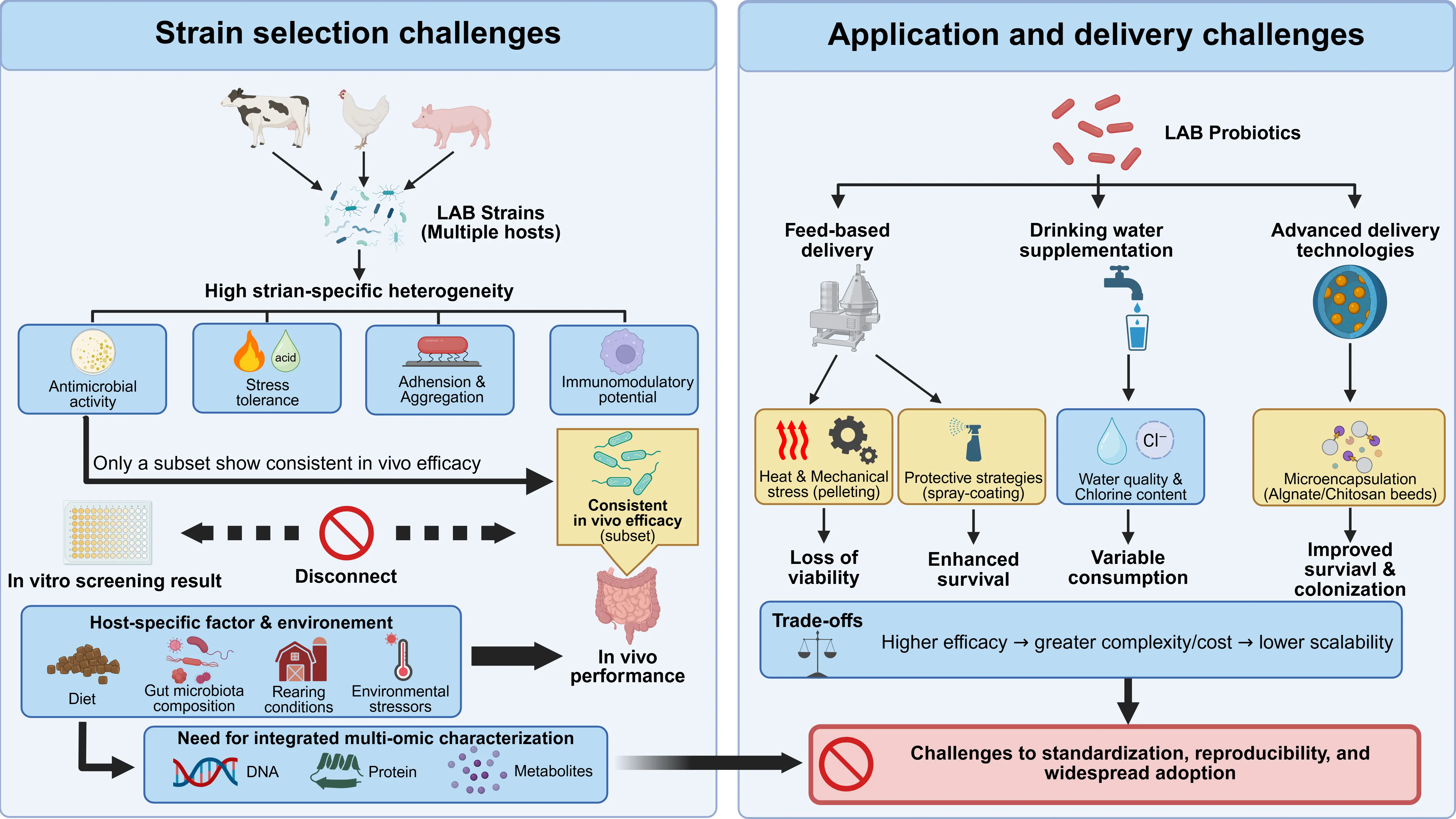
Figure 3. Practical challenges limiting the effective application of LAB probiotics in livestock and poultry.
5.1. Strain Selection
As discussed in Section 3.3, LAB functionality is highly strain-specific and is further influenced by host diet, resident microbiota, rearing conditions, and other environmental factors [113,114,115]. This variability complicates the identification of strains with reproducible efficacy across production systems. Therefore, practical strain selection should move beyond species-level taxonomy or isolated in vitro traits toward an integrated, mechanism-guided framework.
A practical mechanism-guided selection strategy should integrate genomic, metabolic, and host-response features in a stepwise manner. At the first stage, candidate strains should undergo strain-level taxonomic confirmation and genomic safety screening to exclude isolates carrying acquired antimicrobial-resistance genes, virulence-associated determinants, or potentially transferable mobile genetic elements [116]. Among strains that pass this safety screen, functional genomic features related to gastrointestinal survival and host interaction—including genes involved in acid and bile tolerance, mucus adhesion, carbohydrate utilization, exopolysaccharide biosynthesis, bacteriocin production, and other defined probiotic effector functions—can be used to identify candidates with greater functional potential [117]. These genomic predictions should then be verified phenotypically by measuring acid and bile tolerance, epithelial adhesion, aggregation, organic-acid production, bacteriocin activity, exopolysaccharide production, and substrate-utilization capacity [89,108].
The most promising strains should subsequently be evaluated in host-relevant cell models. Practical readouts may include preservation of epithelial barrier function, such as ZO-1 and occludin expression; modulation of inflammatory pathways such as NF-κB and MAPK; dendritic-cell activation and expression of costimulatory molecules; cytokine responses; and, where relevant, APRIL/IgA-associated mucosal immune pathways. Candidates showing consistent activity across these assays should finally be validated in the target livestock species using endpoints such as growth performance, pathogen burden, intestinal morphology, barrier integrity, inflammatory responses, and microbiota/metabolite changes. Thus, strain selection can be organized as a sequential pipeline of safety exclusion, functional prioritization, mechanistic host-cell validation, and target-animal confirmation, rather than relying on a single in vitro probiotic trait.
5.2. Application Methods
Practical deployment of LAB probiotics in animal production hinges on the route and formulation used to deliver viable microbes to the gastrointestinal tract, with each strategy presenting distinct operational considerations.
The most common approach in commercial systems remains dietary inclusion—either through direct mixing into feed or coating onto pellets—enabling large-scale delivery during routine feeding, although thermal and mechanical stress during pelleting can substantially reduce bacterial viability, especially for non-spore-forming strains, thereby limiting functional efficacy unless protective formulations are employed (e.g., spray-coating technologies) to enhance survival through feed processing and storage [118].
Drinking water supplementation represents an alternative that can ensure more uniform intake and has been shown in broiler studies to improve performance and immunological indices compared with feed delivery [119]. However, this route can be constrained by water quality, chlorine content, and variable individual consumption patterns under field conditions.
Beyond feed and water, microencapsulation and targeted delivery technologies—such as alginate or chitosan beads and other protective matrices—have emerged to shield LAB from gastric acidity and improve colonization, yet these approaches introduce additional costs and formulation complexity that must be balanced against scalability for routine use [120].
Collectively, while multiple administration routes can effectively introduce LAB probiotics into livestock and poultry, each method demands tailored strategies to preserve microbial viability, achieve reliable host colonization, and align with the operational realities of large-scale production systems.
5.3. Safety Considerations
Although many LAB have a long history of use as probiotics, their safety should be evaluated at the strain level rather than inferred solely from species identity. A primary concern is the presence and potential transferability of antimicrobial-resistance determinants. Whole-genome sequencing can be used to identify acquired resistance genes, virulence-associated determinants, and mobile genetic elements, whereas phenotypic antimicrobial susceptibility testing remains necessary to confirm that predicted resistance traits are functionally expressed [116,117]. Particular attention should be paid to resistance genes located on plasmids, transposons, or other mobile elements because of their potential horizontal transfer to commensal or pathogenic bacteria.
Safety evaluation should also include phenotypic traits that cannot be fully inferred from genome sequence alone. Candidate LAB strains should be assessed for hemolytic or cytotoxic activity, production of undesirable metabolites such as biogenic amines or excessive D-lactate, and the absence of other strain-specific traits that may adversely affect host health. This is especially important for taxa that contain opportunistic pathogens, such as some Enterococcus strains, for which probiotic functionality and safety must be demonstrated independently at the strain level. In vivo tolerance studies should further confirm that the proposed dose and administration period do not induce adverse clinical, inflammatory, or metabolic responses in the target livestock or poultry species.
Finally, safety assessment should extend beyond the individual animal to consider microbial persistence, environmental dissemination, and the potential transfer of antimicrobial-resistance determinants within farm ecosystems. Therefore, a comprehensive safety framework for LAB application should integrate genomic screening, phenotypic safety testing, host-specific in vivo evaluation, and post-application monitoring. Such an approach is essential to ensure that strains selected for immunomodulatory or production benefits do not introduce unintended risks to animal health, microbial ecology, or antimicrobial-resistance control.
6. Conclusions, Limitations, and Recommendations
This review summarizes the current understanding of how LAB regulate immune function in livestock and poultry through multiple interconnected mechanisms. LAB can directly interact with host pattern-recognition receptors, including TLRs, NOD-like receptors, and other sensing systems, through cell wall components, surface molecules, and secreted factors, thereby modulating epithelial and immune cell responses. LAB also influence intestinal immune homeostasis indirectly by reshaping the gut microbiota and regulating microbial metabolites, including SCFAs and bile acid derivatives, which can further affect epithelial integrity, inflammatory signaling, and systemic immune responses. Representative mechanisms, such as the LAB–EGFR/AKT–APRIL–IgA axis, further illustrate how specific LAB-derived factors can connect microbial signals with mucosal immune regulation. Importantly, these effects are highly strain- and context-dependent and cannot be generalized across LAB strains, hosts, diets, or production conditions. Current evidence is also limited by substantial heterogeneity among experimental systems, incomplete mechanistic validation in target livestock species, insufficient causal evidence linking microbiota or metabolites to immune outcomes, and limited large-scale commercial validation.
Future research should move beyond descriptive assessments toward mechanism-driven, strain-specific, and standardized evaluation of LAB. Integrating strain-level genomic, transcriptomic, metabolomic, and phenotypic data with host immune and microbiome profiling will be important for identifying functional determinants and distinguishing association from causation. In parallel, microbiome-guided precision nutrition may enable LAB interventions to be tailored to host physiological status, diet, and baseline microbial communities. Advances in multi-omics and computational modelling could further support the identification of predictive biomarkers and the rational selection of strain–diet–dose combinations. Together, these approaches may facilitate more precise, reproducible, and biologically informed application of LAB in livestock and poultry production.
Statement of the Use of Generative AI and AI-Assisted Technologies in the Writing Process
During the preparation of this manuscript, the authors used ChatGPT in order to polish English language and improve readability. After using this tool, the authors reviewed and edited the content as needed and take full responsibility for the content of the published article.
Acknowledgments
Thanks to BioRender for the materials (BioRender.com). https://www.biorender.com (accessed on 1 October 2026).
Author Contributions
W.L.: conceptualization, investigation, methodology, writing the original draft; J.M.: investigation, methodology; S.W.: validation, formal analysis; J.Y.: supervision, writing—review; C.L.: supervision, writing—review & editing. All authors reviewed and approved the final version of the manuscript before submission.
Ethics Statement
Not applicable.
Informed Consent Statement
Not applicable.
Data Availability Statement
No data sets were generated or analyzed during the current study.
Funding
This research received no external funding.
Declaration of Competing Interest
The authors declare no competing financial interest.
References
- Robles Ramirez O, Osuna G, Plisson F, Barrientos-Salcedo C. Antimicrobial peptides in livestock: A review with a one health approach. Front. Cell. Infect. Microbiol. 2024, 14, 1339285. DOI:10.3389/fcimb.2024.1339285 [Google Scholar]
- Fishbein SRS, Mahmud B, Dantas G. Antibiotic perturbations to the gut microbiome. Nat. Rev. Microbiol. 2023, 21, 772–788. DOI:10.1038/s41579-023-00933-y [Google Scholar]
- Zamojska D, Nowak A, Nowak I, Macierzyńska-Piotrowska E. Probiotics and Postbiotics as Substitutes of Antibiotics in Farm Animals: A Review. Animals 2021, 11, 3431. DOI:10.3390/ani11123431 [Google Scholar]
- Zhang Y, Zhang Y, Liu F, Mao Y, Zhang Y, Zeng H, et al. Mechanisms and applications of probiotics in prevention and treatment of swine diseases. Porc. Health Manag. 2023, 9, 5. DOI:10.1186/s40813-022-00295-6 [Google Scholar]
- Mansilla FI, Ficoseco CA, Miranda MH, Puglisi E, Nader-Macías MEF, Vignolo GM, et al. Administration of probiotic lactic acid bacteria to modulate fecal microbiome in feedlot cattle. Sci. Rep. 2022, 12, 12957. DOI:10.1038/s41598-022-16786-z [Google Scholar]
- Ahmad R, Yu YH, Hsiao FSH, Dybus A, Ali I, Hsu HC, et al. Probiotics as a Friendly Antibiotic Alternative: Assessment of Their Effects on the Health and Productive Performance of Poultry. Fermentation 2022, 8, 672. DOI:10.3390/fermentation8120672 [Google Scholar]
- Lebeer S, Vanderleyden J, De Keersmaecker SCJ. Host interactions of probiotic bacterial surface molecules: Comparison with commensals and pathogens. Nat. Rev. Microbiol. 2010, 8, 171–184. DOI:10.1038/nrmicro2297 [Google Scholar]
- Hemmi H, Takeuchi O, Kawai T, Kaisho T, Sato S, Sanjo H, et al. A Toll-like receptor recognizes bacterial DNA. Nature 2000, 408, 740–745. DOI:10.1038/35047123 [Google Scholar]
- Girardin SE, Boneca IG, Viala J, Chamaillard M, Labigne A, Thomas G, et al. Nod2 Is a General Sensor of Peptidoglycan through Muramyl Dipeptide (MDP) Detection. J. Biol. Chem. 2003, 278, 8869–8872. DOI:10.1074/jbc.C200651200 [Google Scholar]
- Furnari S, Ciantia R, Garozzo A, Furneri PM, Fuochi V. Lactobacilli-Derived Microbe-Associated Molecular Patterns (MAMPs) in Host Immune Modulation. Biomolecules 2025, 15, 1609. DOI:10.3390/biom15111609 [Google Scholar]
- Takeuchi O, Hoshino K, Kawai T, Sanjo H, Takada H, Ogawa T, et al. Differential Roles of TLR2 and TLR4 in Recognition of Gram-Negative and Gram-Positive Bacterial Cell Wall Components. Immunity 1999, 11, 443–451. DOI:10.1016/S1074-7613(00)80119-3 [Google Scholar]
- Mann ER, Lam YK, Uhlig HH. Short-chain fatty acids: Linking diet, the microbiome and immunity. Nat. Rev. Immunol. 2024, 24, 577–595. DOI:10.1038/s41577-024-01014-8 [Google Scholar]
- Wang Y, Shang N, Huang Y, Gao B, Li P. The Progress of the Biotechnological Production of Class IIa Bacteriocins in Various Cell Factories and Its Future Challenges. Int. J. Mol. Sci. 2024, 25, 5791. DOI:10.3390/ijms25115791 [Google Scholar]
- Wang Z, Du J, Ma W, Diao X, Liu Q, Liu G. Bacteriocins attenuate Listeria monocytogenes–induced intestinal barrier dysfunction and inflammatory response. Appl. Microbiol. Biotechnol. 2024, 108, 384. DOI:10.1007/s00253-024-13228-w [Google Scholar]
- Ismael M, Huang M, Zhong Q. The Bacteriocins Produced by Lactic Acid Bacteria and the Promising Applications in Promoting Gastrointestinal Health. Foods 2024, 13, 3887. DOI:10.3390/foods13233887 [Google Scholar]
- Rimal B, Collins SL, Tanes CE, Rocha ER, Granda MA, Solanki S, et al. Bile salt hydrolase catalyses formation of amine-conjugated bile acids. Nature 2024, 626, 859–863. DOI:10.1038/s41586-023-06990-w [Google Scholar]
- Zhao C, Wu K, Hao H, Zhao Y, Bao L, Qiu M, et al. Gut microbiota-mediated secondary bile acid alleviates Staphylococcus aureus-induced mastitis through the TGR5-cAMP-PKA-NF-κB/NLRP3 pathways in mice. Npj Biofilms Microbiomes 2023, 9, 8. DOI:10.1038/s41522-023-00374-8 [Google Scholar]
- Su X, Gao Y, Yang R. Gut microbiota derived bile acid metabolites maintain the homeostasis of gut and systemic immunity. Front. Immunol. 2023, 14, 1127743. DOI:10.3389/fimmu.2023.1127743 [Google Scholar]
- Lee MH, Nuccio SP, Mohanty I, Hagey LR, Dorrestein PC, Chu H, et al. How bile acids and the microbiota interact to shape host immunity. Nat. Rev. Immunol. 2024, 24, 798–809. DOI:10.1038/s41577-024-01057-x [Google Scholar]
- Ji HF, Li M, Han X, Fan YT, Yang JJ, Long Y, et al. Lactobacilli-Mediated Regulation of the Microbial–Immune Axis: A Review of Key Mechanisms, Influencing Factors, and Application Prospects. Foods 2025, 14, 1763. DOI:10.3390/foods14101763 [Google Scholar]
- Holzapfel WH, Haberer P, Geisen R, Björkroth J, Schillinger U. Taxonomy and important features of probiotic microorganisms in food and nutrition. Am. J. Clin. Nutr. 2001, 73, 365s–373s. DOI:10.1093/ajcn/73.2.365s [Google Scholar]
- Zheng J, Wittouck S, Salvetti E, Franz CMAP, Harris HMB, Mattarelli P, et al. A taxonomic note on the genus Lactobacillus: Description of 23 novel genera, emended description of the genus Lactobacillus Beijerinck 1901, and union of Lactobacillaceae and Leuconostocaceae. Int. J. Syst. Evol. Microbiol. 2020, 70, 2782–2858. DOI:10.1099/ijsem.0.004107 [Google Scholar]
- Makarova KS, Koonin EV. Evolutionary Genomics of Lactic Acid Bacteria. J. Bacteriol. 2007, 189, 1199–1208. DOI:10.1128/JB.01351-06 [Google Scholar]
- Smokvina T, Wels M, Polka J, Chervaux C, Brisse S, Boekhorst J, et al. Lactobacillus paracasei Comparative Genomics: Towards Species Pan-Genome Definition and Exploitation of Diversity. PLoS ONE 2013, 8, e68731. DOI:10.1371/journal.pone.0068731 [Google Scholar]
- Raftis EJ, Salvetti E, Torriani S, Felis GE, O’Toole PW. Genomic Diversity of Lactobacillus salivarius. Appl. Environ. Microbiol. 2011, 77, 954–965. DOI:10.1128/AEM.01687-10 [Google Scholar]
- Salvador PBU, Dalmacio LMM, Kim SH, Kang DK, Balolong MP. Immunomodulatory potential of four candidate probiotic Lactobacillus strains from plant and animal origin using comparative genomic analysis. Access Microbiol. 2021, 3, 000299. DOI:10.1099/acmi.0.000299 [Google Scholar]
- Lee IC, Tomita S, Kleerebezem M, Bron PA. The quest for probiotic effector molecules—Unraveling strain specificity at the molecular level. Pharmacol. Res. 2013, 69, 61–74. DOI:10.1016/j.phrs.2012.09.010 [Google Scholar]
- Spinler JK, Sontakke A, Hollister EB, Venable SF, Oh PL, Balderas MA, et al. From Prediction to Function Using Evolutionary Genomics: Human-Specific Ecotypes of Lactobacillus reuteri Have Diverse Probiotic Functions. Genome Biol. Evol. 2014, 6, 1772–1789. DOI:10.1093/gbe/evu137 [Google Scholar]
- Yu X, Liu Z, Liu Y, Li X, Wang B, Yin J. Antimicrobial roles of probiotics: Molecular mechanisms and application prospects. Trends Food Sci. Technol. 2025, 163, 105146. DOI:10.1016/j.tifs.2025.105146 [Google Scholar]
- Cotter PD, Hill C, Ross RP. Bacteriocins: Developing innate immunity for food. Nat. Rev. Microbiol. 2005, 3, 777–788. DOI:10.1038/nrmicro1273 [Google Scholar]
- Greenacre EJ, Brocklehurst TF, Waspe CR, Wilson DR, Wilson PDG. Salmonella enterica Serovar Typhimurium and Listeria monocytogenes Acid Tolerance Response Induced by Organic Acids at 20°C: Optimization and Modeling. Appl. Environ. Microbiol. 2003, 69, 3945–3951. DOI:10.1128/AEM.69.7.3945-3951.2003 [Google Scholar]
- Arcari T, Feger ML, Guerreiro DN, Wu J, O’Byrne CP. Comparative Review of the Responses of Listeria monocytogenes and Escherichia coli to Low pH Stress. Genes 2020, 11, 1330. DOI:10.3390/genes11111330 [Google Scholar]
- Wu HJ, Wu E. The role of gut microbiota in immune homeostasis and autoimmunity. Gut Microbes 2012, 3, 4–14. DOI:10.4161/gmic.19320 [Google Scholar]
- Loor JJ, Elolimy AA. Immunometabolism in livestock: Triggers and physiological role of transcription regulators, nutrients, and microbiota. Anim. Front. 2022, 12, 13–22. DOI:10.1093/af/vfac061 [Google Scholar]
- Davis E. Immunometabolism and inflammation: A perspective on animal productivity. Anim. Front. 2022, 12, 5–7. DOI:10.1093/af/vfac060 [Google Scholar]
- Yang XJ, Li WL, Feng Y, Yao JH. Effects of immune stress on growth performance, immunity, and cecal microflora in chickens. Poult. Sci. 2011, 90, 2740–2746. DOI:10.3382/ps.2011-01591 [Google Scholar]
- McGill JL, Loving CL, Kehrli ME. Future of Immune Modulation in Animal Agriculture. Annu. Rev. Anim. Biosci. 2025, 13, 255–275. DOI:10.1146/annurev-animal-111523-102209 [Google Scholar]
- Medzhitov R. Recognition of microorganisms and activation of the immune response. Nature 2007, 449, 819–826. DOI:10.1038/nature06246 [Google Scholar]
- Chi H, Pepper M, Thomas PG. Principles and therapeutic applications of adaptive immunity. Cell 2024, 187, 2052–2078. DOI:10.1016/j.cell.2024.03.037 [Google Scholar]
- Weill JC, Weller S, Reynaud CA. B cell diversification in gut-associated lymphoid tissues: From birds to humans. J. Exp. Med. 2023, 220, e20231501. DOI:10.1084/jem.20231501 [Google Scholar]
- Fernandez EM, Valenti V, Rockel C, Hermann C, Pot B, Boneca IG, et al. Anti-inflammatory capacity of selected lactobacilli in experimental colitis is driven by NOD2-mediated recognition of a specific peptidoglycan-derived muropeptide. Gut 2011, 60, 1050–1059. DOI:10.1136/gut.2010.232918 [Google Scholar]
- Muscariello L, De Siena B, Marasco R. Lactobacillus Cell Surface Proteins Involved in Interaction with Mucus and Extracellular Matrix Components. Curr. Microbiol. 2020, 77, 3831–3841. DOI:10.1007/s00284-020-02243-5 [Google Scholar]
- Jurášková D, Ribeiro SC, Silva CCG. Exopolysaccharides Produced by Lactic Acid Bacteria: From Biosynthesis to Health-Promoting Properties. Foods 2022, 11, 156. DOI:10.3390/foods11020156 [Google Scholar]
- Murofushi Y, Villena J, Morie K, Kanmani P, Tohno M, Shimazu T, et al. The toll-like receptor family protein RP105/MD1 complex is involved in the immunoregulatory effect of exopolysaccharides from Lactobacillus plantarum N14. Mol. Immunol. 2015, 64, 63–75. DOI:10.1016/j.molimm.2014.10.027 [Google Scholar]
- Kiššová Z, Tkáčiková Ľ, Mudroňová D, Bhide MR. Immunomodulatory Effect of Lactobacillus reuteri (LimosiLactobacillus reuteri) and Its Exopolysaccharides Investigated on Epithelial Cell Line IPEC-J2 Challenged with Salmonella Typhimurium. Life 2022, 12, 1955. DOI:10.3390/life12121955 [Google Scholar]
- Chen XY, Woodward A, Zijlstra RT, Gänzle MG. Exopolysaccharides Synthesized by Lactobacillus reuteri Protect against Enterotoxigenic Escherichia coli in Piglets. Appl. Env. Microbiol. 2014, 80, 5752–5760. DOI:10.1128/AEM.01782-14 [Google Scholar]
- Wang Y, Wu J, Lv M, Shao Z, Hungwe M, Wang J, et al. Metabolism Characteristics of Lactic Acid Bacteria and the Expanding Applications in Food Industry. Front. Bioeng. Biotechnol. 2021, 9, 612285. DOI:10.3389/fbioe.2021.612285 [Google Scholar]
- Culp EJ, Goodman AL. Cross-feeding in the gut microbiome: Ecology and mechanisms. Cell Host Microbe 2023, 31, 485–499. DOI:10.1016/j.chom.2023.03.016 [Google Scholar]
- Yoon JH, Oh MS, Lee SY. Effectiveness of organic acids for inactivating pathogenic bacteria inoculated in laboratory media and foods: An updated minireview. Food Sci. Biotechnol. 2024, 33, 2715–2728. DOI:10.1007/s10068-024-01618-9 [Google Scholar]
- Małaczewska J, Kaczorek-Łukowska E. Nisin—A lantibiotic with immunomodulatory properties: A review. Peptides 2021, 137, 170479. DOI:10.1016/j.peptides.2020.170479 [Google Scholar]
- Małaczewska J, Kaczorek‐Łukowska E, Wójcik R, Rękawek W, Siwicki AK. In vitro immunomodulatory effect of nisin on porcine leucocytes. Anim. Physiol. Nutr. 2019, 103, 882–893. DOI:10.1111/jpn.13085 [Google Scholar]
- Yuan H, Bai G, Lin Y, Yu X, Yang Q, Dou R, et al. Effects of dietary Nisin on growth performance, immune function, and gut health of broilers challenged by Clostridium perfringens. J. Anim. Sci. 2024, 102, skae017. DOI:10.1093/jas/skae017 [Google Scholar]
- Wu J, Hu S, Cao L. Therapeutic Effect of Nisin Z on Subclinical Mastitis in Lactating Cows. Antimicrob. Agents Chemother. 2007, 51, 3131–3135. DOI:10.1128/AAC.00629-07 [Google Scholar]
- Zhao J, Xie Z, Zheng M, Tang W, Diao H, Yin H. Dietary complex probiotic supplementation changed the composition of intestinal short-chain fatty acids and improved the average daily gain of weaned piglets. Front. Vet. Sci. 2024, 11, 1424855. DOI:10.3389/fvets.2024.1424855 [Google Scholar]
- Liu L, Li Q, Yang Y, Guo A. Biological Function of Short-Chain Fatty Acids and Its Regulation on Intestinal Health of Poultry. Front. Vet. Sci. 2021, 8, 736739. DOI:10.3389/fvets.2021.736739 [Google Scholar]
- Ridlon JM, Gaskins HR. Another renaissance for bile acid gastrointestinal microbiology. Nat. Rev. Gastroenterol. Hepatol. 2024, 21, 348–364. DOI:10.1038/s41575-024-00896-2 [Google Scholar]
- Cheepchirasuk N, Suriyaprom S, Kaewkod T, Intachaisri V, Chitov T, Panya A, et al. Lactic Acid Bacteria Metabolites Modulate Immune Response Against Staphylococcus haemolyticus-Infected RAW264.7 Murine Macrophage: A Novel Approach for Bovine Mastitis. Animals 2025, 15, 3338. DOI:10.3390/ani15223338 [Google Scholar]
- Kang MH, Elnar AG, Kim GB. Review on the Function, Substrate Affinity, and Potential Application of Bile Salt Hydrolase Originated from Probiotic Strains of Lactobacillus, Bifidobacterium, and Enterococcus. Food Sci. Anim. Resour. 2025, 45, 353–374. DOI:10.5851/kosfa.2025.e1 [Google Scholar]
- Hou G, Peng W, Wei L, Li R, Yuan Y, Huang X, et al. Lactobacillus delbrueckii Interfere With Bile Acid Enterohepatic Circulation to Regulate Cholesterol Metabolism of Growing–Finishing Pigs via Its Bile Salt Hydrolase Activity. Front. Nutr. 2020, 7, 617676. DOI:10.3389/fnut.2020.617676 [Google Scholar]
- Lin S, Yang X, Long Y, Zhong H, Wang P, Yuan P, et al. Dietary supplementation with Lactobacillus plantarum modified gut microbiota, bile acid profile and glucose homoeostasis in weaning piglets. Br. J. Nutr. 2020, 124, 797–808. DOI:10.1017/S0007114520001774 [Google Scholar]
- Hang S, Paik D, Yao L, Kim E, Trinath J, Lu J, et al. Bile acid metabolites control TH17 and Treg cell differentiation. Nature 2019, 576, 143–148. DOI:10.1038/s41586-019-1785-z [Google Scholar]
- Zeinali S, Sutton K, Vervelde L. Distribution and spatiotemporal development of organised lymphoid tissues in the chicken intestinal tract. Dev. Comp. Immunol. 2024, 151, 105096. DOI:10.1016/j.dci.2023.105096 [Google Scholar]
- Jørgensen PB, Eriksen LL, Fenton TM, Bailey M, Agace WW, Mörbe UM. The porcine large intestine contains developmentally distinct submucosal lymphoid clusters and mucosal isolated lymphoid follicles. Dev. Comp. Immunol. 2022, 131, 104375. DOI:10.1016/j.dci.2022.104375 [Google Scholar]
- Spivey MA, Dunn-Horrocks SL, Duong T. Epithelial cell adhesion and gastrointestinal colonization of Lactobacillus in poultry. Poult. Sci. 2014, 93, 2910–2919. DOI:10.3382/ps.2014-04076 [Google Scholar]
- Wang M, Wu H, Lu L, Jiang L, Yu Q. Lactobacillus reuteri Promotes Intestinal Development and Regulates Mucosal Immune Function in Newborn Piglets. Front. Vet. Sci. 2020, 7, 42. DOI:10.3389/fvets.2020.00042 [Google Scholar]
- Hosoya S, Villena J, Shimazu T, Tohno M, Fujie H, Chiba E, et al. Immunobiotic lactic acid bacteria beneficially regulate immune response triggered by poly(I:C) in porcine intestinal epithelial cells. Vet. Res. 2011, 42, 111. DOI:10.1186/1297-9716-42-111 [Google Scholar]
- Duarte ME, Deng Z, Kim SW. Effects of dietary Lactobacillus postbiotics and bacitracin on the modulation of mucosa-associated microbiota and pattern recognition receptors affecting immunocompetence of jejunal mucosa in pigs challenged with enterotoxigenic F18+ Escherichia coli. J. Anim. Sci. Biotechnol. 2024, 15, 139. DOI:10.1186/s40104-024-01098-1 [Google Scholar]
- Yuan Z, Huang L, Hu Z, Deng J, Duan Y, Jiang Q, et al. Lactobacillus fermentum ZC529 Protects Intestinal Epithelial Barrier Integrity by Activating the Keap1-Nrf2 Signaling Pathway and Inhibiting the NF-κB Signaling Pathway. Antioxidants 2025, 14, 732. DOI:10.3390/antiox14060732 [Google Scholar]
- Xie Q, Yang M, Duanmu Q, Kang M, Wang J, Tan BE. Ningxiang pig-derived Lactobacillus reuteri improves the gut health of weaned piglets by regulating intestinal barrier function and cytokine profiles. Sci. Rep. 2025, 15, 3993. DOI:10.1038/s41598-025-87105-5 [Google Scholar]
- Abdelaziz K, Nixon T, Joye A, Hassan H, Alizadeh M, Sharif S, et al. Modulation of functional activity of heat-stressed chicken macrophages by poultry-derived probiotic lactobacilli. Can. J. Anim. Sci. 2024, 104, 301–312. DOI:10.1139/cjas-2023-0124 [Google Scholar]
- Yoon JH, Joo SS, An SH, Ban BC, Jung M, Ji W, et al. Inclusion of Lacticaseibacillus paracasei NSMJ15 in broiler diets induces changes in jejunal immune cell population and cecal microbiota. Anim. Biosci. 2024, 37, 2155–2166. DOI:10.5713/ab.24.0162 [Google Scholar]
- Sharma S, Kulkarni RR, Sharif S, Hassan H, Alizadeh M, Pratt S, et al. In ovo feeding of probiotic lactobacilli differentially alters expression of genes involved in the development and immunological maturation of bursa of Fabricius in pre-hatched chicks. Poult. Sci. 2024, 103, 103237. DOI:10.1016/j.psj.2023.103237 [Google Scholar]
- Zheng D, Wang Z, Sui L, Xu Y, Wang L, Qiao X, et al. Lactobacillus johnsonii activates porcine monocyte derived dendritic cells maturation to modulate Th cellular immune response. Cytokine 2021, 144, 155581. DOI:10.1016/j.cyto.2021.155581 [Google Scholar]
- Peng W, Li YH, Yang G, Duan JL, Yang LY, Chen LX, et al. Oral administration of Lactobacillus delbrueckii enhances intestinal immunity through inducing dendritic cell activation in suckling piglets. Food Funct. 2022, 13, 2570–2580. DOI:10.1039/D1FO03864H [Google Scholar]
- Jin YB, Cao X, Shi CW, Feng B, Huang HB, Jiang YL, et al. Lactobacillus rhamnosus GG Promotes Early B Lineage Development and IgA Production in the Lamina Propria in Piglets. J. Immunol. 2021, 207, 2179–2191. DOI:10.4049/jimmunol.2100102 [Google Scholar]
- Cerutti A. The regulation of IgA class switching. Nat. Rev. Immunol. 2008, 8, 421–434. DOI:10.1038/nri2322 [Google Scholar]
- Chorny A, Puga I, Cerutti A. Innate Signaling Networks in Mucosal IgA Class Switching. Adv. Immunol. 2010, 107, 31–69. DOI:10.1016/B978-0-12-381300-8.00002-2 [Google Scholar]
- Ali Q, Ma S, La S, Guo Z, Liu B, Gao Z, et al. Microbial short-chain fatty acids: A bridge between dietary fibers and poultry gut health—A review. Anim. Biosci. 2022, 35, 1461–1478. DOI:10.5713/ab.21.0562 [Google Scholar]
- Corrêa‐Oliveira R, Fachi JL, Vieira A, Sato FT, Vinolo MAR. Regulation of immune cell function by short‐chain fatty acids. Clin. Trans. Imm 2016, 5, e73. DOI:10.1038/cti.2016.17 [Google Scholar]
- Kim CH. Control of lymphocyte functions by gut microbiota-derived short-chain fatty acids. Cell Mol. Immunol. 2021, 18, 1161–1171. DOI:10.1038/s41423-020-00625-0 [Google Scholar]
- Liu H, Lu H, Wang Y, Yu C, He Z, Dong H. Unlocking the power of short-chain fatty acids in ameliorating intestinal mucosal immunity: A new porcine nutritional approach. Front. Cell. Infect. Microbiol. 2024, 14, 1449030. DOI:10.3389/fcimb.2024.1449030 [Google Scholar]
- Breukink E, de Kruijff B. Lipid II as a target for antibiotics. Nat. Rev. Drug Discov. 2006, 5, 321–323. DOI:10.1038/nrd2004 [Google Scholar]
- Xiao X, Wang T, Li X, Li Y, Dai Z, Lewińska A, et al. Effects of dietary nisin on growth performance, meat quality, antioxidant status, and microbial homeostasis in broilers. Anim. Nutr. 2026, 27, 338–350. DOI:10.1016/j.aninu.2026.04.002 [Google Scholar]
- Tao T, Zhang L, Yu T, Ma J, Lu S, Ren J, et al. Exopolysaccharide production by Lactobacillus plantarum T10 is responsible for the probiotic activity in enhancing intestinal barrier function In vitro and in vivo. Food Funct. 2024, 15, 3583–3599. DOI:10.1039/D4FO00526K [Google Scholar]
- Seftiarini W, Pratiwi R, Siregar AR, Nguyen TH, Widodo W. Characterization and immunomodulatory activity of exopolysaccharides produced by Lacticaseibacillus casei strains on macrophage cultures. Food Sci. Biotechnol. 2026, 35, 69–81. DOI:10.1007/s10068-025-02010-x [Google Scholar]
- Tanaka H, Doesburg K, Iwasaki T, Mierau I. Screening of Lactic Acid Bacteria for Bile Salt Hydrolase Activity. J. Dairy Sci. 1999, 82, 2530–2535. DOI:10.3168/jds.S0022-0302(99)75506-2 [Google Scholar]
- Wang Z, Zeng X, Mo Y, Smith K, Guo Y, Lin J. Identification and Characterization of a Bile Salt Hydrolase from Lactobacillus salivarius for Development of Novel Alternatives to Antibiotic Growth Promoters. Appl. Environ. Microbiol. 2012, 78, 8795–8802. DOI:10.1128/AEM.02519-12 [Google Scholar]
- Song Z, Feng S, Zhou X, Song Z, Li J, Li P. Taxonomic identification of bile salt hydrolase‐encoding lactobacilli: Modulation of the enterohepatic bile acid profile. iMeta 2023, 2, e128. DOI:10.1002/imt2.128 [Google Scholar]
- Xu YB, Li DL, Ding XQ, Wang YY, Liang S, Xie LY, et al. Probiotic characterization and comparison of broiler-derived Lactobacillus strains based on technique for order preference by similarity to ideal solution analysis. Poult. Sci. 2023, 102, 102564. DOI:10.1016/j.psj.2023.102564 [Google Scholar]
- Pupa P, Apiwatsiri P, Sirichokchatchawan W, Pirarat N, Maison T, Koontanatechanon A, et al. Use of Lactobacillus plantarum (strains 22F and 25F) and Pediococcus acidilactici (strain 72N) as replacements for antibiotic-growth promotants in pigs. Sci. Rep. 2021, 11, 12028. DOI:10.1038/s41598-021-91427-5 [Google Scholar]
- Ohgi R, Saha S, Zhou B, Sakuma T, Sakurai M, Nakano Y, et al. In vitro evaluation of the immunomodulatory and wakame assimilation properties of Lactiplantibacillus plantarum strains from swine milk. Front. Microbiol. 2024, 15, 1324999. DOI:10.3389/fmicb.2024.1324999 [Google Scholar]
- Yu J, Zuo B, Li Q, Zhao F, Wang J, Huang W, et al. Dietary supplementation with Lactiplantibacillus plantarum P-8 improves the growth performance and gut microbiota of weaned piglets. Microbiol. Spectr. 2024, 12, e02345-22. DOI:10.1128/spectrum.02345-22 [Google Scholar]
- Yang C, Wang S, Li Q, Zhang R, Xu Y, Feng J. Effects of Probiotic Lactiplantibacillus plantarum HJLP-1 on Growth Performance, Selected Antioxidant Capacity, Immune Function Indices in the Serum, and Cecal Microbiota in Broiler Chicken. Animals 2024, 14, 668. DOI:10.3390/ani14050668 [Google Scholar]
- Xiao X, Cui T, Qin S, Wang T, Liu J, Sa L, et al. Beneficial effects of Lactobacillus plantarum on growth performance, immune status, antioxidant function and intestinal microbiota in broilers. Poult. Sci. 2024, 103, 104280. DOI:10.1016/j.psj.2024.104280 [Google Scholar]
- Liu L, Li L, Li C, Wang H, Zhang X, Ren Q, et al. Effects of Lactiplantibacillus plantarum LPJZ-658 Supplementation on the Production, Meat Quality, Intestinal Morphology, and Cecal Microbiota of Broilers Chickens. Microorganisms 2023, 11, 1549. DOI:10.3390/microorganisms11061549 [Google Scholar]
- Jiang X, Xu HJ, Cui ZQ, Zhang YG. Effects of supplementation with Lactobacillus plantarum 299v on the performance, blood metabolites, rumen fermentation and bacterial communities of preweaning calves. Livest. Sci. 2020, 239, 104120. DOI:10.1016/j.livsci.2020.104120 [Google Scholar]
- Zhang D, Wang S, Li G, Chen M, Liu H, Ji H. Two doses of Lactobacillus induced different microbiota profiles and serum immune indices in pigs. J. Funct. Foods 2023, 102, 105405. DOI:10.1016/j.jff.2023.105405 [Google Scholar]
- Wang L, Chen J, Zhang J, Xu F, Luo X, Pang H, et al. Changes in intestinal microbiota, immunity and metabolism caused by mixed Lactiplantibacillus plantarum and Bacillus subtilis-fermented feed in Bamei pigs. Chem. Biol. Technol. Agric. 2024, 11, 76. DOI:10.1186/s40538-024-00593-x [Google Scholar]
- Yang Y, Hong J, Zhang Z, Zheng M, Zhao J, Fang X, et al. Oral supplementation with lactic acid bacteria improve the intestinal epithelial barrier and gut microbiota of broiler chicks to alleviate Salmonella Enteritidis infection. Poult. Sci. 2024, 103, 104385. DOI:10.1016/j.psj.2024.104385 [Google Scholar]
- Yin Y, Peng H, Bai H, Pei Z, Chen Z, Ma C, et al. Enhancing resistance to Salmonella typhimurium in yellow-feathered broilers: A study of a strain of Lactiplantibacillus plantarum as probiotic feed additives. Front. Microbiol. 2024, 15, 1450690. DOI:10.3389/fmicb.2024.1450690 [Google Scholar]
- Duan H, Lu L, Zhang L, Li J, Gu X, Li J. Effects of Lactobacillus Lactis Supplementation on Growth Performance, Hematological Parameters, Meat Quality and Intestinal Flora in Growing-Finishing Pigs. Animals 2023, 13, 1247. DOI:10.3390/ani13071247 [Google Scholar]
- Wang H, Kim IH. Evaluation of Dietary Probiotic (Lactobacillus plantarum BG0001) Supplementation on the Growth Performance, Nutrient Digestibility, Blood Profile, Fecal Gas Emission, and Fecal Microbiota in Weaning Pigs. Animals 2021, 11, 2232. DOI:10.3390/ani11082232 [Google Scholar]
- Chen P, Lv H, Liu W, Wang Y, Zhang K, Che C, et al. Effects of Lactobacillus plantarum HW1 on Growth Performance, Intestinal Immune Response, Barrier Function, and Cecal Microflora of Broilers with Necrotic Enteritis. Animals 2023, 13, 3810. DOI:10.3390/ani13243810 [Google Scholar]
- Lou H, Wang J, Wang Y, Gao Y, Wang W. Protective effects of potential probiotics Lacticaseibacillus rhamnosus SN21-1 and Lactiplantibacillus plantarum SN21-2 against Salmonella typhimurium infection in broilers. Poult. Sci. 2024, 103, 104207. DOI:10.1016/j.psj.2024.104207 [Google Scholar]
- Nazir J, Tabassum S, Tabassum S, Tabassum A, Ha PJ, Ahmad SM. Immunomodulatory Effects Of Lactobacillus plantarum NC8 In Poultry: Enhancing Immune Response Against Salmonellosis. South East. Eur. J. Public Health 2025, XXVI, 1374–1380. DOI:10.70135/seejph.vi.4951 [Google Scholar]
- Casper DP, Hultquist KM, Acharya IP. Lactobacillus plantarum GB LP-1 as a direct-fed microbial for neonatal calves. J. Dairy Sci. 2021, 104, 5557–5568. DOI:10.3168/jds.2020-19438 [Google Scholar]
- Ayyat MS, El-Nagar HA, Wafa WM, Abd El-Latif KM, Mahgoub S, Al-Sagheer AA. Comparable Evaluation of Nutritional Benefits of Lactobacillus plantarum and Bacillus toyonensis Probiotic Supplementation on Growth, Feed Utilization, Health, and Fecal Microbiota in Pre-Weaning Male Calves. Animals 2023, 13, 3422. DOI:10.3390/ani13213422 [Google Scholar]
- Dowarah R, Verma AK, Agarwal N, Singh P, Singh BR. Selection and characterization of probiotic lactic acid bacteria and its impact on growth, nutrient digestibility, health and antioxidant status in weaned piglets. PLoS ONE 2018, 13, e0192978. DOI:10.1371/journal.pone.0192978 [Google Scholar]
- Saeed M, Al-Khalaifah H, Al-Nasser A, Al-Surrayai T. Feeding the future: A new potential nutritional impact of Lactiplantibacillus plantarum and its promising interventions in future for poultry industry. Poult. Sci. 2025, 104, 105130. DOI:10.1016/j.psj.2025.105130 [Google Scholar]
- Yang KM, Jiang ZY, Zheng CT, Wang L, Yang XF. Effect of Lactobacillus plantarum on diarrhea and intestinal barrier function of young piglets challenged with enterotoxigenic Escherichia coli K881. J. Anim. Sci. 2014, 92, 1496–1503. DOI:10.2527/jas.2013-6619 [Google Scholar]
- Pupa P, Apiwatsiri P, Sirichokchatchawan W, Pirarat N, Nedumpun T, Hampson DJ, et al. Microencapsulated probiotic Lactiplantibacillus plantarum and/or Pediococcus acidilactici strains ameliorate diarrhoea in piglets challenged with enterotoxigenic Escherichia coli. Sci. Rep. 2022, 12, 7210. DOI:10.1038/s41598-022-11340-3 [Google Scholar]
- Fernández S, Fraga M, Castells M, Colina R, Zunino P. Effect of the administration of Lactobacillus spp. strains on neonatal diarrhoea, immune parameters and pathogen abundance in pre-weaned calves. Benef Microbes 2020, 11, 477–488. DOI:10.3920/BM2019.0167 [Google Scholar]
- Idowu PA, Mbambalala L, Akinmoladun OF, Idowu AP. Gut Microbiome Modulation by Probiotics: Implications for Livestock Growth Performance and Health—Narrative Review. Appl. Microbiol. 2025, 5, 149. DOI:10.3390/applmicrobiol5040149 [Google Scholar]
- Idowu PA, Mpofu TJ, Magoro AM, Modiba MC, Nephawe KA, Mtileni B. Impact of probiotics on chicken gut microbiota, immunity, behavior, and productive performance—A systematic review. Front. Anim. Sci. 2025, 6, 1562527. DOI:10.3389/fanim.2025.1562527 [Google Scholar]
- Chowdhury MR, Hassan M, Shimosato T. Gut health management in livestock: Roles of probiotics, prebiotics, and synbiotics in growth, immunity, and microbiota modulation. Vet. Res. Commun. 2025, 49, 361. DOI:10.1007/s11259-025-10927-1 [Google Scholar]
- European Food Safety Authority (EFSA). EFSA statement on the requirements for whole genome sequence analysis of microorganisms intentionally used in the food chain. EFSA J. 2024, 22, e8912. DOI:10.2903/j.efsa.2024.8912 [Google Scholar]
- Sevillano E, Lafuente I, Peña N, Cintas LM, Muñoz-Atienza E, Hernández PE, et al. Evaluation of Safety and Probiotic Traits from a Comprehensive Genome-Based In Silico Analysis of LigiLactobacillus salivarius P1CEA3, Isolated from Pigs and Producer of Nisin S. Foods 2023, 13, 107. DOI:10.3390/foods13010107 [Google Scholar]
- Wang A, Lin J, Zhong Q. Spray-coating as a novel strategy to supplement broiler feed pellets with probiotic Lactobacillus salivarius NRRL B-30514. LWT 2021, 137, 110419. DOI:10.1016/j.lwt.2020.110419 [Google Scholar]
- Soumeh EA, Cedeno ADRC, Niknafs S, Bromfield J, Hoffman LC. The Efficiency of Probiotics Administrated via Different Routes and Doses in Enhancing Production Performance, Meat Quality, Gut Morphology, and Microbial Profile of Broiler Chickens. Animals 2021, 11, 3607. DOI:10.3390/ani11123607 [Google Scholar]
- Sebouai M, Hamma-Faradji S, Rezgui A, Sobhi W, Belaouni HA, Ben Salah R, et al. Encapsulated probiotic Lactiplantibacillus strains with promising applications as feed additives for broiler chickens. Comp. Immunol. Microbiol. Infect. Dis. 2024, 111, 102213. DOI:10.1016/j.cimid.2024.102213 [Google Scholar]