
Glycolytic Enzymes as Drivers of Tissue Fibrosis: Metabolic Reprogramming, Lactate Signaling, and Fibrotic Niche Formation

Glycolytic Enzymes as Drivers of Tissue Fibrosis: Metabolic Reprogramming, Lactate Signaling, and Fibrotic Niche Formation
Received: 27 July 2026 Revised: 17 August 2026 Accepted: 01 September 2026 Published: 07 September 2026

© 2026 The authors. This is an open access article under the Creative Commons Attribution 4.0 International License (https://creativecommons.org/licenses/by/4.0/).
1. Introduction
Tissue fibrosis is not an independent disease entity but rather a common pathological endpoint shared by a wide range of chronic disorders, including pulmonary fibrosis, liver cirrhosis, chronic kidney disease, cardiac remodeling, systemic sclerosis, and radiation-induced tissue injury [1,2,3,4,5]. The pathological hallmark of fibrosis is characterized by persistent fibroblast activation, accumulation of myofibroblasts, and excessive deposition of extracellular matrix (ECM) components, including type I/III collagens, fibronectin, and proteoglycans [6,7,8,9,10,11,12,13]. Under physiological conditions, transient fibroblast activation is essential for wound closure and restoration of tissue integrity. However, when injury stimuli, inflammatory mediators, hypoxia, mechanical stress, and metabolic disturbances persist, the reparative program can become dysregulated, leading to sustained and pathological fibrotic responses.
Traditional studies of fibrosis have primarily focused on canonical profibrotic pathways, including transforming growth factor-β (TGF-β), platelet-derived growth factor (PDGF), Wnt/β-catenin signaling, inflammatory cytokines, integrin-mediated signaling, and extracellular matrix stiffening [5,14,15,16,17,18]. Although these pathways remain central regulators of fibrogenesis, they cannot fully explain how profibrotic cells maintain stable pathological phenotypes even after the initiating injury signals fluctuate or decline. Increasing evidence indicates that persistent fibrotic activation requires tight coupling between extracellular profibrotic cues and intracellular metabolic states, which collectively sustain gene expression programs, protein synthesis, and matrix remodeling. Activated fibroblasts require substantial amounts of ATP to support migration, contractility, and secretion, while metabolic intermediates such as NADPH, glycine, proline, UDP-GlcNAc, and other biosynthetic precursors are essential for collagen synthesis, hydroxylation, glycosylation, and cross-linking [19,20,21,22,23]. Therefore, metabolic reprogramming is increasingly recognized not as a passive consequence of fibrosis but as an active determinant of fibrotic niche establishment and maintenance.
Among various metabolic adaptations, enhanced glycolysis has emerged as one of the most consistently observed features across different models of organ fibrosis [24,25,26,27,28,29,30,31,32,33,34,35,36,37,38,39]. However, interpreting glycolysis solely as an ATP-generating pathway fails to capture its broader pathological significance in fibrosis. Glycolytic intermediates can be diverted into anabolic pathways, including the pentose phosphate pathway, the hexosamine biosynthetic pathway, and serine/glycine metabolism, thereby supporting macromolecule synthesis and extracellular matrix production. Meanwhile, lactate, the end product of glycolysis, functions as a multifunctional metabolite rather than merely a metabolic waste product. Its biological effects in fibrosis can be categorized into several distinct mechanisms: serving as a metabolic substrate for oxidative metabolism and biosynthetic processes, inducing extracellular acidification that facilitates latent TGF-β activation, mediating intercellular communication through MCT-dependent lactate transport, and acting as a substrate for protein and histone lactylation [40,41,42,43,44,45,46,47,48].
Collectively, previous studies have established important links between glycolytic reprogramming, lactate metabolism, and lactylation during fibrotic progression. However, these metabolic processes have largely been interpreted as intracellular adaptations within individual profibrotic cells. In this review, we extend this concept by proposing the “glycolysis–lactate–lactylation–fibrotic niche axis” as a multicellular framework that integrates metabolic reprogramming with tissue-level communication networks.
The central concept of the fibrotic niche is that metabolic alterations do not occur in isolation within fibroblasts or myofibroblasts, but instead reshape the surrounding cellular ecosystem through reciprocal interactions among fibroblasts, epithelial cells, endothelial cells, macrophages, and the extracellular matrix [49]. Within this framework, activated fibroblasts serve as major regulators of ECM remodeling and lactate production, while epithelial and endothelial cells contribute to niche formation through injury responses, hypoxia adaptation, and metabolic communication. Meanwhile, macrophages respond to metabolic signals, including lactate-associated cues, to regulate inflammatory and fibrotic transitions [50]. Importantly, ECM remodeling is not merely a consequence of fibrosis but also actively regulates mechanotransduction, including YAP/TAZ signaling, which in turn feeds back to reinforce glycolytic reprogramming [11,51].
Therefore, the novelty of this review does not lie solely in summarizing glycolysis, lactate signaling, or lactylation individually, but rather in integrating these processes into a dynamic metabolic–epigenetic–cellular interaction network. This perspective highlights fibrosis as a self-reinforcing multicellular ecosystem in which metabolic reprogramming may contribute to the establishment and persistence of fibrotic niches through cell-type-specific and context-dependent mechanisms. The conceptual framework of the glycolysis–lactate–lactylation–fibrotic niche axis (Figure 1) illustrates how glycolytic regulators, lactate metabolism, epigenetic regulation, and multicellular communication collectively contribute to the formation and maintenance of the fibrotic niche.
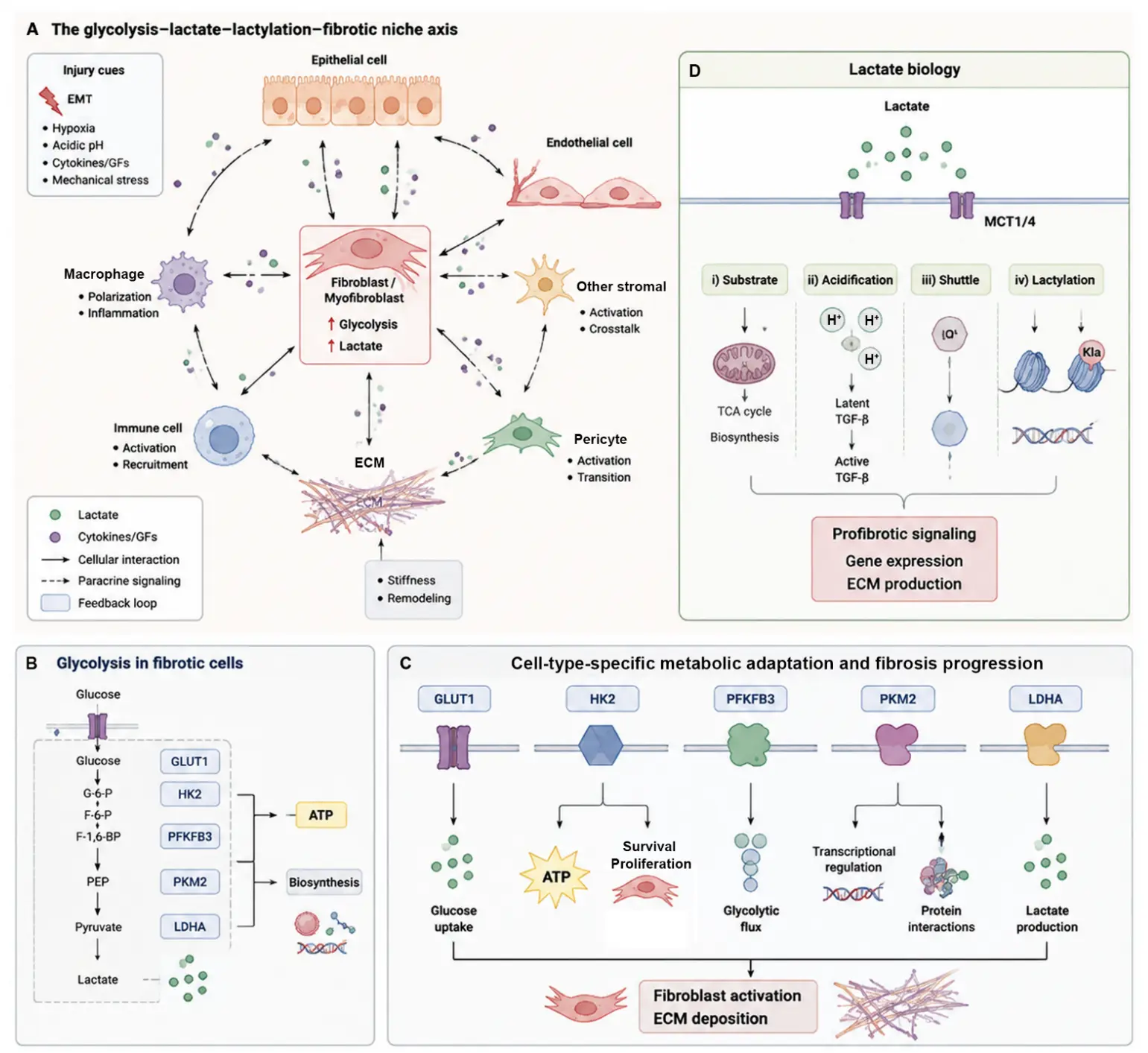
Figure 1. The glycolysis–lactate–lactylation–fibrotic niche axis integrates metabolic reprogramming with multicellular fibrotic progression. (A) Overview of the proposed glycolysis–lactate–lactylation–fibrotic niche framework. Injury-associated stimuli, including hypoxia, inflammatory cytokines, growth factors, acidic stress, and mechanical stress, induce metabolic remodeling in fibrosis-associated cells. Activated fibroblasts/myofibroblasts act as central regulators characterized by enhanced glycolysis and lactate production. Through reciprocal interactions with epithelial cells, endothelial cells, macrophages, immune cells, pericytes, and other stromal populations, glycolytic reprogramming reshapes the fibrotic niche. Extracellular matrix (ECM) accumulation and increased matrix stiffness further activate mechanotransduction pathways, generating a positive feedback loop that reinforces metabolic adaptation and fibrotic progression. (B) Schematic representation of glycolytic reprogramming in fibrotic cells. Increased glucose uptake and enhanced glycolytic activity provide ATP and biosynthetic intermediates to support cellular activation, proliferation, and ECM production. Key glycolytic regulators, including GLUT1, HK2, PFKFB3, PKM2, and LDHA, function as metabolic nodes controlling glucose utilization, glycolytic flux, and lactate generation. (C) Functional integration of major glycolytic regulators in fibrosis. GLUT1 regulates glucose uptake, HK2 controls glycolytic entry and metabolic adaptation, PFKFB3 promotes glycolytic flux, PKM2 participates in both glycolytic metabolism and noncanonical transcriptional/protein-interaction signaling, and LDHA mediates lactate production. These metabolic and signaling functions converge on fibroblast activation and ECM deposition. (D) Multifaceted biological functions of lactate within the fibrotic niche. Lactate acts as more than a glycolytic end product and regulates fibrosis through four distinct mechanisms: metabolic substrate utilization, extracellular acidification and latent TGF-β activation, MCT1/4-dependent intercellular lactate shuttling, and lactylation-mediated transcriptional regulation. These processes collectively contribute to profibrotic signaling, gene expression remodeling, immune regulation, and ECM accumulation.
2. Glycolytic Reprogramming as a Central Component of the Fibrotic Niche
Rather than representing a uniform metabolic switch, glycolytic reprogramming within fibrotic tissues should be considered a coordinated adaptation among multiple cellular populations. Fibroblasts and myofibroblasts exhibit enhanced glycolytic activity to support contractility, proliferation, and ECM production; however, the consequences of this metabolic shift extend beyond individual cells. Through lactate production, transport via MCT1/MCT4, extracellular acidification, and metabolic signaling, glycolytic alterations can influence neighboring epithelial and endothelial cells, as well as immune populations, thereby transforming isolated cellular metabolic changes into a coordinated multicellular fibrotic niche [4,49,51].
In this context, the fibrotic niche represents a metabolically interconnected microenvironment in which different cell types continuously exchange biochemical and mechanical information [49,51]. Fibroblasts function as central regulators of ECM remodeling and metabolic adaptation, whereas epithelial and endothelial cells contribute to niche formation through injury responses, hypoxia adaptation, and altered metabolic communication. Meanwhile, macrophages respond to metabolic and inflammatory cues, thereby regulating the transition from tissue repair to persistent fibrosis [4]. Importantly, ECM accumulation is not merely a consequence of fibrosis but also functions as an active signaling platform. Increased matrix stiffness activates mechanotransduction pathways such as YAP/TAZ, thereby reinforcing glycolytic reprogramming, fibroblast activation, and ECM remodeling, thereby generating a self-reinforcing metabolic–mechanical feedback loop [12].
Glycolysis is a central metabolic pathway for glucose utilization. Under normoxic conditions, most quiescent cells preferentially channel pyruvate into the mitochondria, where it is metabolized through the tricarboxylic acid (TCA) cycle and oxidative phosphorylation to generate ATP. In contrast, under conditions of rapid proliferation or cellular stress, cells may increase glucose uptake and convert a greater proportion of pyruvate into lactate even in the presence of sufficient oxygen, a metabolic phenotype resembling the aerobic glycolysis described by the Warburg effect [19,20,21,22,23,24,25,26,27]. Importantly, enhanced aerobic glycolysis in fibrotic cells does not imply complete suppression of mitochondrial function. Rather, glycolysis, mitochondrial metabolism, glutaminolysis, fatty acid metabolism, and amino acid biosynthesis are frequently rewired in parallel, and their coordinated regulation, in a cell type- and disease stage-dependent manner, collectively supports the initiation and progression of fibrotic responses [28,29,30,31,32,33,48].
From a metabolic perspective, glycolysis consists of an ATP investment phase followed by an ATP generation phase. Glucose is sequentially converted into glyceraldehyde-3-phosphate through the actions of hexokinase (HK), phosphofructokinase (PFK), aldolase A (ALDOA), and triose phosphate isomerase 1 (TPI1). Subsequently, glyceraldehyde-3-phosphate is metabolized to pyruvate through a cascade of enzymes, including glyceraldehyde-3-phosphate dehydrogenase (GAPDH), phosphoglycerate kinase (PGK), phosphoglycerate mutase (PGAM), enolase (ENO), and pyruvate kinase (PK). Pyruvate may either enter the mitochondria for oxidative metabolism or be reduced to lactate by lactate dehydrogenase A (LDHA), thereby regenerating NAD+. Importantly, glycolytic intermediates are not merely transient metabolites but can be diverted into biosynthetic pathways involved in nucleotide, glycoprotein, lipid, and amino acid synthesis, thereby providing the metabolic precursors required for extracellular matrix deposition and tissue remodeling [22,23,48]. However, alterations in the expression of glycolytic enzymes do not necessarily indicate a proportional increase in glycolytic flux. Although increased expression of GLUT1, HK2, PFKFB3, PKM2, or LDHA has frequently been reported in fibrotic tissues and activated profibrotic cells, direct assessment of metabolic flux requires complementary functional approaches, including glucose uptake assays, lactate production measurements, extracellular acidification rate (ECAR) analysis, metabolomic profiling, and stable isotope tracing [31,52]. Therefore, throughout this review, changes in glycolytic enzyme expression are interpreted as indicators of metabolic adaptation, whereas functional metabolic assays are considered stronger evidence for altered glycolytic activity or flux.
Within the fibrotic niche, enhanced glycolysis contributes to microenvironmental remodeling through several interconnected mechanisms. First, it provides ATP and anabolic intermediates to sustain the activation, proliferation, and extracellular matrix production of fibroblasts and myofibroblasts. Second, lactate accumulation and extracellular acidification amplify profibrotic and inflammatory signaling pathways, including transforming growth factor-β (TGF-β) and nuclear factor-κB (NF-κB). Third, metabolic alterations can be linked to transcriptional regulation through post-translational modifications such as histone lactylation. Emerging evidence suggests that lactylation may contribute to sustained expression of profibrotic genes. However, whether this represents true metabolic memory requires further experimental validation [53]. Fourth, monocarboxylate transporter (MCT)-mediated lactate shuttling reshapes intercellular communication among epithelial cells, endothelial cells, macrophages, and stromal cells, indicating that lactate functions as an intercellular signaling metabolite rather than merely a by-product of glycolysis. Therefore, glycolysis in fibrosis should not be regarded as an isolated metabolic pathway but rather as a central metabolic hub that integrates tissue injury signals, immune responses, the vascular microenvironment, mechanical cues, and extracellular matrix deposition. Importantly, the evidence supporting glycolytic regulators in fibrosis varies considerably among different molecules, tissues, and experimental systems. Increased expression of glycolytic enzymes primarily represents an association with metabolic adaptation, whereas causal relationships require stronger evidence from genetic manipulation, pharmacological intervention, metabolic-flux analysis, and, when available, human clinical validation.
3. Pathogenic Roles and Mechanistic Regulation of Key Glycolytic Nodes in Fibrosis
The strength of evidence supporting glycolytic regulators varies substantially among different molecules and disease contexts Table 1.
Table 1. Evidence hierarchy supporting the roles of glycolytic regulators in fibrosis.
|
Glycolytic Regulator |
Evidence Type |
Experimental Evidence |
Fibrosis Context |
|---|---|---|---|
|
GLUT1 |
Expression association + functional inhibition |
Increased expression; glucose uptake changes |
Lung fibrosis |
|
HK2 |
Pharmacological + genetic evidence |
HK2 inhibition reduces fibroblast activation and ECM deposition |
Lung/liver fibrosis |
|
PFKFB3 |
Pharmacological evidence + metabolic analysis |
3PO inhibition decreases glycolytic activity and fibrosis markers |
Lung/kidney/cardiac fibrosis |
|
PKM2 |
Genetic/pharmacological + mechanistic evidence |
Regulation of TGF-β signaling, YAP/TAZ interaction |
Lung/kidney/liver fibrosis |
|
LDHA |
Pharmacological + lactate evidence |
LDHA inhibition decreases lactate production and myofibroblast differentiation |
Lung fibrosis |
|
MCT1/MCT4 |
Functional transport evidence |
Lactate shuttling and intercellular communication |
Multiple fibrosis models |
|
GAPDH/ENO1/TPI1 |
Association/mechanistic evidence |
Noncanonical functions reported |
Limited fibrosis models |
3.1. GLUT1 and HK2
Glucose transporter 1 (GLUT1/SLC2A1) is responsible for basal glucose uptake and represents an upstream rate-limiting determinant of glycolytic reprogramming. Its expression can be induced by multiple fibrogenic stimuli, including hypoxia, transforming growth factor-β (TGF-β), inflammatory cytokines, and PI3K-Akt signaling, thereby enhancing glucose influx into fibrosis-associated cells. The biological significance of GLUT1 lies not in directly controlling individual downstream glycolytic reactions, but rather in determining whether cells possess sufficient substrate availability to enter a high-glycolytic state. In pulmonary and renal fibrosis, increased GLUT1-dependent glucose uptake has been closely associated with fibroblast activation, enhanced glycolytic flux, and excessive extracellular matrix deposition [48,54]. However, the functional consequences of GLUT1 activation are highly cell-type dependent. In epithelial cells, endothelial cells, or immune populations, enhanced glycolysis may represent adaptive responses to injury, inflammation, or metabolic stress rather than direct drivers of fibrosis.
Hexokinase 2 (HK2) catalyzes the phosphorylation of glucose to glucose-6-phosphate, representing the first committed step of glycolysis. HK2 is frequently upregulated in proliferative or stress-responsive cells and can localize to the outer mitochondrial membrane, where it participates in the regulation of energy metabolism, mitochondrial function, and cell survival [55]. In fibrotic diseases, HK2 exerts multifaceted functions. On one hand, HK2 represents an important regulatory node controlling glucose entry into glycolysis and may influence glycolytic flux depending on cellular context and metabolic demand. On the other hand, HK2 promotes lactate production, thereby providing metabolic substrates for lactate-mediated epigenetic regulation.
In pulmonary fibrosis models, TGF-β induces HK2 expression, whereas HK2 inhibition attenuates TGF-β-driven fibroblast proliferation, migration, and the expression of α-smooth muscle actin (α-SMA) and collagen [56]. In liver fibrosis, activated hepatic stellate cells exhibit increased dependence on HK2-mediated glycolysis. Rho et al. reported in Cell Metabolism that HK2-driven lactate accumulation promotes histone lactylation and facilitates hepatic stellate cell activation; both genetic and pharmacological inhibition of HK2 effectively reduced hepatic stellate cell activation and alleviated liver fibrosis [57]. Collectively, these findings indicate that HK2 may represent an important metabolic regulatory node linking glycolytic adaptation, lactate accumulation, and chromatin remodeling during fibrogenesis. Within the fibrotic niche, HK2-mediated metabolic remodeling may influence not only fibroblast activation but also metabolic communication with neighboring immune and epithelial populations.
3.2. PKM2
Pyruvate kinase catalyzes the conversion of phosphoenolpyruvate (PEP) to pyruvate, constituting the final rate-limiting step of glycolysis. Among its isoforms, pyruvate kinase M2 (PKM2) exhibits remarkable structural and functional plasticity. In its tetrameric form, PKM2 functions as a highly active glycolytic enzyme that efficiently promotes pyruvate production. In contrast, the dimeric or monomeric forms are more prone to nuclear translocation, where PKM2 participates in transcriptional regulation, protein–protein interactions, and intracellular signal transduction [58,59,60]. Consequently, PKM2 functions not only as a metabolic enzyme controlling pyruvate generation but also as a signaling regulator whose nuclear localization and protein interactions may contribute to profibrotic phenotypes independently of glycolytic flux. Importantly, PKM2 activity may have distinct consequences depending on cellular identity. While PKM2 activation in fibroblasts or pericytes may support a profibrotic transition, PKM2-dependent metabolic adaptation in immune or epithelial cells may participate in tissue repair responses.
In pulmonary fibrosis, PKM2 has been shown to interact with Smad7, thereby attenuating its negative feedback regulation of TGF-β signaling. This mechanism suggests that PKM2 may promote fibrosis through noncanonical signaling functions in addition to its metabolic role [61]. In addition, PKM2 is closely linked to extracellular matrix stiffness sensing, acting as a mechano-metabolic signaling node that converts matrix stiffening into enhanced glycolytic activity and fibroblast activation [62]. In liver fibrosis, pharmacological promotion of PKM2 tetramerization or inhibition of aberrant PKM2 activation suppresses hepatic stellate cell activation. Moreover, PKM2 regulates fibrogenesis by modulating glycine-dependent metabolism and the availability of metabolic precursors required for collagen biosynthesis, thereby influencing fibrosis across multiple organs [63,64]. In renal fibrosis, PKM2 contributes to pericyte-to-myofibroblast transition and the progression from acute kidney injury (AKI) to chronic kidney disease (CKD). Its nuclear translocation and interactions with mechanotransduction regulators such as Yes-associated protein (YAP) and transcriptional coactivator with PDZ-binding motif (TAZ) are considered key mechanistic events underlying these processes [65,66]. Collectively, the pathogenic role of PKM2 extends well beyond the enhancement of glycolysis itself and is closely associated with its multifunctional identity as a metabolic enzyme, transcriptional coregulator, and mechanosensitive signaling protein. Thus, PKM2 represents a potential metabolic–mechanical interface connecting intracellular glycolysis with tissue-level niche remodeling.
3.3. LDHA and Multifaceted Roles of Lactate in Fibrosis
Lactate dehydrogenase A (LDHA) catalyzes the reduction of pyruvate to lactate while simultaneously regenerating nicotinamide adenine dinucleotide (NAD+), thereby maintaining glycolytic activity under conditions of increased metabolic demand [67]. Across different fibrotic diseases, elevated LDHA expression and lactate accumulation have been observed in activated fibroblasts and other fibrosis-associated cell populations. However, accumulating evidence suggests that lactate should not be interpreted simply as a metabolic endpoint of glycolysis. Instead, lactate acts as a biologically active metabolite that links intracellular metabolic remodeling with extracellular signaling, intercellular communication, and transcriptional regulation within the fibrotic microenvironment.
Beyond its classical role in supporting glycolytic regeneration of NAD+, lactate can participate in metabolic exchange between cells. Through oxidation back to pyruvate, lactate may serve as an alternative carbon source for mitochondrial metabolism in specific cellular contexts, thereby contributing to metabolic flexibility during tissue stress. This concept is consistent with the lactate shuttle theory, which highlights lactate as an intermediary metabolite involved in energy redistribution rather than merely a terminal waste product of glycolysis [40,41]. Although the contribution of lactate utilization to fibrosis remains less extensively characterized than its signaling functions, emerging studies suggest that metabolic coupling between different cell populations may influence the adaptation and persistence of fibrotic tissues.
A more established mechanism linking lactate to fibrosis involves extracellular acidification and regulation of profibrotic signaling. Increased glycolytic activity and LDHA-mediated lactate production enhance proton accumulation and reduce extracellular pH, thereby facilitating the activation of latent TGF-β and promoting myofibroblast differentiation [27]. In pulmonary fibrosis models, elevated lactate levels have been detected in fibrotic tissues and fibroblasts, while exogenous lactate treatment induces the expression of α-smooth muscle actin (α-SMA), calponin, collagen I, and collagen III, at least partly through pH-dependent activation of latent TGF-β [27]. Consistently, inhibition of lactate production attenuates TGF-β-induced fibroblast activation, supporting a functional link between the LDHA–lactate axis and persistent profibrotic signaling [28,29]. Notably, these observations primarily reflect the consequences of lactate-associated extracellular acidification and microenvironmental remodeling, rather than direct evidence for lactate acting as a conventional cytokine-like signal.
In addition to modifying the extracellular environment, lactate also contributes to communication between distinct cellular populations through monocarboxylate transporters (MCTs). MCT1 and MCT4 regulate lactate influx and efflux, allowing metabolic information generated within one cell population to influence neighboring cells, including fibroblasts, epithelial cells, endothelial cells, and immune cells [42,43,44,45,46,47,68,69]. Such lactate exchange provides a potential mechanism through which localized metabolic alterations are propagated across the tissue and contribute to the formation of a metabolically interconnected fibrotic niche. Nevertheless, the biological consequences of lactate transport are likely to depend on the identity and metabolic state of both lactate-producing and lactate-receiving cells.
More recently, lactate has emerged as an important substrate for protein and histone lactylation, providing a potential link between metabolic states and gene regulation. Histone lactylation, particularly H3K18 lactylation, has been implicated in the regulation of fibrosis-associated transcriptional programs, including genes involved in fibroblast activation and inflammatory responses [46,47]. Studies in hepatic and renal fibrosis models have suggested that enhanced glycolytic activity and lactate accumulation may promote lactylation-associated changes in gene expression [57,70]. However, the current evidence remains context-dependent. While increased global lactylation levels have been reported in fibrotic conditions, direct locus-specific evidence linking individual lactylation events to persistent profibrotic transcriptional activation remains limited. Therefore, whether lactylation represents a stable form of metabolic memory requires further experimental validation.
Taken together, the biological significance of lactate in fibrosis extends beyond its production by LDHA. Lactate may influence fibrotic progression through interconnected but mechanistically distinct processes, including metabolic substrate utilization, extracellular acidification, MCT-dependent metabolic communication, and lactylation-mediated regulation of gene expression. The relative contribution of these mechanisms is likely determined by cellular identity, tissue context, and disease stage, emphasizing the need to interpret lactate biology within the broader framework of the fibrotic niche.
3.4. PDK1, Monocarboxylate Transporters, and Other Glycolysis-Associated Regulatory Nodes
Pyruvate dehydrogenase kinase 1 (PDK1) phosphorylates and inhibits pyruvate dehydrogenase (PDH), thereby limiting the entry of pyruvate into the tricarboxylic acid (TCA) cycle and redirecting metabolic flux toward lactate production [71]. In TGF-β-stimulated pulmonary fibroblasts, PDK1 expression is markedly upregulated and promotes the establishment of a glycolytic phenotype. Conversely, treatment with the PDK inhibitor dichloroacetate (DCA) restores pyruvate flux into mitochondrial oxidative metabolism, leading to reduced expression of α-smooth muscle actin (α-SMA) and collagen. These findings suggest that PDK1 contributes to the maintenance of the metabolic state required for myofibroblast activation and persistence [72].
Beyond PFKFB3, additional members of the phosphofructokinase (PFK) family and the monocarboxylate transporter (MCT) system have emerged as critical regulators linking glycolytic flux to fibrotic progression. PFKM and PFKP regulate the irreversible rate-limiting step of glycolysis and thereby influence carbon flux distribution. In addition to controlling glycolytic activity, these enzymes have been implicated in lactate production, protein lactylation, and inflammatory signaling. Meanwhile, MCT1 and MCT4 mediate transmembrane lactate transport, facilitating metabolic communication among fibroblasts, immune cells, and epithelial cells and collectively sustaining a profibrotic microenvironment.
In addition to PFKFB3, other PFK isoforms also contribute to metabolic remodeling during fibrosis. Muscle phosphofructokinase (PFKM) and platelet phosphofructokinase (PFKP) catalyze the conversion of fructose-6-phosphate to fructose-1,6-bisphosphate, constituting a key irreversible and rate-limiting step in glycolysis. Recent studies have shown that PFKP is responsive to TGF-β stimulation, promoting the glycolytic switch in renal tubular epithelial cells and driving renal interstitial fibrosis through enhanced metabolic reprogramming. Conversely, inhibition of PFKP suppresses glycolytic activity in tubular epithelial cells and attenuates fibrotic progression [48]. Moreover, PFKM has been reported to activate the NF-κB signaling axis by promoting lactate accumulation and histone H3 lysine 18 lactylation (H3K18la), thereby amplifying inflammatory responses and accelerating renal fibrosis [73]. Collectively, these findings suggest that the PFK family constitutes an integrated regulatory network linking glycolytic flux, lactate metabolism, protein lactylation, and inflammatory profibrotic signaling [53].
MCT1 and MCT4 are the principal transporters responsible for transmembrane lactate trafficking, maintaining intracellular and extracellular lactate homeostasis while serving as critical transport hubs connecting LDHA activity with paracrine signaling [69]. Through MCT-dependent intercellular transport, lactate facilitates metabolic communication among fibroblasts, immune cells, epithelial cells, and endothelial cells, thereby establishing a “lactate niche” that supports the persistence and progression of fibrosis.
In addition to these well-characterized regulators, several downstream glycolytic enzymes have recently been implicated in the metabolic adaptation associated with fibrosis, although direct mechanistic evidence remains relatively limited.
Triosephosphate isomerase 1 (TPI1) catalyzes the reversible interconversion between dihydroxyacetone phosphate (DHAP) and glyceraldehyde-3-phosphate (GAP), representing an essential node for maintaining glycolytic carbon flux. Beyond its canonical metabolic function, TPI1 also exerts non-metabolic regulatory activities that influence oxidative stress, cell death pathways, and tissue repair. However, compared with its canonical glycolytic function, the contribution of TPI1-mediated noncanonical mechanisms to fibrosis remains less well established. Recent evidence indicates that dopaminylation of endothelial TPI1 suppresses ferroptosis-associated profibrotic paracrine signaling, thereby promoting regenerative repair following lung injury and reducing subsequent fibrotic remodeling. These findings suggest that TPI1 may function as an important regulator balancing tissue regeneration and fibrogenesis [74,75,76].
α-Enolase (ENO1) catalyzes the conversion of 2-phosphoglycerate to phosphoenolpyruvate during the later stages of glycolysis. In addition to its metabolic role, ENO1 has increasingly been recognized for its roles in cell adhesion, migration, and intracellular signaling. These observations indicate that ENO1-associated profibrotic effects cannot be solely attributed to increased glycolytic flux. During fibrosis, ENO1 may facilitate fibroblast activation and extracellular matrix (ECM) deposition through modulation of TGF-β signaling, focal adhesion kinase (FAK)-related pathways, and cell migratory capacity.
Other glycolytic enzymes, including glyceraldehyde-3-phosphate dehydrogenase (GAPDH), phosphoglycerate kinase 1 (PGK1), and phosphoglycerate mutase 1 (PGAM1), have likewise been implicated in metabolic remodeling during fibrosis. Beyond catalyzing glycolytic reactions, GAPDH regulates cell fate by modulating the NAD+/NADH redox balance, cellular oxidative status, and noncanonical protein–protein interactions. Therefore, the contribution of GAPDH to fibrosis may involve both metabolic regulation and glycolysis-independent signaling functions. PGK1 supports the bioenergetic demands of activated fibroblasts through ATP generation, whereas PGAM1 links glycolysis to serine and glycine metabolism, thereby providing biosynthetic precursors required for collagen synthesis [77]. In addition, intermediate glycolytic enzymes such as aldolase A (ALDOA) may contribute to ECM biosynthesis by regulating glycolytic carbon partitioning. Nevertheless, compared with HK2, PFKFB3, PKM2, and LDHA, the roles of these enzymes in fibrosis remain incompletely understood. Future studies integrating single-cell metabolomics, spatial transcriptomics, and stable isotope tracing will be essential for elucidating their precise contributions to fibrotic metabolic reprogramming.
Collectively, glycolytic enzymes involved in fibrosis should not be considered as uniform metabolic regulators. Some enzymes primarily contribute to fibrotic progression by altering glycolytic activity and metabolic flux, whereas others exert additional moonlighting functions via transcriptional regulation, intracellular signaling, or protein–protein interactions. Dissecting these distinct mechanisms is essential for determining whether the profibrotic phenotype is driven by metabolic rewiring or by glycolysis-independent regulatory functions.
4. Mechanisms by Which Glycolytic Enzymes Promote Fibroblast-to-Myofibroblast Transition and Extracellular Matrix Deposition
Glycolytic enzymes contribute to fibrogenesis through at least five interconnected mechanisms. First, they provide the energy and metabolic intermediates required to sustain myofibroblast function. Activated myofibroblasts require substantial bioenergetic and biosynthetic support to maintain stress fiber formation, cell migration, proliferation, and collagen secretion. Enhanced glycolytic flux rapidly supplies ATP while simultaneously fueling the pentose phosphate pathway, serine/glycine biosynthesis, and the hexosamine biosynthetic pathway, thereby supporting the metabolic demands associated with extracellular matrix (ECM) production and tissue remodeling [19,20,21,22,23]. Second, glycolytic enzymes amplify transforming growth factor-β (TGF-β) signaling. TGF-β induces the expression of multiple glycolytic regulators, including HK2, PFKFB3, and LDHA. In turn, lactate accumulation and extracellular acidification promote the activation of latent TGF-β, thereby establishing a self-reinforcing positive feedback loop that sustains profibrotic signaling [27,28,56,78]. Third, glycolytic reprogramming stabilizes hypoxia-inducible factor-1α (HIF-1α) and reinforces hypoxia-like transcriptional programs. Enhanced glycolytic activity, together with alterations in tricarboxylic acid (TCA) cycle intermediates, influences the activity of prolyl hydroxylases, resulting in HIF-1α stabilization. Activated HIF-1α subsequently induces the transcription of glycolysis-associated genes, including GLUT1, HK2, PFKFB3, LDHA, and PDK1, thereby establishing a feed-forward metabolic circuit that further enhances glycolytic reprogramming [26,79]. Fourth, glycolysis is tightly integrated with mechanotransduction. Matrix stiffening activates mechanosensitive pathways such as Yes-associated protein/transcriptional coactivator with PDZ-binding motif (YAP/TAZ), focal adhesion kinase (FAK), and Rho-associated coiled-coil-containing protein kinase (ROCK), which in turn promote glycolytic reprogramming. Enhanced glycolysis subsequently provides the energy and biosynthetic resources necessary for cellular contractility and ECM production, thereby sustaining a reciprocal mechano-metabolic feedback loop that drives progressive fibrosis [12,80,81,82,83,84]. Fifth, glycolytic metabolism contributes to the establishment of epigenetic memory. Lactate serves as the substrate for histone lactylation, which promotes an open chromatin configuration at the promoters or regulatory regions of profibrotic genes, including COL1A1, ACTA2, SOX9, inflammatory mediators, and other fibrosis-associated genes. This epigenetic remodeling stabilizes the myofibroblast phenotype and facilitates the persistence of fibrogenic transcriptional programs [46,47,53,57,85,86,87,88].
Importantly, excessive ECM deposition is not solely determined by increased collagen gene transcription. Collagen biosynthesis is a highly coordinated process involving protein translation, proline and lysine hydroxylation, glycosylation, triple-helix assembly, secretion, extracellular cross-linking, and the dynamic balance between matrix deposition and degradation. Glycolytic enzymes indirectly facilitate collagen maturation by supplying ATP, maintaining the NAD+/NADH redox balance, and generating essential biosynthetic intermediates, including UDP-N-acetylglucosamine (UDP-GlcNAc), NADPH, and amino acid precursors [20,21,22,23]. Furthermore, lactate accumulation and extracellular acidification regulate the activity of matrix metalloproteinases (MMPs), lysyl oxidases (LOXs), integrin activation, and immune cell function, thereby influencing both the rate and direction of ECM remodeling during fibrosis [40,41,42,43,44,45,46,47,88].
5. Organ-Specific Evidence for Glycolytic Reprogramming in Fibrosis
Evidence supporting glycolytic reprogramming is most comprehensively established in pulmonary fibrosis. Studies of lung tissues from patients with idiopathic pulmonary fibrosis (IPF), primary pulmonary fibroblasts, and experimental animal models consistently demonstrate aberrant activation of multiple glycolysis-associated regulators, including HK2, PFKFB3, PKM2, LDHA, PDK1, and the lactate metabolic pathway [26,27,28,29,30,31,32,61,89,90]. Collectively, these findings indicate that metabolic reprogramming is a central feature of pulmonary fibrogenesis and contributes to persistent fibroblast activation and extracellular matrix (ECM) accumulation. In liver fibrosis, activation of hepatic stellate cells (HSCs) is accompanied by coordinated enhancement of both glycolysis and mitochondrial metabolism. Multiple glycolysis-associated regulators, including HK2, PFKFB3, PKM2, MCT1, and protein lactylation, have been demonstrated to participate in HSC activation and the progression of hepatic fibrosis [36,37,38,57,63,85,91]. These observations suggest that metabolic remodeling in activated HSCs involves the integration of glycolytic and mitochondrial pathways rather than a simple shift toward aerobic glycolysis. In renal fibrosis, distinct forms of glycolytic reprogramming have been identified in proximal tubular epithelial cells, pericytes, fibroblasts, and macrophages. Among the reported metabolic regulators, PFKFB3, PKM2, and the lactate–lactylation axis have emerged as particularly important drivers of renal fibrogenesis by coordinating metabolic adaptation with inflammatory and epigenetic responses [34,65,66,70,78,86]. In cardiac fibrosis, ischemic injury, pressure overload, and inflammatory stimuli induce glycolytic activation in cardiac fibroblasts. Glycolysis-associated programs involving PFKFB3, PKM2, and YAP-dependent metabolic signaling contribute to post-myocardial infarction remodeling as well as pressure overload-induced cardiac fibrosis [39,92,93,94]. These findings further support a close interplay between metabolic reprogramming and mechanotransduction during cardiac fibrotic remodeling. In cutaneous fibrosis and keloid formation, enhanced glycolysis has likewise been associated with increased fibroblast proliferation, migration, and collagen synthesis. Several glycolysis-related regulators, including PGK1, LDHA, HIF-1α, and the TGF-β–KLF5 signaling axis, have been implicated in the pathogenesis of pathological scar formation [95,96,97]. Together, these observations indicate that although the dominant metabolic regulators may differ among organs, glycolytic reprogramming represents a conserved pathogenic mechanism underlying fibroblast activation and fibrotic progression across multiple tissues.
6. Therapeutic Implications and Future Challenges
Targeting glycolytic enzymes has emerged as a promising therapeutic strategy for the treatment of fibrosis. A variety of glycolysis-targeting agents, including 2-deoxy-D-glucose (2-DG), 3PO, LDH inhibitors, dichloroacetate (DCA), PKM2 conformation modulators, monocarboxylate transporter (MCT) inhibitors, metformin, AMP-activated protein kinase (AMPK) activators, and several multitarget small-molecule compounds, have demonstrated antifibrotic efficacy in cellular and experimental animal models [28,54,64,72,76,88,91,98,99,100,101]. Despite these encouraging preclinical findings, the clinical translation of glycolysis-targeted therapies faces several major challenges. First, glycolysis is a fundamental metabolic pathway required for normal cellular homeostasis. Consequently, systemic inhibition of glycolytic enzymes may produce undesirable effects on immune cell function, epithelial regeneration, cardiac performance, and skeletal muscle metabolism. Second, the metabolic requirements of fibrotic tissues evolve throughout disease progression. Distinct pathological stages—including the early inflammatory phase, the fibroproliferative phase, and the late-stage fibrotic or sclerotic phase—are likely to exhibit different metabolic vulnerabilities and may therefore require stage-specific therapeutic interventions. Third, the functions of glycolytic enzymes are highly dependent on both cell type and subcellular localization. For example, PFKFB3 exerts distinct, and in some contexts even divergent, biological functions in fibroblasts, renal tubular epithelial cells, endothelial cells, and myeloid cells. Accordingly, future therapeutic strategies should emphasize cell type-specific drug delivery, temporally optimized intervention, and rational combination therapies to maximize therapeutic efficacy while minimizing systemic toxicity.
An ideal research framework should incorporate multiple complementary approaches. Single-cell transcriptomics, spatial metabolomics, and stable isotope tracing should be integrated to comprehensively map glucose metabolic flux across distinct cellular populations within fibrotic tissues. Genetic models with cell type-specific manipulation of glycolytic enzymes will be essential for dissecting their functions in fibroblasts, epithelial cells, endothelial cells, and immune cells. In parallel, mechanically tunable extracellular matrix (ECM) models should be employed to elucidate the reciprocal interactions between matrix stiffness and glycolytic reprogramming. Finally, there remains a pressing need to develop in vivo imaging approaches and robust biomarkers that simultaneously monitor glycolytic activity, lactate accumulation, protein lactylation, and ECM deposition, thereby facilitating mechanistic investigation and therapeutic evaluation in fibrotic diseases [102,103,104,105,106,107].
7. Conclusions
Glycolytic enzymes play multifaceted roles in tissue fibrosis that extend far beyond their canonical metabolic functions, encompassing metabolic catalysis, signal transduction, epigenetic regulation, and intercellular communication. Among these regulators, HK2 governs glucose entry into the glycolytic pathway and drives the lactate–lactylation axis; PFKFB3 promotes glycolytic activity and supports fibroblast activation; PKM2 integrates metabolic regulation with TGF-β signaling and mechanotransduction through its conformational plasticity and nuclear functions; whereas LDHA and monocarboxylate transporters (MCTs) coordinate lactate production, transport, and microenvironmental acidification, thereby further promoting TGF-β activation, immune modulation, and histone lactylation. Collectively, these glycolytic regulators constitute a central pathogenic network that links tissue injury, metabolic reprogramming, myofibroblast stabilization, and extracellular matrix (ECM) deposition during fibrogenesis. Looking forward, therapeutic strategies targeting glycolytic enzymes should not simply aim for global suppression of glycolysis. Instead, future interventions should focus on selectively disrupting pathological metabolism–phenotype coupling according to organ context, cell type, disease stage, and metabolic flux characteristics. Such precision metabolic targeting is expected to maximize antifibrotic efficacy while minimizing adverse effects associated with systemic metabolic inhibition, ultimately providing a more rational framework for the development of metabolism-based therapies for fibrotic diseases.
The major metabolic targets and their reported antifibrotic mechanisms Table 2.
Table 2. Glycolytic enzymes and lactate-associated pathways as therapeutic targets in fibrosis.
|
Target |
Main Mechanism in Fibrosis |
Antifibrotic Effects |
References |
|---|---|---|---|
|
HK2 |
Promotes glycolysis, lactate production, and H3K18 lactylation to activate fibroblasts/HSCs |
Reduced fibroblast activation and ECM deposition |
|
|
PFKFB3 |
Drives glycolytic reprogramming and lactate-mediated profibrotic signaling |
Decreased collagen synthesis and fibrosis progression |
|
|
PKM2 |
Regulates glycolysis, TGF-β signaling, and collagen biosynthesis |
Reduced fibroblast transition and fibrosis |
|
|
LDHA/MCT axis |
Controls lactate production and transport, regulating fibrotic communication |
Reduced myofibroblast differentiation and ECM accumulation |
|
|
PDK1 |
Redirects metabolism toward glycolysis by inhibiting mitochondrial oxidation |
Restored oxidative metabolism and reduced fibrosis |
|
|
Other glycolytic enzymes (GAPDH, ENO1, PGK1, PGAM1) |
Participate in noncanonical regulation of migration and transcription |
Potential antifibrotic targets |
Statement of the Use of Generative AI and AI-Assisted Technologies in the Writing Process
During the preparation of this manuscript, the authors used [chatgpt] to improve the language and readability of the manuscript. After using this tool, the authors carefully reviewed and edited the content as necessary and take full responsibility for the content of the published article.
Author Contributions
Writing—Original Draft Preparation, H.Q.; Visualization, H.Q.; Conceptualization, S.C., Y.L., Q.Y., W.D. and X.Z.; Supervision, Y.C. and C.L. All authors have read and approved the final manuscript.
Ethics Statement
Not applicable.
Informed Consent Statement
Not applicable.
Data Availability Statement
Data sharing not applicable to this article as no datasets were generated or analysed during the current study. All information is derived from publicly available articles and datasets.
Funding
This work was supported by the grants from National Natural Science Foundation of China (82500087), Suzhou Municipal Basic Research Special Project (SSD2024031, SSD2025056).
Declaration of Competing Interest
All the authors declared no competing interests.
References
- Wynn T. Cellular and molecular mechanisms of fibrosis. J. Pathol. 2008, 214, 199–210. DOI:10.1002/path.2277 [Google Scholar]
- Wynn TA, Ramalingam TR. Mechanisms of fibrosis: Therapeutic translation for fibrotic disease. Nat. Med. 2012, 18, 1028–1040. DOI:10.1038/nm.2807 [Google Scholar]
- Rockey DC, Bell PD, Hill JA. Fibrosis—A Common Pathway to Organ Injury and Failure. N. Engl. J. Med. 2015, 372, 1138–1149. DOI:10.1056/NEJMra1300575 [Google Scholar]
- Henderson NC, Rieder F, Wynn TA. Fibrosis: From mechanisms to medicines. Nature 2020, 587, 555–566. DOI:10.1038/s41586-020-2938-9 [Google Scholar]
- Frangogiannis NG. Transforming growth factor–β in tissue fibrosis. J. Exp. Med. 2020, 217, e20190103. DOI:10.1084/jem.20190103 [Google Scholar]
- Tomasek JJ, Gabbiani G, Hinz B, Chaponnier C, Brown RA. Myofibroblasts and mechano-regulation of connective tissue remodelling. Nat. Rev. Mol. Cell Biol. 2002, 3, 349–363. DOI:10.1038/nrm809 [Google Scholar]
- Hinz B, Phan SH, Thannickal VJ, Galli A, Bochaton-Piallat ML, Gabbiani G. The myofibroblast: One function, multiple origins. Am. J. Pathol. 2007, 170, 1807–1816. DOI:10.2353/ajpath.2007.070112 [Google Scholar]
- Klingberg F, Hinz B, White ES. The myofibroblast matrix: Implications for tissue repair and fibrosis. J. Pathol. 2013, 229, 298–309. DOI:10.1002/path.4104 [Google Scholar]
- Frangogiannis NG. Fibroblast—Extracellular Matrix Interactions in Tissue Fibrosis. Curr. Pathobiol. Rep. 2016, 4, 11–18. DOI:10.1007/s40139-016-0099-1 [Google Scholar]
- DeLeon-Pennell KY, Barker TH, Lindsey ML. Fibroblasts: The arbiters of extracellular matrix remodeling. Matrix Biol. 2020, 91–92, 1–7. DOI:10.1016/j.matbio.2020.05.006 [Google Scholar]
- Parker MW, Rossi D, Peterson M, Smith K, Sikström K, White ES, et al. Fibrotic extracellular matrix activates a profibrotic positive feedback loop. J. Clin. Investig. 2014, 124, 1622–1635. DOI:10.1172/JCI71386 [Google Scholar]
- Liu F, Lagares D, Choi KM, Stopfer L, Marinković A, Vrbanac V, et al. Mechanosignaling through YAP and TAZ drives fibroblast activation and fibrosis. Am. J. Physiol.-Lung Cell. Mol. Physiol. 2015, 308, L344–L357. DOI:10.1152/ajplung.00300.2014 [Google Scholar]
- Younesi FS, Miller AE, Barker TH, Rossi FMV, Hinz B. Fibroblast and myofibroblast activation in normal tissue repair and fibrosis. Nat. Rev. Mol. Cell Biol. 2024, 25, 617–638. DOI:10.1038/s41580-024-00716-0 [Google Scholar]
- Gabbiani G. The myofibroblast in wound healing and fibrocontractive diseases. J. Pathol. 2003, 200, 500–503. DOI:10.1002/path.1427 [Google Scholar]
- Hinz B. Mechanical Aspects of Lung Fibrosis: A Spotlight on the Myofibroblast. Proc. Am. Thorac. Soc. 2012, 9, 137–147. DOI:10.1513/pats.201202-017AW [Google Scholar]
- Wight TN, Potter-Perigo S. The extracellular matrix: An active or passive player in fibrosis? Am. J. Physiol.-Gastrointest. Liver Physiol. 2011, 301, G950–G955. DOI:10.1152/ajpgi.00132.2011 [Google Scholar]
- Zhao X, Chen J, Sun H, Zhang Y, Zou D. New insights into fibrosis from the ECM degradation perspective: The macrophage-MMP-ECM interaction. Cell Biosci. 2022, 12, 56. DOI:10.1186/s13578-022-00856-w [Google Scholar]
- Schuster R, Younesi F, Ezzo M, Hinz B. The Role of Myofibroblasts in Physiological and Pathological Tissue Repair. Cold Spring Harb. Perspect. Biol. 2023, 15, a041231. DOI:10.1101/cshperspect.a041231 [Google Scholar]
- Warburg O. On the Origin of Cancer Cells. Science 1956, 123, 309–314. DOI:10.1126/science.123.3191.309 [Google Scholar]
- Vander Heiden MG, Cantley LC, Thompson CB. Understanding the Warburg Effect: The Metabolic Requirements of Cell Proliferation. Science 2009, 324, 1029–1033. DOI:10.1126/science.1160809 [Google Scholar]
- Lunt SY, Vander Heiden MG. Aerobic Glycolysis: Meeting the Metabolic Requirements of Cell Proliferation. Annu. Rev. Cell Dev. Biol. 2011, 27, 441–464. DOI:10.1146/annurev-cellbio-092910-154237 [Google Scholar]
- Liberti MV, Locasale JW. The Warburg Effect: How Does it Benefit Cancer Cells? Trends Biochem. Sci. 2016, 41, 211–218. DOI:10.1016/j.tibs.2015.12.001 [Google Scholar]
- Kay EJ, Koulouras G, Zanivan S. Regulation of Extracellular Matrix Production in Activated Fibroblasts: Roles of Amino Acid Metabolism in Collagen Synthesis. Front. Oncol. 2021, 11, 719922. DOI:10.3389/fonc.2021.719922 [Google Scholar]
- DeBerardinis RJ, Chandel NS. Fundamentals of cancer metabolism. Sci. Adv. 2016, 2, e1600200. DOI:10.1126/sciadv.1600200 [Google Scholar]
- Pavlova NN, Thompson CB. The Emerging Hallmarks of Cancer Metabolism. Cell Metab. 2016, 23, 27–47. DOI:10.1016/j.cmet.2015.12.006 [Google Scholar]
- Xie N, Tan Z, Banerjee S, Cui H, Ge J, Liu RM, et al. Glycolytic Reprogramming in Myofibroblast Differentiation and Lung Fibrosis. Am. J. Respir. Crit. Care Med. 2015, 192, 1462–1474. DOI:10.1164/rccm.201504-0780OC [Google Scholar]
- Kottmann RM, Kulkarni AA, Smolnycki KA, Lyda E, Dahanayake T, Salibi R, et al. Lactic Acid Is Elevated in Idiopathic Pulmonary Fibrosis and Induces Myofibroblast Differentiation via pH-Dependent Activation of Transforming Growth Factor-β. Am. J. Respir. Crit. Care Med. 2012, 186, 740–751. DOI:10.1164/rccm.201201-0084OC [Google Scholar]
- Kottmann RM, Trawick E, Judge JL, Wahl LA, Epa AP, Owens KM, et al. Pharmacologic inhibition of lactate production prevents myofibroblast differentiation. Am. J. Physiol.-Lung Cell. Mol. Physiol. 2015, 309, L1305–L1312. DOI:10.1152/ajplung.00058.2015 [Google Scholar]
- Judge JL, Owens KM, Pollock SJ, Woeller CF, Thatcher TH, Williams JP, et al. Ionizing radiation induces myofibroblast differentiation via lactate dehydrogenase. Am. J. Physiol.-Lung Cell. Mol. Physiol. 2015, 309, L879–L887. DOI:10.1152/ajplung.00153.2015 [Google Scholar]
- Para R, Romero F, George G, Summer R. Metabolic Reprogramming as a Driver of Fibroblast Activation in Pulmonary Fibrosis. Am. J. Med. Sci. 2019, 357, 394–398. DOI:10.1016/j.amjms.2019.02.003 [Google Scholar]
- Hamanaka RB, Mutlu GM. Metabolic requirements of pulmonary fibrosis: Role of fibroblast metabolism. FEBS J. 2021, 288, 6331–6352. DOI:10.1111/febs.15693 [Google Scholar]
- Yan P, Liu J, Li Z, Wang J, Zhu Z, Wang L, et al. Glycolysis Reprogramming in Idiopathic Pulmonary Fibrosis: Unveiling the Mystery of Lactate in the Lung. Int. J. Mol. Sci. 2023, 25, 315. DOI:10.3390/ijms25010315 [Google Scholar]
- Li J, Zhai X, Sun X, Cao S, Yuan Q, Wang J. Metabolic reprogramming of pulmonary fibrosis. Front. Pharmacol. 2022, 13, 1031890. DOI:10.3389/fphar.2022.1031890 [Google Scholar]
- Zhu X, Jiang L, Long M, Wei X, Hou Y, Du Y. Metabolic Reprogramming and Renal Fibrosis. Front. Med. 2021, 8, 746920. DOI:10.3389/fmed.2021.746920 [Google Scholar]
- Wang S, Liang Y, Dai C. Metabolic Regulation of Fibroblast Activation and Proliferation during Organ Fibrosis. Kidney Dis. 2022, 8, 115–125. DOI:10.1159/000522417 [Google Scholar]
- Hou W, Syn WK. Role of Metabolism in Hepatic Stellate Cell Activation and Fibrogenesis. Front. Cell Dev. Biol. 2018, 6, 150. DOI:10.3389/fcell.2018.00150 [Google Scholar]
- Smith-Cortinez N, van Eunen K, Heegsma J, Serna-Salas SA, Sydor S, Bechmann LP, et al. Simultaneous Induction of Glycolysis and Oxidative Phosphorylation during Activation of Hepatic Stellate Cells Reveals Novel Mitochondrial Targets to Treat Liver Fibrosis. Cells 2020, 9, 2456. DOI:10.3390/cells9112456 [Google Scholar]
- Horn P, Tacke F. Metabolic reprogramming in liver fibrosis. Cell Metab. 2024, 36, 1439–1455. DOI:10.1016/j.cmet.2024.05.003 [Google Scholar]
- Hoque MM, Gbadegoye JO, Hassan FO, Raafat A, Lebeche D. Cardiac fibrogenesis: An immuno-metabolic perspective. Front. Physiol. 2024, 15, 1336551. DOI:10.3389/fphys.2024.1336551 [Google Scholar]
- Brooks GA. The Science and Translation of Lactate Shuttle Theory. Cell Metab. 2018, 27, 757–785. DOI:10.1016/j.cmet.2018.03.008 [Google Scholar]
- Rabinowitz JD, Enerbäck S. Lactate: The ugly duckling of energy metabolism. Nat. Metab. 2020, 2, 566–571. DOI:10.1038/s42255-020-0243-4 [Google Scholar]
- Liang H, Xu L, Yang Y. Lactate and lactylation: Novel perspectives on fibrosis pathogenesis and therapeutic directions. J. Transl. Med. 2025, 23, 705. DOI:10.1186/s12967-025-06748-0 [Google Scholar]
- Zhang L, Li Q, Deng Y, Zou Y, Wang L, Li J. Glycolysis to lactylation: Unraveling the metabolic and epigenetic landscape in tissue fibrosis. Mol. Med. Rep. 2025, 32, 290. DOI:10.3892/mmr.2025.13655 [Google Scholar]
- Wei X, Long M, Yu J, Du Y. The lactate-lactylation axis in renal fibrosis: Potential mechanisms in diabetic kidney disease. Ann. Med. 2025, 57, 2587326. DOI:10.1080/07853890.2025.2587326 [Google Scholar]
- Li B, Li XH, Zhou Y, Feng DD, Luo ZQ, Cheng HP. Lactylation in pulmonary fibrosis: Current understanding and challenges. J. Transl. Med. 2026, 24, 514. DOI:10.1186/s12967-026-07991-9 [Google Scholar]
- Zhang D, Tang Z, Huang H, Zhou G, Cui C, Weng Y, et al. Metabolic regulation of gene expression by histone lactylation. Nature 2019, 574, 575–580. DOI:10.1038/s41586-019-1678-1 [Google Scholar]
- Wu S, Li J, Zhan Y. H3K18 lactylation accelerates liver fibrosis progression through facilitating SOX9 transcription. Exp. Cell Res. 2024, 440, 114135. DOI:10.1016/j.yexcr.2024.114135 [Google Scholar]
- Yang S, Wu H, Li Y, Li L, Xiang J, Kang L, et al. Inhibition of PFKP in renal tubular epithelial cell restrains TGF-β induced glycolysis and renal fibrosis. Cell Death Dis. 2023, 14, 816. DOI:10.1038/s41419-023-06347-1 [Google Scholar]
- Li L, Fu H, Liu Y. The fibrogenic niche in kidney fibrosis: Components and mechanisms. Nat. Rev. Nephrol. 2022, 18, 545–557. DOI:10.1038/s41581-022-00590-z [Google Scholar]
- Froom ZSCS, Callaghan NI, Davenport Huyer L. Cellular crosstalk in fibrosis: Insights into macrophage and fibroblast dynamics. J. Biol. Chem. 2025, 301, 110203. DOI:10.1016/j.jbc.2025.110203 [Google Scholar]
- Duffield JS, Lupher M, Thannickal VJ, Wynn TA. Host Responses in Tissue Repair and Fibrosis. Annu. Rev. Pathol. Mech. Dis. 2013, 8, 241–276. DOI:10.1146/annurev-pathol-020712-163930 [Google Scholar]
- Hui S, Ghergurovich JM, Morscher RJ, Jang C, Teng X, Lu W, et al. Glucose feeds the TCA cycle via circulating lactate. Nature 2017, 551, 115–118. DOI:10.1038/nature24057 [Google Scholar]
- Qiu Z, Zhang Y, Li X, Xie Z. Lactylation in tissue fibrosis: Epigenetic mechanisms, metabolic crosstalk, and therapeutic opportunities. J. Transl. Med. 2026, 24, 633. DOI:10.1186/s12967-026-08046-9 [Google Scholar]
- Cho SJ, Moon JS, Lee CM, Choi AMK, Stout-Delgado HW. Glucose Transporter 1–Dependent Glycolysis Is Increased during Aging-Related Lung Fibrosis, and Phloretin Inhibits Lung Fibrosis. Am. J. Respir. Cell Mol. Biol. 2017, 56, 521–531. DOI:10.1165/rcmb.2016-0225OC [Google Scholar]
- Roberts DJ, Miyamoto S. Hexokinase II integrates energy metabolism and cellular protection: Akting on mitochondria and TORCing to autophagy. Cell Death Differ. 2015, 22, 248–257. DOI:10.1038/cdd.2014.173 [Google Scholar]
- Yin X, Choudhury M, Kang JH, Schaefbauer KJ, Jung MY, Andrianifahanana M, et al. Hexokinase 2 couples glycolysis with the profibrotic actions of TGF-β. Sci. Signal. 2019, 12, eaax4067. DOI:10.1126/scisignal.aax4067 [Google Scholar]
- Rho H, Terry AR, Chronis C, Hay N. Hexokinase 2-mediated gene expression via histone lactylation is required for hepatic stellate cell activation and liver fibrosis. Cell Metab. 2023, 35, 1406–1423.e8. DOI:10.1016/j.cmet.2023.06.013 [Google Scholar]
- Christofk HR, Vander Heiden MG, Harris MH, Ramanathan A, Gerszten RE, Wei R, et al. The M2 splice isoform of pyruvate kinase is important for cancer metabolism and tumour growth. Nature 2008, 452, 230–233. DOI:10.1038/nature06734 [Google Scholar]
- Israelsen WJ, Vander Heiden MG. Pyruvate kinase: Function, regulation and role in cancer. Semin. Cell Dev. Biol. 2015, 43, 43–51. DOI:10.1016/j.semcdb.2015.08.004 [Google Scholar]
- Yang W, Lu Z. Pyruvate kinase M2 at a glance. J. Cell Sci. 2015, 128, 1655–1660. DOI:10.1242/jcs.166629 [Google Scholar]
- Gao S, Li X, Jiang Q, Liang Q, Zhang F, Li S, et al. PKM2 promotes pulmonary fibrosis by stabilizing TGF-β1 receptor I and enhancing TGF-β1 signaling. Sci. Adv. 2022, 8, eabo0987. DOI:10.1126/sciadv.abo0987 [Google Scholar]
- Wang Y, Hu R, Zhang X, Cai G, Ding Q, Che P. PKM2 mediates the glycolytic response to matrix stiffness in lung fibroblasts. Front. Cell Dev. Biol. 2026, 14, 1771078. DOI:10.3389/fcell.2026.1771078 [Google Scholar]
- Zheng D, Jiang Y, Qu C, Yuan H, Hu K, He L, et al. Pyruvate Kinase M2 Tetramerization Protects against Hepatic Stellate Cell Activation and Liver Fibrosis. Am. J. Pathol. 2020, 190, 2267–2281. DOI:10.1016/j.ajpath.2020.08.002 [Google Scholar]
- Satyanarayana G, Turaga RC, Sharma M, Wang S, Mishra F, Peng G, et al. Pyruvate kinase M2 regulates fibrosis development and progression by controlling glycine auxotrophy in myofibroblasts. Theranostics 2021, 11, 9331–9341. DOI:10.7150/thno.60385 [Google Scholar]
- Chen Y, Bai X, Chen J, Huang M, Hong Q, Ouyang Q, et al. Pyruvate kinase M2 regulates kidney fibrosis through pericyte glycolysis during the progression from acute kidney injury to chronic kidney disease. Cell Prolif. 2024, 57, e13548. DOI:10.1111/cpr.13548 [Google Scholar]
- Kosakai W, Inoue T, Sato T, Okada H. PKM2 inhibitor suppresses kidney fibrogenesis by disrupting YAP-TEAD-CCN2 transcriptional signaling following ischemia–reperfusion injury. J. Biol. Chem. 2026, 302, 111029. DOI:10.1016/j.jbc.2025.111029 [Google Scholar]
- Valvona CJ, Fillmore HL, Nunn PB, Pilkington GJ. The Regulation and Function of Lactate Dehydrogenase A: Therapeutic Potential in Brain Tumor. Brain Pathol. 2016, 26, 3–17. DOI:10.1111/bpa.12299 [Google Scholar]
- Chen J, Huang X, Huang Z, Cao Y. Activated Hepatic Stellate Cells Promote the M1 to M2 Macrophage Transformation and Liver Fibrosis by Elevating the Histone Acetylation Level. Dis. Markers 2022, 2022, 9883831. DOI:10.1155/2022/9883831 [Google Scholar]
- Halestrap AP. The SLC16 gene family—Structure, role and regulation in health and disease. Mol. Asp. Med. 2013, 34, 337–349. DOI:10.1016/j.mam.2012.05.003 [Google Scholar]
- Wang Y, Li H, Jiang S, Fu D, Lu X, Lu M, et al. The glycolytic enzyme PFKFB3 drives kidney fibrosis through promoting histone lactylation-mediated NF-κB family activation. Kidney Int. 2024, 106, 226–240. DOI:10.1016/j.kint.2024.04.016 [Google Scholar]
- Sugden MC, Holness MJ. Recent advances in mechanisms regulating glucose oxidation at the level of the pyruvate dehydrogenase complex by PDKs. Am. J. Physiol.-Endocrinol. Metab. 2003, 284, E855–E862. DOI:10.1152/ajpendo.00526.2002 [Google Scholar]
- Goodwin J, Choi H, Hsieh MH, Neugent ML, Ahn JM, Hayenga HN, et al. Targeting Hypoxia-Inducible Factor-1α/Pyruvate Dehydrogenase Kinase 1 Axis by Dichloroacetate Suppresses Bleomycin-induced Pulmonary Fibrosis. Am. J. Respir. Cell Mol. Biol. 2018, 58, 216–231. DOI:10.1165/rcmb.2016-0186OC [Google Scholar]
- Chen Y, Wang W, Cheng M, Wei L, Sang Y, Gao Y, et al. The glycolytic enzyme PFKM promotes renal fibrosis by activating the NF-κB pathway via lactate-mediated H3K18 lactylation. Cell. Mol. Life Sci. 2026, 83, 121. DOI:10.1007/s00018-026-06118-z [Google Scholar]
- Mo C, Li H, Yan M, Xu S, Wu J, Li J, et al. Dopaminylation of endothelial TPI1 suppresses ferroptotic angiocrine signals to promote lung regeneration over fibrosis. Cell Metab. 2024, 36, 1839–1857.e12. DOI:10.1016/j.cmet.2024.07.008 [Google Scholar]
- Li Y, Shi X, Zhang F, Zhou X, Zhu X, Chen J, et al. HDAC11-Mediated Deacetylation of Triosephosphate Isomerase 1 Promotes Idiopathic Pulmonary Fibrosis. Research 2025, 8, 0953. DOI:10.34133/research.0953 [Google Scholar]
- Chen J, Li Y, Fu K, Qiu H, Cui Y, Chen Z, et al. TWIST2-dependent transcriptional activation of TPI1 mediates TGF-β1-driven fibroblast activation in pulmonary fibrosis. Cell. Signal. 2026, 148, 112844. DOI:10.1016/j.cellsig.2026.112844 [Google Scholar]
- Turner E, Roda G, Londono P, Herrera JA, Evans CM, Rangarajan S. Inhibition of Distal Glycolytic Enzymes Suppresses Fibroblast Activation without Deleterious Bioenergetic Effects. Am. J. Respir. Cell Mol. Biol. 2024, 70, 519–521. DOI:10.1165/rcmb.2023-0193LE [Google Scholar]
- Yang Q, Huo E, Cai Y, Zhang Z, Dong C, Asara JM, et al. PFKFB3-Mediated Glycolysis Boosts Fibroblast Activation and Subsequent Kidney Fibrosis. Cells 2023, 12, 2081. DOI:10.3390/cells12162081 [Google Scholar]
- Semenza GL. HIF-1 and mechanisms of hypoxia sensing. Curr. Opin. Cell Biol. 2001, 13, 167–171. DOI:10.1016/S0955-0674(00)00194-0 [Google Scholar]
- Dupont S, Morsut L, Aragona M, Enzo E, Giulitti S, Cordenonsi M, et al. Role of YAP/TAZ in mechanotransduction. Nature 2011, 474, 179–183. DOI:10.1038/nature10137 [Google Scholar]
- Tschumperlin DJ, Ligresti G, Hilscher MB, Shah VH. Mechanosensing and fibrosis. J. Clin. Investig. 2018, 128, 74–84. DOI:10.1172/JCI93561 [Google Scholar]
- Chang Y, Lee JWN, Holle AW. The mechanobiology of fibroblast activation in disease. APL Bioeng. 2025, 9, 021505. DOI:10.1063/5.0272393 [Google Scholar]
- Ganzleben I, Medoff BD. Mechanobiology and the extracellular matrix in pulmonary fibrosis. iScience 2025, 28, 113993. DOI:10.1016/j.isci.2025.113993 [Google Scholar]
- Tiskratok W, Chuinsiri N, Limraksasin P, Kyawsoewin M, Jitprasertwong P. Extracellular Matrix Stiffness: Mechanotransduction and Mechanobiological Response-Driven Strategies for Biomedical Applications Targeting Fibroblast Inflammation. Polymers 2025, 17, 822. DOI:10.3390/polym17060822 [Google Scholar]
- Khanal S, Liu Y, Bamidele AO, Wixom AQ, Washington AM, Jalan-Sakrikar N, et al. Glycolysis in hepatic stellate cells coordinates fibrogenic extracellular vesicle release spatially to amplify liver fibrosis. Sci. Adv. 2024, 10, eadn5228. DOI:10.1126/sciadv.adn5228 [Google Scholar]
- Nishima N, Tanaka S. Lactate: A missing link between metabolism and inflammation in CKD progression? Kidney Int. 2024, 106, 183–185. DOI:10.1016/j.kint.2024.05.016 [Google Scholar]
- Qiu Y, Shao X. Histone Lactylation in Diseases: Regulation by Traditional Chinese Medicine and Therapeutic Implications. Drug Des. Dev. Ther. 2025, 19, 6435–6459. DOI:10.2147/DDDT.S524008 [Google Scholar]
- Karsdal MA, Nielsen SH, Leeming DJ, Langholm LL, Nielsen MJ, Manon-Jensen T, et al. The good and the bad collagens of fibrosis—Their role in signaling and organ function. Adv. Drug Deliv. Rev. 2017, 121, 43–56. DOI:10.1016/j.addr.2017.07.014 [Google Scholar]
- Hu X, Xu Q, Wan H, Hu Y, Xing S, Yang H, et al. PI3K-Akt-mTOR/PFKFB3 pathway mediated lung fibroblast aerobic glycolysis and collagen synthesis in lipopolysaccharide-induced pulmonary fibrosis. Lab. Investig. 2020, 100, 801–811. DOI:10.1038/s41374-020-0404-9 [Google Scholar]
- Chen W, Zhang J, Zhong W, Liu Y, Lu Y, Zeng Z, et al. Anlotinib Inhibits PFKFB3-Driven Glycolysis in Myofibroblasts to Reverse Pulmonary Fibrosis. Front. Pharmacol. 2021, 12, 744826. DOI:10.3389/fphar.2021.744826 [Google Scholar]
- Mejias M, Gallego J, Naranjo-Suarez S, Ramirez M, Pell N, Manzano A, et al. CPEB4 Increases Expression of PFKFB3 to Induce Glycolysis and Activate Mouse and Human Hepatic Stellate Cells, Promoting Liver Fibrosis. Gastroenterology 2020, 159, 273–288. DOI:10.1053/j.gastro.2020.03.008 [Google Scholar]
- Wang F, Yin X, Fan YM, Zhang X, Ma C, Jia K, et al. Upregulation of glycolytic enzyme PFKFB3 by deubiquitinase OTUD4 promotes cardiac fibrosis post myocardial infarction. J. Mol. Med. 2023, 101, 743–756. DOI:10.1007/s00109-023-02323-6 [Google Scholar]
- Yang Q, Zong X, Zhuang L, Pan R, Tudi X, Fan Q, et al. PFKFB3 Inhibitor 3PO Reduces Cardiac Remodeling after Myocardial Infarction by Regulating the TGF-β1/SMAD2/3 Pathway. Biomolecules 2023, 13, 1072. DOI:10.3390/biom13071072 [Google Scholar]
- Tsai CR, Liu L, Zhao Y, Kim JH, Czarnewski P, Li RG, et al. YAP-Induced Glycolysis Drives Fibroinflammation and Disrupts Fibroblast Fidelity. Circ. Res. 2025, 137, 1443–1458. DOI:10.1161/CIRCRESAHA.125.326480 [Google Scholar]
- Wang P, Wang Q, Yang X, An Y, Wang J, Nie F, et al. Targeting the Glycolytic Enzyme PGK1 to Inhibit the Warburg Effect: A New Strategy for Keloid Therapy. Plast. Reconstr. Surg. 2023, 151, 970e–980e. DOI:10.1097/PRS.0000000000010137 [Google Scholar]
- Deng CC, Hu YF, Zhu DH, Cheng Q, Gu JJ, Feng QL, et al. Single-cell RNA-seq reveals fibroblast heterogeneity and increased mesenchymal fibroblasts in human fibrotic skin diseases. Nat. Commun. 2021, 12, 3709. DOI:10.1038/s41467-021-24110-y [Google Scholar]
- Wang K, Wen D, Xu X, Zhao R, Jiang F, Yuan S, et al. Extracellular matrix stiffness—The central cue for skin fibrosis. Front. Mol. Biosci. 2023, 10, 1132353. DOI:10.3389/fmolb.2023.1132353 [Google Scholar]
- Rangarajan S, Bone NB, Zmijewska AA, Jiang S, Park DW, Bernard K, et al. Metformin reverses established lung fibrosis in a bleomycin model. Nat. Med. 2018, 24, 1121–1127. DOI:10.1038/s41591-018-0087-6 [Google Scholar]
- Sato N, Takasaka N, Yoshida M, Tsubouchi K, Minagawa S, Araya J, et al. Metformin attenuates lung fibrosis development via NOX4 suppression. Respir. Res. 2016, 17, 107. DOI:10.1186/s12931-016-0420-x [Google Scholar]
- Tang CJ, Xu J, Ye HY, Wang XB. Metformin prevents PFKFB3‑related aerobic glycolysis from enhancing collagen synthesis in lung fibroblasts by regulating AMPK/mTOR pathway. Exp. Ther. Med. 2021, 21, 581. DOI:10.3892/etm.2021.10013 [Google Scholar]
- Liu Q, Li J, Li X, Zhang L, Yao S, Wang Y, et al. Advances in the understanding of the role and mechanism of action of PFKFB3‑mediated glycolysis in liver fibrosis (Review). Int. J. Mol. Med. 2024, 54, 105. DOI:10.3892/ijmm.2024.5429 [Google Scholar]
- Kuppe C, Ibrahim MM, Kranz J, Zhang X, Ziegler S, Perales-Patón J, et al. Decoding myofibroblast origins in human kidney fibrosis. Nature 2021, 589, 281–286. DOI:10.1038/s41586-020-2941-1 [Google Scholar]
- Tsukui T, Sun KH, Wetter JB, Wilson-Kanamori JR, Hazelwood LA, Henderson NC, et al. Collagen-producing lung cell atlas identifies multiple subsets with distinct localization and relevance to fibrosis. Nat. Commun. 2020, 11, 1920. DOI:10.1038/s41467-020-15647-5 [Google Scholar]
- Travaglini KJ, Nabhan AN, Penland L, Sinha R, Gillich A, Sit RV, et al. A molecular cell atlas of the human lung from single-cell RNA sequencing. Nature 2020, 587, 619–625. DOI:10.1038/s41586-020-2922-4 [Google Scholar]
- Ramachandran P, Dobie R, Wilson-Kanamori JR, Dora EF, Henderson BEP, Luu NT, et al. Resolving the fibrotic niche of human liver cirrhosis at single-cell level. Nature 2019, 575, 512–518. DOI:10.1038/s41586-019-1631-3 [Google Scholar]
- Mayorca-Guiliani AE, Leeming DJ, Henriksen K, Mortensen JH, Nielsen SH, Anstee QM, et al. ECM formation and degradation during fibrosis, repair, and regeneration. npj Metab. Health Dis. 2025, 3, 25. DOI:10.1038/s44324-025-00063-4 [Google Scholar]
- Li Y, Cui Y, Qiu H, Fu K, Shao C, Chen Z, et al. CUDC-907 attenuates pulmonary fibrosis by reversing HDAC1-mediated SMAD4 deacetylation at lysine 45. Biol. Direct 2026, 21, 158. DOI:10.1186/s13062-026-00951-9 [Google Scholar]