
Small Extracellular Vesicles-Mediated Immunoregulation in Melanoma Progression and Its Implications for Immunotherapy

Small Extracellular Vesicles-Mediated Immunoregulation in Melanoma Progression and Its Implications for Immunotherapy
Xiaodong Li
1,†
Wanli Chen
2,†
Wanjun Li
2
Jionghan Zhuang
1
Zhongrui Liu
3
Jinheng Wang
1,*

Received: 24 April 2026 Revised: 14 July 2026 Accepted: 13 August 2026 Published: 26 August 2026

© 2026 The authors. This is an open access article under the Creative Commons Attribution 4.0 International License (https://creativecommons.org/licenses/by/4.0/).
1. Introduction
Melanoma is a malignant skin tumor derived from melanocytes. The morbidity has risen steadily in recent years, and it is extremely deadly, accounting for 73% of skin cancer related deaths [1]. Surgical resection remains the mainstay of treatment for localized disease, whereas advanced melanoma has historically been managed with chemotherapy and radiotherapy. However, melanoma is characterized by an early and aggressive potential for distant metastasis. Low sensitivity to radiation and chemotherapy combined with high medication resistance results in poor efficacy and a poor prognosis. Owing to the emergence of immunotherapy, especially immune checkpoint inhibitor (ICI) therapy, the prognosis for melanoma patients has dramatically improved [2]. At present, the main immune checkpoint inhibitors are monoclonal antibodies against programmed cell death protein-1 (PD-1), programmed cell death ligand-1 (PD-L1), and cytotoxic T lymphocyte-associated antigen-4 (CTLA-4). While immunotherapy has advantages, issues like immune-related adverse events (irAEs) and medication resistance of immune checkpoint inhibitors have also surfaced. According to statistics, in the treatment of malignant melanoma patients with PD-1 inhibitors, up to 50% of patients have no obvious therapeutic effect [3]. irAEs are a series of unique toxic reactions caused by T cell invasion after the use of immune checkpoint blockers. Its in vivo mechanism of action and potential factors contributing to drug resistance are not fully elucidated, and changes in the immune microenvironment are considered important factors underlying irAEs and tumor drug resistance.
Small extracellular vesicles (sEVs), a crucial medium for information exchange between cells, may modulate the melanoma immune microenvironment [4]. sEVs are extracellular vesicles with diameters ranging from 30 to 150 nm, secreted by diverse cell types [5]. They mediate intercellular communication by carrying bioactive cargo, such as lipids, proteins, and nucleic acids, and play critical roles in shaping the tumor immune microenvironment and facilitating tumor progression [6]. In accordance with the MISEV 2018 guidelines [7] issued by the International Society for Extracellular Vesicles (ISEV), the term “exosomes” mentioned herein falls within the broad category of small extracellular vesicles (sEVs). To ensure consistent compliance with the current standardized EV nomenclature recommended by the international research community, we uniformly adopt the term sEVs in the following discussion. With advances in single-vesicle sequencing and multi-omics technologies, accumulating evidence reveals that sEVs exhibit marked heterogeneity, and distinct subpopulations play divergent roles in immune regulation [8,9]. In particular, non-coding RNAs carried by sEVs (such as miRNAs, lncRNAs, and circRNAs), as well as metabolism-related proteins, have been demonstrated to play critical roles in regulating T cell function, inducing immunosuppression, and promoting tumor immune evasion [10]. Additionally, sEVs are considered a critical link between tumor metabolic reprogramming and immunosuppression. Accumulating recent studies have revealed that tumor-derived PD-L1-bearing sEVs not only act as “molecular decoys” to attenuate the therapeutic efficacy of anti-PD-1/PD-L1 antibodies, but also serve as a critical bridge linking tumor metabolic reprogramming and immunosuppression [11,12]. Combined therapeutic strategies targeting these sEVs hold great promise for reversing acquired resistance to single-agent antibody therapy [13,14,15,16]. Moreover, the latest research has concluded that circulating extracellular vesicles expressing PD-1 and PD-L1 can predict response to metastatic melanoma to checkpoint inhibitor immunotherapy and mediate its drug resistance [17]. Thus, a deeper understanding of the regulatory roles of sEVs in the crosstalk between tumor cells and immune cells, as well as the key mechanisms underlying immune evasion and therapeutic resistance, may provide a novel theoretical basis and potential intervention strategies to overcome resistance to immune checkpoint inhibitors.
Distinct from cutaneous melanoma, uveal melanoma (UM) represents the most common primary intraocular malignancy in adults. It is characterized by exclusive GNAQ/GNA11 mutational signatures, a prominent predilection for hepatic metastasis, and inherent resistance to immunotherapy [18]. In recent years, accumulating attention has been paid to the pivotal roles of UM-derived sEVs in immune suppression, pre-metastatic niche formation, and liquid biopsy application. Under hypoxic conditions, UM cells facilitate sEV-mediated lactate transport via CD63 upregulation, thereby inducing M2 macrophage polarization and CD8+ T cell exhaustion [19]. Moreover, UM-derived sEVs can activate hepatic stellate cells and establish a pro-fibrotic and pro-angiogenic hepatic pre-metastatic niche by enriching functional molecules such as MIF [20]. In terms of liquid biopsy, inflammation-associated proteins encapsulated in circulating sEVs exhibit promising potential as novel biomarkers for UM diagnosis and metastasis prediction [21]. Although current investigations regarding UM-derived sEVs remain less comprehensive than those of CM sEVs, their unique molecular features and substantial translational value have been preliminarily confirmed.
Therefore, given the role of sEVs as mediators of intra- and intercellular communication, this review retrospectively focuses on how sEVs derived from melanoma or immune cells modulate the tumor microenvironment. In this review, we specifically focus on the latest regulatory mechanisms and emphasize the feasibility of utilizing sEVs as a tool to identify potential therapeutic targets, thereby offering insights and a theoretical foundation for clinical translation.
2. Immunoregulation Mediated by sEVs Derived from Melanoma
Melanoma-derived sEVs primarily target immune cells, reshaping the tumor immune microenvironment via changes in immune cell abundance and composition and consequently suppressing their antitumor activity [22,23,24]. Melanoma-derived sEVs bidirectionally modulate innate and adaptive immunity (Figure 1), thereby remodeling the immune system and promoting melanoma progression via immune escape [25,26].
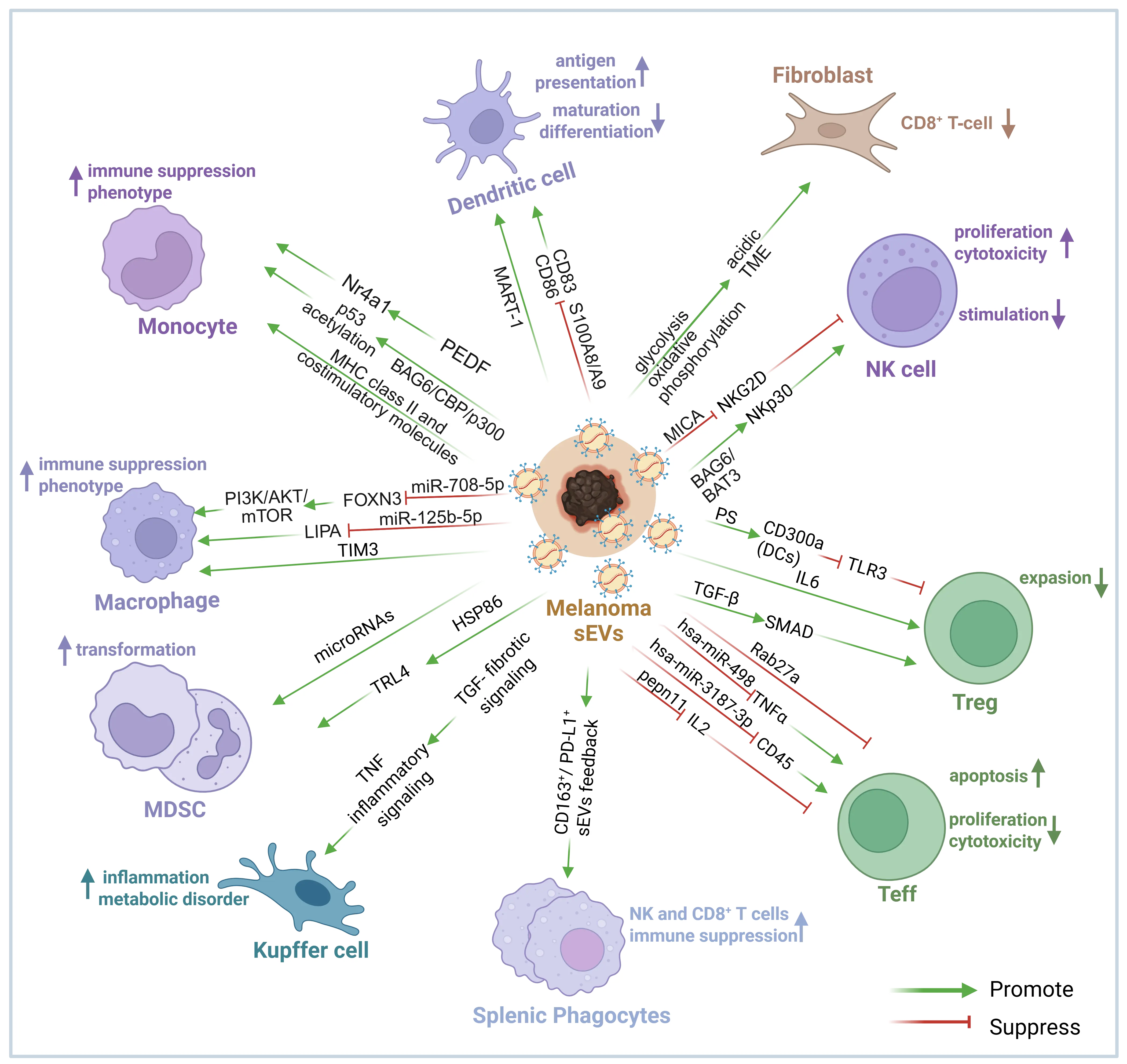
Figure 1. The melanoma sEVs-mediated immunoregulation. Melanoma-derived sEVs transfer proteins, lipids, and regulatory RNAs to multiple immune and stromal cell populations, thereby remodeling both the local tumor micro-environment and systemic immunity. These sEVs can enhance monocyte-mediated immune surveillance, dendritic-cell antigen presentation, and NK-cell cytotoxicity through pathways such as PEDF–NR4A1, MART-1–MHC-I, and BAG6/BAT3–NKp30. Conversely, they can suppress dendritic-cell maturation and costimulatory signaling, promote immune suppressive macrophage polarization and MDSC expansion, induce fibroblast metabolic reprogramming and an acidic microenvironment, impair NKG2D-dependent NK-cell activity, and alter Treg and effector T-cell differentiation, survival, and function. Melanoma-derived sEVs also affect Kupffer cells and splenic phagocytes, contributing to inflammatory, fibrotic, and systemic immune suppressive feedback. The overall effect depends on tumor stage, vesicle subtype, cargo composition, and recipient-cell context. Green arrows indicate promotion or activation, whereas red T-bars indicate suppression or inhibition. Abbreviations: DC: dendritic cell; MDSC: myeloid-derived suppressor cell; NK: natural killer; Teff: effector T cell; Treg: regulatory T cell; TME: tumor micro-environment. An upward arrow represents an increase and a downward arrow represents a decrease. The figure was created with BioRender.com/lr1pu1i accessed on 9 August 2026.
2.1. Monocytes and Macrophages
Circulating monocytes can be divided into classical Ly6Chigh (Ly6ChighPMo) and nonclassical Ly6Clow (Ly6ClowPMo) subsets based on surface-marker expression; the latter are often associated with vascular patrolling and immune surveillance [27]. Melanoma-derived sEVs dynamically regulate monocyte fate, with their effects dependent on both tumor stage and the molecular cargo of these vesicles. Distinct sEVs subpopulations exert divergent functions, directing monocyte differentiation toward either an immunosuppressive or immunostimulatory phenotype. Interestingly, a recent study has concluded that the immune-suppressive phenotype of monocytes might be associated with the sEVs derived from melanoma. Luong et al. demonstrated that melanoma-derived sEVs drive monocyte conversion toward an immunosuppressive phenotype, evidenced by the downregulation of MHC class II and costimulatory molecules [28]. By contrast, Plebanek, MP et al. reported that sEVs from nonmetastatic melanoma deliver pigment epithelium-derived factor (PEDF) to bone marrow myeloid precursors, induce the orphan nuclear receptor NR4A1, expand Ly6Clow patrolling monocytes, and recruit natural killer (NK) cells and macrophages to suppress metastasis [29]. In addition, melanoma-derived extracellular vesicles inhibit tumor metastasis to the lung by recruiting Ly6ClowPMo, whereas the formation of these antitumor vesicles depends on p53 acetylation mediated by the BAG6/CBP/p300-acetylase complex [30]. Interestingly, BAG6 gene ablation and perturbation of its associated signaling pathway trigger the secretion of a distinct extracellular vesicle subtype that recruits pro-tumor neutrophils to the pre-metastatic niche instead of inhibiting metastasis [30]. These findings indicate that while sEVs can induce an immunosuppressive monocyte phenotype, loss of BAG6 leads to the release of a distinct sEVs subtype that actively promotes metastasis. However, when derived from non-metastatic melanoma that contains PEDF, sEVs can elicit an innate immune response [29]. Thus, the function of melanoma-derived sEVs is dependent on the tumor’s intrinsic state, underscoring the need to elucidate this correlation to stratify patients.
Upon migrating into peripheral tissues, monocytes can further differentiate into macrophages and dendritic cells (DCs), and macrophages are categorized into the antitumor M1 phenotype and the M2 phenotype, the latter of which is also known as tumor-associated macrophages (TAMs) [31,32]. Previous studies have shown that macrophages co-cultured with melanoma-derived sEVs exhibit a mixed M1/M2 phenotype [31]. However, we are still trying to identify the decisive factors for each phenotype determined by sEVs. Mechanistically, the depletion of TIM-3 in melanoma-derived sEVs has been shown to impair TAM proliferation [33]. Additionally, sEVs-delivered miR-125b-5p promotes TAM polarization by repressing lysosomal acid lipase A (LIPA) in recipient macrophages [34]. Specifically, melanoma-derived sEVs drive the generation of TAMs either by directly targeting macrophages via miR-125b-5p or by indirectly acting on CD4+ T cells through TIM-3 [24]. Furthermore, miR-708-5p, which is highly enriched in melanoma-derived sEVs, directly targets FOXN3, thereby activating the PI3K/AKT/mTOR pathway in macrophages and promoting their polarization toward the M2 phenotype [35].
In short, PEDF and BAG6 are critical mediators of innate anti-tumor immunity, while TIM-3, miR-125b-5p, and miR-708-5p represent promising molecular therapeutic targets that restrain tumor progression by blocking M2 macrophage polarization. Furthermore, these molecules may serve as potential biomarkers for monitoring tumor burden and progression. Gao et al. indicated that TIM-3 expression in sEVs isolated from patients with non-small cell lung cancer is higher than that in healthy subjects, and this trend is consistent with tumor size and metastatic status [36]. What’s more, while modulating macrophage phenotypes, melanoma-derived sEVs can recruit relevant immune cells, including macrophages and neutrophils [21,34,37,38,39], which are associated with innate immunity and promote pre-metastatic niche formation [40].
In summary, melanoma-derived sEVs exert dual and opposing regulatory effects on the monocyte–macrophage lineage, driving both immunosuppressive and anti-tumor immune responses. The contradictory functional outcomes are primarily determined by the molecular cargo profiles of sEVs and the intrinsic status of parental tumors. sEVs derived from non-metastatic melanoma are enriched in PEDF, which potentiates immune surveillance via the Nr4a1 signaling pathway. In contrast, advanced-stage melanoma exhibits BAG6 deficiency, which reprograms sEVs toward a pro-metastatic phenotype, indicating that the functional integrity of the p53/BAG6 complex is critical for sustaining the production of anti-tumor sEVs. Furthermore, sEVs-mediated regulation differs substantially between monocyte precursors and mature macrophages, and the functional molecules carried by distinct sEVs subpopulations ultimately dictate their immunomodulatory direction. Collectively, these findings suggest that stratified sEV-targeted therapeutic strategies should be implemented according to tumor staging. Specifically, targeting miR-708-5p or TIM-3 can effectively block M2 macrophage polarization in advanced melanoma, whereas preserving the PEDF/BAG6 axis is favorable for maintaining endogenous immune surveillance in early-stage patients. Meanwhile, the sEVs cargo signature holds great promise as a liquid biopsy biomarker for tumor staging and prognostic evaluation in melanoma.
2.2. Myeloid-Derived Suppressor Cells
Myeloid-derived suppressor cells (MDSCs) are a population of immunosuppressive cells originating from the bone marrow and serve as progenitors of DCs and macrophages. According to the resource, MDSCs can be separated into polymorphonuclear MDSCs (PMN-MDSCs) and mononuclear MDSCs (M-MDSCs), both of which have the ability to inhibit the immune system [41]. Studies have revealed that melanoma cell-derived sEVs carrying miRNAs alter MDSC function, promoting their expansion and enhancing their immunosuppressive phenotype. Studies have revealed that melanoma cell-derived sEVs carrying miRNAs alter MDSC function, promoting their expansion and enhancing their immunosuppressive phenotype [42]. According to the analysis of clinical data of microRNA transcription, it was further found that a group of microRNAs (miR-146a, miR-155, miR-125b, miR-100, let-7e, miR-125a, miR-146b, miR-99b) participated in the transformation of monocytes into MDSCs mediated by melanoma sEVs [43]. In other words, sEVs derived from melanoma may directly affect the production of MDSCs by microRNA. In addition, Fleming et al. reported that melanoma-derived sEVs predominantly engage on myeloid cells through HSP86, reprogramming normal myeloid cells into MDSC, which led to NF-kB activation and PD-L1 expression up-regulation [44]. Thus, melanoma-derived sEVs can expand and activate MDSCs through both microRNAs and protein cargo.
Current studies have reached a consistent core conclusion regarding the regulatory effects of melanoma-derived sEVs on MDSCs, whereby sEVs potently promote MDSC expansion and augment their immunosuppressive functions. Nevertheless, distinct molecular mechanisms have been reported across different studies. Several investigations highlight the regulatory role of an eight-miRNA signature, whereas others emphasize the critical involvement of the HSP86/TLR4 signaling axis. Such mechanistic discrepancies can be largely attributed to the inherent subpopulation heterogeneity of sEVs, as distinct sEVs subpopulations package unique molecular cargos. Despite the diversity in upstream regulatory molecules, all these divergent signaling cascades ultimately converge on common downstream events, including NF-κB activation and PD-L1 upregulation, which synergistically amplify immunosuppression within the tumor microenvironment.
2.3. DCs
Melanoma-derived sEVs can exert certain antitumor immune effects by carrying tumor antigens to initiate antigen-specific immune responses in vivo, yet they can also directly inhibit DC maturation or modulate T-cell responses by educating DCs, ultimately promoting tumor progression [45]. As the most critical antigen-presenting cells, DCs are also the primary recipients of melanoma-derived sEVs that carry tumor antigens [46,47]. Melanoma-derived sEVs deliver tumor antigens such as MART-1 into DCs, thereby boosting tumor cell killing by inducing the proliferation of MART-1-specific cytotoxic T lymphocytes (CTLs) [22]. Leveraging this mechanism, Morishita et al. engineered GALA-modified sEVs to boost immune responses by facilitating MHC-I antigen presentation within DCs [48]. In addition to affecting antigen presentation, melanoma secretions can block the development of mature DCs by inhibiting molecules such as CD83 and CD86. This inhibition of DC maturation may be related to the presence of VEGF-A in the primary tumor microenvironment, which can trigger the expression of S100A8/A9 [49,50,51,52].
Therefore, the bidirectional regulation of dendritic cells (DCs) by melanoma-derived sEVs is strictly dictated by their molecular cargo composition. Specifically, tumor antigen-containing sEVs potentiate cytotoxic T lymphocyte (CTL)-mediated anti-tumor immunity, whereas sEVs carrying VEGF-A/S100A8/A9 suppress DC maturation. These opposing functional outcomes demonstrate that sEVs subpopulation heterogeneity determines their immunomodulatory polarity. Accordingly, the rational design of sEVs-based immunotherapeutic strategies should prioritize targeted approaches to redirect sEVs toward pro-antigen-presenting phenotypes and circumvent their intrinsic immunosuppressive risks. Such strategies include GALA peptide modification and combination treatment with S100A8/A9 inhibitors.
2.4. Natural Killer Cells
Natural killer cells (NK cells) are the core cells of the innate immune system. They can activate and transmit functional information through the molecular receptor ligand recognition mechanism on the cell surface to participate in the anti-tumor immune response [52,53]. NKG2D supports immune surveillance and cytotoxicity, whereas melanoma-derived sEVs bearing MICA can downregulate NKG2D through a TGF-β–dependent mechanism, thereby suppressing NK-cell activity [54,55,56,57,58]. In addition to inhibiting NK cells function, sEVs can also carry BAG6 and activate NK cells by binding to the NKP30 receptor [30]. Stimulation of RIG-I in melanoma cells also generates an antitumor sEVs population enriched in the NKp30 ligands BAG6/BAT3, which promotes NKp30-dependent melanoma-cell lysis [59,60]. Therefore, melanoma-derived sEVs may bidirectionally regulate the function and quantity of NK cells in different ways. However, the irradiated melanoma-derived sEVs can inhibit tumor growth by producing interferon, which can lead to an increase in tumor-infiltrating NK cells [61,62]. Collectively, these studies demonstrate that the bidirectional regulation of NK cell activity by melanoma-derived sEVs is primarily dictated by sEVs subtypes and their encapsulated molecular cargo. Specifically, the MICA/TGF-β axis mediates immunosuppressive effects, whereas BAG6/NKp30 signaling and RIG-I stimulation elicit NK cell-activating responses. External interventions such as irradiation can remodel the cargo profile of melanoma sEVs, thereby switching their functional phenotype from immune-suppressive to immune-stimulatory. Such regulatory plasticity suggests that combinatorial regimens incorporating RIG-I agonists or radiotherapy, rather than sole blockade of sEVs secretion, may represent more potent strategies for NK cell-based anti-melanoma immunotherapy. Therefore, the effect of sEVs from melanoma on NK cells is multifaceted. Further elucidation of relevant mechanisms will help the development of NK cells therapy. The mechanisms by which these receptor-ligand recognitions bind to and affect NK cell function can serve as targets for the clinical design of antagonists or agonists, providing the groundwork for improving NK cell performance in immunotherapy.
2.5. Regulatory T Cells
Early studies have shown that melanoma-derived sEVs promote the expansion of regulatory T cells (Tregs), thereby facilitating immune escape [63]. Previous evidence highlights that dendritic cells (DCs) internalize melanoma-derived sEVs, in which sEVs-associated phosphatidylserine (PS) engages the inhibitory receptor CD300a. This interaction critically represses the TLR3-TRIF-IRF3 signaling axis typically activated by sEVs-derived RNA. Consequently, this blockade impairs IFN-β production by DCs, resulting in a marked reduction in tumor-infiltrating Tregs [64]. This finding underscores CD300a as a pivotal checkpoint in sEVs-mediated immunosuppressive networks. However, little evidence exists regarding a direct relationship between melanoma-derived sEVs and Tregs. In other researches, tumor-derived sEVs can promote the transformation of Tregs into Th17 cells through an IL-6-dependent pathway [65]. Additionally, TGF-β enriched within tumor-derived sEVs engages receptors on the T cell surface and drives their differentiation into regulatory T cells via activation of the downstream SMAD signaling pathway, alongside elevated FOXP3 expression [66].
Thus, Melanoma-derived sEVs regulate regulatory T cell (Treg) biology through multiple synergistic rather than antagonistic mechanisms. sEVs directly promote Treg differentiation via the TGF-β/SMAD signaling cascade, induce Treg-to-Th17 phenotypic conversion in an IL-6-dependent manner, and indirectly modulate Treg function by targeting CD300a expressed on dendritic cells. This regulatory network exhibits prominent signaling redundancy. The differentiation bias of distinct Treg subsets is shaped by the crosstalk between sEVs molecular cargo composition and microenvironmental cytokine cues, which collectively facilitates tumor immune evasion in melanoma.
2.6. Effector T Cells
By controlling the proliferation and death processes, sEVs from melanoma can increase the variety of effector T cells (Teffs). Zhou et al. demonstrated that melanoma-derived sEVs trigger CD4+ T cell apoptosis both in vitro and in vivo, while Rab27a knockdown suppresses sEVs secretion, elevates intratumoral T cell abundance, and attenuates tumor progression [58,67,68]. As a result, sEVs from melanoma may inhibit CD4+ T cell proliferation and induce apoptosis [58]. Furthermore, interleukins, which mediate intercellular immune communication, are enriched in melanoma-derived sEVs compared with those from healthy donors [21,69]. Wu et al. reported that melanoma-derived sEV carry PTPN11 protein and mRNA, which disrupt IL-2 signaling cascades and restrain T cell proliferation [70]. Thus, melanoma-derived sEVs deliver relevant proteins that modulate cytokine signaling pathways, thereby influencing the proliferation and apoptosis of Teffs [58].
Lactate accumulation and lipid metabolism reprogramming have been demonstrated to promote effector T cell exhaustion, and this ‘metabolic-immune’ coupling mechanism has emerged as a research hotspot in tumor immunology in recent years [71]. Melanoma-derived sEVs indirectly modulate T cell function and survival by regulating cellular metabolism [72]. Shu et al. demonstrated that melanoma-derived sEVs enhance aerobic glycolysis and suppress oxidative phosphorylation in human adult dermal fibroblasts (HADFs). This metabolic shift acidifies the extracellular microenvironment and triggers CD8+ T cell death. This also reveals the hypoxia and acidic tumor microenvironment and confirms the tumor’s depressing effect on immune cell function [73].
Melanoma-derived sEVs also directly manipulate the function of T cells. It has been found that melanoma-derived sEVs carry hsa-miR-3187-3p and hsa-miR-498. The former targets CD45 and suppresses its expression, thereby dephosphorylating Src protein family tyrosine kinases and enhancing T cell intracellular signaling, whereas hsa-miR-498 directly targets TNF-α and inhibits its expression in CD8+ T cells [74]. Additionally, melanoma-derived sEVs carry PD-L1, which recruits tyrosine protein phosphatase SHP2 to the cytoplasmic region of PD-1 to mediate dephosphorylation of the intracellular domains of TCR and CD28, thereby inducing immunosuppression [13,52]. Moreover, Guan et al. clarified that phosphorylation of hepatocyte growth factor–regulated tyrosine kinase substrate (HRS) in tumor cells regulates anti-tumor immunity by inducing PD-L1+ immunosuppressive sEVs, and suggested that blocking HRS phosphorylation could serve as a potential strategy to improve the efficacy of cancer immunotherapy [75].
In conclusion, melanoma-derived sEVs can regulate T cell proliferation, differentiation, apoptosis, and function through receptor-ligand recognition (PD-1 and PD-L1, etc.) or by delivering related cytokines (IL-6 and IL-2, etc.). These multiple mechanisms cooperatively mediate global immunosuppression, reflecting the redundancy and complexity underlying tumor immune evasion. Rather than operating in isolation, these signaling axes constitute a positive feedback loop: sEVs-driven metabolic reprogramming acidifies the tumor microenvironment to further cripple T cell function, while PD-L1 delivers direct inhibitory signals via the phosphatase SHP2. Accordingly, monotherapy targeting only a single molecular node rarely yields satisfactory therapeutic outcomes, highlighting the necessity of combinatorial regimens that simultaneously block PD-L1 signaling, reverse metabolic suppression, and neutralize immunosuppressive microRNAs. Based on the clinical data, Porcelli et al. have confirmed a model that higher levels of tumor-derived, DC-derived, and CD8+ T cell-derived uPAR+ sEVs in non-responders may represent a new biomarker of innate resistance to immunotherapy with checkpoint inhibitors [76]. Serratì et al. further demonstrated that the immunoregulatory molecular profile of circulating sEVs (e.g., PD-1+ sEVs) can serve as a liquid biopsy biomarker for dynamically assessing patients’ responses to immunotherapy and predicting prognosis [77].
2.7. Fibroblasts
Cancer-associated fibroblasts (CAFs) constitute the most abundant stromal cell population within the melanoma microenvironment, and their secreted extracellular vesicles (sEVs) exert critical functions in mediating immunosuppression and therapeutic resistance. CAF-derived sEVs encapsulate diverse immunomodulatory molecules, which recruit immunosuppressive cell subsets and simultaneously impair the effector function of cytotoxic T lymphocytes via signaling axes such as TGF-β and IL-6 [78,79]. Existing studies have revealed that periostin (POSTN) carried by CAF-derived sEVs activates immune checkpoint molecules through the β-catenin pathway, thereby facilitating the formation of an immune “cold” tumor microenvironment [80,81]. Moreover, CAF subpopulations with high expression of FGF2 and PDPN exhibit superior capacity to suppress T cell activity [82,83]. On the contrary, other evidence demonstrates that sEVs secreted by CD9-positive CAFs restrain the proliferation of malignant melanoma cells, indicating that sEVs derived from distinct CAF subsets exert bidirectional biological effects [84].
sEVs-mediated crosstalk between melanoma cells and CAFs also merits intensive attention. Melanoma cells deliver non-coding RNAs, including miR-155-5p via sEVs, which reprogram normal fibroblasts into pro-angiogenic CAFs [85]. These CD9/CD63 double-positive sEVs not only modulate intracellular gene expression but also significantly correlate with the 5-year disease-free survival of melanoma patients [86]. In addition, fibroblast-remodeled melanosomes loaded with AKT1 are internalized by macrophages to trigger robust VEGF secretion, which drives angiogenesis and distant metastasis [87]. Therapeutic targeting of fibroblast activation protein (FAP), a specific marker of CAFs, markedly boosts the anti-tumor efficacy of anti-PD-1 antibodies, which suggests that CAF-derived sEVs and their downstream signaling cascades serve as promising targets for overcoming immunotherapy resistance [88,89].
The conflicting functional phenotypes of CAF-derived sEVs stem from CAF subpopulation heterogeneity and disparities in their molecular cargo: POSTN/TGF-β-positive subsets promote immunosuppression, whereas CD9-positive subsets confer anti-proliferative activity against tumor cells. Melanoma cells harness sEVs to remodel resident fibroblasts into pro-tumorigenic CAFs, establishing a bidirectional paracrine feedback loop. Selectively targeting pathogenic CAF subpopulations and their sEVs (represented by FAP) rather than pan-inhibition of all CAFs emerges as a viable strategy to overcome ICI resistance in melanoma.
2.8. Kupffer Cells
The liver ranks among the most prevalent distant metastatic sites for melanoma. As tissue-resident hepatic macrophages, Kupffer cells exert central functions in sEVs-facilitated pre-metastatic niche formation. Melanoma-derived sEVs are internalized by hepatic Kupffer cells, which subsequently secrete TGF-β. This cytokine upregulates fibronectin production in hepatic stellate cells and recruits additional macrophages, ultimately fostering the construction of a hepatic metastatic microenvironment [90,91]. For uveal melanoma (UM), tumor-derived sEVs can be taken up by organ-specific resident cells, including Kupffer cells, laying the groundwork for pre-metastatic niche establishment in the liver [92]. Furthermore, saturated fatty acids (e.g., palmitic acid) encapsulated within tumor-derived sEVs stimulate Kupffer cells to secrete TNF in a TLR4-dependent manner, thereby triggering hepatic inflammation and metabolic dysfunction [93,94]. Collectively, these observations suggest that blocking Kupffer cell uptake of tumor-derived sEVs or inhibiting downstream inflammatory signaling cascades may act as novel therapeutic strategies to curb liver metastasis of melanoma.
Cumulative evidence unanimously validates that Kupffer cells initiate hepatic pre-metastatic niche development via internalization of tumor sEVs followed by secretion of TGF-β and TNF. Notably, sEVs originating from cutaneous melanoma (CM) versus uveal melanoma (UM) may elicit divergent magnitudes of Kupffer cell responses due to distinct cargo compositions. In addition, whether TGF-β-mediated fibrotic signaling and TNF-driven inflammatory pathways act synergistically or antagonistically remains poorly characterized. Strategies targeting sEVs uptake and downstream signal blockade each possess distinct advantages and limitations. Combined intervention regimens hold the greatest translational potential, yet a delicate balance between target specificity and systemic biosafety must be achieved.
2.9. Splenic Phagocytes
As the largest secondary lymphoid organ in the body, the spleen serves as a central hub for systemic immune surveillance and represents a critical target organ of melanoma-derived sEVs [95]. Tumor-originated sEVs traffic to the spleen, markedly reducing the frequencies of splenic NK cells and CD8+ T cells while elevating the proportion of regulatory T cells, thereby remodeling the splenic immune landscape [96]. Melanoma-derived sEVs also directly suppress splenocyte proliferation, trigger cellular apoptosis, and elicit cell-cycle arrest [97]. Systemic administration of tumor-derived sEVs induces splenomegaly, disrupts hematopoietic homeostasis, drives extramedullary hematopoiesis, and expands immature erythroid progenitors within the spleen [98].
Notably, splenic phagocytes themselves exert immunomodulatory functions via secreted sEVs. Melanoma-derived sEVs are internalized by splenic macrophages, driving their polarization toward a regulatory macrophage phenotype that further represses splenocyte proliferation [99]. In addition, CD163+ sEVs derived from the spleen are markedly enriched in the tumor tissues of melanoma patients and tightly correlate with systemic immunosuppression [100]. PD-L1-positive sEVs disseminate to draining lymph nodes and the spleen to suppress immune cell function, whereas blockade of tumor sEVs biogenesis and secretion abolishes acquired resistance to PD-L1 inhibitors [100].
Collectively, these data identify the spleen as the central hub for sEVs-mediated systemic immune regulation in melanoma. On one hand, tumor sEVs directly reshape splenic immune compartments by decreasing NK and CD8+ T cell abundance, enriching Treg populations, and initiating extramedullary hematopoiesis. On the other hand, splenic phagocytes undergo phenotypic education toward a regulatory profile upon uptake of tumor sEVs; their subsequent secretion of CD163+ or PD-L1+ sEVs further amplifies local and systemic immunosuppression. The initiation of this positive feedback loop relies on prior “education” of splenic cells by tumor sEVs, and splenocyte-derived sEVs reciprocally reinforce this suppressive phenotype. Distinct sEVs subsets (PD-L1+ and CD163+) may possess divergent capacities to remodel splenic immune homeostasis, yet the underlying molecular mechanisms remain unclarified. One key current technical bottleneck is that researchers cannot distinguish the individual immunomodulatory contributions of tumor-intrinsic sEVs from those released by splenocytes, which complicates the rational and precise design of targeted therapeutic regimens.
3. Immunoregulation Mediated by Immune Cell-Derived sEVs
sEVs secreted by immune cells within the tumor microenvironment and immune organs act as critical intercellular messengers in melanoma progression. These sEVs facilitate bidirectional communication between immune cells and melanoma cells, transferring bioactive molecules that modulate tumor growth, metastasis, and immune evasion (Figure 2). By reshaping the tumor microenvironment and altering melanoma cell behavior, this crosstalk represents a key regulatory axis that drives melanoma development, offering potential targets for novel therapeutic interventions.
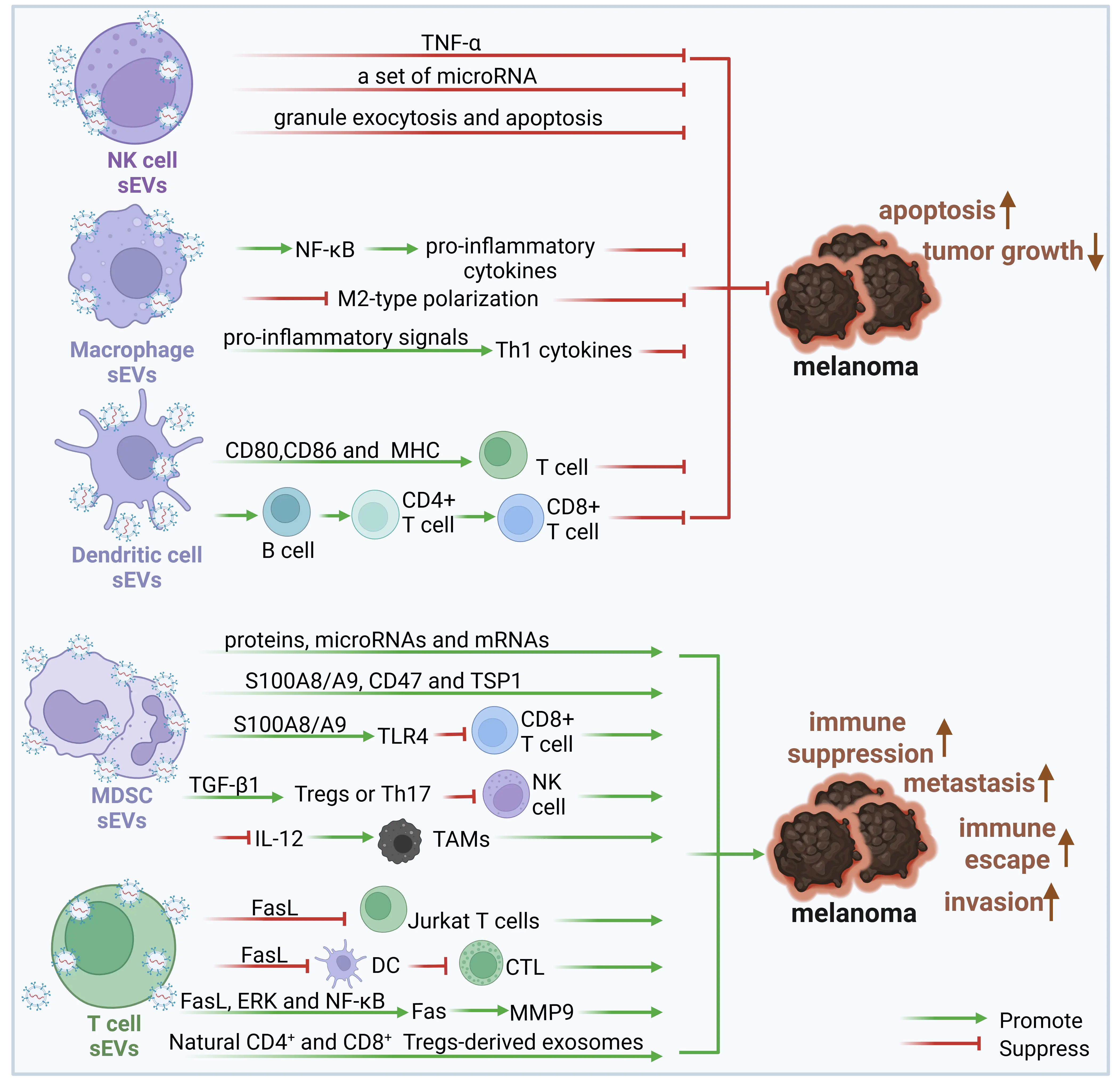
Figure 2. The sEVs-mediated immunoregulation by immune cell-derived sEVs in melanoma progression. Immune cell-derived sEVs exert both melanoma-inhibitory and melanoma-promoting effects. NK-cell-derived sEVs deliver TNF-α, regulatory microRNAs, and cytotoxic molecules associated with granule exocytosis and apoptosis, thereby inducing melanoma-cell death and restricting tumor growth. M1 macrophage-derived sEVs activate NF-κB-dependent pro-inflammatory signaling, inhibit M2 polarization, and enhance Th1 cytokine responses. Dendritic-cell-derived sEVs carry MHC molecules and the costimulatory molecules CD80 and CD86, facilitating B-cell and CD4+/CD8+ T-cell activation and strengthening antitumor immunity. In contrast, MDSC-derived sEVs containing S100A8/A9, CD47, TSP1, TGF-β1, and other regulatory cargo suppress CD8+ T-cell and NK-cell functions while promoting Treg/Th17 differentiation, TAM polarization, immune escape, and metastasis. Activated T-cell- and Treg-derived sEVs can suppress DC–CTL responses and CD8+ antitumor immunity and may activate Fas/ERK/NF-κB/MMP9-related invasive pathways. Green arrows indicate promotion or activation, whereas red T-bars indicate suppression or inhibition. Abbreviations: CTL: cytotoxic T lymphocyte; DC: dendritic cell; MDSC: myeloid-derived suppressor cell; NK: natural killer; TAM: tumor-associated macrophage; Treg: regulatory T cell. An upward arrow represents an increase and a downward arrow represents a decrease. The figure was created with BioRender.com/z6xiqkc accessed on 9 August 2026.
3.1. NK Cell-Derived sEVs
NK cell-derived sEVs exert cytotoxic effects against melanoma cells. As a key component of the innate immune system, NK cells target infected cells without prior sensitization and modulate other immune cells, including DCs, neutrophils, and T lymphocytes, through the secretion of cytokines and growth factors [101]. Notably, NK cells kill tumor cells not only through the classical pathways of granule exocytosis and death receptor-mediated apoptosis [102], but also by transferring microRNAs with tumor-suppressive functions as sEVs cargo to cancer cells, thereby inhibiting tumor growth. Specifically, miRNAs such as miR-10b-5p and miR-92a-3p carried by NK cell-derived sEVs may indirectly enhance the antitumor immune response by promoting Th1 polarization [103]. Accumulating evidence indicates that NK cell-derived sEVs carry characteristic markers (e.g., CD63) and cytotoxic molecules such as perforin [104,105,106], while containing an even higher FasL protein content than NK cells themselves [107]. Furthermore, NK cell-derived sEVs can modulate the proliferation, survival, and apoptosis of melanoma cells through TNF-α delivery [107], an effect that is also observed at low doses [108]. In conclusion, NK cell-derived sEVs represent a potential immunotherapeutic strategy for melanoma.
Accumulating studies have validated that NK cell-derived sEVs exhibit a stable and consistent functional phenotype. Carrying FasL, perforin, TNF-α, and tumor-suppressive microRNAs, these sEVs exert direct anti-tumor cytotoxic effects. Nevertheless, their therapeutic potency is modulated by the Fas sensitivity of melanoma cells: Fas-sensitive melanoma clones undergo apoptosis upon treatment, whereas Fas-resistant variants may acquire enhanced invasive capacity in response to FasL stimulation. Accordingly, clinical translation of NK-sEVs requires prior evaluation of tumoral Fas status, or combinatorial administration with Fas pathway modulators to improve therapeutic efficacy.
3.2. M1 Macrophage-Derived sEVs
M1 macrophage-derived sEVs can induce antitumor immune responses. On one hand, sEVs secreted by M1 macrophages can activate the NF-κB pathway in M0 macrophages, promote the polarization of M0 macrophages toward the M1 phenotype, and induce the release of abundant pro-inflammatory cytokines (IL-6, IL-12, and TNF-α), thereby establishing a local inflammatory microenvironment [109]; on the other hand, these sEVs can also reverse the immunosuppressive microenvironment by inhibiting the M2-type polarization of tumor-associated macrophages [110]. Studies have demonstrated that sEVs derived from M1 macrophages preferentially accumulate within lymph nodes, where they transmit pro-inflammatory signals to resident macrophages and DCs and trigger secretion of Th1-type cytokines [56]. Moreover, as simplified and homologous replicas of their parental cells, M1 macrophage-derived sEVs can directly initiate immune responses by stimulating the activation of immature macrophages and DCs [111].
Therefore, M1 macrophage-derived sEVs may serve as a novel class of immune adjuvants for melanoma and other cancers. A related study demonstrated that, unlike traditional aluminum salt or emulsion adjuvants, which function by delivering antigens and triggering local inflammation at the injection site, sEVs adjuvants can traffic to lymph nodes and establish a local inflammatory microenvironment that favors the induction of an adaptive Th1 immune response [112]. In preclinical studies, M1 macrophage–derived sEVs enhanced the activity of TRP-2 peptide vaccines delivered in lipid calcium phosphate nanoparticles and strengthened antigen-specific CTL responses [111,113,114].
Collectively, M1 macrophage-derived sEVs exert consistent pro-tumorigenic immune effects in anti-tumor regulation, which promote M1 macrophage polarization, reverse M2-mediated immunosuppression via the NF-κB signaling cascade, target lymphoid tissues, and enhance CTL responses by serving as immune vaccine adjuvants. Although these regulatory mechanisms function through distinct pathways, they converge on a unified core principle: the remodeling of immunosuppressive tumor microenvironments. Such functional synergism indicates that M1-sEVs possess multiple cooperative immunostimulatory mechanisms, endowing them with considerable translational potential for vaccine development and combinatorial immunotherapy.
3.3. DC-Derived sEVs
DC-derived sEVs may be more suitable for tumor therapy than DCs. Given that DCs, as professional antigen-presenting cells, can elicit potent adaptive immune responses in vivo [115], DC-based immunotherapy offers distinct advantages. However, in vitro-generated DCs are vulnerable to immunosuppressive factors secreted within the tumor microenvironment; these factors drive DC dysfunction, thereby blunting downstream immune responses. In contrast, as a comparatively stable subset of extracellular vesicles, DC-derived sEVs exhibit functional resistance to direct modulation by the tumor microenvironment [116]. Studies have reported that DC-derived sEVs have a longer half-life in vivo than DCs after injection [117]. Additionally, DC-derived sEVs not only serve as antigen delivery vehicles but can also directly participate in T cell activation by carrying costimulatory molecules (such as CD80 and CD86) as well as MHC molecules [118]. Therefore, DC-derived sEVs may be more suitable for tumor therapy than DCs.
DC-derived sEVs have been employed as therapeutic antitumor vaccines in patients with malignant melanoma, as verified in a phase I clinical trial. Sepideh et al. demonstrated the safety of DC-derived sEVs administration and showed that DC-derived sEVs vaccines trigger recall responses of both adaptive (T lymphocytes) and innate (natural killer cells) immune cells [119].
In the context of DC-derived sEVs-based immunotherapy, the role of B cells within the tumor microenvironment warrants particular attention. Experimental studies indicate that these vesicles induce CD8+ T-cell responses in a CD4+ T-cell–dependent manner and that B cells are required for maximal CD8+ T-cell activation, suggesting that B cell–mediated support of CD4+ T cells contributes to cytotoxic immunity [120]. In B16 melanoma tumor models, where tumor eradication depends on CD8+ T cells, B cell depletion promotes melanoma growth [121].
DC-derived sEVs possess dual capacities for antigen delivery and direct T cell activation. Cumulative evidence has consistently validated their promising anti-tumor potential, and phase I clinical trials have preliminarily verified the safety profile of DC-derived sEVs-based therapies. Nevertheless, several critical scientific issues remain to be addressed for their further clinical translation. Current studies have not systematically elucidated the functional heterogeneity of DC-derived sEVs secreted by dendritic cells at distinct maturation stages. In addition, DC-derived sEVs can shape CD8+ T cell immune responses by modulating B cell function, highlighting the essential role of T–B cell crosstalk in DC-derived sEVs-triggered anti-tumor immune activation. Furthermore, as inherently inert extracellular vesicles, the structural stability and functional superiority of DC-derived sEVs within the complex tumor microenvironment require further validation through rigorous in vitro and in vivo investigations.
3.4. MDSC-Derived sEVs
MDSC-derived sEVs (MDSCs-sEVs), with a diameter ranging from 25 nm to 30 nm, contain proteins involved in recruiting MDSCs [122,123,124,125,126]. Given their homology with parental cells, MDSCs-sEVs can promote tumor immunosuppression, metastasis, and angiogenesis, and even enhance the drug resistance of malignant tumors [124,127,128], thereby skewing antitumor immunity toward a tumor-promoting type 2 immune response [122]. Studies have shown that MDSCs-sEVs contain proteins, microRNAs, and mRNAs that alter the function of recipient cells and ultimately mediate tumor immunosuppression [124].
MDSCs-sEVs enhance tumor immunosuppression by modulating the levels of bioactive molecules in MDSCs and other immune cells [129]. For example, the pro-inflammatory heterodimer S100A8/A9, transported by MDSC-sEVs, exerts chemotactic activity toward MDSCs and critically contributes to their recruitment into tumor tissues and to the formation of the pre-metastatic niche [130]. Meanwhile, MDSCs-sEVs are enriched in other chemotactic proteins, such as CD47 and TSP1, which, together with S100A8/A9, mediate the immunosuppressive function of MDSCs [122]. Moreover, S100A8/A9 interacts with TLR4, thereby inhibiting glycolysis, proliferation, and IFN-γ production in CD8+ T cells, ultimately inducing their exhaustion [131]. In addition, TGF-β1-enriched MDSC-sEVs induce the differentiation of Tregs or Th17 cells and suppress NK cell cytotoxicity, thereby potentiating tumor immunosuppression [124]. Similar to MDSCs, MDSCs-sEVs can also transform macrophages into TAMs by reducing IL-12 generation in macrophages [122].
Therefore, immunotherapies targeting MDSC-sEVs and their clinical applications hold great potential for malignant and non-malignant diseases, although studies exploring their clinical use remain scarce. For instance, plasma levels of S100A9 derived from MDSCs-sEVs are significantly higher in colorectal cancer patients than in healthy controls, and are also elevated in patients with tumor recurrence compared to those who underwent successful tumor resection. Therefore, S100A9 derived from MDSCs-sEVs may serve as a biomarker for predicting the occurrence and progression of colorectal cancer [132]. However, to date, the clinical application and safety of MDSC-sEVs in cancer patients remain to be further investigated.
These vesicles further amplify systemic immunosuppression within the tumor microenvironment by secreting S100A8/A9 to recruit MDSCs via chemotaxis, releasing TGF-β1 to drive Treg and Th17 cell differentiation, and markedly impairing the anti-tumor functions of NK cells and CD8+ T cells. However, accumulating data have revealed substantial discrepancies in the molecular cargo profiles of MDSC-derived sEVs across different tumor models, such as colorectal cancer and melanoma, indicating prominent tumor-specific functional characteristics of MDSC-sEVs. From a clinical translation perspective, MDSC-sEVs inherit the pro-tumor and immunosuppressive properties of their parental MDSCs. Compared with direct MDSC targeting, specific intervention against sEVs can largely avoid disrupting normal myeloid cell development, thereby conferring superior targeting specificity. Nevertheless, the safety and clinical feasibility of this sEVs-targeting strategy remain to be comprehensively validated, which currently constitutes the major bottleneck limiting its clinical implementation.
3.5. T Cell-Derived sEVs
Activated T cell-derived sEVs inhibit the antitumor response of immune cells. Previous studies have reported that Fas signaling can induce tumor apoptosis and inhibit tumor growth when activated by FasL in vivo [133,134,135]. However, activated T cells that release FasL+ sEVs can also induce apoptosis in Jurkat T cells [136]. In addition, FasL+ sEVs derived from CD8+ T cells can suppress the ability of dendritic cells to stimulate CTL responses [137], thus inhibiting the killing of the tumor. Moreover, long-term studies have demonstrated that Fas signaling can drive proliferation of Fas-resistant tumor cells at certain FasL concentrations [138,139].
Activated T cell-derived sEVs enhance tumor cell motility and invasiveness [140,141,142,143,144]. Matrix metalloproteinase 9 (MMP9) promotes tumor invasion and metastasis in the B16 melanoma model [145]. Specifically, CD8+ T cell-derived sEVs are considered the core factor inducing MMP-9-mediated degradation of extracellular matrix proteins, thereby providing essential conditions for the proliferation and metastasis of Fas+ tumor cells [146]. Further studies have demonstrated that sEVs derived from activated CD8+ T cells activate the ERK and NF-κB pathways in melanoma cells, upregulate MMP9 expression via Fas signaling, and thereby promote melanoma metastasis in vivo, underscoring the important role of CD8+ T cell-derived sEVs in tumor immune evasion [147]. Meanwhile, FasL in activated T cell-derived sEVs activates Fas signaling in tumor cells, thereby inducing MMP9 expression and ultimately enhancing the invasiveness of Fas-resistant tumor cells, a mechanism that may provide a new avenue for understanding tumor immune evasion [145].
Natural CD4+CD25+ and CD8+CD25+ Treg-derived sEVs may serve as an alternative immunotherapy for tumors and autoimmune diseases [148]. Natural CD4+CD25+ and CD8+CD25+ Tregs have been shown to inhibit autoimmune diseases [149]. Natural CD8+CD25+ Tregs-derived sEVs inhibited the antitumor immune response of CD8+ T cells, as did the parental cells [148]. However, the specific mechanism of the immune response remains to be further researched.
Activated CD8+ T cell-derived sEVs exert bidirectional effects mediated by the FasL/Fas signaling cascade. These sEVs trigger apoptosis in Fas-sensitive tumor cells, whereas they upregulate MMP9 expression through activation of the ERK/NF-κB pathway in Fas-resistant tumor cells to facilitate invasion and metastasis. Meanwhile, such sEVs can also impair dendritic cell function and dampen anti-tumor immune responses. This dual “double-edged sword” effect is primarily determined by the sensitivity of Fas and the drug-resistance status of tumor cells. Clinically, T cell activation strengthens anti-tumor immunity but may concurrently stimulate the release of invasion-promoting sEVs. Accordingly, combinatorial regimens incorporating Fas pathway modulators or MMP9 inhibitors represent potential strategies to circumvent the pro-metastatic risk while preserving the anti-tumor therapeutic efficacy of T cell-based immunotherapy.
4. Diverse Functions of sEVs and Summary of Cargo–Pathway–Target Cell Regulatory Networks in Melanoma
Melanoma cell-derived and immune cell-derived sEVs form a complex and bidirectional intercellular communication network within the melanoma immune microenvironment. The biological consequences of these vesicles are determined by their cellular origin, molecular cargo, recipient cell type, and the downstream signaling pathways they activate or suppress. The principal sEVs cargo–pathway–target cell networks discussed in Sections 2 and 3 are summarized in the Table 1.
Table 1. Major sEVs cargo–pathway–target cell networks involved in melanoma immunoregulation.
|
Source Cell |
Transferred sEVs Cargo or Factor |
Target Genes or Pathways |
Target Cell |
Function or Effect |
References |
|---|---|---|---|---|---|
|
Melanoma cell |
Unspecified sEVs cargo |
Downregulation of MHC class II and costimulatory molecules |
Monocyte |
Induces an immunosuppressive monocyte phenotype |
[29] |
|
PEDF |
NR4A1 induction |
Ly6Clow patrolling monocyte |
Enhances immune surveillance and suppresses metastasis |
[30] |
|
|
BAG6-associated cargo |
CBP/p300-dependent p53 acetylation |
Ly6Clow patrolling monocyte; NK cell; macrophage |
Supports antitumor sEVs formation and limits pulmonary metastasis |
[31] |
|
|
TIM-3 |
TIM-3-dependent signaling |
CD4+ T cell/macrophage |
Promotes TAM proliferation and M2-like polarization |
[25,34] |
|
|
miR-125b-5p |
LIPA repression |
Macrophage |
Promotes TAM/M2 polarization |
[35] |
|
|
miR-708-5p |
FOXN3 inhibition and PI3K/AKT/mTOR activation |
Macrophage |
Promotes M2 polarization and tumor-supportive macrophage activity |
[36] |
|
|
miR-146a, miR-155, miR-125b, miR-100, let-7e, miR-125a, miR-146b and miR-99b |
Convergent myeloid reprogramming |
Monocyte/MDSC |
Promotes monocyte-to-MDSC conversion and immunosuppression |
[43,44] |
|
|
HSP86 |
TLR4 → NF-κB → PD-L1 |
Myeloid cell/MDSC |
Expands immunosuppressive MDSCs and increases PD-L1 expression |
[45] |
|
|
MART-1 and other tumor antigens |
MHC-I antigen presentation |
Dendritic cell and CTL |
Enhances antigen-specific CTL proliferation and tumor-cell killing |
[23,46–49] |
|
|
VEGF-A/S100A8/A9-associated signals |
Reduction of CD83 and CD86 |
Dendritic cell |
Inhibits DC maturation and weakens antigen presentation |
[50–53] |
|
|
MICA and TGF-β-associated signals |
NKG2D downregulation |
NK cell |
Suppresses NK-cell activation and cytotoxicity |
[55–59] |
|
|
BAG6/BAT3 |
NKp30 activation |
NK cell |
Promotes NKp30-dependent melanoma-cell lysis |
[31,60,61] |
|
|
Phosphatidylserine (PS) |
CD300a inhibition of TLR3-TRIF-IRF3 and IFN-β production |
Dendritic cell/Treg |
Indirectly alters tumor-infiltrating Treg accumulation |
[65] |
|
|
IL-6-associated cargo |
IL-6-dependent signaling |
Treg |
Promotes Treg-to-Th17 phenotypic conversion |
[66] |
|
|
TGF-β |
TGF-β receptor → SMAD → FOXP3 |
T cell |
Promotes differentiation into regulatory T cells |
[67] |
|
|
PTPN11 protein and mRNA |
Disruption of IL-2 signaling |
Effector T cell |
Restrains T-cell proliferation |
[71] |
|
|
miR-3187-3p |
CD45 repression and altered Src-family kinase signaling |
CD4+/CD8+ T cell |
Perturbs T-cell intracellular signaling and function |
[75] |
|
|
miR-498 |
TNF-α repression |
CD8+ T cell |
Suppresses effector cytokine expression |
[75] |
|
|
PD-L1 |
PD-1 → SHP2-mediated dephosphorylation of TCR/CD28 |
Effector T cell |
Induces T-cell dysfunction and immunosuppression; contributes to ICI resistance |
[14,53,76] |
|
|
Metabolism-modifying sEVs cargo |
Aerobic glycolysis ↑; oxidative phosphorylation ↓; extracellular acidification |
Fibroblast → CD8+ T cell |
Creates a metabolically suppressive microenvironment and promotes T-cell death/exhaustion |
[72–74] |
|
|
NK cell |
Perforin, FasL and TNF-α |
Granule/death-receptor and TNF signaling |
Melanoma cell |
Induces apoptosis and reduces melanoma-cell survival and proliferation |
[105–109] |
|
miR-10b-5p and miR-92a-3p |
Th1-polarizing regulatory networks |
Immune cells/melanoma microenvironment |
Enhances antitumor immunity and inhibits tumor growth |
[104] |
|
|
M1 macrophage |
Pro-inflammatory sEVs cargo |
NF-κB activation; IL-6, IL-12 and TNF-α production |
M0 macrophage |
Promotes M1 polarization and establishes a pro-inflammatory microenvironment |
[110] |
|
Pro-inflammatory signals |
Suppression of M2-type polarization |
TAM/macrophage |
Reverses macrophage-mediated immunosuppression |
[111] |
|
|
Lymph-node-targeting sEVs cargo |
Th1 cytokine induction and antigen-specific CTL activation |
Macrophage/DC/CTL |
Acts as an immune adjuvant and enhances melanoma vaccine efficacy |
[57,112–115] |
|
|
Dendritic cell |
MHC-I, MHC-II, CD80, CD86 and tumor antigens |
Antigen presentation and costimulatory signaling |
CD4+ T cell, CD8+ T cell, NK cell and B cell |
Activates adaptive and innate antitumor responses; supports vaccine activity |
[117–122] |
|
MDSC |
S100A8/A9, CD47 and TSP1 |
Chemotactic and suppressive signaling |
MDSC/premetastatic niche |
Recruits MDSCs and amplifies tumor-associated immunosuppression |
[123,130,131] |
|
S100A8/A9 |
TLR4 signaling; glycolysis, proliferation and IFN-γ production |
CD8+ T cell |
Induces CD8+ T-cell metabolic dysfunction and exhaustion |
[132] |
|
|
TGF-β1 |
TGF-β-dependent differentiation and cytotoxicity suppression |
Treg, Th17 cell and NK cell |
Promotes Treg/Th17 differentiation and suppresses NK-cell cytotoxicity |
[125] |
|
|
Factors reducing macrophage IL-12 |
IL-12 production ↓ |
Macrophage |
Promotes conversion into TAMs |
[123] |
|
|
Activated T cell/CD8+ T cell |
FasL |
Fas signaling |
T cell and dendritic cell |
Induces T-cell apoptosis and suppresses DC-dependent CTL responses |
[137,138] |
|
Activated CD8+ T cell |
FasL and associated sEVs signals |
Fas → ERK/NF-κB → MMP9 |
Fas-resistant melanoma cell |
Enhances extracellular-matrix degradation, motility, invasion and metastasis |
[141–148] |
|
Natural CD4+CD25+ or CD8+CD25+ Treg |
Immunosuppressive sEVs cargo not fully defined |
Mechanism incompletely characterized |
CD8+ T cell |
Suppresses the antitumor T-cell response |
[149,150] |
5. sEVs-Driven Immunotherapy Resistance Mechanisms and Combination Intervention Strategies
Although ICI has markedly improved clinical outcomes in patients with melanoma, 40–50% of individuals fail to respond to ICI monotherapy or develop acquired resistance following treatment [150]. In recent years, tumor-derived sEVs have been identified as critical regulators mediating ICI resistance [151]. In contrast to the localized regulatory action of tumor cell membrane-bound PD-L1, multiple immunosuppressive cargo molecules enclosed in sEVs travel throughout the body via circulating body fluids to lymphoid organs, remodel the systemic host immune microenvironment, and eventually form a systemic immunosuppressive phenotype [152].
5.1. sEVs Surface PD-L1 Acts as a “Molecular Decoy” to Mediate Systemic Immunosuppression
The most central and direct mechanism underlying sEVs-mediated ICI resistance relies on PD-L1, which is abundantly enriched on the surface of tumor-derived sEVs. Distinct from membrane-bound PD-L1 on tumor cells, sEVs-associated PD-L1 circulates systemically to distant lymphoid tissues and suppresses T cell activation at sites far from primary tumors. Work from Chen et al. demonstrated that PD-L1+ extracellular vesicles derived from metastatic melanoma dampen anti-tumor immunity by inducing exhaustion of tumor-specific CD8+ T cells [153]. More importantly, surface PD-L1 on sEVs acts as a “molecular decoy” that binds to administered anti-PD-1/PD-L1 antibodies, thereby reducing the concentration of therapeutic antibodies available to target PD-L1 on tumor cell membranes [154]. This decoy mechanism provides a compelling rationale for the observation that some patients with high circulating PD-L1+ sEVs levels fail to respond to ICI despite positive tumoral PD-L1 expression.
5.2. sEVs-Mediated T Cell Dysfunction and Metabolic Reprogramming
Beyond the decoy effect, melanoma-derived sEVs trigger T cell dysfunction through multiple convergent signaling cascades that collectively establish a profoundly immunosuppressive microenvironment.
On one hand, sEVs deliver immunosuppressive microRNAs to directly impair effector functions of T cells. Vignard et al. [74] reported that melanoma-derived sEVs carry hsa-miR-3187-3p and hsa-miR-498. The former targets CD45 to downregulate its expression, thereby further promoting dephosphorylation of Src family protein tyrosine kinases and disrupting intracellular T cell signaling. The latter directly targets TNF-α and suppresses its production in CD8+ T cells. In addition, glycolysis-regulated exosomal LINC01214 represses CD8+ T cell function and mediates anti-PD-1 resistance in melanoma through modulating the miR-4492/PPP1R11 axis [155].
On the other hand, sEVs drive T cell exhaustion by eliciting metabolic reprogramming. A study published in Science Translational Medicine by Ma et al. [150] uncovered that PD-L1 contained within tumor-derived sEVs induces T cell senescence and functional suppression via lipid metabolic rewiring. Tumor-derived sEVs-associated PD-L1 triggers robust DNA damage and hyperactive lipid metabolism in both human and murine T cells. Genetic ablation of PD-L1 within tumor-derived sEVs alleviates senescence and lipid accumulation in adoptively transferred T cells, demonstrating that surface PD-L1 on tumor-derived sEVs impairs T cell function by activating the aforementioned signaling axes. Research from Dratkiewicz et al. [156] illustrated that melanoma-originated sEVs upregulate aerobic glycolysis and downregulate oxidative phosphorylation in human dermal fibroblasts, leading to extracellular acidification and subsequent CD8+ T cell death. Bland et al. [72] further verified that sEVs secreted by B16F0 melanoma remodel the transcriptome of cytotoxic T lymphocytes, disrupt mitochondrial respiration, and compromise metabolic fitness in T cells. This metabolism-immune crosstalk represents an emerging paradigm wherein sEVs-mediated metabolic reprogramming of both tumor stromal cells and immune cells synergistically fuels T cell exhaustion and ICI resistance.
Furthermore, sEVs facilitate ICI resistance by shuttling immunosuppressive proteins and cytokines. Accumulating evidence has demonstrated that melanoma-derived sEVs contain both PTPN11 protein and its mRNA, which disrupt IL-2 pathway signaling and constrain T cell proliferation [157]. Meanwhile, sEVs-associated immunosuppressive cytokines such as TGF-β boost the differentiation of Tregs and block the expansion of effector T cells, ultimately blunting anti-tumor immune responses and conferring resistance to ICI [158].
5.3. sEVs-Mediated Myeloid Cell Reprogramming
sEVs drive ICI resistance by reprogramming myeloid cells toward an immunosuppressive phenotype. Fleming et al. [44] demonstrated that melanoma-derived sEVs predominantly trigger TLR4 signaling in myeloid cells via HSP86, thereby reprogramming naive myeloid cells into MDSCs and facilitating NF-κB activation and PD-L1 upregulation. sEVs-induced MDSC expansion further amplifies immune suppression through S100A8/A9-mediated chemotaxis and subsequent T cell inhibition. Through clinical transcriptomic analysis of microRNA profiles, Huber et al. [43] identified a panel of microRNAs that mediate the sEVs-driven transformation of monocytes into MDSCs in melanoma, which serves as a predictive signature for ICI resistance in melanoma patients.
Reprogramming of tumor-associated macrophage (TAM) polarization represents another pivotal mechanism underlying sEVs-induced ICI resistance. Melanoma-derived sEVs carrying miR-125b-5p promote pro-tumor TAM polarization by targeting lysosomal acid lipase A in macrophages, while sEVs-delivered miR-708-5p induce M2-type macrophage polarization by directly targeting FOXN3 and activating the PI3K/AKT/mTOR signaling pathway [35]. These sEVs-educated TAMs secrete abundant immunosuppressive cytokines, which further impair the cytotoxic function of cytotoxic T lymphocytes (CTLs) and ultimately establish an immune-excluded tumor phenotype closely correlated with poor ICI therapeutic responses. In addition, TAM-derived sEVs exhibit high PD-L1 expression, which potently inhibits the proliferation and effector function of CD8+ T cells. Collectively, these alterations reshape the tumor microenvironment into an immune-excluded state, ultimately leading to ICI treatment failure [100].
5.4. Combined Intervention Strategies
Given the multi-target and multi-pathway regulatory characteristics of sEVs in ICI resistance, monotherapy is insufficient to dismantle the complex sEVs-governed immunosuppressive network. Accordingly, multi-node combined intervention strategies targeting the biogenesis, secretion, and functional activity of sEVs, as well as sEV-induced downstream immune cell reprogramming, represent a core direction for reversing ICI resistance and improving immunotherapeutic efficacy in melanoma.
5.4.1. Targeting sEVs Biogenesis and Secretion
Inhibition of key regulators involved in sEVs generation, including Rab27a, neutral sphingomyelinase, and core components of the ESCRT complex, can reduce the systemic burden of immunosuppressive sEVs [159]. Guan et al. [75] demonstrated that blockade of HRS phosphorylation suppresses the secretion of immunosuppressive PD-L1+ sEVs and rescues anti-tumor immune responses. Furthermore, high-throughput screening has identified multiple sEV PD-L1 inhibitors, such as ibuprofen, which exert synergistic anti-tumor effects with immunotherapy [160]. Emerging evidence reveals that targeting protein palmitoylation enables dual downregulation of PD-L1 on tumor cell membranes and sEVs alike, establishing a novel therapeutic paradigm for tumors with low responsiveness to PD-L1 antibody monotherapy [161].
5.4.2. Dual Targeting of Tumor-Derived and sEVs-Derived PD-L1
Combining conventional PD-L1 antibodies with agents that block sEVs PD-L1 secretion or neutralize circulating sEVs PD-L1 effectively restores the concentration of functional therapeutic antibodies at tumor sites [162]. A recently developed pH-responsive sEVs permeabilization strategy (ExoPERM) selectively disrupts sEVs within the tumor microenvironment via pH-sensitive PEGylated peptides. This approach efficiently blocks the interaction between sEVs PD-L1 and PD-1 on CD8+ T cells, thereby restoring T cell effector functions [163,164]. In murine melanoma models, the combination of ExoPERM with immune checkpoint blockade markedly enhances the infiltration and activation of tumor-infiltrating CD8+ T cells, thereby converting immune “cold” tumors into T cell-inflamed “hot” tumors [164]. Such a dual-targeting strategy is particularly beneficial for patients with high baseline sEVs PD-L1 levels, who are predicted to be refractory to single-agent ICI therapy.
5.4.3. Reversing sEVs-Driven Myeloid Cell Reprogramming
Inhibition of TLR4 signaling, S100A8/A9 antagonism, or blockade of miRNA-mediated macrophage polarization can constrain the formation of immunosuppressive myeloid phenotypes and reinstate anti-tumor immune surveillance [165]. In addition, therapeutic strategies targeting MDSCs depletion or reprogramming of TAMs toward the M1-like pro-inflammatory phenotype exert synergistic effects with ICI treatment [166,167].
5.4.4. Engineered sEVs-Based Therapeutic Agents
Owing to their outstanding biocompatibility and inherent capacity for targeted delivery, sEVs serve as versatile therapeutic vectors to reshape the tumor immune microenvironment and enhance ICI efficacy. Morishita et al. constructed GALA peptide-modified sEVs that enhance MHC-I antigen presentation in dendritic cells, thereby amplifying systemic anti-tumor immune responses [168]. Notably, a newly developed inhalable bispecific exosomal T-cell activator (BEAT) system utilizes the Alix sorting domain to co-display PD-1 and FZD8 at a 1:1 ratio on individual sEVs surfaces, enabling simultaneous blockade of the PD-L1 and Wnt7b signaling pathways [169]. In ICI-resistant melanoma mouse models, inhalable BEAT efficiently recruits and activates intratumoral CD8+ T cells, remodels the immunosuppressive tumor microenvironment, and elicits robust anti-tumor effects, that are superior to combination treatment with PD-L1 and Wnt7b bispecific antibodies [169]. Moreover, engineered sEVs can be loaded with functional miRNAs (e.g., miR-155 to potentiate T cell function) or PD-L1-targeting siRNAs to silence tumoral PD-L1 expression, thereby generating synergistic anti-tumor effects with ICI.
5.4.5. sEVs-Based Liquid Biopsy for Precise Stratified Therapy
Monitoring circulating levels of PD-L1+ sEVs, PD-1+ sEVs, uPAR+ sEVs, or specific sEVs miRNA signatures enables the identification of patients at high risk of ICI resistance and guides early combination intervention [75,76,77]. Furthermore, sEVs-based liquid biopsy enables dynamic evaluation of therapeutic responses and facilitates adaptive adjustments to individualized treatment strategies during clinical follow-up [170].
6. Conclusions and Future Perspectives
6.1. sEVs-Mediated Immune Regulatory Networks: Dual Effects and Determinant Factors
This review systematically elaborates the immunomodulatory functions of sEVs derived from melanoma cells and multiple immune cell subsets (NK cells, macrophages, DCs, MDSCs, and T cells) within the tumor microenvironment.
6.1.1. Melanoma Cell-Derived sEVs Exert Bidirectional Immunomodulatory Effects
At the innate immune level, these sEVs either suppress NK cell function via the MICA/TGF-β axis or activate NK cells through the BAG6/NKp30 signaling cascade. Similarly, they can induce DC maturation to stimulate CTL responses by delivering tumor antigens, while simultaneously impairing DC maturation via VEGF-A/S100A8/A9 signaling. With regard to adaptive immunity, sEVs cooperatively suppress effector T cells through multiple cascades, including PD-L1/SHP2, miR-3187-3p/miR-498, PTPN11/IL-2, and metabolic reprogramming. Meanwhile, they drive Tregs expansion and MDSCs accumulation to establish a systemic immunosuppressive milieu. The ultimate functional bias of this bidirectional regulation is determined by the integrated interplay of tumor stage, sEVs subtypes, cargo composition, p53/BAG6 expression status, and microenvironmental cues such as hypoxia and local TGF-β concentration.
6.1.2. sEVs Secreted by Immune Cells Predominantly Mediate Anti-Tumor Activities
NK cell-derived sEVs carry FasL, perforin, TNF-α, and tumor-suppressive microRNAs to elicit direct cytotoxicity. M1 macrophage-derived sEVs exert anti-tumor functions by polarizing M0 macrophages toward an M1 phenotype, transporting pro-inflammatory cytokines, and acting as vaccine adjuvants. DC-derived sEVs possess dual capacities for antigen delivery and direct T cell activation, and their safety profile has been validated in phase I clinical trials. In contrast, MDSC-derived sEVs amplify immune suppression via effector molecules, including S100A8/A9 and TGF-β1. T cell-derived sEVs exhibit FasL-dependent dual bioactivities: they trigger apoptosis in drug-sensitive tumor cells yet promote invasiveness of therapy-resistant tumor clones.
Collectively, sEVs construct intricate immune regulatory networks in the melanoma microenvironment, and their biological functions are tightly governed by the crosstalk among parental cell origin, molecular cargo, and contextual microenvironmental conditions.
6.2. Translational Challenges and Future Perspectives
Despite the promising applications of sEVs in the diagnosis, prognostic evaluation, and immunotherapeutic intervention of melanoma, multiple translational bottlenecks remain to be addressed before robust clinical implementation.
6.2.1. Lack of Unified Standards for sEVs Isolation and Purification
Currently, ultracentrifugation, size-exclusion chromatography, immunoaffinity enrichment, and polymer-based precipitation constitute the mainstream techniques for sEVs isolation. However, these methods differ substantially in product purity, sample recovery rate, and experimental reproducibility, and no universally accepted gold-standard protocol has been established in the field [171]. sEVs isolated via distinct purification strategies exhibit inherent variations in particle size distribution and the abundance of functional protein and nucleic acid cargos. Such technical biases impair cross-laboratory data comparability and compromise the stability and reliability of results from sEVs-based liquid biopsies, highlighting the urgent need to develop standardized operating procedures and full-process quality control systems for sEVs research.
6.2.2. Imperfect Quantitative Systems for sEVs Cargo Detection
Precise quantitative detection of sEVs surface biomarkers (e.g., PD-L1), microRNAs, and functional proteins still faces prominent technical limitations. For the representative biomarker PD-L1, enzyme-linked immunosorbent assay (ELISA) and flow cytometry are the predominant quantitative approaches. Nevertheless, unified operating guidelines and standardized internal reference criteria are currently unavailable for these assays. Moreover, the absence of authoritative reference materials and universal data normalization principles severely restricts the routine clinical application of sEVs-derived biomarkers.
6.2.3. In Vivo Stability and Pharmacokinetic Limitations
Intravenously administered sEVs are rapidly cleared by hepatic Kupffer cells and splenic macrophages, with an in vivo circulation half-life ranging from several minutes to hours, thereby drastically reducing the effective dose delivered to target lesions [172]. Engineered strategies, including PEGylation, targeted ligand conjugation, and hybrid sEVs-liposome construction, can prolong sEVs circulation time; however, their potential impacts on biosafety and immunogenicity require further rigorous evaluation [173].
6.2.4. Off-Target Toxicity Risks
Beyond the delivery of therapeutic cargos, surface proteins and internal contents of sEVs may be non-specifically internalized by normal tissues, triggering aberrant immune activation and inflammatory responses. Furthermore, insufficient removal of oncogenic cargos from tumor-derived sEVs may conversely promote tumor progression. Therefore, systematic assessment of the biodistribution, immunogenicity, and long-term safety of engineered sEVs is indispensable for clinical translation.
6.2.5. Imperfect Clinical Trial System
DC-derived sEVs vaccines have demonstrated favorable safety profiles in phase I clinical trials for melanoma and can induce NK and T cell immune memory responses. However, large-scale phase II/III randomized controlled clinical data are lacking, and the definitive therapeutic efficacy remains to be validated. Additionally, most engineered sEVs formulations, including miRNA-loaded, PD-L1-silenced, and drug-loaded sEVs, are still in the preclinical stage, with considerable challenges remaining in large-scale production and standardized quality control.
6.2.6. Advances in Single-Vesicle Analysis and Breakthroughs in Immune Regulation Research
Traditional bulk analysis of sEVs averages signals from tens of thousands of vesicles, masking molecular heterogeneity between individual vesicles and obscuring rare but critical biological information originating from pathological cells. Rapid advances in single-vesicle analytical technology have overcome this fundamental limitation. Current mainstream platforms include single-particle tracking microscopy, single-vesicle RNA sequencing, nano-flow cytometry, nanoplasmonic sensing, immune droplet digital detection, microfluidics, and nanomaterial-based biosensing [174]. In terms of methodological innovation, FRET-enabled tetrahedral DNA nanoprobes combined with nano-flow cytometry enable precise quantification of single-vesicle miRNAs at high resolution [175]. The Protein Interaction Coupling (PICO) reference-free quantitative assay integrates DNA-barcoded antibodies with digital PCR, achieving multiplexed protein profiling of intact single vesicles using merely 1 µL of sample [176]. High-throughput single-vesicle imaging platforms have also realized direct profiling of human plasma specimens across 191 clinical samples [177].
Single-vesicle resolution is essential for deciphering sEVs-mediated immune regulation in melanoma. Tumor-derived sEVs exhibit prominent heterogeneity, and distinct sEVs subpopulations carry completely different immunomodulatory molecules. Specifically, PD-L1+/PD-L1− and BAG6+/BAG6− sEVs subsets exert opposite immune-activating or immunosuppressive effects, which are strictly determined by the molecular signature of individual subpopulations. Single-vesicle multi-omics analysis enables accurate identification of key sEVs subpopulations driving tumor immune evasion and characterizes their co-expression patterns of PD-L1, immunosuppressive miRNAs, and metabolic regulators, providing unprecedented resolution for elucidating the precise molecular mechanisms underlying sEVs-mediated immunosuppression and ICI resistance. Furthermore, single-vesicle technologies facilitate the discovery of novel diagnostic biomarkers and therapeutic targets. In the future, the deep integration of single-vesicle analysis with artificial intelligence-driven data interpretation is expected to achieve precise prediction and dynamic monitoring of ICI therapeutic responses in melanoma patients via liquid biopsy.
6.2.7. Future Research Directions
Subsequent research can be focused on five core directions: (1) establishing standardized platforms for sEVs isolation, purification, and multi-dimensional characterization to unify full-process quality control specifications; (2) conducting large-sample validation based on prospective clinical cohorts to clarify the clinical value of diverse sEVs biomarkers for prognostic stratification and ICI response prediction in melanoma patients; (3) optimizing the manufacturing protocols of engineered sEVs to promote translational evaluation of modified vesicle therapeutics; (4) exploring synergistic combination regimens of sEVs intervention with ICI therapy and dissecting the underlying mechanisms of combinatorial efficacy enhancement; (5) characterizing the distinct molecular profiles of sEVs in rare melanoma subtypes (e.g., uveal melanoma) to develop subtype-specific diagnostic and targeted therapeutic strategies. Collectively, the clinical translation of sEVs-based basic research into individualized immunotherapeutic regimens for melanoma requires interdisciplinary collaboration across immunology, pharmaceutics, clinical medicine, and bioengineering, as well as sustained in-depth scientific investigation.
Statement of the Use of Generative AI and AI-Assisted Technologies in the Writing Process
During the preparation of this manuscript, the author used Doubao (seed-2.0-Pro) for language polishing. The authors subsequently reviewed and edited the content as needed and take full responsibility for the accuracy and integrity of the published article.
Author Contributions
J.W. designed the manuscript outline and provided the writing framework. X.L., W.C. and W.L. conceived and wrote the manuscript. X.L., W.C. and J.Z. jointly conceived and designed the figures. J.W. and Z.L. reviewed the content and provided revision suggestions. All authors have made contributions to the revision of the manuscript and have read and approved the submitted version.
Ethics Statement
Not applicable.
Informed Consent Statement
Not applicable.
Data Availability Statement
Not applicable.
Funding
This work was supported by the Guangdong Basic and Applied Basic Research Foundation (2024A1515013250 to J.W.) and the Guangdong Provincial Key Areas Special Projects for Regular Higher Education Institutions (2024ZDZX2054 to J.W.).
Declaration of Competing Interest
The authors declare that they have no known competing financial interests or personal relationships that could have appeared to influence the work reported in this paper.
References
- Hossain SM, Gimenez G, Stockwell PA, Tsai P, Print CG, Rys J, et al. Innate immune checkpoint inhibitor resistance is associated with melanoma sub-types exhibiting invasive and de-differentiated gene expression signatures. Front. Immunol. 2022, 13, 955063. DOI:10.3389/fimmu.2022.955063 [Google Scholar]
- van den Bulk J, Verdegaal EME, van der Ploeg M, Visser M, Nunes JB, de Ru AH, et al. Neoantigen Targetability in Progressive Advanced Melanoma. Clin. Cancer Res. 2023, 29, 4278–4288. DOI:10.1158/1078-0432.CCR-23-1106 [Google Scholar]
- Wang C, Liu S, Li X, Cui K, Zhang W, Du Y. Baseline neutrophil-to- ratio combined with the change during treatment provides risk stratification for metastatic malignant melanoma patients treated with PD-1 inhibitors in a Chinese population. Front. Oncol. 2023, 13, 1118301. DOI:10.3389/fonc.2023.1118301 [Google Scholar]
- Du Z, Feng Y, Zhang H, Liu J, Wang J. Melanoma-derived small extracellular vesicles remodel the systemic onco-immunity via disrupting hematopoietic stem cell proliferation and differentiation. Cancer Lett. 2022, 545, 215841. DOI:10.1016/j.canlet.2022.215841 [Google Scholar]
- Du Z, Zhang H, Feng Y, Zhan D, Li S, Tu C, et al. Tumour-derived small extracellular vesicles contribute to the tumour progression through reshaping the systemic immune macroenvironment. Br. J. Cancer 2023, 128, 1249–1266. DOI:10.1038/s41416-023-02175-4 [Google Scholar]
- van Niel G, Carter DRF, Clayton A, Lambert DW, Raposo G, Vader P. Challenges and directions in studying cell–cell communication by extracellular vesicles. Nat. Rev. Mol. Cell Biol. 2022, 23, 369–382. DOI:10.1038/s41580-022-00460-3 [Google Scholar]
- Théry C, Witwer KW, Aikawa E, Alcaraz MJ, Anderson JD, Andriantsitohaina R, et al. Minimal information for studies of extracellular vesicles 2018 (MISEV2018): A position statement of the International Society for Extracellular Vesicles and update of the MISEV2014 guidelines. J. Extracell. Vesicles 2018, 7, 1535750. DOI:10.1080/20013078.2018.1535750 [Google Scholar]
- Silva TF, Hutchins E, Zhao W, Ciani Y, Kim M, Ko E, et al. Extracellular vesicle heterogeneity through the lens of multiomics. Cell Rep. Med. 2025, 6, 102161. DOI:10.1016/j.xcrm.2025.102161 [Google Scholar]
- Wang T, Ng CY, Ng BZJ, Toh WS, Hui JHP. Multi-omics analysis of small extracellular vesicles in osteoarthritis: Bridging the gap between molecular insights and clinical applications. Burn. Trauma 2025, 13, tkaf023. DOI:10.1093/burnst/tkaf023 [Google Scholar]
- Yeat NY, Chen RH. Extracellular vesicles: Biogenesis mechanism and impacts on tumor immune microenvironment. J. Biomed. Sci. 2025, 32, 85. DOI:10.1186/s12929-025-01182-2 [Google Scholar]
- Mandal K, Barik GK, Santra MK. Overcoming resistance to anti-PD-L1 immunotherapy: Mechanisms, combination strategies, and future directions. Mol. Cancer 2025, 24, 246. DOI:10.1186/s12943-025-02400-z [Google Scholar]
- Zhang H, Du Z, Tu C, Zhou X, Menu E, Wang J. Hypoxic Bone Marrow Stromal Cells Secrete miR-140–5p and miR-28–3p That Target SPRED1 to Confer Drug Resistance in Multiple Myeloma. Cancer Res. 2024, 84, 39–55. DOI:10.1158/0008-5472.CAN-23-0189 [Google Scholar]
- Dean E, Lorigan P. Advances in the management of melanoma: Targeted therapy, immunotherapy and future directions. Expert Rev. Anticancer. Ther. 2012, 12, 1437–1448. DOI:10.1586/era.12.124 [Google Scholar]
- Chen G, Huang AC, Zhang W, Zhang G, Wu M, Xu W, et al. Exosomal PD-L1 contributes to immunosuppression and is associated with anti-PD-1 response. Nature 2018, 560, 382–386. DOI:10.1038/s41586-018-0392-8 [Google Scholar]
- Yokoi A, Ochiya T. Exosomes and extracellular vesicles: Rethinking the essential values in cancer biology. Semin. Cancer Biol. 2021, 74, 79–91. DOI:10.1016/j.semcancer.2021.03.032 [Google Scholar]
- Bustos SO, Leal Santos N, Chammas R, Andrade LNDS. Secretory Autophagy Forges a Therapy Resistant Microenvironment in Melanoma. Cancers 2022, 14, 234. DOI:10.3390/cancers14010234 [Google Scholar]
- Serratì S, Guida M, Di Fonte R, De Summa S, Strippoli S, Iacobazzi RM, et al. Circulating extracellular vesicles expressing PD1 and PD-L1 predict response and mediate resistance to checkpoint inhibitors immunotherapy in metastatic melanoma. Mol. Cancer 2022, 21, 20. DOI:10.1186/s12943-021-01490-9 [Google Scholar]
- Pilch J, Mizera J, Tota M, Donizy P. GNAQ/GNA11-Related Benign and Malignant Entities—A Common Histoembriologic Origin or a Tissue-Dependent Coincidence. Cancers 2024, 16, 3672. DOI:10.3390/cancers16213672 [Google Scholar]
- Dong L, Zheng S, Feng Y. Hypoxia-induced immunosuppression in uveal melanoma is mediated by CD63+ exosomes delivering lactate to reprogram immune cells. Front. Mol. Biosci. 2026, 12, 1743009. DOI:10.3389/fmolb.2025.1743009 [Google Scholar]
- Seitz T, John N, Sommer J, Dietrich P, Thasler WE, Hartmann A, et al. Role of Fibroblast Growth Factors in the Crosstalk of Hepatic Stellate Cells and Uveal Melanoma Cells in the Liver Metastatic Niche. Int. J. Mol. Sci. 2022, 23, 11524. DOI:10.3390/ijms231911524 [Google Scholar]
- Wróblewska JP, Lach MS, Kulcenty K, Galus Ł, Suchorska WM, Rösel D, et al. The Analysis of Inflammation-Related Proteins in a Cargo of Exosomes Derived from the Serum of Uveal Melanoma Patients Reveals Potential Biomarkers of Disease Progression. Cancers 2021, 13, 3334. DOI:10.3390/cancers13133334 [Google Scholar]
- Hood JL. Natural melanoma-derived extracellular vesicles. Semin. Cancer Biol. 2019, 59, 251–265. DOI:10.1016/j.semcancer.2019.06.020 [Google Scholar]
- Tucci M, Mannavola F, Passarelli A, Stucci LS, Cives M, Silvestris F. Exosomes in melanoma: A role in tumor progression, metastasis and impaired immune system activity. Oncotarget 2018, 9, 20826–20837. DOI:10.18632/oncotarget.24846 [Google Scholar]
- Zebrowska A, Widlak P, Whiteside T, Pietrowska M. Signaling of Tumor-Derived sEV Impacts Melanoma Progression. Int. J. Mol. Sci. 2020, 21, 5066. DOI:10.3390/ijms21145066 [Google Scholar]
- Simiczyjew A, Dratkiewicz E, Mazurkiewicz J, Ziętek M, Matkowski R, Nowak D. The Influence of Tumor Microenvironment on Immune Escape of Melanoma. Int. J. Mol. Sci. 2020, 21, 8359. DOI:10.3390/ijms21218359 [Google Scholar]
- Basile MS, Mazzon E, Fagone P, Longo A, Russo A, Fallico M, et al. Immunobiology of Uveal Melanoma: State of the Art and Therapeutic Targets. Front. Oncol. 2019, 9, 1145. DOI:10.3389/fonc.2019.01145 [Google Scholar]
- Bianchini M, Duchêne J, Santovito D, Schloss MJ, Evrard M, Winkels H, et al. PD-L1 expression on nonclassical monocytes reveals their origin and immunoregulatory function. Sci. Immunol. 2019, 4, eaar3054. DOI:10.1126/sciimmunol.aar3054 [Google Scholar]
- Luong N, Lenz JA, Modiano JF, Olson JK. Extracellular Vesicles Secreted by Tumor Cells Promote the Generation of Suppressive Monocytes. Immunohorizons 2021, 5, 647–658. DOI:10.4049/immunohorizons.2000017 [Google Scholar]
- Plebanek MP, Angeloni NL, Vinokour E, Li J, Henkin A, Martinez-Marin D, et al. Pre-metastatic cancer exosomes induce immune surveillance by patrolling monocytes at the metastatic niche. Nat. Commun. 2017, 8, 1319. DOI:10.1038/s41467-017-01433-3 [Google Scholar]
- Schuldner M, Dörsam B, Shatnyeva O, Reiners KS, Kubarenko A, Hansen HP, et al. Exosome-dependent immune surveillance at the metastatic niche requires BAG6 and CBP/p300-dependent acetylation of p53. Theranostics 2019, 9, 6047–6062. DOI:10.7150/thno.36378 [Google Scholar]
- Bardi GT, Smith MA, Hood JL. Melanoma exosomes promote mixed M1 and M2 macrophage polarization. Cytokine 2018, 105, 63–72. DOI:10.1016/j.cyto.2018.02.002 [Google Scholar]
- Qian Y, Qiao S, Dai Y, Xu G, Dai B, Lu L, et al. Molecular-Targeted Immunotherapeutic Strategy for Melanoma via Dual-Targeting Nanoparticles Delivering Small Interfering RNA to Tumor-Associated Macrophages. ACS Nano 2017, 11, 9536–9549. DOI:10.1021/acsnano.7b05465 [Google Scholar]
- Li X, Liu Y, Yang L, Jiang Y, Qian Q. TIM-3 shuttled by MV3 cells-secreted exosomes inhibits CD4+ T cell immune function and induces macrophage M2 polarization to promote the growth and metastasis of melanoma cells. Transl. Oncol. 2022, 18, 101334. DOI:10.1016/j.tranon.2021.101334 [Google Scholar]
- Gerloff D, Lützkendorf J, Moritz RKC, Wersig T, Mäder K, Müller LP, et al. Melanoma-Derived Exosomal miR-125b-5p Educates Tumor Associated Macrophages (TAMs) by Targeting Lysosomal Acid Lipase A (LIPA). Cancers 2020, 12, 464. DOI:10.3390/cancers12020464 [Google Scholar]
- Xu M, He B, Zhou X, Shu L, Ma D. Melanoma exosomal miR-708-5p promotes macrophage M2 polarization and cancer metastasis. Cell Death Dis. 2026, 17, 346. DOI:10.1038/s41419-026-08597-1 [Google Scholar]
- Gao J, Qiu X, Li X, Fan H, Zhang F, Lv T, et al. Expression profiles and clinical value of plasma exosomal Tim-3 and Galectin-9 in non-small cell lung cancer. Biochem. Biophys. Res. Commun. 2018, 498, 409–415. DOI:10.1016/j.bbrc.2018.02.114 [Google Scholar]
- Das SK, Sarkar D, Emdad L, Fisher PB. MDA-9/Syntenin: An emerging global molecular target regulating cancer invasion and metastasis. Adv. Cancer Res. 2019, 144, 137–191. DOI:10.1016/bs.acr.2019.03.011 [Google Scholar]
- Gener Lahav T, Adler O, Zait Y, Shani O, Amer M, Doron H, et al. Melanoma‐derived extracellular vesicles instigate proinflammatory signaling in the metastatic microenvironment. Int. J. Cancer 2019, 145, 2521–2534. DOI:10.1002/ijc.32521 [Google Scholar]
- Bardi GT, Al-Rayan N, Richie JL, Yaddanapudi K, Hood JL. Detection of Inflammation-Related Melanoma Small Extracellular Vesicle (sEV) mRNA Content Using Primary Melanocyte sEVs as a Reference. Int. J. Mol. Sci. 2019, 20, 1235. DOI:10.3390/ijms20051235 [Google Scholar]
- Liu Y, Gu Y, Han Y, Zhang Q, Jiang Z, Zhang X, et al. Tumor Exosomal RNAs Promote Lung Pre-metastatic Niche Formation by Activating Alveolar Epithelial TLR3 to Recruit Neutrophils. Cancer Cell 2016, 30, 243–256. DOI:10.1016/j.ccell.2016.06.021 [Google Scholar]
- Liu Q, Chen Y, Li J, Xiao L, Zhang W, Zhao J, et al. Bone marrow cells are differentiated into MDSCs by BCC‐Ex through down‐regulating the expression of CXCR4 and activating STAT3 signalling pathway. J. Cell. Mol. Med. 2021, 25, 5497–5510. DOI:10.1111/jcmm.16559 [Google Scholar]
- Xie Z, Zhou Z, Yang S, Zhang S, Shao B. Epigenetic regulation and therapeutic targets in the tumor microenvironment. Mol. Biomed. 2023, 4, 17. DOI:10.1186/s43556-023-00126-2 [Google Scholar]
- Huber V, Vallacchi V, Fleming V, Hu X, Cova A, Dugo M, et al. Tumor-derived microRNAs induce myeloid suppressor cells and predict immunotherapy resistance in melanoma. J. Clin. Investig. 2018, 128, 5505–5516. DOI:10.1172/JCI98060 [Google Scholar]
- Fleming V, Hu X, Weller C, Weber R, Groth C, Riester Z, et al. Melanoma Extracellular Vesicles Generate Immunosuppressive Myeloid Cells by Upregulating PD-L1 via TLR4 Signaling. Cancer Res. 2019, 79, 4715–4728. DOI:10.1158/0008-5472.CAN-19-0053 [Google Scholar]
- Leary N, Walser S, He Y, Cousin N, Pereira P, Gallo A, et al. Melanoma‐derived extracellular vesicles mediate lymphatic remodelling and impair tumour immunity in draining lymph nodes. J. Extracell. Vesicles 2022, 11, e12197. DOI:10.1002/jev2.12197 [Google Scholar]
- Pretti MAM, Bernardes SS, da Cruz JGV, Boroni M, Possik PA. Extracellular vesicle-mediated crosstalk between melanoma and the immune system: Impact on tumor progression and therapy response. J. Leukoc. Biol. 2020, 108, 1101–1115. DOI:10.1002/JLB.3MR0320-644R [Google Scholar]
- Kim WS, Choi D, Park JM, Song HY, Seo HS, Lee DE, et al. Comparison of Exosomes Derived from Non- and Gamma-Irradiated Melanoma Cancer Cells as a Potential Antigenic and Immunogenic Source for Dendritic Cell-Based Immunotherapeutic Vaccine. Vaccines 2020, 8, 699. DOI:10.3390/vaccines8040699 [Google Scholar]
- Morishita M, Takahashi Y, Nishikawa M, Ariizumi R, Takakura Y. Enhanced Class I Tumor Antigen Presentation via Cytosolic Delivery of Exosomal Cargos by Tumor-Cell-Derived Exosomes Displaying a pH-Sensitive Fusogenic Peptide. Mol. Pharm. 2017, 14, 4079–4086. DOI:10.1021/acs.molpharmaceut.7b00760 [Google Scholar]
- Ruhland MK, Roberts EW, Cai E, Mujal AM, Marchuk K, Beppler C, et al. Visualizing Synaptic Transfer of Tumor Antigens among Dendritic Cells. Cancer Cell 2020, 37, 786–799.e5. DOI:10.1016/j.ccell.2020.05.002 [Google Scholar]
- Maus RLG, Jakub JW, Nevala WK, Christensen TA, Noble-Orcutt K, Sachs Z, et al. Human Melanoma-Derived Extracellular Vesicles Regulate Dendritic Cell Maturation. Front. Immunol. 2017, 8, 358. DOI:10.3389/fimmu.2017.00358 [Google Scholar]
- Maus RLG, Jakub JW, Hieken TJ, Nevala WK, Christensen TA, Sutor SL, et al. Identification of novel, immune-mediating extracellular vesicles in human lymphatic effluent draining primary cutaneous melanoma. Oncoimmunology 2019, 8, e1667742. DOI:10.1080/2162402X.2019.1667742 [Google Scholar]
- Khan AQ, Akhtar S, Prabhu KS, Zarif L, Khan R, Alam M, et al. Exosomes: Emerging Diagnostic and Therapeutic Targets in Cutaneous Diseases. Int. J. Mol. Sci. 2020, 21, 9264. DOI:10.3390/ijms21239264 [Google Scholar]
- Zhan D, Du Z, Zhang S, Huang J, Zhang J, Zhang H, et al. Targeting Caveolin‐1 in Multiple Myeloma Cells Enhances Chemotherapy and Natural Killer Cell‐Mediated Immunotherapy. Adv. Sci. 2025, 12, e2408373. DOI:10.1002/advs.202408373 [Google Scholar]
- Li S, Wang Y, Yan H. Valproate up-regulates the expression of NKG2DL through the MEK/ERK signaling pathway to enhance the killing effect of NK cells on A375 human melanoma cells. Chin. J. Cell. Mol. Immunol. 2022, 38, 32–38. Available online: https://europepmc.org/article/med/35078573 (accessed on 24 July 2026).
- Touboul R, Zaravinos A, Bonavida B. Defective Natural Killer Cells in Melanoma: Role of NKG2D in Pathogenesis and Immunotherapy. Crit. Rev. Immunol. 2021, 41, 45–76. DOI:10.1615/CritRevImmunol.2021037186 [Google Scholar]
- Gate D, Saligrama N, Leventhal O, Yang AC, Unger MS, Middeldorp J, et al. Clonally expanded CD8 T cells patrol the cerebrospinal fluid in Alzheimer’s disease. Nature 2020, 577, 399–404. DOI:10.1038/s41586-019-1895-7 [Google Scholar]
- Ferrari de Andrade L, Kumar S, Luoma AM, Ito Y, Alves da Silva PH, Pan D, et al. Inhibition of MICA and MICB Shedding Elicits NK-Cell–Mediated Immunity against Tumors Resistant to Cytotoxic T Cells. Cancer Immunol. Res. 2020, 8, 769–780. DOI:10.1158/2326-6066.CIR-19-0483 [Google Scholar]
- Sharma P, Diergaarde B, Ferrone S, Kirkwood JM, Whiteside TL. Melanoma cell-derived exosomes in plasma of melanoma patients suppress functions of immune effector cells. Sci. Rep. 2020, 10, 92. DOI:10.1038/s41598-019-56542-4 [Google Scholar]
- Jiang Y, Zhang H, Wang J, Chen J, Guo Z, Liu Y, et al. Exploiting RIG-I-like receptor pathway for cancer immunotherapy. J. Hematol. Oncol. 2023, 16, 8. DOI:10.1186/s13045-023-01405-9 [Google Scholar]
- Heidegger S, Stritzke F, Dahl S, Daßler-Plenker J, Joachim L, Buschmann D, et al. Targeting nucleic acid sensors in tumor cells to reprogram biogenesis and RNA cargo of extracellular vesicles for T cell-mediated cancer immunotherapy. Cell Rep. Med. 2023, 4, 101171. DOI:10.1016/j.xcrm.2023.101171 [Google Scholar]
- Jella KK, Nasti TH, Li Z, Lawson DH, Switchenko JM, Ahmed R, et al. Exosome-Containing Preparations from Postirradiated Mouse Melanoma Cells Delay Melanoma Growth In Vivo by a Natural Killer Cell–Dependent Mechanism. Int. J. Radiat. Oncol. Biol. Phys. 2020, 108, 104–114. DOI:10.1016/j.ijrobp.2020.06.016 [Google Scholar]
- Martinović KM, Milićević M, Larsen AK, Džodić R, Jurišić V, Konjević G, et al. Effect of cytokines on NK cell activity and activating receptor expression in high-risk cutaneous melanoma patients. Eur. Cytokine Netw. 2019, 30, 160–167. DOI:10.1684/ecn.2019.0440 [Google Scholar]
- Wieckowski EU, Visus C, Szajnik M, Szczepanski MJ, Storkus WJ, Whiteside TL. Tumor-Derived Microvesicles Promote Regulatory T Cell Expansion and Induce Apoptosis in Tumor-Reactive Activated CD8+ T Lymphocytes. J. Immunol. 2009, 183, 3720–3730. DOI:10.4049/jimmunol.0900970 [Google Scholar]
- Nakazawa Y, Nishiyama N, Koizumi H, Kanemaru K, Nakahashi-Oda C, Shibuya A. Tumor-derived extracellular vesicles regulate tumor-infiltrating regulatory T cells via the inhibitory immunoreceptor CD300a. eLife 2021, 10, e61999. DOI:10.7554/eLife.61999 [Google Scholar]
- Guo H, Li B, Li N, Liu X, Gao H, Sun X, et al. Exosomes: Potential executors of IL‐35 gene‐modified adipose‐derived mesenchymal stem cells in inhibiting acute rejection after heart transplantation. Scand. J. Immunol. 2022, 96, e13171. DOI:10.1111/sji.13171 [Google Scholar]
- Wang YA, Li XL, Mo YZ, Fan CM, Tang L, Xiong F, et al. Effects of tumor metabolic microenvironment on regulatory T cells. Mol. Cancer 2018, 17, 168. DOI:10.1186/s12943-018-0913-y [Google Scholar]
- Xiao D, Li X, Rouchka EC, Waigel S, Zacharias W, McMasters KM, et al. Comparative gene expression analysis in melanocytes driven by tumor cell-derived exosomes. Exp. Cell Res. 2020, 386, 111690. DOI:10.1016/j.yexcr.2019.111690 [Google Scholar]
- Zhou J, Yang Y, Wang W, Zhang Y, Chen Z, Hao C, et al. Melanoma-released exosomes directly activate the mitochondrial apoptotic pathway of CD4+ T cells through their microRNA cargo. Exp. Cell Res. 2018, 371, 364–371. DOI:10.1016/j.yexcr.2018.08.030 [Google Scholar]
- Briukhovetska D, Dörr J, Endres S, Libby P, Dinarello CA, Kobold S. Interleukins in cancer: From biology to therapy. Nat. Rev. Cancer 2021, 21, 481–499. DOI:10.1038/s41568-021-00363-z [Google Scholar]
- Wu Y, Deng W, McGinley EC, Klinke DJ. Melanoma exosomes deliver a complex biological payload that upregulates PTPN11 to suppress T lymphocyte function. Pigment. Cell Melanoma Res. 2017, 30, 203–218. DOI:10.1111/pcmr.12564 [Google Scholar]
- Dong S, Li T. Metabolic reprogramming in cancer: Signaling pathways and therapeutic targets. Mol. Cancer 2026, 25, 60. DOI:10.1186/s12943-026-02582-0 [Google Scholar]
- Bland CL, Byrne‐Hoffman CN, Fernandez A, Rellick SL, Deng W, Klinke DJ. Exosomes derived from B16F0 melanoma cells alter the transcriptome of cytotoxic T cells that impacts mitochondrial respiration. FEBS J. 2018, 285, 1033–1050. DOI:10.1111/febs.14396 [Google Scholar]
- Shu SL, Yang Y, Allen CL, Maguire O, Minderman H, Sen A, et al. Metabolic reprogramming of stromal fibroblasts by melanoma exosome microRNA favours a pre-metastatic microenvironment. Sci. Rep. 2018, 8, 12905. DOI:10.1038/s41598-018-31323-7 [Google Scholar]
- Vignard V, Labbé M, Marec N, André-Grégoire G, Jouand N, Fonteneau JF, et al. MicroRNAs in Tumor Exosomes Drive Immune Escape in Melanoma. Cancer Immunol. Res. 2020, 8, 255–267. DOI:10.1158/2326-6066.CIR-19-0522 [Google Scholar]
- Guan L, Wu B, Li T, Beer LA, Sharma G, Li M, et al. HRS phosphorylation drives immunosuppressive exosome secretion and restricts CD8+ T-cell infiltration into tumors. Nat. Commun. 2022, 13, 4078. DOI:10.1038/s41467-022-31713-6 [Google Scholar]
- Porcelli L, Guida M, De Summa S, Di Fonte R, De Risi I, Garofoli M, et al. uPAR+ extracellular vesicles: A robust biomarker of resistance to checkpoint inhibitor immunotherapy in metastatic melanoma patients. J. Immunother. Cancer 2021, 9, e002372. DOI:10.1136/jitc-2021-002372 [Google Scholar]
- Serratì S, Di Fonte R, Porcelli L, De Summa S, De Risi I, Fucci L, et al. Circulating extracellular vesicles are monitoring biomarkers of anti-PD1 response and enhancer of tumor progression and immunosuppression in metastatic melanoma. J. Exp. Clin. Cancer Res. 2023, 42, 251. DOI:10.1186/s13046-023-02808-9 [Google Scholar]
- Li Y, Wang L, Ma W, Wu J, Wu Q, Sun C. Paracrine signaling in cancer-associated fibroblasts: Central regulators of the tumor immune microenvironment. J. Transl. Med. 2025, 23, 697. DOI:10.1186/s12967-025-06744-4 [Google Scholar]
- Zhang Q, Cao Z, Yan S, Chen B, Wu H, Cao H, et al. Metabolic and immune crosstalk between cancer-associated fibroblasts and pancreatic cancer cells. J. Transl. Med. 2025, 23, 1118. DOI:10.1186/s12967-025-07164-0 [Google Scholar]
- Li J, Huang Y, Fu L, Shi M, Hu G, Du F, et al. Role of exosomal non‑coding RNAs in cancer‑associated fibroblast‑mediated therapy resistance (Review). Int. J. Oncol. 2025, 67, 68. DOI:10.3892/ijo.2025.5774 [Google Scholar]
- Chen J, Zhou C, Chen Y, Huang K, Wang Y, Wang J. Exosomal POSTN from cancer-associated fibroblasts drives progression of microinvasive lung adenocarcinoma: Insights from single-cell and tissue exosome sequencing analysis. Front. Immunol. 2026, 17, 1767771. DOI:10.3389/fimmu.2026.1767771 [Google Scholar]
- Wong KY, Cheung AH, Chen B, Chan WN, Yu J, Lo KW, et al. Cancer‐associated fibroblasts in nonsmall cell lung cancer: From molecular mechanisms to clinical implications. Int. J. Cancer 2022, 151, 1195–1215. DOI:10.1002/ijc.34127 [Google Scholar]
- Koppensteiner L, Mathieson L, O’Connor RA, Akram AR. Cancer Associated Fibroblasts—An Impediment to Effective Anti-Cancer T Cell Immunity. Front. Immunol. 2022, 13, 887380. DOI:10.3389/fimmu.2022.887380 [Google Scholar]
- Nedaeinia R, Najafgholian S, Salehi R, Goli M, Ranjbar M, Nickho H, et al. The role of cancer-associated fibroblasts and exosomal miRNAs-mediated intercellular communication in the tumor microenvironment and the biology of carcinogenesis: A systematic review. Cell Death Discov. 2024, 10, 380. DOI:10.1038/s41420-024-02146-5 [Google Scholar]
- Zhou X, Yan T, Huang C, Xu Z, Wang L, Jiang E, et al. Melanoma cell-secreted exosomal miR-155-5p induce proangiogenic switch of cancer-associated fibroblasts via SOCS1/JAK2/STAT3 signaling pathway. J. Exp. Clin. Cancer Res. 2018, 37, 242. DOI:10.1186/s13046-018-0911-3 [Google Scholar]
- Zemanek T, Danisovic L, Nicodemou A. Exosomes and solid cancer therapy: Where are we now? Med. Oncol. 2025, 42, 77. DOI:10.1007/s12032-025-02626-3 [Google Scholar]
- Parikh R, Parikh S, Berzin D, Vaknine H, Ovadia S, Likonen D, et al. Recycled melanoma-secreted melanosomes regulate tumor-associated macrophage diversification. EMBO J. 2024, 43, 3553–3586. DOI:10.1038/s44318-024-00103-7 [Google Scholar]
- Roma-Rodrigues C, Rivas-García L, Baptista PV, Fernandes AR. Gene Therapy in Cancer Treatment: Why Go Nano? Pharmaceutics 2020, 12, 233. DOI:10.3390/pharmaceutics12030233 [Google Scholar]
- Yuan J, Zhang R, Geng Z. Cancer-associated fibroblasts in lymph node metastasis: Insights and therapeutic strategies. Chin. Med. J. 2026, 139, 502–514. DOI:10.1097/CM9.0000000000003833 [Google Scholar]
- Zhang X, Yuan X, Shi H, Wu L, Qian H, Xu W. Exosomes in cancer: Small particle, big player. J. Hematol. Oncol. 2015, 8, 83. DOI:10.1186/s13045-015-0181-x [Google Scholar]
- Lin S, Shu L, Guo Y, Yuan J, Zhang J, Wang Y, et al. Cargo-eliminated osteosarcoma-derived small extracellular vesicles mediating competitive cellular uptake for inhibiting pulmonary metastasis of osteosarcoma. J. Nanobiotechnol. 2024, 22, 360. DOI:10.1186/s12951-024-02636-9 [Google Scholar]
- Quaranta V, Schmid MC. Macrophage-Mediated Subversion of Anti-Tumour Immunity. Cells 2019, 8, 747. DOI:10.3390/cells8070747 [Google Scholar]
- Wang X, Luo X, Xiao R, Liu X, Zhou F, Jiang D, et al. Targeting metabolic-epigenetic-immune axis in cancer: Molecular mechanisms and therapeutic implications. Signal Transduct. Target. Ther. 2026, 11, 28. DOI:10.1038/s41392-025-02334-4 [Google Scholar]
- Zhang J, Li J, Zheng Y, Zhu C, Wu Z, Wang T, et al. Lipid metabolism and lipid signaling in extracellular vesicles ontogeny: From biogenesis to functional execution. J. Nanobiotechnol. 2025, 23, 672. DOI:10.1186/s12951-025-03769-1 [Google Scholar]
- Yuan T, Liu J, Zhang C, Lv X, Tan G, Xue L, et al. Tumor-Induced Rewiring of Splenic Niches: From Immune Organ to Cancer Accomplice. Int. J. Biol. Sci. 2026, 22, 2533–2556. DOI:10.7150/ijbs.127169 [Google Scholar]
- Benito-Martín A, Jasiulionis MG, García-Silva S. Extracellular vesicles and melanoma: New perspectives on tumor microenvironment and metastasis. Front. Cell Dev. Biol. 2023, 10, 1061982. DOI:10.3389/fcell.2022.1061982 [Google Scholar]
- Kohan Z, Shayegh J, Kazemi T, Ahmadpour E, Babaei S, Shekari N. Immune modulation by melanoma-derived exosomes: Suppression of BALB/c mice splenic cell proliferation, induction of apoptosis, and cell cycle arrest. Mol. Biol. Rep. 2025, 52, 794. DOI:10.1007/s11033-025-10899-0 [Google Scholar]
- Mamand DR, Bazaz S, Mohammad DK, Liang X, Pavlova S, Mim C, et al. Extracellular vesicles originating from melanoma cells promote dysregulation in haematopoiesis as a component of cancer immunoediting. J. Extracell. Vesicles 2024, 13, e12471. DOI:10.1002/jev2.12471 [Google Scholar]
- Kluszczynska K, Czyz M. Extracellular Vesicles-Based Cell-Cell Communication in Melanoma: New Perspectives in Diagnostics and Therapy. Int. J. Mol. Sci. 2023, 24, 965. DOI:10.3390/ijms24020965 [Google Scholar]
- Zhong W, Lu Y, Han X, Yang J, Qin Z, Zhang W, et al. Upregulation of exosome secretion from tumor-associated macrophages plays a key role in the suppression of anti-tumor immunity. Cell Rep. 2023, 42, 113224. DOI:10.1016/j.celrep.2023.113224 [Google Scholar]
- Hellström I, Hellström KE. Cytotoxic effect of lymphocytes from pregnant mice on cultivated tumor cells. II. Blocking and unblocking of cytotoxicity. Int. J. Cancer 1975, 15, 30–38. DOI:10.1002/ijc.2910150105 [Google Scholar]
- Buri MC, Shoeb MR, Bykov A, Repiscak P, Baik H, Dupanovic A, et al. Natural Killer Cell–Mediated Cytotoxicity Shapes the Clonal Evolution of B-cell Leukemia. Cancer Immunol. Res. 2025, 13, 430–446. DOI:10.1158/2326-6066.CIR-24-0189 [Google Scholar]
- Gui Y, Peng M, Huang Y, Chen Z, Chen J, Xiao J, et al. New insights into extracellular vesicles in metastatic cancer: From mechanisms to diagnostics and targeted therapies. Mol. Ther. 2025, 33, 4731–4747. DOI:10.1016/j.ymthe.2025.07.016 [Google Scholar]
- Gao W, Liu H, Yuan J, Wu C, Huang D, Ma Y, et al. Exosomes derived from mature dendritic cells increase endothelial inflammation and atherosclerosis via membrane TNF‐α mediated NF‐κB pathway. J. Cell. Mol. Med. 2016, 20, 2318–2327. DOI:10.1111/jcmm.12923 [Google Scholar]
- Kalimuthu S, Gangadaran P, Li XJ, Oh JM, Lee HW, Jeong SY, et al. In Vivo therapeutic potential of mesenchymal stem cell-derived extracellular vesicles with optical imaging reporter in tumor mice model. Sci. Rep. 2016, 6, 30418. DOI:10.1038/srep30418 [Google Scholar]
- Lee JK, Park SR, Jung BK, Jeon YK, Lee YS, Kim MK, et al. Exosomes Derived from Mesenchymal Stem Cells Suppress Angiogenesis by Down-Regulating VEGF Expression in Breast Cancer Cells. PLoS ONE 2013, 8, e84256. DOI:10.1371/journal.pone.0084256 [Google Scholar]
- Zhu L, Kalimuthu S, Gangadaran P, Oh JM, Lee HW, Baek SH, et al. Exosomes Derived From Natural Killer Cells Exert Therapeutic Effect in Melanoma. Theranostics 2017, 7, 2732–2745. DOI:10.7150/thno.18752 [Google Scholar]
- Lugini L, Cecchetti S, Huber V, Luciani F, Macchia G, Spadaro F, et al. Immune Surveillance Properties of Human NK Cell-Derived Exosomes. J. Immunol. 2012, 189, 2833–2842. DOI:10.4049/jimmunol.1101988 [Google Scholar]
- Lyu C, Sun H, Sun Z, Liu Y, Wang Q. Roles of exosomes in immunotherapy for solid cancers. Cell Death Dis. 2024, 15, 106. DOI:10.1038/s41419-024-06494-z [Google Scholar]
- Wu Y, Song Y, Wang R, Wang T. Molecular mechanisms of tumor resistance to radiotherapy. Mol. Cancer 2023, 22, 96. DOI:10.1186/s12943-023-01801-2 [Google Scholar]
- Cheng L, Wang Y, Huang L. Exosomes from M1-Polarized Macrophages Potentiate the Cancer Vaccine by Creating a Pro-inflammatory Microenvironment in the Lymph Node. Mol. Ther. 2017, 25, 1665–1675. DOI:10.1016/j.ymthe.2017.02.007 [Google Scholar]
- O’Hagan DT, De Gregorio E. The path to a successful vaccine adjuvant—‘The long and winding road’. Drug Discov. Today 2009, 14, 541–551. DOI:10.1016/j.drudis.2009.02.009 [Google Scholar]
- Iessi E, Logozzi M, Lugini L, Azzarito T, Federici C, Spugnini EP, et al. Acridine Orange/exosomes increase the delivery and the effectiveness of Acridine Orange in human melanoma cells: A new prototype for theranostics of tumors. J. Enzym. Inhib. Med. Chem. 2017, 32, 648–657. DOI:10.1080/14756366.2017.1292263 [Google Scholar]
- Singh M, Agarwal S, Agarwal V, Mall S, Pancham P, Mani S. Current theranostic approaches for metastatic cancers through hypoxia-induced exosomal packaged cargo. Life Sci. 2021, 286, 120017. DOI:10.1016/j.lfs.2021.120017 [Google Scholar]
- Steinman RM. Decisions About Dendritic Cells: Past, Present, and Future. Annu. Rev. Immunol. 2012, 30, 1–22. DOI:10.1146/annurev-immunol-100311-102839 [Google Scholar]
- Zitvogel L, Regnault A, Lozier A, Wolfers J, Flament C, Tenza D, et al. Eradication of established murine tumors using a novel cell-free vaccine: Dendritic cell derived exosomes. Nat. Med. 1998, 4, 594–600. DOI:10.1038/nm0598-594 [Google Scholar]
- Luketic L, Delanghe J, Sobol PT, Yang P, Frotten E, Mossman KL, et al. Antigen Presentation by Exosomes Released from Peptide-Pulsed Dendritic Cells Is not Suppressed by the Presence of Active CTL. J. Immunol. 2007, 179, 5024–5032. DOI:10.4049/jimmunol.179.8.5024 [Google Scholar]
- Schioppa T, Gaudenzi C, Zucchi G, Piserà A, Vahidi Y, Tiberio L, et al. Extracellular vesicles at the crossroad between cancer progression and immunotherapy: Focus on dendritic cells. J. Transl. Med. 2024, 22, 691. DOI:10.1186/s12967-024-05457-4 [Google Scholar]
- Nikfarjam S, Rezaie J, Kashanchi F, Jafari R. Dexosomes as a cell-free vaccine for cancer immunotherapy. J. Exp. Clin. Cancer Res. 2020, 39, 258. DOI:10.1186/s13046-020-01781-x [Google Scholar]
- Näslund TI, Gehrmann U, Qazi KR, Karlsson MCI, Gabrielsson S. Dendritic Cell–Derived Exosomes Need to Activate Both T and B Cells to Induce Antitumor Immunity. J. Immunol. 2013, 190, 2712–2719. DOI:10.4049/jimmunol.1203082 [Google Scholar]
- DiLillo DJ, Yanaba K, Tedder TF. B Cells Are Required for Optimal CD4+ and CD8+ T Cell Tumor Immunity: Therapeutic B Cell Depletion Enhances B16 Melanoma Growth in Mice. J. Immunol. 2010, 184, 4006–4016. DOI:10.4049/jimmunol.0903009 [Google Scholar]
- Burke M, Choksawangkarn W, Edwards N, Ostrand-Rosenberg S, Fenselau C. Exosomes from Myeloid-Derived Suppressor Cells Carry Biologically Active Proteins. J. Proteome Res. 2014, 13, 836–843. DOI:10.1021/pr400879c [Google Scholar]
- Burke MC, Oei MS, Edwards NJ, Ostrand-Rosenberg S, Fenselau C. Ubiquitinated Proteins in Exosomes Secreted by Myeloid-Derived Suppressor Cells. J. Proteome Res. 2014, 13, 5965–5972. DOI:10.1021/pr500854x [Google Scholar]
- Geis-Asteggiante L, Belew AT, Clements VK, Edwards NJ, Ostrand-Rosenberg S, El-Sayed NM, et al. Differential Content of Proteins, mRNAs, and miRNAs Suggests that MDSC and Their Exosomes May Mediate Distinct Immune Suppressive Functions. J. Proteome Res. 2018, 17, 486–498. DOI:10.1021/acs.jproteome.7b00646 [Google Scholar]
- Chauhan S, Danielson S, Clements V, Edwards N, Ostrand-Rosenberg S, Fenselau C. Surface Glycoproteins of Exosomes Shed by Myeloid-Derived Suppressor Cells Contribute to Function. J. Proteome Res. 2017, 16, 238–246. DOI:10.1021/acs.jproteome.6b00811 [Google Scholar]
- Geis-Asteggiante L, Dhabaria A, Edwards N, Ostrand-Rosenberg S, Fenselau C. Top–down analysis of low mass proteins in exosomes shed by murine myeloid-derived suppressor cells. Int. J. Mass. Spectrom. 2015, 378, 264–269. DOI:10.1016/j.ijms.2014.08.035 [Google Scholar]
- Deng Z, Rong Y, Teng Y, Zhuang X, Samykutty A, Mu J, et al. Exosomes miR-126a released from MDSC induced by DOX treatment promotes lung metastasis. Oncogene 2017, 36, 639–651. DOI:10.1038/onc.2016.229 [Google Scholar]
- Zhu D, Tian J, Wu X, Li M, Tang X, Rui K, et al. G-MDSC-derived exosomes attenuate collagen-induced arthritis by impairing Th1 and Th17 cell responses. Biochim. Et. Biophys. Acta (BBA) Mol. Basis Dis. 2019, 1865, 165540. DOI:10.1016/j.bbadis.2019.165540 [Google Scholar]
- Chen Z, Yuan R, Hu S, Yuan W, Sun Z. Roles of the Exosomes Derived From Myeloid-Derived Suppressor Cells in Tumor Immunity and Cancer Progression. Front. Immunol. 2022, 13, 817942. DOI:10.3389/fimmu.2022.817942 [Google Scholar]
- Eisenblaetter M, Flores-Borja F, Lee JJ, Wefers C, Smith H, Hueting R, et al. Visualization of Tumor-Immune Interaction—Target-Specific Imaging of S100A8/A9 Reveals Pre-Metastatic Niche Establishment. Theranostics 2017, 7, 2392–2401. DOI:10.7150/thno.17138 [Google Scholar]
- Ning Y, Lei K, Gao X, Kong Y, Shan Y, Tian T, et al. Targeting neutrophil‐driven immunosuppression: A strategy to overcome immune checkpoint inhibitor resistance. Clin. Transl. Med. 2026, 16, e70582. DOI:10.1002/ctm2.70582 [Google Scholar]
- Wang Y, Yin K, Tian J, Xia X, Ma J, Tang X, et al. Granulocytic Myeloid‐Derived Suppressor Cells Promote the Stemness of Colorectal Cancer Cells through Exosomal S100A9. Adv. Sci. 2019, 6, 1901278. DOI:10.1002/advs.201901278 [Google Scholar]
- Li Y, Raffo AJ, Drew L, Mao Y, Tran A, Petrylak DP, et al. Fas-mediated apoptosis is dependent on wild-type p53 status in human cancer cells expressing a temperature-sensitive p53 mutant alanine-143. Cancer Res. 2003, 63, 1527–1533. Available online: https://aacrjournals.org/cancerres/article-abstract/63/7/1527/511105 (accessed on 24 July 2026).
- Peng SL, Robert ME, Hayday AC, Craft J. A tumor-suppressor function for Fas (CD95) revealed in T cell-deficient mice. J. Exp. Med. 1996, 184, 1149–1154. DOI:10.1084/jem.184.3.1149 [Google Scholar]
- Rokhlin OW, Glover RA, Cohen MB. Fas-mediated apoptosis in human prostatic carcinoma cell lines occurs via activation of caspase-8 and caspase-7. Cancer Res. 1998, 58, 5870–5875. Available online: https://aacrjournals.org/cancerres/article-abstract/58/24/5870/504747(accessed on 24 July 2026).
- Martínez-Lorenzo MJ, Anel A, Gamen S, Monleón I, Lasierra P, Larrad L, et al. Activated Human T Cells Release Bioactive Fas Ligand and APO2 Ligand in Microvesicles. J. Immunol. 1999, 163, 1274–1281. DOI:10.4049/jimmunol.163.3.1274 [Google Scholar]
- Xie Y, Zhang H, Li W, Deng Y, Munegowda MA, Chibbar R, et al. Dendritic Cells Recruit T Cell Exosomes via Exosomal LFA-1 Leading to Inhibition of CD8+ CTL Responses through Downregulation of Peptide/MHC Class I and Fas Ligand-Mediated Cytotoxicity. J. Immunol. 2010, 185, 5268–5278. DOI:10.4049/jimmunol.1000386 [Google Scholar]
- Zhang Y, Liu Q, Zhang M, Yu Y, Liu X, Cao X. Fas Signal Promotes Lung Cancer Growth by Recruiting Myeloid-Derived Suppressor Cells via Cancer Cell-Derived PGE2. J. Immunol. 2009, 182, 3801–3808. DOI:10.4049/jimmunol.0801548 [Google Scholar]
- Chen L, Park SM, Tumanov AV, Hau A, Sawada K, Feig C, et al. CD95 promotes tumour growth. Nature 2010, 465, 492–496. DOI:10.1038/nature09075 [Google Scholar]
- Mitsiades CS, Poulaki V, Fanourakis G, Sozopoulos E, McMillin D, Wen Z, et al. Fas Signaling in Thyroid Carcinomas Is Diverted from Apoptosis to Proliferation. Clin. Cancer Res. 2006, 12, 3705–3712. DOI:10.1158/1078-0432.CCR-05-2493 [Google Scholar]
- Shinohara H, Yagita H, Ikawa Y, Oyaizu N. Fas drives cell cycle progression in glioma cells via extracellular signal-regulated kinase activation. Cancer Res. 2000, 60, 1766–1772. Available online: https://aacrjournals.org/cancerres/article-abstract/60/6/1766/507215 (accessed on 24 July 2026).
- Barnhart BC, Legembre P, Pietras E, Bubici C, Franzoso G, Peter ME. CD95 ligand induces motility and invasiveness of apoptosis‐resistant tumor cells. EMBO J. 2004, 23, 3175–3185. DOI:10.1038/sj.emboj.7600325 [Google Scholar]
- Nijkamp MW, Hoogwater FJH, Steller EJA, Westendorp BF, van der Meulen TA, Leenders MWH, et al. CD95 is a key mediator of invasion and accelerated outgrowth of mouse colorectal liver metastases following radiofrequency ablation. J. Hepatol. 2010, 53, 1069–1077. DOI:10.1016/j.jhep.2010.04.040 [Google Scholar]
- Li H, Fan X, Stoicov C, Liu JH, Zubair S, Tsai E, et al. Human and Mouse Colon Cancer Utilizes CD95 Signaling for Local Growth and Metastatic Spread to Liver. Gastroenterology 2009, 137, 934–944.e4. DOI:10.1053/j.gastro.2009.06.004 [Google Scholar]
- van Deventer HW, Wu QP, Bergstralh DT, Davis BK, O’Connor BP, Ting JPY, et al. C-C Chemokine Receptor 5 on Pulmonary Fibrocytes Facilitates Migration and Promotes Metastasis via Matrix Metalloproteinase 9. Am. J. Pathol. 2008, 173, 253–264. DOI:10.2353/ajpath.2008.070732 [Google Scholar]
- Essola JM, Zhang M, Yang H, Li F, Xia B, Mavoungou JF, et al. Exosome regulation of immune response mechanism: Pros and cons in immunotherapy. Bioact. Mater. 2024, 32, 124–146. DOI:10.1016/j.bioactmat.2023.09.018 [Google Scholar]
- Zhou Q, Wei S, Wang H, Li Y, Fan S, Cao Y, et al. T cell-derived exosomes in tumor immune modulation and immunotherapy. Front. Immunol. 2023, 14, 1130033. DOI:10.3389/fimmu.2023.1130033 [Google Scholar]
- Xie Y, Zhang X, Zhao T, Li W, Xiang J. Natural CD8+25+ regulatory T cell-secreted exosomes capable of suppressing cytotoxic T lymphocyte-mediated immunity against B16 melanoma. Biochem. Biophys. Res. Commun. 2013, 438, 152–155. DOI:10.1016/j.bbrc.2013.07.044 [Google Scholar]
- Zwar TD, Read S, van Driel IR, Gleeson PA. CD4+CD25+ Regulatory T Cells Inhibit the Antigen-Dependent Expansion of Self-Reactive T Cells In Vivo. J. Immunol. 2006, 176, 1609–1617. DOI:10.4049/jimmunol.176.3.1609 [Google Scholar]
- Ma F, Liu X, Zhang Y, Tao Y, Zhao L, Abusalamah H, et al. Tumor extracellular vesicle–derived PD-L1 promotes T cell senescence through lipid metabolism reprogramming. Sci. Transl. Med. 2025, 17, eadm7269. DOI:10.1126/scitranslmed.adm7269 [Google Scholar]
- Xiao BL, Wang XL, Xia HF, Zhang LZ, Wang KM, Chen ZK, et al. HRS Regulates Small Extracellular Vesicle PD-L1 Secretion and Is Associated with Anti–PD-1 Treatment Efficacy. Cancer Immunol. Res. 2023, 11, 228–240. DOI:10.1158/2326-6066.CIR-22-0277 [Google Scholar]
- Yu ZL, Liu JY, Chen G. Small extracellular vesicle PD-L1 in cancer: The knowns and unknowns. NPJ Precis. Oncol. 2022, 6, 42. DOI:10.1038/s41698-022-00287-3 [Google Scholar]
- Chen J, Song Y, Miao F, Chen G, Zhu Y, Wu N, et al. PDL1‐positive exosomes suppress antitumor immunity by inducing tumor‐specific CD8+ T cell exhaustion during metastasis. Cancer Sci. 2021, 112, 3437–3454. DOI:10.1111/cas.15033 [Google Scholar]
- Zhao Y, Qu Y, Hao C, Yao W. PD-1/PD-L1 axis in organ fibrosis. Front. Immunol. 2023, 14, 1145682. DOI:10.3389/fimmu.2023.1145682 [Google Scholar]
- Ding Z, Wu B, Yang J, Wang D, Qiao J, Guo F. Glycolysis regulated exosomal LINC01214 inhibited CD8+ T cell function and induced anti-PD1 resistance in melanoma via modulating miR-4492/PPP1R11 axis. Non-Coding RNA Res. 2025, 10, 242–251. DOI:10.1016/j.ncrna.2024.10.005 [Google Scholar]
- Dratkiewicz E, Simiczyjew A, Mazurkiewicz J, Ziętek M, Matkowski R, Nowak D. Hypoxia and Extracellular Acidification as Drivers of Melanoma Progression and Drug Resistance. Cells 2021, 10, 862. DOI:10.3390/cells10040862 [Google Scholar]
- Schwarzenbach H, Gahan PB. Exosomes in Immune Regulation. ncRNA 2021, 7, 4. DOI:10.3390/ncrna7010004 [Google Scholar]
- Fagoonee S, Bolontrade MF, Defilippi P, Weiskirchen R. The Role of Extracellular Vesicles in Mediating Signaling in Biliary Epithelial Cell Activation and Cholangiopathies. Cells 2025, 14, 1274. DOI:10.3390/cells14161274 [Google Scholar]
- Lee JC, Ray RM, Scott TA. Prospects and challenges of tissue-derived extracellular vesicles. Mol. Ther. 2024, 32, 2950–2978. DOI:10.1016/j.ymthe.2024.06.025 [Google Scholar]
- Chen ZK, Zheng S, Long Y, Wang KM, Xiao BL, Li JB, et al. High-throughput screening identifies ibuprofen as an sEV PD-L1 inhibitor for synergistic cancer immunotherapy. Mol. Ther. 2024, 32, 3580–3596. DOI:10.1016/j.ymthe.2024.08.027 [Google Scholar]
- Li L, Li Y, Yang C, Radford DC, Wang J, Janát‐Amsbury M, et al. Inhibition of Immunosuppressive Tumors by Polymer‐Assisted Inductions of Immunogenic Cell Death and Multivalent PD‐L1 Crosslinking. Adv. Funct. Mater. 2020, 30, 1908961. DOI:10.1002/adfm.201908961 [Google Scholar]
- Park SS, Kim JI, Lee CH, Bae JH, Park JM, Choe EJ, et al. Temsirolimus Enhances Anti-Cancer Immunity by Inducing Autophagy-Mediated Degradation of the Secretion of Small Extracellular Vesicle PD-L1. Cancers 2022, 14, 4081. DOI:10.3390/cancers14174081 [Google Scholar]
- Ou W, Jiang L, Gu Y, Soe ZC, Kim BK, Gautam M, et al. Regulatory T Cells Tailored with pH-Responsive Liposomes Shape an Immuno-Antitumor Milieu against Tumors. ACS Appl. Mater. Interfaces 2019, 11, 36333–36346. DOI:10.1021/acsami.9b11371 [Google Scholar]
- Kim CH, Ko H, Lee JA, Kim SH, Park M, Lee CH, et al. Combating small extracellular vesicle–mediated immunological barriers in the tumor microenvironment via strategically activatable PEGylated peptides. Signal Transduct. Target. Ther. 2026, 11, 200. DOI:10.1038/s41392-026-02736-y [Google Scholar]
- Chen X, Zha W, Shen J, Li L, Fu Y, Tian P, et al. S100A8/A9 promotes renal fibrosis by driving macrophage-to-myofibroblast transition via the TLR4/NF-κB pathway. Int. Immunopharmacol. 2026, 179, 116646. DOI:10.1016/j.intimp.2026.116646 [Google Scholar]
- Zhang S, Yu B, Sheng C, Yao C, Liu Y, Wang J, et al. SHISA3 Reprograms Tumor‐Associated Macrophages Toward an Antitumoral Phenotype and Enhances Cancer Immunotherapy. Adv. Sci. 2024, 11, e2403019. DOI:10.1002/advs.202403019 [Google Scholar]
- Zhao Y, Du J, Shen X. Targeting myeloid-derived suppressor cells in tumor immunotherapy: Current, future and beyond. Front. Immunol. 2023, 14, 1157537. DOI:10.3389/fimmu.2023.1157537 [Google Scholar]
- Gong Z, Cheng C, Sun C, Cheng X. Harnessing engineered extracellular vesicles for enhanced therapeutic efficacy: Advancements in cancer immunotherapy. J. Exp. Clin. Cancer Res. 2025, 44, 138. DOI:10.1186/s13046-025-03403-w [Google Scholar]
- Liu S, Liu M, Wang Z, Hu S, Zhang K, Lu C, et al. Engineering bispecific exosome activators of T cells to target immune checkpoint inhibitor-resistant metastatic melanoma. Nat. Biotechnol. 2026, 1–12. DOI:10.1038/s41587-025-02890-8 [Google Scholar]
- Dong X, Bai X, Ni J, Zhang H, Duan W, Graham P, et al. Exosomes and breast cancer drug resistance. Cell Death Dis. 2020, 11, 987. DOI:10.1038/s41419-020-03189-z [Google Scholar]
- Xu J, Zhao Y, Chen Z, Wei L. Clinical Application of Different Liquid Biopsy Components in Hepatocellular Carcinoma. J. Pers. Med. 2024, 14, 420. DOI:10.3390/jpm14040420 [Google Scholar]
- Fuster-Matanzo A, Gessler F, Leonardi T, Iraci N, Pluchino S. Acellular approaches for regenerative medicine: On the verge of clinical trials with extracellular membrane vesicles? Stem Cell Res. Ther. 2015, 6, 227. DOI:10.1186/s13287-015-0232-9 [Google Scholar]
- Liang G, Zeng M, Wang Q, Wang W, Liu J, Gao M, et al. Exosomal miR-218 secreted from endothelial progenitor cells mitigates acute lung injury in sepsis mice by inhibiting HMGA1 in alveolar macrophages. Stem Cell Res. Ther. 2025, 16, 431. DOI:10.1186/s13287-025-04558-1 [Google Scholar]
- Tran HL, Zheng W, Issadore DA, Im H, Cho YK, Zhang Y, et al. Extracellular Vesicles for Clinical Diagnostics: From Bulk Measurements to Single-Vesicle Analysis. ACS Nano 2025, 19, 28021–28109. DOI:10.1021/acsnano.5c00706 [Google Scholar]
- Serrano DR, Juste F, Anaya BJ, Ramirez BI, Sánchez-Guirales SA, Quispillo JM, et al. Exosome-Based Drug Delivery: A Next-Generation Platform for Cancer, Infection, Neurological and Immunological Diseases, Gene Therapy and Regenerative Medicine. Pharmaceutics 2025, 17, 1336. DOI:10.3390/pharmaceutics17101336 [Google Scholar]
- Wei Z, Zhou N, Jing M, Cai Y, Zhang Y, Wang X, et al. SVAtlas: A comprehensive single extracellular vesicle omics resource. Nucleic Acids Res. 2026, 54, D1807–D1816. DOI:10.1093/nar/gkaf1189 [Google Scholar]
- Han C, Melkonian AV, Rolando JC, Whiteman S, Walt DR. High-throughput single-vesicle imaging platform for direct extracellular vesicle profiling of human plasma. Nat. Commun. 2026, 17, 5600. DOI:10.1038/s41467-026-72179-0 [Google Scholar]