
The Relevance of Neurocircuitry-Based TMS EEG Biomarkers and EVs in Next Generation Versatile Therapeutic Approaches of the Human Glymphatic System: A Hypothesis-Driven Perspective
The Relevance of Neurocircuitry-Based TMS EEG Biomarkers and EVs in Next Generation Versatile Therapeutic Approaches of the Human Glymphatic System: A Hypothesis-Driven Perspective
Agustín Castañeyra-Perdomo
1,†
 José Luis González-Mora
2,†
Risto Ilmoniemi
3,†
Nikos Makris
4,*,†
Stefano Pallanti
5,6,†
José Luis González-Mora
2,†
Risto Ilmoniemi
3,†
Nikos Makris
4,*,†
Stefano Pallanti
5,6,†
Received: 31 January 2026 Revised: 11 May 2026 Accepted: 22 July 2026 Published: 06 August 2026

© 2026 The authors. This is an open access article under the Creative Commons Attribution 4.0 International License (https://creativecommons.org/licenses/by/4.0/).
1. Introduction
The present Perspective follows a logical progression from fundamental biology to therapeutic innovation. Given the hypothesis-driven rationale and the multifactorial nature of the processes involved, we examine the emerging convergence of glymphatic physiology, extracellular vesicle (EV) biology, and TMS, and we identify key knowledge gaps and critically review the mechanistic and experimental evidence supporting the interaction of EVs and TMS. We present a logical framework that begins with the functional anatomy of the glymphatic system (GS), followed by its dysfunction in neurodegenerative and autoimmune disorders and the current neuroimaging approaches used to assess its function, with particular emphasis on diffusion tensor image analysis along the perivascular space (DTI-ALPS). We then examine the therapeutic potential of transcranial magnetic stimulation (TMS) and extracellular vesicles (EVs) for restoring GS function, focusing on the mechanistic basis of their actions in the central nervous system and, in particular, their effects and interactions with glymphatic physiology. Furthermore, we highlight the potential of a neurocircuitry-guided, TMS-EEG biomarker approach to enable personalized interventions targeting glymphatic dysfunction, and, finally we discuss existing evidence gaps, alternative hypotheses and testable predictions in the context of the present framework for restoring glymphatic function with TMS and EVs. The overall plan of this Perspective is illustrated in Table 1.
Table 1. Overall plan of this Perspective in an integral logical flow of concepts and central themes.
|
Step |
Concept |
Central Theme |
|---|---|---|
|
1 |
GS Biology |
Functional anatomy and physiology of the glymphatic system |
|
2 |
GS Dysfunction |
Impaired glymphatic function in neurodegenerative and autoimmune disorders |
|
3 |
Assessment |
Neuroimaging biomarkers of GS function (DTI-ALPS) |
|
4 |
TMS |
Neuromodulation to restore glymphatic function |
|
5 |
EVs |
Extracellular vesicles as regenerative and immunomodulatory therapeutics |
|
6 |
TMS & EVs |
Putative synergistic effects on glymphatic restoration |
|
7 |
Precision and Personalized Medicine |
TMS EEG biomarkers for patient stratification, treatment optimization, and monitoring for precision medicine and subject-specific circuit-based neuroimaging-guided TMS EEG for personalized medicine |
The flow of fluids in the central nervous system (CNS) is essential for maintaining physiological and mental processes in the brain and maintaining its homeostatic balance. This continuous brain fluid flow, which is mainly driven by arterial pulsations, is characterized by the influx of cerebrospinal fluid (CSF) from the subarachnoid space into the Virchow–Robin spaces and then to the brain parenchyma, facilitated by the astrocytic aquaporin-4 (AQP4) water channels en route to the perivenous spaces and finally to the meningeal lymphatic vessels and cervical lymph nodes [1,2]. Because of its functional similarity to the peripheral lymphatic system, namely performing perivascular exchange with waste clearing, it has been named “glymphatic system” (GS) [1,2]. The activity of the GS is noticeably enhanced during restorative sleep [3]. In addition to sleep, metabolite clearance from the adult brain is facilitated by exercise. Over time, impaired glymphatic function due to insufficient or non-restorative sleep has been associated with accelerated cognitive decline, increased burden of neuroinflammation, and heightened incidence of neurological disorders. In the context of neuromodulation research, these findings underscore the importance of considering sleep quality and glymphatic–immune interactions as key modulators of therapeutic efficacy, thereby reinforcing the potential of network-targeted TMS interventions to restore physiological mechanisms of brain clearance and neuroimmune regulation. Thus, the GS and its flow dynamics, which were originally discovered and described in the murine brain [1,2,4,5], are currently an active field of study in humans, especially using non-invasive techniques such as neuroimaging. EVs have also been proposed as potential glymphatic system therapeutic agents, given their capability to traverse the blood-brain barrier (BBB), reduce neuroinflammation, increase cellular communication and central nervous system (CNS) homeostasis, and promote healing. Given its role in clearance and removal of amyloid beta, tau protein and several other noxious or waste substances and metabolic by-products from the brain parenchyma as well as in immune surveillance [6], the study of the GS is currently considered critically important for developing treatments of neurodegenerative diseases such as Alzheimer’s disease (AD) or Parkinson’s disease (PD) as well as brain tumors such as glioblastoma. Collectively, we envision that the synergistic combination of TMS and EV-based therapies represents a promising next-generation, versatile neurotherapeutic strategy for targeting the human glymphatic system, with broad potential applications in the treatment of neurodegenerative and autoimmune disorders.
2. Relevant Sections
2.1. The Glymphatic System (GS) in Health and Disease
2.1.1. The Structural and Functional Anatomy of the Human Glymphatic System (GS) and Its Relevance in Brain Clearance and Neuroimmune Interactions
The brain’s fluid system comprises several compartments and barriers (e.g., [7]), including the intracellular fluid (ICF; 60–68%), interstitial fluid (ISF) or extracellular fluid (12–20%), blood (10%), and cerebrospinal fluid (CSF; 10%) compartments [7,8,9]. The blood–brain barrier (BBB) separates blood from the ISF, whereas the blood–CSF barrier separates blood from CSF, tightly regulating the passage of macromolecules into the brain parenchyma. The glymphatic system (GS) is a brain-wide perivascular clearance pathway functionally analogous to the peripheral lymphatic system [1,2]. Driven primarily by arterial pulsations, CSF flows from the subarachnoid space into the Virchow–Robin spaces, enters the brain parenchyma through astrocytic aquaporin-4 (AQP4) water channels, and exits via the perivenous spaces to the meningeal lymphatic vessels and cervical lymph nodes [1,2,4]. This process is critically dependent on perivascular astroglial AQP4 channels. Recent advances have substantially refined the structural and functional anatomy of the GS, identifying distinct anatomical zones that regulate fluid circulation and are selectively vulnerable to pathological disruption, resulting in impaired waste clearance, edema formation, and immune dysregulation (see e.g., [5,6,10]). The GS consists of four functional segments as illustrated in Figure 1. S1, periarterial influx, corresponding to the Virchow–Robin space between the arterial vascular adventitia (AVA) and pia mater (PM); S2, CSF–ISF exchange, where the pia mater gradually disappears, permitting direct interaction between CSF and ISF; S3, perivenous efflux, where CSF–ISF exits along perivenous spaces bordered by the external limiting membrane (ELM) and venous vascular adventitia (VVA); and S4, meningeal perivenous drainage, in which fluid continues through subarachnoid perivenous spaces before draining into the meningeal lymphatic vessels (MLVs). Each segment has distinct structural and functional roles in CSF–ISF exchange [10]. Because the GS clears amyloid-β, tau, and other neurotoxic metabolites while contributing to immune surveillance [6], it has become a major therapeutic target in neurodegenerative disorders such as Alzheimer’s disease (AD), Parkinson’s disease (PD), and brain tumors, including glioblastoma. Glymphatic activity is markedly enhanced during restorative sleep [3] and facilitated by exercise. Conversely, sleep fragmentation, chronic insomnia, and sleep-disordered breathing, including obstructive sleep apnea, impair glymphatic influx and clearance by disrupting slow-wave sleep, adrenergic downregulation, and intracranial pressure dynamics. Consequently, impaired clearance of amyloid-β, phosphorylated tau, and inflammatory mediators promotes chronic neuroinflammation, accelerates cognitive decline, and increases susceptibility to neurodegenerative disease.
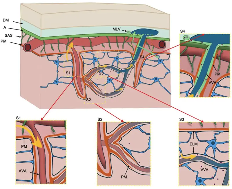
Figure 1. The anatomy of the glymphatic system (GS) with its four compartments (i.e., S1, S2, S3, and S4). Abbreviations—A: arachnoid; AVA: arterial vascular adventitia; DM: dura mater; ELM: external limiting membrane; MLV: meningeal lymphatic vessel; PM: pia mater; SAS: subarachnoid space; VVA: venous vascular adventitia. See text for further details. Adapted with permission from [10].
2.1.2. GS Role in Neurological, Psychiatric, and Neuroimmune Disorders
The GS’s role in brain waste clearance, its interplay with neuroimmune mechanisms, including immune surveillance and neuroinflammation, and their crosstalk contribute to neurodegenerative disease pathophysiology. Brain stimulation approaches, such as transcranial magnetic stimulation (TMS), have been successfully used recently in GS therapeutics, applied to cortically located targets in different regions throughout the cerebral cortex of the human brain, particularly the dorsolateral prefrontal cortex (DLPFC) and parietal cortex. An alternative mechanism in modulating the GS function could be via TMS of autonomic brainstem centers responsible for its innervation, in particular, the centers of the parasympathetic system which are responsible for vasodilation and the facilitation of glymphatic fluid flow dynamics. There are currently several clinical disorders in which the GS is involved due to its abnormal clearance. Neurodegenerative diseases are prominent among these disorders. More specifically, in Alzheimer’s disease (AD), Aβ- and tau- protein accumulation, sleep abnormalities, and AQP4 depolarization alter glymphatic clearance (e.g., [11,12,13]). A recent study [14] investigated changes in the DTI analysis along the perivascular space (DTI-ALPS) index following theta-burst stimulation (TBS) in older adults with mild cognitive impairment (MCI) [14]. Likewise, in Parkinson’s disease (PD) [15,16], Huntington’s disease (HD), and amyotrophic lateral sclerosis (ALS), there have been demonstrated glymphatic fluid dynamic abnormalities as shown in detail in Table 2. Furthermore, glymphatic dysfunction has also been implicated in various acute central nervous system (CNS) injuries, such as stroke, traumatic brain injury (TBI) and idiopathic normal pressure hydrocephalus (iNPH), as well as pathologies related to inflammation levels, summarized in Table 2 (from [11,12,17], modified). Last but not least, anatomical and functional disturbances in the GS are increasingly recognized as contributors to the pathophysiology of glioblastoma (GBM). In particular, tumor-induced compression, glial proliferation, and vascular disorganization disrupt directional perivascular flow and glymphatic clearance, changes that are most pronounced in the CSF–ISF exchange (S2) and perivenous efflux (S3) regions, with reduced meningeal outflow in the newly defined S4 zone as reported in glioma [10,17,18,19,20]. Advanced MRI techniques have revealed significantly reduced glymphatic activity in GBM patients, correlating with interstitial fluid retention and peritumoral edema [21]. These disruptions are closely associated with changes in AQP4 expression and distribution. In healthy brain tissue, AQP4 is highly polarized at astrocytic end-feet lining the perivascular spaces, facilitating the exchange of CSF and ISF and the clearance of metabolic waste [22,23]. In GBM, however, AQP4 becomes disorganized—often redistributed throughout the astrocytic membrane or even overexpressed in tumor cells—thereby compromising tumor-associated perivascular fluid transport [10,23,24]. Finally, it should be noted that current neuroimaging appears to be a potential avenue for assessing GS fluid dynamics, both in normality and in clinical conditions (e.g., [14]).
Table 2. Neurological conditions with demonstrated glymphatic fluid dynamic abnormalities. Modified from [11,12,17].
|
Neurodegenerative Diseases |
|---|
|
Alzheimer’s Disease: Aβ and tau accumulation; sleep disturbances and AQP4 depolarization impair glymphatic clearance. |
|
Parkinson’s Disease: α-syn accumulation; dopaminergic neuron loss; glymphatic dysfunction linked to REM sleep and circadian disruption. |
|
ALS: TDP-43 deposition; AQP4 mis localization; reactive gliosis impairs waste clearance. |
|
Huntington’s Disease: Not detailed, but protein aggregation likely involves glymphatic dysfunction. |
|
Acute CNS Injuries |
|
Traumatic Brain Injury (TBI): Impaired glymphatic clearance of Aβ, tau, GFAP; AQP4 depolarization; sleep disruption worsens accumulation. |
|
Stroke: CSF circulation blockage; AQP4 expression changes; local ischemia exacerbates protein deposition and neuroinflammation. |
|
iNPH: CSF accumulation, AQP4 depolarization, sleep apnea; impaired glymphatic clearance contributes to dementia symptoms. |
|
Sleep alterations, including post-COVID-19 sleep disorder |
|
Glymphatic dysfunction linked to sleep disturbances, depression, bipolar disorder, and migraines. |
|
Astrocyte dysfunction and altered AQP4 expression contribute to protein accumulation and abnormal neuronal signaling. |
2.2. The Role of Neuroimaging in GS Assessment and Clinical Research
Recent reports indicate that GS flow can be assessed by current neuroimaging. The understanding of this field surged from experimental animal studies using neuroimaging to study glymphatic system flow dynamics. Specifically, Iliff and colleagues in three subsequent studies in rodents using in-vivo two-photon microscopy [1,2] and dynamic contrast-enhanced magnetic resonance imaging (DCE-MRI) [4] demonstrated the flow of perivascular CSF influx in the brain tissue and ISF clearance and the association of this flow to the astroglial water channel AQP4 as well as the physiological dependence of this flow to cerebral arterial pulsation. Furthermore, in a fourth study, they showed clinical relevance of their discovery, indicating that “perivascular CSF influx and interstitial solute efflux, including the clearance of amyloid beta, are more rapid in the sleeping compared to the waking brain” [3]. These findings in the murine experimental animal model epitomize in a succinct fashion the relevance of the glymphatic system flow dynamics in normality and clinical conditions. In translating to humans from the experimental animal model using neuroimaging, the most direct representation has been the utilization of intrathecal gadolinium-based contrast agents [2,6,25]. Furthermore, the activity of the glymphatic system is noticeably enhanced during sleep [3] and facilitated by exercise. Translation of these original observations in rodents has been achieved in humans, where MRI studies have demonstrated waves of CSF flow during deep sleep paired with neural and vascular oscillations [6,26,27]. Given these realizations, the glymphatic system and its flow dynamics are currently active areas of study in humans, particularly using neuroimaging. These methods could be used for early diagnosis, prevention, and monitoring of treatments of different clinical conditions [28]. Neuroimaging for clinical glymphatic examinations in humans is feasible, but we must keep in mind that we need to develop acquisition protocols that are convenient for patients. Although contrast agents, such as gadolinium, can be used in humans, several practical limitations make their use challenging [28]. Instead, diffusion-weighted imaging (DWI) techniques seem more promising in the study of glymphatic system flow dynamics. These include a diffusion tensor imaging (DTI) technique that measures water diffusion along the perivascular space, thus named diffusion tensor imaging analysis along the perivascular space (DTI-ALPS), a high index of which indicates an effective waste clearance through the glymphatic system. It has also been demonstrated using DTI that water diffusivity increases during night sleep in healthy subjects [29]. Nevertheless, it should be noted that although MRI research in glymphatic flow dynamics is still in its initial phases, several imaging modalities such as intravenous GBCA-enhanced MRI, PET/single-photon emission computed tomography (SPECT), and contrast-free MRI techniques [6] seem potentially worthwhile techniques in studying CSF and glymphatic flow dynamics. Overall, there is active research in imaging cranial fluid dynamics and experimental mechanistic models of the glymphatic flow, trying to translate our understanding from animal research to human clinical implementation, in particular using contrast-free MRI techniques [30]. Importantly, phase contrast MRI (PC-MRI) can measure cerebral blood or CSF flow dynamics and could potentially be used for the detection of glymphatic system flow abnormalities in clinical conditions [28,31,32,33]. Apparently, diffusion-weighted imaging (DWI) techniques are the most popular approach for studying glymphatic system flow dynamics in humans, given the possibility of retrospective analysis of accessible DTI datasets [6]. Prospectively, DWI appears to have additional potential to address a critically important mechanistic aspect of the GS flow, namely the flow at the level of astroglial AQP4 water channels, once technological improvements allow the implementation of DWI acquisition protocols with appropriate spatial resolution, effectively solving this challenging problem.
2.3. Putative Therapeutic Approaches of the Human Glymphatic System—The Effects of TMS and EVs in the CNS and a Hypothesis for a Next Generation Combinatorial Therapeutic Approach in Human GS Dysfunctions
2.3.1. The Effects of TMS in the CNS and the Human GS and Neurocircuitry TMS EEG-Based Biomarkers for GS Treatments
Recent advances in an array of fields, such as bioengineering and molecular biology, have brought us to the realization that we can make unprecedented breakthroughs in medicine. With respect to the topic of this perspective paper, there have been significant recent discoveries in non-invasive neuromodulation technology (TMS) and magnetic resonance imaging (MRI) of the central nervous system (CNS) that address novel therapeutic approaches for GS in humans. The TMS electroencephalography (TMS EEG) biomarkers approach has recently become a means to obtain a new understanding of the causal chains of neuronal signaling. Importantly, TMS EEG biomarkers can be implemented for the diagnosis of diseases and the assessment of treatment in neurotherapeutics, specifically using neuromodulation. Brain stimulation approaches, such as TMS, act on brain circuits that have been strongly considered candidate endophenotypes and actual targets in neuromodulation interventions, as revealed by multimodal neuroimaging. Novel technological advancements in TMS, such as multi-locus TMS (mTMS) [34], demonstrate the capability to engage with established neuroanatomical circuits. Furthermore, improved access to specific brain circuits by mTMS, coupled with recently achieved precise mapping of specific cortical-subcortical circuits using multimodal neuroimaging [35], enables new experimental possibilities in the neuromodulation of specific brain networks, including those involved in modulating the GS. At a mechanistic level, the glymphatic system is regulated predominantly by neurochemical mechanisms rather than by direct neural innervation. Its activity is dynamically modulated by autonomic signaling, the sleep–wake cycle, aging, genetic factors, and body posture. Glymphatic function is enhanced during sleep, when reduced noradrenergic tone promotes astrocytic relaxation, expansion of perivascular spaces, and increased cerebrospinal fluid–interstitial fluid exchange, thereby facilitating metabolic waste clearance [36,37,38,39]. In contrast, heightened sympathetic activity during wakefulness suppresses glymphatic transport, whereas parasympathetic signaling promotes glymphatic flow by modulating cerebral vascular pulsatility and cerebrospinal fluid dynamics [36]. In normality, the autonomic nervous system (ANS) pathways play an important role in reducing glymphatic function during wakefulness via sympathetic activation and increasing glymphatic flow via parasympathetic (vagal) activation, which is more pronounced during sleep. Importantly, these ANS circuits have been demonstrated recently in humans using multimodal neuroimaging, specifically T1W/T2W MRI and diffusion MRI (dMRI) tractography [40,41]. Although still challenging, by modulating these circuits using TMS and, necessarily, with the aid of multimodal neuroimaging for their precise localization and targeting, we could potentially enhance glymphatic function. Furthermore, the targeted interventions may improve sleep dynamics, with consequent boost to glymphatic activity. It should be noted that stimulation of subcortical nuclei can be done directly or indirectly by targeting cortical areas with known connections to specific subcortical structures. Integrating TMS EEG biomarkers with neuroimaging has the potential to enable circuit-specific neuromodulation with unprecedented precision, facilitating personalized treatment strategies tailored to an individual's clinical characteristics, biological profile, preferences, lifestyle, and social context while strengthening the physician–patient relationship. By further incorporating genetic, molecular, phenotypic, and environmental data, this framework could stratify patients into biologically defined subgroups, thereby optimizing prevention and therapeutic interventions. Rather than designing unique treatments for every individual, this precision and personalized medicine approach aims to deliver the most effective intervention to patients who share common biological characteristics. Accordingly, neuroimaging-guided TMS-EEG biomarkers that identify circuit-specific therapeutic targets may represent a key foundation for advancing precision and personalized neuromodulatory interventions (Table 1). By applying theta-burst stimulation (TBS), a type of TMS pulse sequence, over the left parietal cortex, a therapeutic effect has been demonstrated recently, characterized by memory improvement, in a population of subjects with mild cognitive impairment (MCI) [14]. Importantly, Sundman and colleagues (2025) reported that memory improvements correlated with high indices in DTI-ALPS, indicating a significant improvement in waste removal and, thus, in increased clearance efficiency of the GS in these subjects [14]. Neurocircuitry TMS EEG-based biomarkers characterized by neuroimaging. One of the principal goals of modern neuroscience is to elucidate the neurobiological basis of behavior [42]. In this conceptual framework, the brain circuits are considered the elementary hardwired units in the brain that underlie every behavior carried out by an organism. Fundamentally, systems biology is thought through as the endophenotypes or biomarkers acting as an interface between behavior and the genome [43,44,45,46,47] (Figure 2). In terms of functional neuroanatomy assessment of behavior responses, the combination of structural circuit anatomy and the electrophysiological response of this circuit are two necessary parameters. This can be achieved with combined neuroimaging characterization of brain circuits and subsequent TMS EEG examination, an approach that allows the determination of a brain circuit both, structurally and functionally. This approach allows us to stimulate a specific brain circuit based on its identity and not just in terms of the topographic location or proximity of the cortical target zone of stimulation [35,48]. This is a conceptual matter of significant relevance and consequences, given that the true biomarker associated with a specific behavior is not the cortical area of stimulation but, instead the stimulated specific brain circuit. The TMS electroencephalography (TMS EEG) biomarkers approach has become recently a means to obtain new understanding of the causal chains of neuronal signaling in the brain [35]. The determination of the target and brain circuit associated with that target can be done by multimodal neuroimaging, specifically T1W/T2W MRI and diffusion MRI tractography [35]. Thus, the efficient application of this approach requires neuroimaging acquisitions and analysis prior to the TMS intervention. We have elaborated in detail on this topic in a recent publication of our group [35].
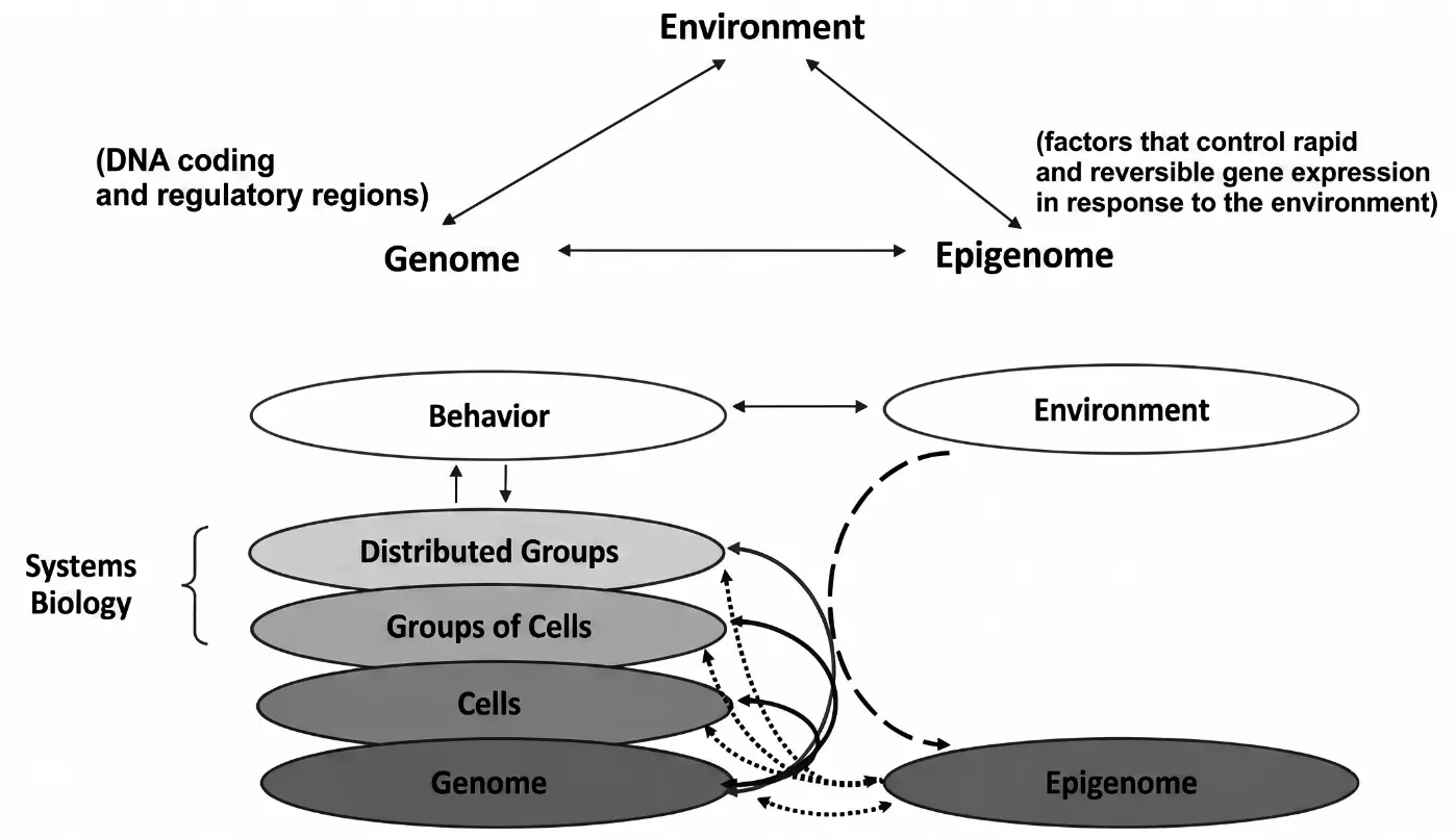
Figure 2. Systems-biology model illustrating the reciprocal interactions among the genome, epigenome, environment, cellular organization, distributed neural networks, and behavior. Environmental factors may induce rapid and potentially reversible epigenetic changes, whereas neural activity and behavior can, in turn, influence gene-expression processes. Adapted with permission from [47].
2.3.2. The Effects of EVs in the CNS and the Human GS
On a different front, advances in molecular biology, stem cell research (MSCs and EVs), and tissue engineering have fostered the development of novel therapeutic strategies for CNS regeneration and repair in experimental non-human primates and rodents, with remarkable results and significant translational potential (e.g., [49,50,51]). Extracellular vesicles (EVs) [52] are nano-sized (30–100 nm), bilipid membrane vesicles released by mesenchymal stem cells (MSCs) that mediate paracrine signaling by delivering bioactive molecules to nearby cells. Unlike MSCs, which are multipotent adult stem cells found in tissues such as bone marrow, umbilical cord, and adipose tissue, EVs are cell-free agents, making them more versatile for injectable therapies. Although first identified in the 1980s, EVs were recognized in the early 2000s as carriers of functional RNA capable of modifying recipient cells [53]. EVs are now regarded as key mediators of intercellular communication with anti-inflammatory, homeostatic, and neuroregenerative properties. They transport messenger RNA (mRNA), microRNA (miRNA), small interfering RNA (siRNA), long non-coding RNA (lncRNA), extrachromosomal DNA, proteins, lipids, and other signaling molecules [54], thereby promoting cell proliferation, inhibiting apoptosis, and enhancing tissue repair [55,56,57,58,59,60,61,62]. Their ability to cross the blood-brain barrier (BBB), reduce neuroinflammation, improve cellular communication, and restore CNS homeostasis has led to their proposal as promising glymphatic system therapeutic agents. Numerous preclinical studies support the therapeutic potential of EVs (e.g., [49,50]). For example, Moore and colleagues (2019) demonstrated that intravenous administration of MSC-derived EVs following experimentally induced cortical injury in non-human primates led to complete recovery of fine hand motor function within 3–5 weeks, likely through attenuation of inflammation [51]. Early human clinical trials, primarily using intravenous or intranasal EV administration, have also reported encouraging outcomes in neurodegenerative disorders, including AD, PD, Lewy body dementia, amyotrophic lateral sclerosis (ALS), as well as stroke and COVID-19 [63,64,65,66,67]. Despite these promising findings, important challenges remain before EVs can achieve routine clinical use. Safety, standardization, manufacturing, and regulatory concerns must be resolved before approval by the FDA (U.S. Food and Drug Administration) or EMA (European Medicines Agency) (e.g., [68]). Moreover, although EVs exert beneficial immunomodulatory effects, their role in immune regulation remains incompletely understood. Both immune- and non-immune cell-derived EVs may exacerbate disease by modulating immune responses, inflammatory signaling, and tissue injury; their effects depend on cellular origin, molecular cargo, and microenvironment. (e.g., [68]). In a recent review, Wang and colleagues (2025) indicated that EVs are promising therapeutic mediators that modulate disease processes by suppressing cancer progression, reducing tissue damage, and promoting tissue repair in neurodegenerative, infectious, metabolic, cardiovascular, and renal diseases [68]. As indicated in Table 3, EVs have been shown to be effective therapeutic agents at a preclinical phase, which, however, has not been sufficiently corroborated yet at a clinical phase. Nevertheless, being aware of this important limitation for clinical use individually or in combination with TMS, their broad regenerative potential positions EVs among the most promising next-generation therapies for CNS repair.
Table 3. Levels of Evidence for TMS, Extracellular Vesicle (EV)-Based Interventions, and Their Proposed Combination.
|
Domain |
TMS |
EVs |
TMS & EVs |
Evidence |
Gap |
|---|---|---|---|---|---|
|
Mechanism |
Established |
Established |
Emerging |
Plasticity; immune modulation |
Synergy unknown |
|
Preclinical |
Strong |
Strong |
Moderate |
Improved recovery |
Need comparative studies |
|
Glymphatic |
Indirect |
Indirect |
Hypothesis |
Autonomic/vascular effects |
No direct human proof |
|
Neuroimmune |
Strong |
Strong |
Moderate |
Reduced inflammation |
Relative contribution unclear |
|
Clinical |
Established |
Early trials |
None |
TMS effective in selected disorders |
No combination trials |
|
Biomarkers |
TMS EEG/MRI |
EV cargo |
None |
Promising markers |
Need multimodal validation |
|
Safety |
Established |
Favorable |
Unknown |
Independent safety |
Combined safety unknown |
|
Translation |
High |
Moderate |
Proof-of-concept |
Strong rationale |
Optimize dose, timing, biomarkers |
Abbreviations: EVs, extracellular vesicles; TMS, transcranial magnetic stimulation.
2.4. A Hypothesis for a Synergistic TMS–EV Therapeutic Strategy Targeting the Human Glymphatic System
In this Perspective, we propose that the combination of transcranial magnetic stimulation (TMS) and extracellular vesicles (EVs) represents a next-generation, versatile therapeutic strategy for restoring glymphatic system (GS) function. Our central hypothesis is that TMS and EVs interact synergistically, producing therapeutic effects that exceed those achievable with either intervention alone. Recent advances in molecular and cell-free EV biology, systems neuroscience, neuroimaging, brain circuit-based endophenotyping, and non-invasive neuromodulation support this hypothesis. Although EV-based therapies have not yet entered routine clinical practice, extensive preclinical studies have demonstrated their remarkable regenerative potential and translational promise. We propose that the efficacy of TMS–EV combination therapy is determined by the degree of biological synergism between these interventions and further optimized by treatment-related factors, including TMS stimulation parameters, EV source and cargo, dosage, route of administration, and treatment timing.
Growing experimental evidence supports this concept. TMS combined with mesenchymal stem cells (MSCs), the principal source of therapeutic EVs, enhances neuronal survival, suppresses neuroinflammation, regulates ferroptosis-associated pathways, and improves functional recovery following spinal cord injury and cerebral ischemia [69,70,71]. Cheng et al. (2024) further demonstrated that repetitive TMS (rTMS) and MSCs synergistically inhibit neuroinflammation and neuronal PANoptosis during the subacute phase of cerebral infarction, potentially through rTMS-mediated downregulation of the Repressor Element-1 Silencing Transcription factor (REST), although the precise molecular pathways remain to be established [71]. Mechanistically, the proposed synergism arises from complementary actions operating at multiple biological scales. TMS primarily modulates neuronal circuits, autonomic activity, and neurovascular function, whereas EVs act at the molecular level by delivering bioactive cargo—including mRNAs, microRNAs, proteins, and lipids—that regulate gene expression, suppress apoptosis, attenuate neuroinflammation, and promote tissue repair [53,54,55,56,57,58,59,60,61,62]. Importantly, TMS may also enhance glymphatic transport through autonomic regulation. Reduced sympathetic noradrenergic tone together with increased parasympathetic activity promotes astrocytic relaxation, enlargement of perivascular spaces, and increased cerebrospinal fluid–interstitial fluid exchange, thereby facilitating metabolic waste clearance [36,37,38,39]. Because EVs readily cross the blood–brain barrier and restore cellular homeostasis, improved glymphatic transport may enhance their distribution throughout the brain while simultaneously promoting the clearance of inflammatory mediators and neurotoxic proteins. Conversely, the anti-inflammatory and neurorestorative effects of EVs may potentiate the neurophysiological changes induced by TMS, creating a reciprocal positive feedback loop that amplifies neuroprotection, neuroplasticity, and tissue repair.
The glymphatic system itself is regulated predominantly through neurochemical rather than direct neural innervation. Its activity is dynamically influenced by autonomic signaling, the sleep–wake cycle, aging, genetic factors, and body posture. Glymphatic function is maximal during sleep, when reduced noradrenergic activity facilitates astrocytic relaxation and perivascular fluid exchange, whereas sympathetic activation during wakefulness suppresses glymphatic transport. In contrast, parasympathetic activation enhances cerebral vascular pulsatility and cerebrospinal fluid dynamics, thereby promoting glymphatic flow [36,37,38,39]. These physiological mechanisms provide a plausible biological substrate through which TMS could enhance glymphatic function. Collectively, these observations support a unified therapeutic framework in which TMS optimizes the neural, autonomic, vascular, and glymphatic environment, while EVs provide regenerative molecular signals that reinforce tissue repair. Their integration has the potential to reduce neuroinflammation, protein aggregation, ferroptosis, and other forms of regulated cell death while enhancing waste clearance, neuroplasticity, and functional recovery. Such a multimodal strategy may accelerate the development of safe, mechanism-based therapies for neurological disorders characterized by glymphatic dysfunction, including stroke, Alzheimer's disease, Parkinson's disease, amyotrophic lateral sclerosis, Lewy body dementia, and potentially other neuroinflammatory conditions [18,63,64,65,66,67,72,73,74,75]. Although EV therapy remains largely preclinical, ongoing clinical trials support its translational potential. We therefore propose that combining precision TMS—particularly targeting autonomic and parasympathetic circuits—with EV therapy represents a promising next-generation approach for restoring glymphatic and neuroimmune homeostasis and improving clinical outcomes across a broad spectrum of central nervous system diseases. The proposed framework is illustrated in Figure 3, and its levels of evidence supporting TMS, EV-based therapies, and their proposed combination are depicted in Table 3.
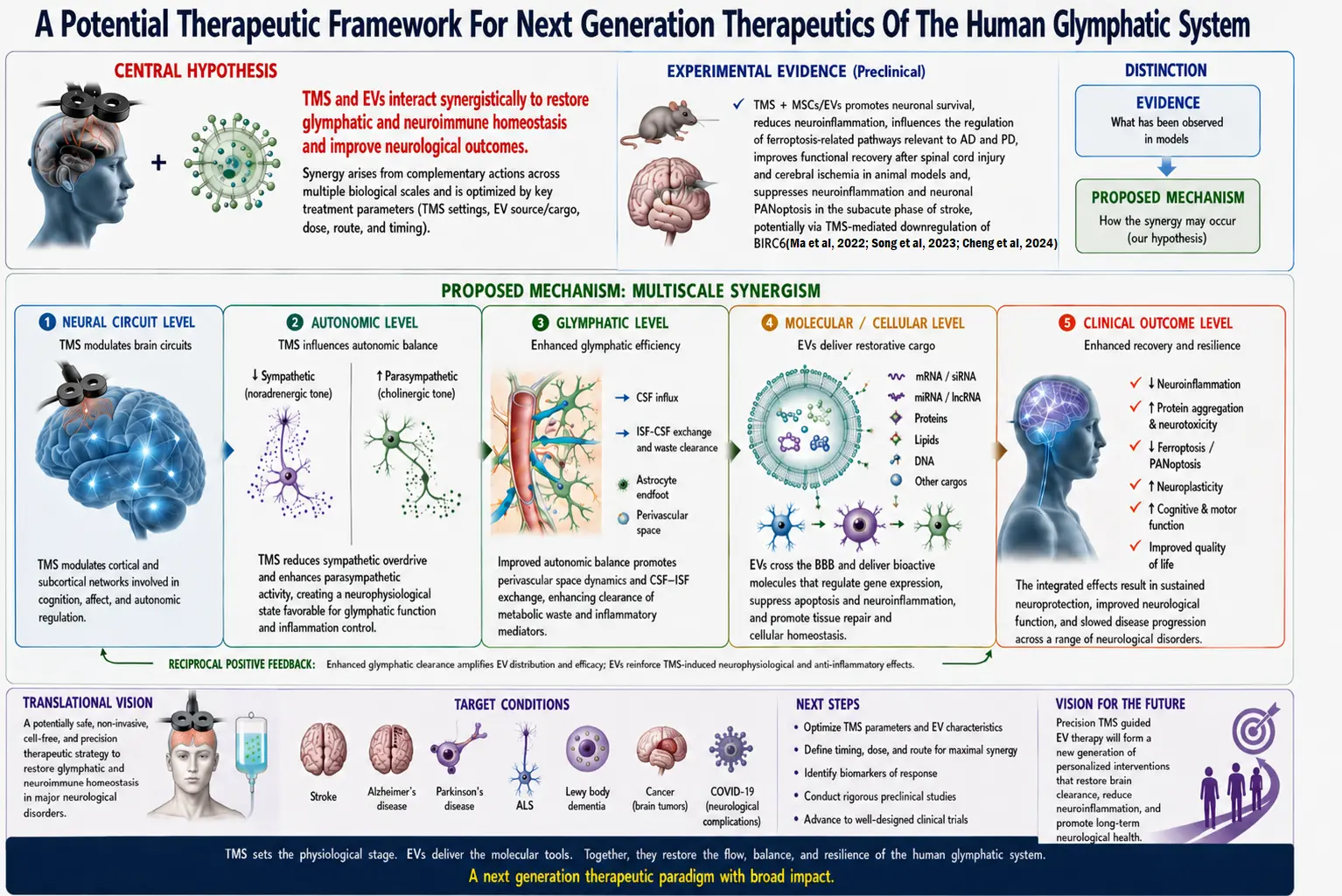
Figure 3. A potential framework based on a hypothesis of a synergistic TMS-EV therapeutic intervention targeting the human glymphatic system. In this model, TMS would exert vasodilatation and anti-inflammatory effects by downregulating the orthosympathetic system and upregulating the parasympathetic system. EVs would exert a direct anti-inflammatory effect on the GS. Abbreviations: TMS = transcranial magnetic stimulation. “ChatGPT (OpenAI, GPT-5.5) was used to assist in drafting and refining conceptual figures and text. All scientific content interpretation, critical evaluation, and final editing were performed and verified by the authors” [69,70,71].
2.5. Evidence Gaps, Alternative Hypotheses, and Testable Predictions in the Context of the Present Framework for Restoring Glymphatic Function with TMS and EVs
Evidence gaps although accumulating evidence implicates glymphatic dysfunction in neurological disease, direct evidence that transcranial magnetic stimulation (TMS) enhances glymphatic function in humans remains incomplete. The mechanisms underlying TMS–glymphatic and extracellular vesicle (EV)–glymphatic interactions are not fully defined, the optimal parameters for combined TMS and EV therapy remain unknown, and DTI-ALPS and TMS-EEG require further validation as biomarkers of glymphatic function and treatment response. Alternative hypotheses The therapeutic effects of TMS may primarily reflect modulation of neural circuits, neurovascular coupling, or sleep-dependent processes independent of glymphatic function, whereas EVs may act predominantly through immunomodulatory and neuroregenerative, and neurorepair mechanisms. Moreover, the combined effects of TMS and EVs may be additive rather than synergistic, and glymphatic dysfunction may represent a consequence rather than a primary driver of disease. Testable predictions If the proposed framework is correct, combined TMS and EV therapy should enhance glymphatic function beyond either intervention alone, with concomitant improvements in DTI-ALPS and TMS-EEG biomarkers, reductions in pathological protein accumulation and neuroinflammation, and superior clinical outcomes. Furthermore, patients with the greatest baseline glymphatic impairment should derive the greatest therapeutic benefit. Finally, it should be noted that the successful clinical translation of a combined TMS–EV therapeutic strategy will require systematic optimization of its organizational and methodological framework, including treatment protocols, temporal coordination between neuromodulation and EV administration, dose selection, treatment frequency, and compatibility among administration routes. Defining these parameters will be essential for ensuring reproducibility, safety, and therapeutic efficacy across clinical settings. The evidence gaps, alternative hypotheses, and testable predictions of the proposed framework in this Perspective are illustrated in Figure 4.
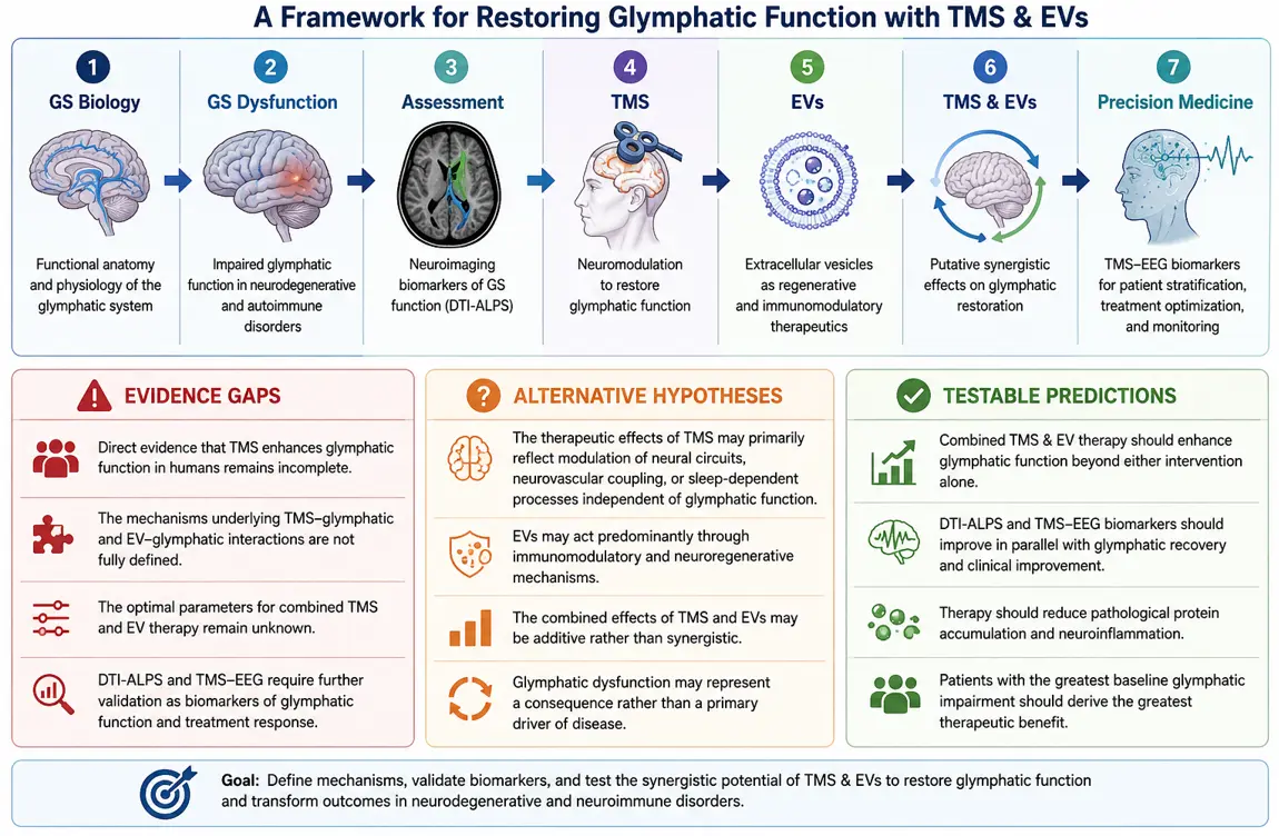
Figure 4. Evidence gaps, alternative hypotheses and testable predictions in the context of the present framework for restoring glymphatic function with TMS and EVs. Abbreviations: GS = glymphatic system; TMS = transcranial magnetic stimulation; EVs = extracellular vesicles. “ChatGPT (OpenAI, GPT-5.5) was used to assist in drafting and refining conceptual figures and text. All scientific content interpretation, critical evaluation, and final editing were performed and verified by the authors”.
3. Conclusions
The discovery of the glymphatic system (GS) has fundamentally advanced our understanding of brain homeostasis by revealing a dynamic clearance network that is tightly coupled to neuroimmune regulation. Increasing evidence indicates that glymphatic dysfunction and neuroinflammation reinforce one another in a self-perpetuating cycle that contributes to the pathogenesis and progression of numerous neurological disorders, including Alzheimer's disease, Parkinson's disease, multiple sclerosis, stroke, and other neurodegenerative conditions [16,76]. Consequently, therapeutic strategies that simultaneously restore glymphatic function and neuroimmune homeostasis represent an important opportunity for disease modification.
In this Perspective, we propose a potential next-generation therapeutic framework based on the putative synergistic integration of transcranial magnetic stimulation (TMS) and extracellular vesicles (EVs). As illustrated in Figure 3, we hypothesize that TMS and EVs act through complementary mechanisms across multiple biological scales. TMS primarily modulates neural circuits, autonomic function, and glymphatic dynamics, thereby creating a physiological environment that favors waste clearance and tissue repair. In parallel, EVs deliver regenerative molecular cargo that suppresses neuroinflammation, promotes neuroprotection and neuroplasticity, and enhances cellular homeostasis. Their interaction may establish a reciprocal positive feedback loop in which improved glymphatic transport facilitates EV distribution and therapeutic efficacy, while EV-mediated repair amplifies the neurophysiological effects induced by TMS. We propose that this multiscale synergism may ultimately translate into superior clinical outcomes compared with either intervention alone. Although EV-based therapies remain largely preclinical and require further evaluation to establish their safety, efficacy, and regulatory approval for human use, accumulating experimental evidence supports their considerable translational potential. Future studies should define the biological mechanisms underlying TMS–EV synergism, optimize stimulation protocols and EV characteristics, identify predictive biomarkers—including electrophysiological and neuroimaging measures—and validate this integrated strategy in well-designed clinical trials. Collectively, we propose that combining precision TMS with cell-free EV therapy constitutes a potential and versatile, mechanism-based therapeutic platform for restoring glymphatic and neuroimmune homeostasis. Beyond treating glymphatic dysfunction, this framework may offer a broadly applicable strategy to enhance neuroprotection, promote brain repair, and slow disease progression across a wide spectrum of central nervous system disorders.
Statement of the Use of Generative AI and AI-Assisted Technologies in the Writing Process
During the preparation of this manuscript, the authors used ChatGPT (GPT-5.5) to create graphical summaries intended to synthesize and clarify the content of the manuscript. Following the use of this tool, the authors reviewed and edited the generated content as necessary and take full responsibility for the content of the published article.
Acknowledgements
OpenAI’s ChatGPT (GPT-5.5) was used to assist with language editing and the iterative design of Figure 3 and Figure 4 and Table 1. All scientific concepts, interpretations, and final content were developed, verified, and approved by the authors. We would like to thank Universidad de La Laguna de Tenerife for hosting the “International Special Seminar” on “Biomarker Discovery and Neurotherapeutics using Transcranial Magnetic Stimulation (TMS)” (https://meet.google.com/gxv-pbnn-ugg) in the Institute of Biomedical Technology (ITB) in La Laguna de Tenerife on 24 November 2025, the contents of which are reflected in this perspective paper.
Author Contributions
All authors contributed equally to all aspects of the work.
Ethics Statement
Not applicable.
Informed Consent Statement
Not applicable.
Data Availability Statement
Not applicable.
Funding
This work was supported by the following grants: R01MH112748 (N.M.), R01AG042512 (N.M.), K24MH116366, R01MH132610, R01MH125860 (N.M.), R21NS136960 (N.M.), and R01NS125307 (N.M.).
Declaration of Competing Interest
The authors declare that they have no known competing financial interests or personal relationships that could have influenced the work reported in this article.
References
-
Iliff JJ, Wang M, Liao Y, Plogg BA, Peng W, Gundersen GA, et al. A Paravascular Pathway Facilitates CSF Flow through the Brain Parenchyma and the Clearance of Interstitial Solutes, Including Amyloid β. Sci. Transl. Med. 2012, 4, 147ra111. DOI:10.1126/scitranslmed.3003748 [Google Scholar]
-
Iliff JJ, Lee H, Yu M, Feng T, Logan J, Nedergaard M, et al. Brain-Wide Pathway for Waste Clearance Captured by Contrast-Enhanced MRI. J. Clin. Investig. 2013, 123, 1299–1309. DOI:10.1172/JCI67677 [Google Scholar]
-
Xie L, Kang H, Xu Q, Chen MJ, Liao Y, Thiyagarajan M, et al. Sleep Drives Metabolite Clearance from the Adult Brain. Science 2013, 342, 373–377. DOI:10.1126/science.1241224 [Google Scholar]
-
Iliff JJ, Wang M, Zeppenfeld DM, Venkataraman A, Plog BA, Liao Y, et al. Cerebral Arterial Pulsation Drives Paravascular CSF-Interstitial Fluid Exchange in the Murine Brain. J. Neurosci. 2013, 33, 18190–18199. DOI:10.1523/JNEUROSCI.1592-13.2013 [Google Scholar]
-
Iliff JJ, Chen MJ, Plog BA, Zeppenfeld DM, Soltero M, Yang L, et al. Impairment of Glymphatic Pathway Function Promotes Tau Pathology after Traumatic Brain Injury. J. Neurosci. 2014, 34, 16180–16193. DOI:10.1523/JNEUROSCI.3020-14.2014 [Google Scholar]
-
Kipnis J, Benveniste H, Eichmann A, Thomas J-L, Reich DS, Lewis LD, et al. Resolving the Mysteries of Brain Clearance and Immune Surveillance. Neuron 2025, 113, 3908–3923. DOI:10.1016/j.neuron.2025.10.036 [Google Scholar]
-
Jessen NA, Munk ASF, Lundgaard I, Nedergaard M. The Glymphatic System: A Beginner’s Guide. Neurochem. Res. 2015, 40, 2583–2599. DOI:10.1007/s11064-015-1581-6 [Google Scholar]
-
Thrane AS, Rangroo Thrane V, Nedergaard M. Drowning Stars: Reassessing the Role of Astrocytes in Brain Edema. Trends Neurosci. 2014, 37, 620–628. DOI:10.1016/j.tins.2014.08.010 [Google Scholar]
-
Johanson CE. Choroid Plexus–Cerebrospinal Fluid Circulatory Dynamics: Impact on Brain Growth, Metabolism, and Repair. In Neuroscience in Medicine; Humana Press: Totowa, NJ, USA, 2008; pp. 173–200. ISBN 9781603274548. [Google Scholar]
-
Castañeyra-Ruiz L, González-Marrero I, García-Abad LH, Gonzalez-Arnay E, Camacho M, Carmona-Calero EM, et al. Aquaporin-4 in Glioblastoma: A Nexus of Glymphatic Dysfunction, Edema, Immune Evasion, and Treatment Resistance. Front. Cell. Neurosci. 2025, 19, 1685491. DOI:10.3389/fncel.2025.1685491 [Google Scholar]
-
Tang M, Diao F, Dong Y, Lin J, Wang T, Yang L, et al. Systemic Inflammatory Response Index Mediates the Association between Stroke and Hypertension: A Cross-Sectional Study from NHANES 2001 to 2016. Front. Neurol. 2025, 16, 1603241. DOI:10.3389/fneur.2025.1603241 [Google Scholar]
-
Zhang D, Li X, Li B. Glymphatic System Dysfunction in Central Nervous System Diseases and Mood Disorders. Front. Aging Neurosci. 2022, 14, 873697. DOI:10.3389/fnagi.2022.873697 [Google Scholar]
-
Yang C-C, Huang K-Y, Hsu J-L, Hu C-J, Lu YH, Kuan Y-C. Effects of Intermittent Theta-Burst Stimulation on Cognition and Glymphatic System Activity in Mild Cognitive Impairment and Very Mild Alzheimer’s Disease: A Randomized Controlled Trial. J. Neuroeng. Rehabil. 2025, 22, 195. DOI:10.1186/s12984-025-01738-1 [Google Scholar]
-
Sundman MH, Liu Y, Chen N-K, Chou Y-H. The Glymphatic System as a Therapeutic Target: TMS-Induced Modulation in Older Adults. Front. Aging Neurosci. 2025, 17, 1597311. DOI:10.3389/fnagi.2025.1597311 [Google Scholar]
-
Lin R, Cai G, Chen Y, Zheng J, Wang S, Xiao H, et al. Association of Glymphatic System Function with Peripheral Inflammation and Motor Symptoms in Parkinson’s Disease. NPJ Park. Dis. 2025, 11, 62. DOI:10.1038/s41531-025-00909-0 [Google Scholar]
-
Rajai Firouzabadi S, Mohammadi I, Aghajanian S, Aarabi A, Oraee S, Soltani SM, et al. Glymphatic Dysfunction in Parkinson’s Disease: A Systematic Review and Meta-Analysis of Neuroimaging Studies. Mov. Disord. Clin. Pract. 2026, 13, 331–343. DOI:10.1002/mdc3.70296 [Google Scholar]
-
Rasmussen MK, Mestre H, Nedergaard M. The Glymphatic Pathway in Neurological Disorders. Lancet Neurol. 2018, 17, 1016–1024. DOI:10.1016/S1474-4422(18)30318-1 [Google Scholar]
-
Gao Y, Liu K, Zhu J. Glymphatic System: An Emerging Therapeutic Approach for Neurological Disorders. Front. Mol. Neurosci. 2023, 16, 1138769. DOI:10.3389/fnmol.2023.1138769 [Google Scholar]
-
Gao M, Liu Z, Zang H, Wu X, Yan Y, Lin H, et al. A Histopathologic Correlation Study Evaluating Glymphatic Function in Brain Tumors by Multiparametric MRI. Clin. Cancer Res. 2024, 30, 4876–4886. DOI:10.1158/1078-0432.CCR-24-0150 [Google Scholar]
-
Scalia G, Ferini G, Silven MP, Noto M, Costanzo R, Maugeri R, et al. Alterations of the Glymphatic System in Glioblastomas: A Systematic Review. Anticancer. Res. 2024, 44, 3223–3230. DOI:10.21873/anticanres.17140 [Google Scholar]
-
Mueller SM, McFarland White K, Fass SB, Chen S, Shi Z, Ge X, et al. Evaluation of Gliovascular Functions of AQP4 Readthrough Isoforms. Front. Cell. Neurosci. 2023, 17, 1272391. DOI:10.3389/fncel.2023.1272391 [Google Scholar]
-
McCoy ES, Haas BR, Sontheimer H. Water Permeability through Aquaporin-4 Is Regulated by Protein Kinase C and Becomes Rate-Limiting for Glioma Invasion. Neuroscience 2010, 168, 971–981. DOI:10.1016/j.neuroscience.2009.09.020 [Google Scholar]
-
Mou K, Chen M, Mao Q, Wang P, Ni R, Xia X, et al. AQP-4 in Peritumoral Edematous Tissue Is Correlated with the Degree of Glioma and with Expression of VEGF and HIF-Alpha. J. Neurooncol. 2010, 100, 375–383. DOI:10.1007/s11060-010-0205-x [Google Scholar]
-
Noell S, Ritz R, Wolburg-Buchholz K, Wolburg H, Fallier-Becker P. An Allograft Glioma Model Reveals the Dependence of Aquaporin-4 Expression on the Brain Microenvironment. PLoS ONE 2012, 7, e36555. DOI:10.1371/journal.pone.0036555 [Google Scholar]
-
Ringstad G, Valnes LM, Vatnehol SAS, Pripp AH, Eide PK. Prospective T1 Mapping to Assess Gadolinium Retention in Brain after Intrathecal Gadobutrol. Neuroradiology 2023, 65, 1321–1331. DOI:10.1007/s00234-023-03198-7 [Google Scholar]
-
Picchioni D, Özbay PS, Mandelkow H, de Zwart JA, Wang Y, van Gelderen P, et al. Autonomic Arousals Contribute to Brain Fluid Pulsations during Sleep. Neuroimage 2022, 249, 118888. DOI:10.1016/j.neuroimage.2022.118888 [Google Scholar]
-
Fultz NE, Bonmassar G, Setsompop K, Stickgold RA, Rosen BR, Polimeni JR, et al. Coupled Electrophysiological, Hemodynamic, and Cerebrospinal Fluid Oscillations in Human Sleep. Science 2019, 366, 628–631. DOI:10.1126/science.aax5440 [Google Scholar]
-
Boyd ED, Kaur J, Ding G, Chopp M, Jiang Q. Clinical Magnetic Resonance Imaging Evaluation of Glymphatic Function. NMR Biomed. 2024, 37, e5132. DOI:10.1002/nbm.5132 [Google Scholar]
-
Tuura RO, Volk C, Callaghan F, Jaramillo V, Huber R. Sleep-Related and Diurnal Effects on Brain Diffusivity and Cerebrospinal Fluid Flow. Neuroimage 2021, 241, 118420. DOI:10.1016/j.neuroimage.2021.118420 [Google Scholar]
-
Bohr T, Hjorth PG, Holst SC, Hrabětová S, Kiviniemi V, Lilius T, et al. The Glymphatic System: Current Understanding and Modeling. iScience 2022, 25, 104987. DOI:10.1016/j.isci.2022.104987 [Google Scholar]
-
Korbecki A, Zimny A, Podgórski P, Sąsiadek M, Bladowska J. Imaging of Cerebrospinal Fluid Flow: Fundamentals, Techniques, and Clinical Applications of Phase-Contrast Magnetic Resonance Imaging. Pol. J. Radiol. 2019, 84, e240–e250. DOI:10.5114/pjr.2019.86881 [Google Scholar]
-
Kiviniemi V, Wang X, Korhonen V, Keinänen T, Tuovinen T, Autio J, et al. Ultra-Fast Magnetic Resonance Encephalography of Physiological Brain Activity - Glymphatic Pulsation Mechanisms? J. Cereb. Blood Flow Metab. 2016, 36, 1033–1045. DOI:10.1177/0271678X15622047 [Google Scholar]
-
Kiviniemi V, Helakari H, Raitamaa L, Huotari N, Rajna Z, Järvelä M, et al. Ultrafast Imaging of Physiological Brain Pulsations with Magnetic Resonance Encephalography-from Noise to Predictive Clinical Biomarker. NMR Biomed. 2025, 38, e70092. DOI:10.1002/nbm.70092 [Google Scholar]
-
Koponen LM, Nieminen JO, Ilmoniemi RJ. Multi-Locus Transcranial Magnetic Stimulation-Theory and Implementation. Brain Stimul. 2018, 11, 849–855. DOI:10.1016/j.brs.2018.03.014 [Google Scholar]
-
Lioumis P, Roine T, Granö I, Aydogan DB, Ukharova E, Souza VH, et al. Optimization of TMS Target Engagement: Current State and Future Perspectives. Front. Neurosci. 2025, 19, 1517228. DOI:10.3389/fnins.2025.1517228 [Google Scholar]
-
Nycz B, Mandera M. The Features of the Glymphatic System. Auton. Neurosci. 2021, 232, 102774. DOI:10.1016/j.autneu.2021.102774 [Google Scholar]
-
Hauglund NL, Andersen M, Tokarska K, Radovanovic T, Kjaerby C, Sørensen FL, et al. Norepinephrine-Mediated Slow Vasomotion Drives Glymphatic Clearance during Sleep. Cell 2025, 188, 606–622.e17. DOI:10.1016/j.cell.2024.11.027 [Google Scholar]
-
Wang X, Huang P, Haacke EM, Wu P, Zhang X, Zhang H, et al. MRI Index of Glymphatic System Mediates the Influence of Locus Coeruleus on Cognition in Parkinson’s Disease. Parkinsonism Relat. Disord. 2024, 123, 106558. DOI:10.1016/j.parkreldis.2024.106558 [Google Scholar]
-
Falgàs N, Tort-Colet N, Martín-Sobrino I, Mayà G, Peña-González M, Rudilosso S, et al. Relationship between Locus Coeruleus and Slow-Wave Sleep in Aging and Alzheimer’s Disease. Alzheimers. Dement. 2026, 22, e71183. DOI:10.1002/alz.71183 [Google Scholar]
-
Makris N, Toppa PH, Rushmore RJ, Haggerty K, Papadimitriou G, Tobet S, et al. Combined T1-Weighted MRI and Diffusion MRI Tractography of Paraventricular, Locus Coeruleus, and Dorsal Vagal Complex Connectivity in Brainstem-Hypothalamic Nuclei. J. Biol. Methods 2024, 11, e99010036. DOI:10.14440/jbm.2024.0043 [Google Scholar]
-
Kikinis Z, Castañeyra-Perdomo A, González-Mora JL, Rushmore RJ, Toppa PH, Haggerty K, et al. Investigating the Structural Network Underlying Brain-Immune Interactions Using Combined Histopathology and Neuroimaging: A Critical Review for Its Relevance in Acute and Long COVID-19. Front. Psychiatry 2024, 15, 1337888. DOI:10.3389/fpsyt.2024.1337888 [Google Scholar]
-
Cowan WM, Harter DH, Kandel ER. The Emergence of Modern Neuroscience: Some Implications for Neurology and Psychiatry. Annu. Rev. Neurosci. 2000, 23, 343–391. DOI:10.1146/annurev.neuro.23.1.343 [Google Scholar]
-
Gottesman II, Gould TD. The Endophenotype Concept in Psychiatry: Etymology and Strategic Intentions. Am. J. Psychiatry 2003, 160, 636–645. DOI:10.1176/appi.ajp.160.4.636 [Google Scholar]
-
Meyer-Lindenberg A, Weinberger DR. Intermediate Phenotypes and Genetic Mechanisms of Psychiatric Disorders. Nat. Rev. Neurosci. 2006, 7, 818–827. DOI:10.1038/nrn1993 [Google Scholar]
-
Hyman SE, Nestler EJ. The Molecular Foundations of Psychiatry; American Psychiatric Press: Washington, DC, USA, 1993; ISBN 9780880483537. [Google Scholar]
-
Makris N, Biederman J, Monuteaux MC, Seidman LJ. Towards Conceptualizing a Neural Systems-Based Anatomy of Attention-Deficit/Hyperactivity Disorder. Dev. Neurosci. 2009, 31, 36–49. DOI:10.1159/000207492 [Google Scholar]
-
Breiter HC, Gasic GP, Makris N. Imaging the Neural Systems for Motivated Behavior and Their Dysfunction in Neuropsychiatric Illness. In Complex Systems Science in Biomedicine; Deisboeck TS, Kresh JY, Eds.; Springer US: Boston, MA, USA, 2006; pp. 763–810. ISBN 9780387335322. [Google Scholar]
-
Kasthuri N, Hayworth KJ, Berger DR, Schalek RL, Conchello JA, Knowles-Barley S, et al. Saturated Reconstruction of a Volume of Neocortex. Cell 2015, 162, 648–661. DOI:10.1016/j.cell.2015.06.054 [Google Scholar]
-
Tsintou M, Dalamagkas K, Makris N. Taking Central Nervous System Regenerative Therapies to the Clinic: Curing Rodents versus Nonhuman Primates versus Humans. Neural Regen. Res. 2020, 15, 425–437. DOI:10.4103/1673-5374.266048 [Google Scholar]
-
Tsintou M, Dalamagkas K, Moore TL, Rathi Y, Kubicki M, Rosene DL, et al. The Use of Hydrogel-Delivered Extracellular Vesicles in Recovery of Motor Function in Stroke: A Testable Experimental Hypothesis for Clinical Translation Including Behavioral and Neuroimaging Assessment Approaches. Neural Regen. Res. 2021, 16, 605–613. DOI:10.4103/1673-5374.295269 [Google Scholar]
-
Moore TL, Bowley BGE, Pessina MA, Calderazzo SM, Medalla M, Go V, et al. Mesenchymal Derived Exosomes Enhance Recovery of Motor Function in a Monkey Model of Cortical Injury. Restor. Neurol. Neurosci. 2019, 37, 347–362. DOI:10.3233/RNN-190910 [Google Scholar]
-
Edgar JR. Q&A: What Are Exosomes, Exactly? BMC Biol. 2016, 14, 46. DOI:10.1186/s12915-016-0268-z [Google Scholar]
-
Valadi H, Ekström K, Bossios A, Sjöstrand M, Lee JJ, Lötvall JO. Exosome-Mediated Transfer of MRNAs and MicroRNAs Is a Novel Mechanism of Genetic Exchange between Cells. Nat. Cell Biol. 2007, 9, 654–659. DOI:10.1038/ncb1596 [Google Scholar]
-
Kalra H, Simpson RJ, Ji H, Aikawa E, Altevogt P, Askenase P, et al. Vesiclepedia: A Compendium for Extracellular Vesicles with Continuous Community Annotation. PLoS Biol. 2012, 10, e1001450. DOI:10.1371/journal.pbio.1001450 [Google Scholar]
-
Zhou Y, Xu H, Xu W, Wang B, Wu H, Tao Y, et al. Exosomes Released by Human Umbilical Cord Mesenchymal Stem Cells Protect against Cisplatin-Induced Renal Oxidative Stress and Apoptosis in Vivo and in Vitro. Stem Cell Res. Ther. 2013, 4, 34. DOI:10.1186/scrt194 [Google Scholar]
-
Tan CY, Lai RC, Wong W, Dan YY, Lim S-K, Ho HK. Mesenchymal Stem Cell-Derived Exosomes Promote Hepatic Regeneration in Drug-Induced Liver Injury Models. Stem Cell Res. Ther. 2014, 5, 76. DOI:10.1186/scrt465 [Google Scholar]
-
Nakamura Y, Miyaki S, Ishitobi H, Matsuyama S, Nakasa T, Kamei N, et al. Mesenchymal-Stem-Cell-Derived Exosomes Accelerate Skeletal Muscle Regeneration. FEBS Lett. 2015, 589, 1257–1265. DOI:10.1016/j.febslet.2015.03.031 [Google Scholar]
-
Zhang J, Guan J, Niu X, Hu G, Guo S, Li Q, et al. Exosomes Released from Human Induced Pluripotent Stem Cells-Derived MSCs Facilitate Cutaneous Wound Healing by Promoting Collagen Synthesis and Angiogenesis. J. Transl. Med. 2015, 13, 49. DOI:10.1186/s12967-015-0417-0 [Google Scholar]
-
Zhang ZG, Chopp M. Exosomes in Stroke Pathogenesis and Therapy. J. Clin. Investig. 2016, 126, 1190–1197. DOI:10.1172/JCI81133 [Google Scholar]
-
Nong K, Wang W, Niu X, Hu B, Ma C, Bai Y, et al. Hepatoprotective Effect of Exosomes from Human-Induced Pluripotent Stem Cell-Derived Mesenchymal Stromal Cells against Hepatic Ischemia-Reperfusion Injury in Rats. Cytotherapy 2016, 18, 1548–1559. DOI:10.1016/j.jcyt.2016.08.002 [Google Scholar]
-
Qi X, Zhang J, Yuan H, Xu Z, Li Q, Niu X, et al. Exosomes Secreted by Human-Induced Pluripotent Stem Cell-Derived Mesenchymal Stem Cells Repair Critical-Sized Bone Defects through Enhanced Angiogenesis and Osteogenesis in Osteoporotic Rats. Int. J. Biol. Sci. 2016, 12, 836–849. DOI:10.7150/ijbs.14809 [Google Scholar]
-
Lee YS, Jung WY, Heo H, Park MG, Oh S-H, Park B-G, et al. Exosome-Mediated Ultra-Effective Direct Conversion of Human Fibroblasts into Neural Progenitor-like Cells. ACS Nano 2018, 12, 2531–2538. DOI:10.1021/acsnano.7b08297 [Google Scholar]
-
Lotfy A, AboQuella NM, Wang H. Mesenchymal Stromal/Stem Cell (MSC)-Derived Exosomes in Clinical Trials. Stem Cell Res. Ther. 2023, 14, 66. DOI:10.1186/s13287-023-03287-7 [Google Scholar]
-
Crose JJ, Crose A, Ransom JT, Lightner AL. Bone Marrow Mesenchymal Stem Cell-Derived Extracellular Vesicle Infusion for Amyotrophic Lateral Sclerosis. Neurodegener. Dis. Manag. 2024, 14, 111–117. DOI:10.1080/17582024.2024.2344396 [Google Scholar]
-
Wang X-S, Wang Y, Xu Y, Zhang S-R, Zhang Y, Peng L-L, et al. Effectiveness of Mesenchymal Stem Cell-Derived Extracellular Vesicles Therapy for Parkinson’s Disease: A Systematic Review of Preclinical Studies. World J. Stem Cells 2025, 17, 102421. DOI:10.4252/wjsc.v17.i4.102421 [Google Scholar]
-
Xie X, Song Q, Dai C, Cui S, Tang R, Li S, et al. Clinical Safety and Efficacy of Allogenic Human Adipose Mesenchymal Stromal Cells-Derived Exosomes in Patients with Mild to Moderate Alzheimer’s Disease: A Phase I/II Clinical Trial. Gen. Psychiatr. 2023, 36, e101143. DOI:10.1136/gpsych-2023-101143 [Google Scholar]
-
Prodromos CC, Del Villar R, Jin MY, Abd-Elsayed A, Candido K. Exosome-Rich Mesenchymal Stem Cell Secretome Improves Strength in Patients with Amyotrophic Lateral Sclerosis, Kennedy Disease, Congenital Myasthenic Syndrome and Lewy Body Dementia. Am. J. Stem Cells 2025, 14, 217–229. DOI:10.62347/FTXA8845 [Google Scholar]
-
Wang X, Liu M, Zhao M, Shen H, Jin Q, Rao D, et al. Extracellular Vesicles in Laboratory Medicine: A Review and Outlook. Front. Cell Dev. Biol. 2025, 13, 1709461. DOI:10.3389/fcell.2025.1709461 [Google Scholar]
-
Ma Y-H, Liang Q-Y, Ding Y, Han I, Zeng X. Multimodal Repair of Spinal Cord Injury with Mesenchymal Stem Cells. Neurospine 2022, 19, 616–629. DOI:10.14245/ns.2244272.136 [Google Scholar]
-
Song Q-F, Cui Q, Wang Y-S, Zhang L-X. Mesenchymal Stem Cells, Extracellular Vesicles, and Transcranial Magnetic Stimulation for Ferroptosis after Spinal Cord Injury. Neural Regen. Res. 2023, 18, 1861–1868. DOI:10.4103/1673-5374.367838 [Google Scholar]
-
Cheng S, Lu Q, Liu Q, Ma Y, Chen J, Lu D, et al. Synergistic Effects of Repeated Transcranial Magnetic Stimulation and Mesenchymal Stem Cells Transplantation on Alleviating Neuroinflammation and PANoptosis in Cerebral Ischemia. J. Neuroinflamm. 2024, 21, 311. DOI:10.1186/s12974-024-03302-5 [Google Scholar]
-
Zou K, Deng Q, Zhang H, Huang C. Glymphatic System: A Gateway for Neuroinflammation. Neural Regen. Res. 2024, 19, 2661–2672. DOI:10.4103/1673-5374.391312 [Google Scholar]
-
Hablitz LM, Nedergaard M. The Glymphatic System: A Novel Component of Fundamental Neurobiology. J. Neurosci. 2021, 41, 7698–7711. DOI:10.1523/JNEUROSCI.0619-21.2021 [Google Scholar]
-
Ding J, Zhao C, Hao X, Jiao H. Glymphatic and Meningeal Lymphatic Dysfunction in Alzheimer’s Disease: Mechanisms and Therapeutic Perspectives. Alzheimers. Dement. 2025, 21, e70709. DOI:10.1002/alz.70709 [Google Scholar]
-
Lin Y, Jin J, Lv R, Luo Y, Dai W, Li W, et al. Repetitive Transcranial Magnetic Stimulation Increases the Brain’s Drainage Efficiency in a Mouse Model of Alzheimer’s Disease. Acta Neuropathol. Commun. 2021, 9, 102. DOI:10.1186/s40478-021-01198-3 [Google Scholar]
-
Alghanimy A, Work LM, Holmes WM. The Glymphatic System and Multiple Sclerosis: An Evolving Connection. Mult. Scler. Relat. Disord. 2024, 83, 105456. DOI:10.1016/j.msard.2024.105456 [Google Scholar]