
Protective Proteins Can Improve Cell-Free System Performance in Austere Environments
Protective Proteins Can Improve Cell-Free System Performance in Austere Environments

Received: 26 April 2026 Revised: 09 June 2026 Accepted: 09 July 2026 Published: 21 July 2026

© 2026 The authors. This is an open access article under the Creative Commons Attribution 4.0 International License (https://creativecommons.org/licenses/by/4.0/).
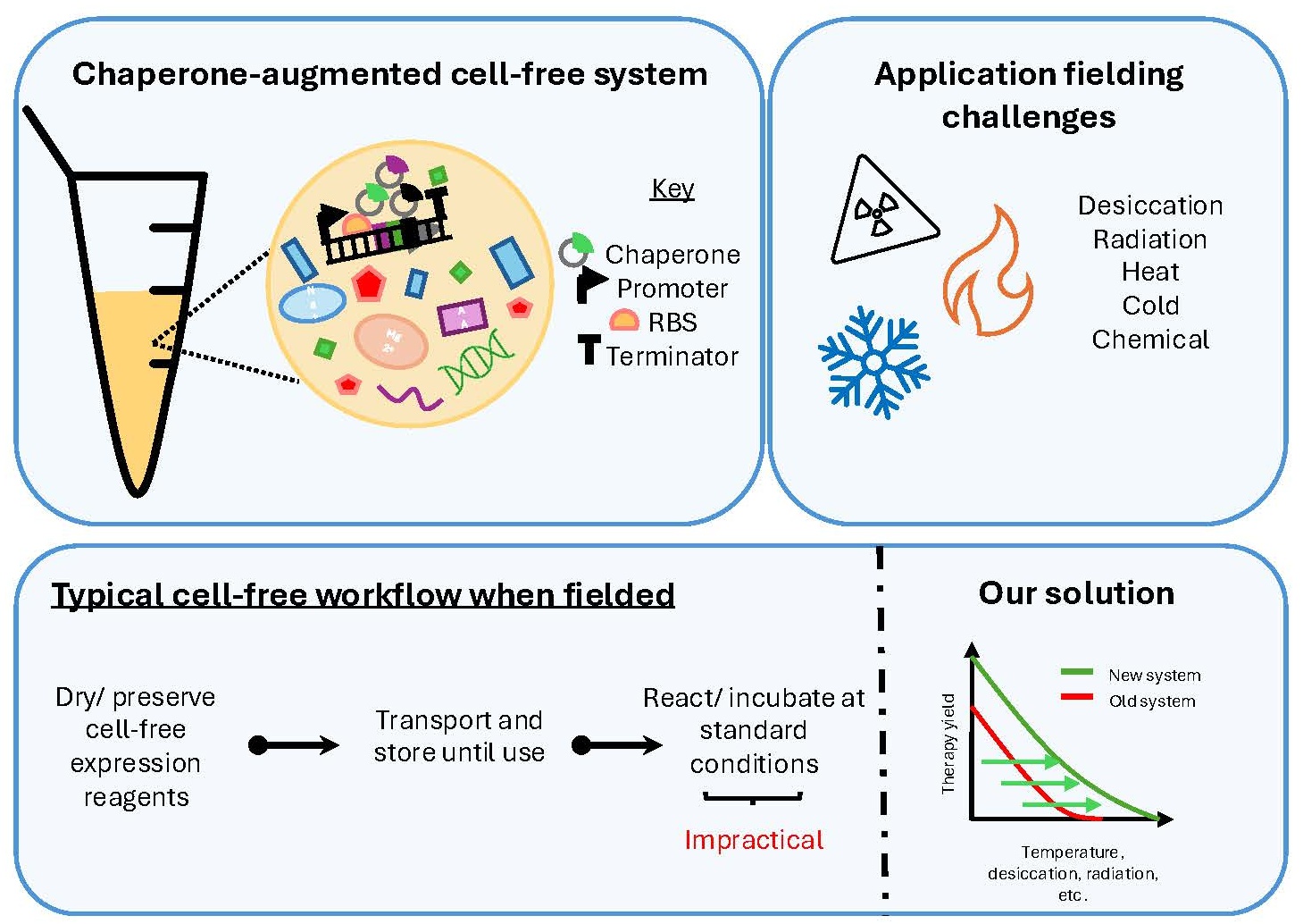
Graphical Abstract
1. Introduction
Maintaining the therapeutic protein lifecycle—generation, transport, and storage—requires large amounts of capital, infrastructure, and well-trained technical staff. As a result, these medicines are out of reach for many people worldwide. Cell-free protein expression technologies offer the potential for addressing this protein lifecycle burden. Established methods for preserving cell-free systems enable transportation and storage without requiring specialized conditions, e.g., cold-chain management, thereby greatly expanding accessibility and ease of deployment [1,2,3,4,5,6]. However, when the systems are reconstituted for application use, their broad utility is impacted, with most cell-free reactions still carried forward at standard incubation temperatures, e.g., 16 °C to 37 °C [7]. This inconsistency severely limits where these systems can be deployed and operated, diminishing the advantages over traditional therapeutic protein platforms. By broadening the operating range of cell-free systems (CFS), the application potential for these technologies would be significantly improved, paving the way for more fieldable protein synthesis applications.
To our knowledge, no cell-free research work has attempted to broaden a cell-free system’s operating range during protein expression. Instead, cell-free researchers have given considerable focus to preserving cell-free system parts, including the extract and reaction buffer, the latter comprising all the energy resources necessary for translation. Traditionally, these systems are lyophilized together or separately, often with a preserving agent, e.g., a small or large sugar molecule or complex [2,3,4,5,6].
Other efforts have sought to leverage molecular chaperones in cell-free systems, with the intention of improving the activity of the intended protein product [8,9,10]. For example, Nishimura et al. developed an extract preparative workflow wherein heat-shock chaperones were intentionally upregulated by exposing the primary culture to an elevated incubation temperature, specifically 42 °C [9]. These authors pay special attention to the pitfalls of culturing Escherichia coli at temperatures outside the norm, including a diminished growth rate and dry cell weight, which are important considerations in extract workflows. Extract efficacy has long been attributed to a culture’s growth rate or lack thereof [9,10,11]. Extracts derived from cultures harvested during rapid growth periods have higher levels of translationally active proteins and free ribosomes [9]. Despite these challenges, the findings of Nishimura et al. support the use of chaperone proteins in cell-free workflows, given that these authors report higher levels of protein expression when using lysates that have chaperone proteins. Similarly, Yamane et al. show that their chaperone-inspired system, using DnaK and GroEL, both heat-shock chaperones, generated higher levels of solubilized and properly folded reporter protein [10]. Together, these authors successfully demonstrate the functionality of chaperone components in cell-free systems, specifically that they help properly fold protein targets, a hallmark of molecular chaperones. However, these two approaches have limitations. First, for chaperone expression to occur, cultures must be grown in stressful environments, making culture optimization and extract preparation more difficult. Second, the only chaperones that can be used are those native to the cell line being cultured.
To address these limitations, we developed methods that enable the convenient expression of non-native chaperones, such as WZY2 and DHN1α, both of which are dehydration-resistant chaperones, in cell-free workflows. This approach extends chaperone inquiry beyond those from E. coli, enabling rapid integration of recombinant molecular chaperones into cell-free systems and facilitating their study at elevated temperatures via fluorescence monitoring.
This work investigates whether chaperones from various sources can function in cell-free systems to enhance reporter output at high incubation temperatures relevant to fielded conditions. The goals of this work were threefold. First, identify heat-shock chaperone candidates and then identify the gene regulatory elements controlling the chaperone’s expression, such as ribosomal binding sites, promoters, and terminators. Second, test whether these components can be added to cell-free systems to achieve higher enhanced green fluorescent protein (EGFP) fluorescence output at temperatures above standard laboratory conditions. Third, determine whether dual-chaperone systems result in higher system performance. We report an over 100-fold improvement in EGFP fluorescence when chaperones are used in cell-free systems and detect fluorescence in the chaperone-inspired systems 4.5 °C higher than in the control. Together, these efforts offer a generic yet robust approach to identifying chaperones that may improve system performance in fielded conditions.
2. Materials and Methods
2.1. Chaperone Selection and Plasmids
All plasmids were designed and built using the software services IDT DNA, De Novo DNA, and SnapGene. IDT DNA was used for codon optimization, De Novo DNA was used to design ribosomal binding sites for strong translation efficiency, and SnapGene was used to verify and validate our plasmid design. The designed chaperone expression plasmids were synthesized by Twist Biosciences (South San Francisco, CA, USA), and chaperone plasmid DNA was prepared using the Qiagen Midi prep kit (Germantown, MD, USA).
2.2. 2xYPTG (Yeast Extract, Phosphate Buffer, Tryptone, and Glycerol)
The phosphate buffer and glycerol solution were prepared independently of the yeast extract and tryptone solution. The yeast-tryptone solution was prepared by adding 16 g of tryptone, 10 g of yeast extract, and 5 g of sodium chloride to a 2 L flask, then adding MilliQ water to bring the final volume to 1 L. The phosphate buffer was prepared by adding 2.64 g of monosodium phosphate and 5.68 g of disodium phosphate to a 250 mL flask, then adding MilliQ water to obtain a final volume of 100 mL. Lastly, the glycerol solution was made by adding 3.75 mL of 100% glycerol to 96.25 mL of MilliQ water in a 250 mL flask. All three solutions were autoclaved, and 2XYPTG media were made by combining each of the three solutions.
2.3. Buffer A
1 L of Buffer A was prepared by combining these reagents in a 2 L bottle: 12.194 g potassium acetate, 3.002 g magnesium acetate, 0.5 mL 2-mercaptoethanol (2-ME), 0.155 g dithiothreitol (DTT), 0.489 g Tris-acetate, and 0.884 g Tris. MilliQ water was added up to 1 L, and the solution was filtered using a 0.22-micron filter.
2.4. Buffer B
1 L of Buffer B was prepared by combining these reagents in a 2 L bottle: 12.194 g potassium acetate, 3.002 g magnesium acetate, 0.155 g dithiothreitol (DTT), 0.489 g Tris-acetate, and 0.884 g Tris. MilliQ water was added up to 1 L, and the solution was filtered using a 0.22-micron filter.
2.5. Extract
To begin, BL21 Star (DE3) cells (Invitrogen, Carlsbad, CA, USA) were grown overnight in a 500 mL Erlenmeyer flask containing 100 mL of 2xYPTG media with shaking at 225 rpm at 37 °C. 5 mL of this culture was transferred to a 2 L Erlenmeyer flask containing 500 mL of 2xYPTG. This primary culture was left to grow with shaking at 225 rpm at 37 °C for 4 h. Isopropyl β-d-1-thiogalactopyranoside (IPTG) was added at the first hour before the log phase growth of the culture was reached. Following the four-hour growth period, cultures were transferred to 250 mL Nalgene bottles and immediately chilled on ice. The Nalgene bottles containing the culture were centrifuged for 20 min at 4 °C and 4000 relative centrifugal units (RCF). The supernatant was decanted, and the Nalgene bottles containing the wet cell pellet were weighed. The wet cell pellets were reconstituted using Buffer A. 20 mL of Buffer A was used for each gram of wet cell pellet. Following reconstitution, the bottles were centrifuged for 20 min at 4 °C and 4000 RCF. This wash step was repeated twice. Buffer A was decanted, and the weight of the wet cell pellets was measured. The bottles were stored overnight at −80 °C. The next day, the pellets were left to thaw in a room-temperature water bath, then reconstituted with 1.27 mL of Buffer B per gram of pellet. The reconstitution was aliquoted in 1.5 mL amounts to 2 mL microcentrifuge tubes. The cell suspensions were sonicated on ice for seven cycles at 50% amplitude. The resulting solution was centrifuged for 10 min at 4 °C and 12,000 RCF. The supernatant was collected and stored at −80 °C.
Traditional cell-free extract workflows recommend run-off reactions or lengthy incubation periods following the last centrifugation step. Kwon and colleagues in 2015 demonstrated that run-off reaction times for extract workflows using BL21 Star (DE3) cells contribute to worse downstream experimental outcomes [11]. In light of this finding, we opted to omit the run-off reaction. Additionally, the same paper shows that total energy imparted to the sample during sonication is a worthwhile indicator of extract efficacy and that energies between 556 J and 1 kJ are best [11]. This is why each sample was sonicated for seven on/off cycles.
2.6. Reaction Buffer
The reaction buffer used for each of these experiments contained the following concentrated reagents: 90 mM potassium glutamate, 80 mM ammonium acetate, 32 mM magnesium acetate, 34 µg/mL folinic acid, 1.2 mM ATP, 0.85 mM of GTP, CTP and UTP, 2 mM DTT, 0.64 mM cAMP, 28.5 mM HEPES-KOH, 4 mM of cysteine, 2.1 mM of every other 19 amino acids, 2% PEG (8000), 67 mM creatine phosphate, and 3.2 µg/mL creatine kinase.
2.7. EGFP Expression Assay
System efficacy was measured by assaying EGFP fluorescence. Each of the experiments indicated above was done using this assay. To begin, chaperone plasmid DNA was added to a 96-well plate at the necessary concentrations. They equaled those in the figure legends when combined with the reaction mixture and reporter plasmid. The reaction mixture comprised 65% reaction buffer and 35% extract. The reaction mixture was added to each well containing the chaperone and was left to react for 20 min at room temperature. The remaining reaction mixture was aliquoted into the few remaining wells during these 20 min. The plate was placed in the thermal cycler, and the samples were given 10 additional minutes to react, but at the experimental temperatures, not at room temperature. After these 30 min, EGFP DNA was added to each of the wells for an in-solution concentration of 10 ng/µL, and water was added to the background samples. These reactions were incubated for 4 h at the experimental temperature, and kanamycin (500 µg/mL) was added to arrest reporter expression. EGFP fluorescence was assayed at 37 °C using a Biotek Cytation 5 at excitation and emission spectra equaling 479 ± 20 nm and 520 ± 20 nm, respectively.
3. Results
3.1. Gene Regulatory Components Used in Chaperone Plasmid Design
In line with our core research goal of broadening the operating range of cell-free systems at higher incubation temperatures, we initially focused on three chaperones identified as heat-shock or desiccation-resistant chaperones that have been shown to improve E. coli viability at temperatures as high as 50 °C [12,13,14]. These chaperones are PtDRG1, WZY2, and CsLEA11 from the organisms Pyropia tenera (nori), Triticum aestivum (wheat), and Cucumis sativus (cucumber), respectively (Table 1) [12,13,14]. The last two chaperones, WZY2 and CsLEA11, both dehydrins, are late embryogenesis abundant proteins but differ in their protein segment composition, specifically the Y-segment [12,13]. PtDRG1, however, is not classically a dehydrin but functions in the same manner as WZY2 and CsLEA11 [14]. Prior to expressing these components in cell-free systems derived from E. coli, strong ribosomal binding sites were designed in-silico using the RBS Calculator (De Novo DNA) [15]. Predicted translation initiation rates (TIR) equaling 85,768.52, 726,371.24, and 525,472.86 were obtained for PtDRG1, WZY2, and CsLEA11, respectively (Table 2). In addition, a T7Tet promoter, controllable via the Tet operator, and a strong terminator, BBa_1006, were chosen (Table 3). Together, these components permitted control over the chaperone’s expression in the cell-free system.
Table 1. Listed are the experimental chaperone components and their native function.
|
Host Species |
Chaperone |
Protein Class |
Type of Protection |
|---|---|---|---|
|
Pyropia tenera (nori) |
PtDRG1 |
Desiccation response gene (DRG) |
Heat |
|
Triticum aestivum (wheat) |
WZY2 |
Dehydrin (DHN) |
Heat |
|
Cucumis sativus (cucumber) |
CsLEA11 |
DHN |
Heat |
Table 2. Translation initiation rates are given for each of the chaperone components in addition to their in-silico RBS design.
|
Plasmid |
Promoter |
Terminator |
RBS Sequence |
TIR |
|---|---|---|---|---|
|
pT7Tet-PtDRG1-Bba_1006 |
T7Tet |
Bba_1006 |
CCCTTTAATTCTAAATACGATATTACGGAGGTAATTTA |
85,768.52 |
|
pT7Tet-WZY2-Bba_1006 |
T7Tet |
Bba_1006 |
GGGGGCCCTATAGTAAAAGCGTCTTGTTGGACGCAAAGTAAGGAGGTTTTTT |
726,371.24 |
|
pT7Tet-CsLEA11-Bba_1006 |
T7Tet |
Bba_1006 |
AGGGGGGGAGATAATAGGTAGGAGAATTAAGGAGGTAATTA |
525,472.86 |
Table 3. Gene regulatory components that are shared across the three chaperone plasmids.
3.2. Higher Reporter Fluorescence Levels Were Observed at Elevated Incubation Temperatures with the Use of the Chaperones WZY2, CsLEA11, and PtDRG1
To begin, three heat-shock chaperones, including WZY2, CsLEA11, and PtDRG1, were tested in cell-free reactions at different plasmid DNA concentrations. These entry-level experiments, described in Figure 1, were used to screen the efficacy of the chaperone components relative to EGFP fluorescence output. Our chaperone-augmented cell-free system (CACFS), irrespective of which chaperone was used, outperformed the traditional cell-free system (TCFS), denoted as ‘EGFP control’, at all temperatures tested, even at 37 °C (Figure 1b–d). These results support the hypothesis that these chaperones can serve as a worthwhile cell-free additive across the incubation temperatures tested, with respect to EGFP fluorescence.
3.3. Chaperone Effects Are Dose- and Temperature-Dependent
It was hypothesized that higher levels of chaperone DNA would denote lower EGFP fluorescence due to the competing expression of the chaperone component and the fluorescent reporter. In anticipation of false negatives, six chaperone plasmid DNA concentrations ranging from 2.5 ng/µL to 15 ng/µL were studied. In all three CACFSs, chaperone plasmid DNA exceeding 10 ng/µL resulted in the lowest EGFP fluorescence, whereas 2.5 ng/µL for the chaperones WZY2 and CsLEA11 and 7.5 ng/µL for the chaperone PtDRG1 gave the highest (Figure 1b–d). These concentrations were identified as the optimal DNA concentrations for our three systems following the trapezoidal integration of their EGFP expression curves (Supplementary Figure S5). In some cases, the CACFSs underperformed compared to the EGFP control (Figure 1b,d). For example, chaperone plasmid DNA concentrations of 12.5 and 15 ng/µL for the chaperones WZY2 and PtDRG1 resulted in lower fluorescence output at temperatures as high as 45 °C (Figure 1b,d). Thus, it was found that chaperone effects are dose- and temperature-dependent.
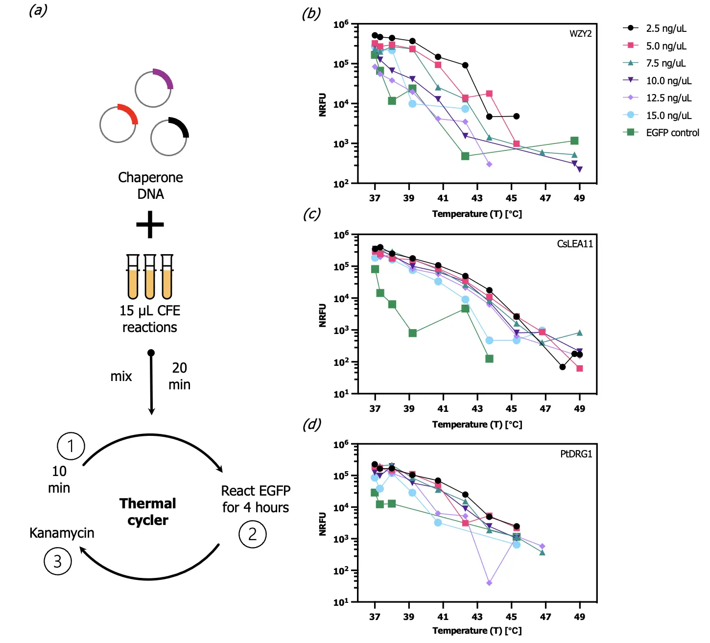
Figure 1. Chaperone functionality characterization. (a) The EGFP expression assay is described. First, chaperone plasmid DNA was added to CFE reactions for 20 min at room temperature and 10 min at the experimental incubation temperatures. Next, EGFP DNA was added and incubated for 4 h at the experimental temperatures. Kanamycin was added after the 4-h incubation period to arrest reporter expression. Fluorescence was assayed in a Biotek Cytation 5. (b–d) Chaperones WZY2, CsLEA11, and PtDRG1 were studied at concentrations ranging from 2.5 ng/µL to 15.0 ng/µL (n = 1). Their effects on fluorescent output at high incubation temperatures were measured using the EGFP expression assay. All data points represent fluorescence values normalized to background samples containing extract, reaction buffer, and water as described in the methods section.
3.4. Chaperone Components Broaden the Cell-Free System Operating Range by 4.5 °C and Increase Fluorescence Output up to 100-Fold
Following the initial screening experiments (Figure 1), we performed further characterizations (Figure 2) using the optimized chaperone DNA concentrations—2.5 ng/µL for WZY2 and CsLEA11, and 7.5 ng/µL for PtDRG1—and the same EGFP expression assay as shown in Figure 1a. The WZY2 system outperformed the EGFP control, with 100-fold and 21-fold increases in EGFP output at 40.7 °C and 42.3 °C, respectively (Figure 2a). The CsLEA11 and PtDRG1 systems outperformed the EGFP control up to 40.7 °C and outpaced the control by 16-fold and 36-fold at 38 °C and 40.7 °C, respectively (Figure 2b,c). To facilitate comparisons, Figure 2d shows the incubation temperature at which the systems reach EGFP fluorescence, which approximates 10% of the maximum EGFP fluorescence observed in the EGFP control experiments. Additionally, these stated expression thresholds are over an order of magnitude above the detection threshold. All three CACFSs met this threshold at 43.7 °C, whereas the threshold was reached at 39.2 °C for the EGFP control (Figure 2d). Thus, based on this metric, chaperones extended the operating temperature range by 4.5 °C. Supplementary Figure S6 demonstrates this system productivity shift using polynomial regression models generated in R (R version 4.4.0 (24 April 24)) [19]. System productivity, as a function of chaperone usage and incubation temperature, can be predicted using these models with a high degree of certainty (Supplementary Figure S6).
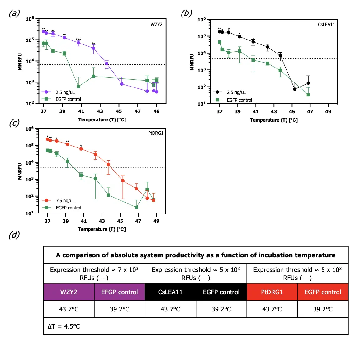
Figure 2. Optimized chaperone system characterization at high incubation temperatures and operating range assessment. (a–c) Chaperone effects on EGFP expression, in triplicate, were characterized for WZY2, CsLEA11, and PtDRG1 at the optimized plasmid DNA concentrations 2.5 ng/μL, 2.5 ng/μL, and 7.5 ng/μL, respectively (n = 3). (d) An expression threshold of 7 × 10³ (WZY2) and 5 × 10³ MNFRUs (CsLEA11 & PtDRG1) was identified and set to make system comparisons easier and assert that the CACFSs enable the broadening of the cell-free system’s operating range by 4.5 °C. All data points represent fluorescence values normalized to background samples containing extract, reaction buffer, and water. Data points represent the mean fluorescence of three replicates, and error bars represent the standard deviation. Reported statistical significance was found using a Student’s t-test. Significance thresholds were determined according to these p-value inequalities: * p < 0.05, ** p < 0.01, and *** p < 0.001.
3.5. Dual-Chaperone Systems Exhibit Increased Fluorescence Compared to Single-Chaperone Systems
Since each chaperone tested has a unique structure and composition, we examined whether co-expression of chaperones could benefit system performance. Using the same EGFP expression assay indicated in Figure 1a, two chaperones were expressed simultaneously at equal concentrations. The chaperone DNA totals provided in the Figure 3 legend represent the gross total amounts, with each chaperone component contributing equally. The shaded region, representing the area under the single chaperone’s best-averaged curve across four experiments, was used to illustrate the changes in fluorescence when two chaperones are expressed instead of one. In all cases, the dual-chaperone systems, shown by the line segments in Figure 3, produce higher fluorescence than single-chaperone systems at higher incubation temperatures. For example, the WZY2-CsLEA11 system, compared to the optimized WZY2 system, achieves a 1.4-fold and 2.3-fold increase in fluorescence at 37 °C and 45.3 °C, respectively (Figure 3a). The WZY2-CsLEA11 system, compared to the CsLEA11 system, shows 2-fold and 6-fold increases in fluorescence at those same temperatures (Figure 3a). The WZY2-PtDRG1 system leads to 3-fold and 6-fold increases in fluorescence at 45.3 °C compared to the WZY2 and PtDRG1 systems, respectively (Figure 3b). Lastly, the CsLEA11-PtDRG1 system exhibits 4-fold and 3-fold increases in fluorescence at 45.3 °C compared to the CsLEA11 and PtDRG1 systems, respectively (Figure 3c). Dual-chaperone systems result in an increase in fluorescence, but more testing is required to gauge whether these dual-chaperone results hold in subsequent cell-free workflows.
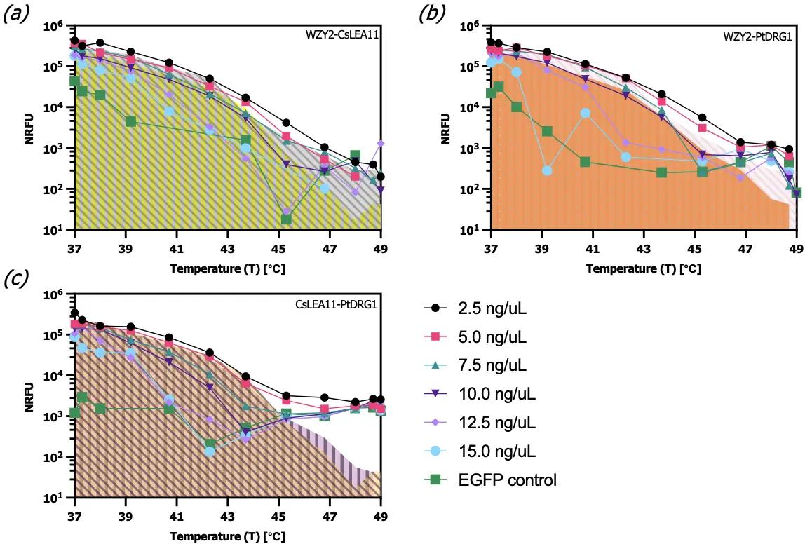
Figure 3. Dual chaperone characterization relative to CFS expressive capacity at high incubation temperatures. Chaperones were expressed jointly at equal concentrations, with total DNA concentrations indicated in the legend. CFS productivity was quantified using the assay shown in Figure 2a: (a) WZY2 and CsLEA11; (b) WZY2 and PtDRG1 (c) CsLEA11 and PtDRG1 (n = 1). All data points represent fluorescence values normalized to background samples containing extract, reaction buffer, and water. Shaded regions represent each of the chaperones’ highest performing measure when expressed independently. These data are derived from Figure 2 and Figure 3 and account for inter- and intra-experiment variability.
4. Discussion
In this work, we show that protective proteins can be used to expand the operating range of traditional E. coli-derived cell-free systems relative to high incubation temperatures. More specifically, we show that the chaperones WZY2, CsLEA11, and PtDRG1 improve fluorescence output, in some cases, by 100-fold and push these systems’ workable temperature range by 4.5 °C, up to 43.7 °C. To our knowledge, no other efforts to date have sought to expand the temperature range over which these cell-free protein expression systems can operate. Additionally, we offer an experimentally validated approach to enable additional fieldable protein expression efforts, such as biosensing operations, that are complicated by technological gaps, as indicated in the graphical abstract.
The observed enhancement of output protein activity (EGFP fluorescence) could arise from stabilization of the output protein and/or of the transcription/translation components. Based on the nature of our study design, coupled with previous indications of the broad specificity of protective capabilities of the chosen proteins, our results suggest that protection of expression components is a significant factor in the observed enhancement of fluorescence at higher temperatures, if not the primary driver [12,13,14]. Temperatures > 78 °C are required for irreversible denaturation of EGFP, which are far above the temperatures used in our assays [20]. To address the fact that EGFP brightness is still temperature dependent, we conducted all of our fluorescence assays at a common temperature (37 °C). Therefore, by arresting translation immediately after expression and conducting all fluorescence assays at a common temperature, we allow restoration of any EGFP that may have been reversibly denatured in our higher temperature tests.
Nonetheless, future efforts to probe the precise nature of protection can inform paths to further optimization of performance at elevated temperatures. Towards this goal, RNA and protein quantification, expression of different reporters, and therapeutic proteins with different folding and stability requirements would be informative. In addition, characterizations using purified protective proteins can provide a more accurate quantification of full protective enhancement.
Though the information from these potential future characterizations would certainly be valuable, we accomplished a key, broader goal of developing a simple screening procedure for assesssing diverse protective protein candidates. By adopting the simple approach of co-expressing protective proteins with the output protein, we avoid the time-consuming and costly step of purifying each protein candidate. While we aimed for simplicity and scalability in the method for assessing protective proteins, our choice of addressing performance at elevated temperatures is more challenging than most other forms of stress. By comparison, many other forms of environmental stress can be ones for which a simple chemical addition can rapidly establish the test environment. We carefully considered the scale and timing of reactions, leveraged a gradient thermocycler, and introduced a translational inhibition step to carefully control temperature changes and expression duration across many samples simultaneously.
Beyond screening, in the context of applications at scale, our co-expression approach exhibits tradeoffs with respect to the alternative of adding purified protective proteins to conventional extracts. On one hand, yield of the output protein is expected to be lower for our co-expression system. When protective proteins are expressed in the cell-free system, fewer reaction resources are available to express the output protein. On the other hand, we expect scalability advantages by avoiding the potentially high costs of large-scale protein purification.
An appealing alternative that can combine the relative advantages of these two approaches is to express the proteins in the live E. coli used to produce extract, similar to how strains that express T7 RNA polymerase are often used to make extracts, avoiding the need to add purified T7 RNA polymerase for T7 promoter expression. Though the full development and optimization of this alternative are beyond the scope of this study, we piloted this approach and investigated other basic deployment considerations. Supplementary Figure S1, Figure S2 and Figure S3 suggest that chaperone components can be expressed in the extract and combined with the reaction buffer to improve system stability over time and capacity during operation. This exciting approach suggests that chaperone components can act as stabilizing agents for preservation efforts when expressed in the extract. If this is the case, this experimental approach can address each technological challenge facing fieldable protein expression—preservation, storage, and the regulation of system operating temperatures. For this to be realized, a series of optimizations must occur during extract preparation to make this approach realistic. For example, in our experiments, when E. coli is transformed with a chaperone, growth is significantly slowed, portending diminished translational efficacy and outright extract yield (Supplementary Figure S4).
This study establishes a practical workflow for incorporating heterologous protective proteins into cell-free systems and highlights their effectiveness in broadening the operating range, with implications for field-deployable systems and their applications. Our methodology can be extended to identify proteins for other forms of protection, for example, robustness to different pH or salinity ranges, oxidative damage, or radiation. The fact that our results suggest that protective proteins can have additive effects points to the exciting possibility of further extending the operating range with respect to not only temperature but also other stressors simultaneously. Therefore, our work embodies a step towards greatly expanding the practical application contexts of cell-free synthetic biology. Take, for example, protein expression in space. As other-worldly travel becomes more frequent, the need for a system that can reliably express therapeutic proteins while accounting for capsule size, capacity, and radiation exposure will be great. In this case, cell-free systems can be deployed with radiation-resistant chaperones expressed in extract, combined with the reaction buffer, to afford routine and worthwhile therapeutic expression.
Supplementary Materials
The following supporting information can be found at: https://www.sciepublish.com/article/pii/1123, Figure S1: Cell-free system productivity as a function of temperature and chaperone expression in the extract; Figure S2: Optimization of chaperone-transformed extract; Figure S3: Chaperone effects on cell-free system preservation; Figure S4: Effect of WZY2 on E. coli cell growth; Figure S5: Integration result of the chaperone expression curves from Figure 1; Figure S6: Polynomial regression models for the chaperone-augmented cell-free systems, including WZY2, CsLEA11, and PtDRG1.
Acknowledgments
The authors thank all members of the Karig lab for their administrative and technical support.
Author Contributions
Conceptualization: D.K.K. and M.R.B.; Methodology: M.R.B. and D.K.K.; Software: M.R.B.; Validation: M.R.B., S.L. and D.K.K.; Formal Analysis: M.R.B.; Investigation: M.R.B.; Resources: D.K.K.; Data Curation: M.R.B.; Writing—Original Draft Preparation: M.R.B. and D.K.K.; Writing—Review and Editing: M.R.B., S.L. and D.K.K.; Visualization: M.R.B.; Supervision: D.K.K.; Project Administration: D.K.K.; Funding Acquisition: Not applicable.
Ethics Statement
Not applicable.
Informed Consent Statement
Not applicable.
Data Availability Statement
All data can be found in the main manuscript text and in the supplementary materials.
Funding
Clemson University internal startup funds were used to fund this research.
Declaration of Competing Interest
The authors declare that they have no known competing financial interests or personal relationships that could have appeared to influence the work reported in this paper.
References
- Smith MT, Berkheimer SD, Werner CJ, Bundy BC. Lyophilized Escherichia Coli-Based Cell-Free Systems for Robust, High-Density, Long-Term Storage. BioTechniques 2014, 56, 186–193. DOI:10.2144/000114158 [Google Scholar]
- Wilding KM, Zhao EL, Earl CC, Bundy BC. Thermostable lyoprotectant-enhanced cell-free protein synthesis for on-demand endotoxin-free therapeutic production. New Biotechnol. 2019, 53, 73–80. DOI:10.1016/j.nbt.2019.07.004 [Google Scholar]
- Yang J, Cui Y, Cao Z, Ma S, Lu Y. Strategy exploration for developing robust lyophilized cell-free systems. Biotechnol. Notes 2021, 2, 44–50. DOI:10.1016/j.biotno.2021.08.004 [Google Scholar]
- Gregorio NE, Kao WY, Williams LC, Hight CM, Patel P, Watts KR, et al. Unlocking Applications of Cell-Free Biotechnology through Enhanced Shelf Life and Productivity of E. coli Extracts. ACS Synth. Biol. 2020, 9, 766–778. DOI:10.1021/acssynbio.9b00433 [Google Scholar]
- Warfel KF, Williams A, Wong DA, Sobol SE, Desai P, Li J, et al. A Low-Cost, Thermostable, Cell-Free Protein Synthesis Platform for On-Demand Production of Conjugate Vaccines. ACS Synth. Biol. 2023, 12, 95–107. DOI:10.1021/acssynbio.2c00392 [Google Scholar]
- Karig DK, Bessling S, Thielen P, Zhang S, Wolfe J. Preservation of protein expression systems at elevated temperatures for portable therapeutic production. J. R. Soc. Interface 2017, 14, 20161039. DOI:10.1098/rsif.2016.1039 [Google Scholar]
- Silverman AD, Karim AS, Jewett MC. Cell-free gene expression: An expanded repertoire of applications. Nat. Rev. Genet. 2020, 21, 151–170. DOI:10.1038/s41576-019-0186-3 [Google Scholar]
- Higuchi K, Yabuki T, Ito M, Kigawa T. Cold shock proteins improve E. coli cell‐free synthesis in terms of soluble yields of aggregation‐prone proteins. Biotech. Bioeng. 2020, 117, 1628–1639. DOI:10.1002/bit.27326 [Google Scholar]
- Nishimura N, Kitaoka Y, Niwano M. Cell-free system derived from heat-shocked Escherichia coli: Synthesis of enzyme protein possessing higher specific activity. J. Ferment. Bioeng. 1995, 79, 131–135. DOI:10.1016/0922-338X(95)94079-7 [Google Scholar]
- Yamane T, Ikeda Y, Nagasaka T, Nakano H. Enhanced Cell-Free Protein Synthesis Using a S30 Extract from Escherichia coli Grown Rapidly at 42 °C in an Amino Acid Enriched Medium. Biotechnol. Prog. 2008, 21, 608–613. DOI:10.1021/bp0400238 [Google Scholar]
- Kwon YC, Jewett MC. High-throughput preparation methods of crude extract for robust cell-free protein synthesis. Sci. Rep. 2015, 5, 8663. DOI:10.1038/srep08663 [Google Scholar]
- Yang W, Zhang L, Lv H, Li H, Zhang Y, Xu Y, et al. The K-segments of wheat dehydrin WZY2 are essential for its protective functions under temperature stress. Front. Plant Sci. 2015, 6, 406. DOI:10.3389/fpls.2015.00406 [Google Scholar]
- Zhou Y, He P, Xu Y, Liu Q, Yang Y, Liu S. Overexpression of CsLEA11, a Y3SK2-type dehydrin gene from cucumber (Cucumis sativus), enhances tolerance to heat and cold in Escherichia coli. AMB Express 2017, 7, 182. DOI:10.1186/s13568-017-0483-1 [Google Scholar]
- Na Y, Lee HN, Wi J, Jeong WJ, Choi DW. PtDRG1, a Desiccation Response Gene from Pyropia tenera (Rhodophyta), Exhibits Chaperone Function and Enhances Abiotic Stress Tolerance. Mar. Biotechnol. 2018, 20, 584–593. DOI:10.1007/s10126-018-9828-2 [Google Scholar]
- Salis HM, Mirsky EA, Voigt CA. Automated design of synthetic ribosome binding sites to control protein expression. Nat. Biotechnol. 2009, 27, 946–950. DOI:10.1038/nbt.1568 [Google Scholar]
- Studier FW, Moffatt BA. Use of bacteriophage T7 RNA polymerase to direct selective high-level expression of cloned genes. J. Mol. Biol. 1986, 189, 113–130. DOI:10.1016/0022-2836(86)90385-2 [Google Scholar]
- Karig DK, Iyer S, Simpson ML, Doktycz MJ. Expression optimization and synthetic gene networks in cell-free systems. Nucleic Acids Res. 2012, 40, 3763–3774. DOI:10.1093/nar/gkr1191 [Google Scholar]
- Cambray G, Guimaraes JC, Mutalik VK, Lam C, Mai QA, Thimmaiah T, et al. Measurement and modeling of intrinsic transcription terminators. Nucleic Acids Res. 2013, 41, 5139–5148. DOI:10.1093/nar/gkt163 [Google Scholar]
- R Core Team. R: A Language and Environment for Statistical Computing; R Foundation for Statistical Computing: Vienna, Austria, 2024. Available online: https://www.R-project.org/ (accessed on 24 April 2024).
- Melnik BS, Molochkov NV, Prokhorov DA, Uversky VN, Kutyshenko VP. Molecular mechanisms of the anomalous thermal aggregation of green fluorescent protein. Biochim. Et Biophys. Acta (BBA)-Proteins Proteom. 2011, 1814, 1930–1939. DOI:10.1016/j.bbapap.2011.07.017 [Google Scholar]