
Lactononadecapeptide Upregulates Gene Expression of Neuroplasticity and Cholinergic Activation in Human iPSC-Derived Neurons
Lactononadecapeptide Upregulates Gene Expression of Neuroplasticity and Cholinergic Activation in Human iPSC-Derived Neurons
Junko Kimura 1,† Asako Kusumi 2 Koki Sato 3 Fumiya Nakamura 2 Kazuhito Ohsawa 2 Yoshihisa Kato 1 Shizuo Yamada 1,*
Received: 27 March 2026 Revised: 20 May 2026 Accepted: 25 June 2026 Published: 30 June 2026

© 2026 The authors. This is an open access article under the Creative Commons Attribution 4.0 International License (https://creativecommons.org/licenses/by/4.0/).
Graphical Abstract
1. Introduction
The rapid increase in the number of dementia patients, particularly those with Alzheimer’s disease (AD), represents a major social and medical challenge worldwide [1]. However, no definitive preventive or therapeutic agents for AD have been developed yet. Brain-derived neurotrophic factor (BDNF) and nerve growth factor (NGF) are key regulators of synaptic plasticity and memory [2,3]. In addition, cholinergic components such as choline acetyltransferase (ChAT) and the muscarinic M1 receptor (CHRM1), as well as the N-methyl-D-aspartate receptor (NMDAR) subunit NR1, play essential roles in synaptic transmission and cognitive function [4,5,6,7]. Moreover, TrkB, the receptor for BDNF, is a critical mediator of BDNF signaling in neuronal survival and plasticity [8].
Fermented milk products are traditional foods with various health benefits [9]. Recently, functional food peptides have gained significant attention for their neuroprotective potential, often modulating central nervous functions via gut–brain axis signaling. Previously, Ohsawa et al. [10] evaluated the effects of whey from a Lactobacillus helveticus-fermented dairy product, Calpis™ sour milk, a fermented milk produced during the manufacturing process of CalpisTM, on short-term memory impairment and found that it improved scopolamine-induced memory impairment in mice. Subsequently, they identified the active ingredient: lactononadecapeptide (LNDP: NIPPLTQTPVVVPPFLQPE) derived from β-casein [11]. More recently, Ohsawa et al. [12], Sasai et al. [13], and Yamada et al. [14] reported that LNDP-containing foods improved cognitive and memory functions in healthy middle-aged or elderly Japanese subjects. Indeed, our previous preclinical and clinical studies have revealed that LNDP-containing foods consistently improved cognitive and memory functions [10,11,12,13,14]. However, the molecular mechanisms by which LNDP improves cognitive function remain unclear, as memory formation is strictly regulated by the expression of various memory-related genes [15]. Based on the previously observed in vivo effects of LNDP, we focused on specific memory-related genes involved in neurotrophic support and cholinergic transmission. While traditional animal-derived cell models often present a translational gap due to species-specific differences, human induced pluripotent stem cell (hiPSC)-derived neurons accurately reflect human physiological receptor profiles and functional networks, serving as a superior translational model. Therefore, the primary objective of this study was to examine the direct effects of LNDP on the gene expression of memory-related neuroplasticity and cholinergic signaling in hiPSC-derived neurons. Additionally, we utilized PC12 cells to perform a preliminary evaluation of the safety and cytotoxicity profile of LNDP.
2. Materials and Methods
LNDP was chemically synthesized (GenScript, Piscataway, NJ, USA) and dissolved in sterile water immediately before use. The hiPSC-derived neurons ‘ReproNeuro’ (ReproCELL, Yokohama, Japan) were differentiated and maintained following the manufacturer’s instructions and as previously described [16]. PC12 cells (RIKEN BioResource Center, Tsukuba, Japan) were cultured in DMEM supplemented with horse serum and fetal bovine serum under standard conditions, as previously described [17]. PC12 cells were selected as a well-established and robust mammalian neuronal model for preliminary cytotoxicity screening prior to evaluating the highly sensitive human neurons. Cytotoxicity of LNDP was evaluated in PC12 cells using the CCK-8 assay (Dojindo, Kumamoto, Japan). Cells were treated with 0.1–10 μM LNDP for 24 h in a CO2 incubator. Absorbance was measured at 450 nm using a Multiskan™ GO microplate reader (Thermo Fisher Scientific, Waltham, MA, USA). For gene expression analysis, hiPSC-derived neurons were treated with 0.1 or 1 μM LNDP, or vehicle, for 0.5–24 h. These concentrations were selected based on their physiological relevance and blood circulation levels estimated from previous clinical trials [12,13,14]. Gene expression was analyzed by qRT-PCR using SYBR® Green Cells-to-CT™ Kits (Thermo Fisher Scientific) according to the manufacturer’s instructions. Reverse transcription and amplification were performed as described previously [16]. Gene-specific primers (Takara Bio, Kusatsu, Japan) are listed in Supporting Information (Table 1), with ACTB as an internal control. Relative expression levels were calculated by the 2−ΔΔCT method [18]. Statistical significance was assessed by one-way ANOVA followed by Tukey’s multiple comparison test with GraphPad Prism 5.0 (GraphPad Software, Inc., La Jolla, CA, USA). p value < 0.05 was considered significant.
Table 1. Sequences of the primers for qRT-PCR analysis.
|
Gene |
Forward Primer (5′–3′) |
Reverse Primer (5′–3′) |
Product Size (bp) |
|---|---|---|---|
|
BDNF |
gaactcccagtgccgaactacc |
ttatgaatcgccagccaattctc |
83 |
|
TrkB |
cctggcatcgtggcatttc |
tcagtcccacataagcttcaacatc |
133 |
|
NGF |
atgctggacccaagctca |
tgatcagagtgtagaacaacatgga |
123 |
|
CHRM1 |
atcaagtcccgaggcagca |
acttgactcccactcccaggaa |
85 |
|
ChAT |
agccctgccgtgatctttg |
gcacagtcagtgggaatggagt |
134 |
|
NR1 |
gagggtaccagatgtccacca |
cttgcatgtcccatcactca |
95 |
|
ACTB |
tggcacccagcacaatgaa |
ctaagtcatagtccgcctagaagca |
186 |
3. Results
The cytotoxicity of LNDP was evaluated using the CCK-8 assay. As shown in Figure 1, no significant alteration in the number of viable cells was detected when PC12 cells were exposed to 0.1, 1, or 10 μM LNDP compared to the vehicle control.
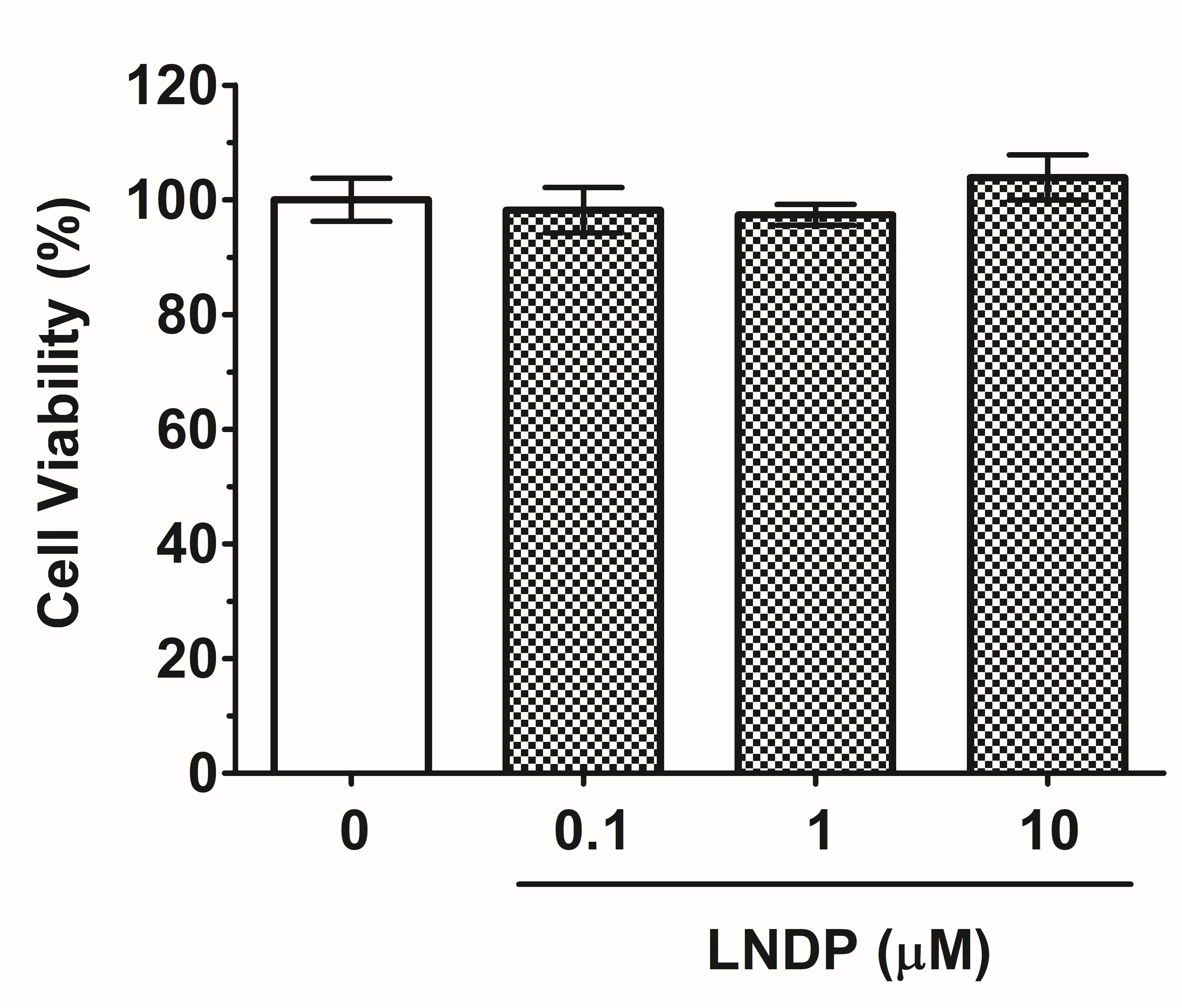
Figure 1. Effects of LNDP on PC12 cell viability. PC12 cells were treated with various concentrations of LNDP (0.1, 1, or 10 μM) for 24 h. Cell viability was assessed using the CCK-8 assay. Results are shown as the mean ± S.E.M. (n = 5). Statistical analyses were performed using one-way ANOVA followed by Tukey’s multiple comparison test. No significant differences were detected against the vehicle control (p ≥ 0.1).
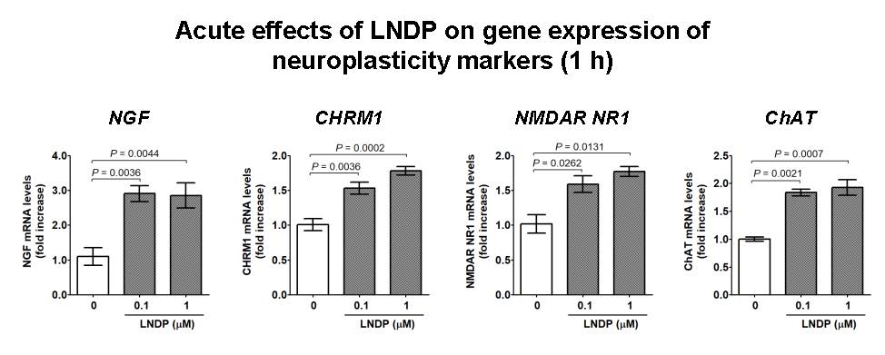
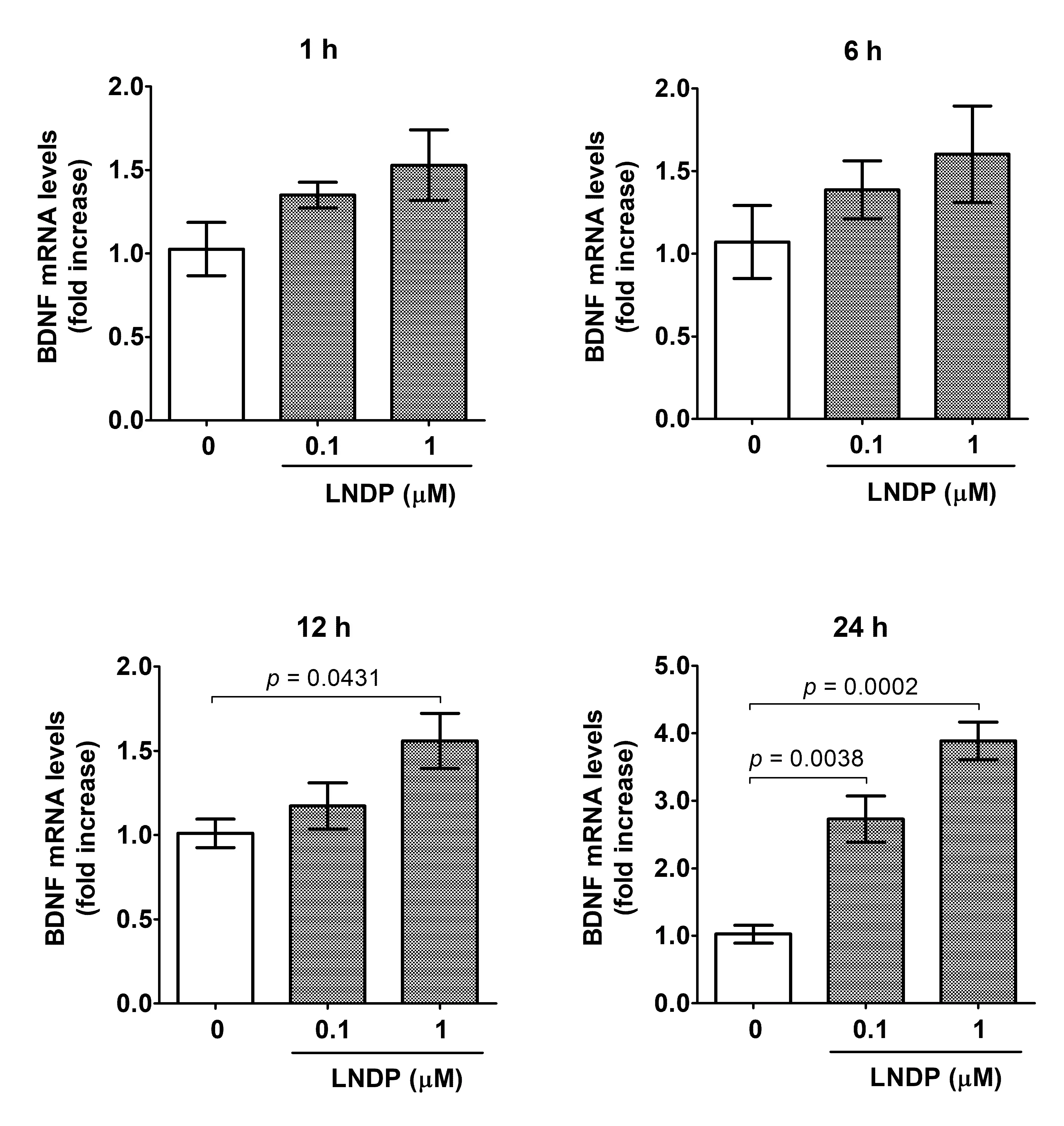
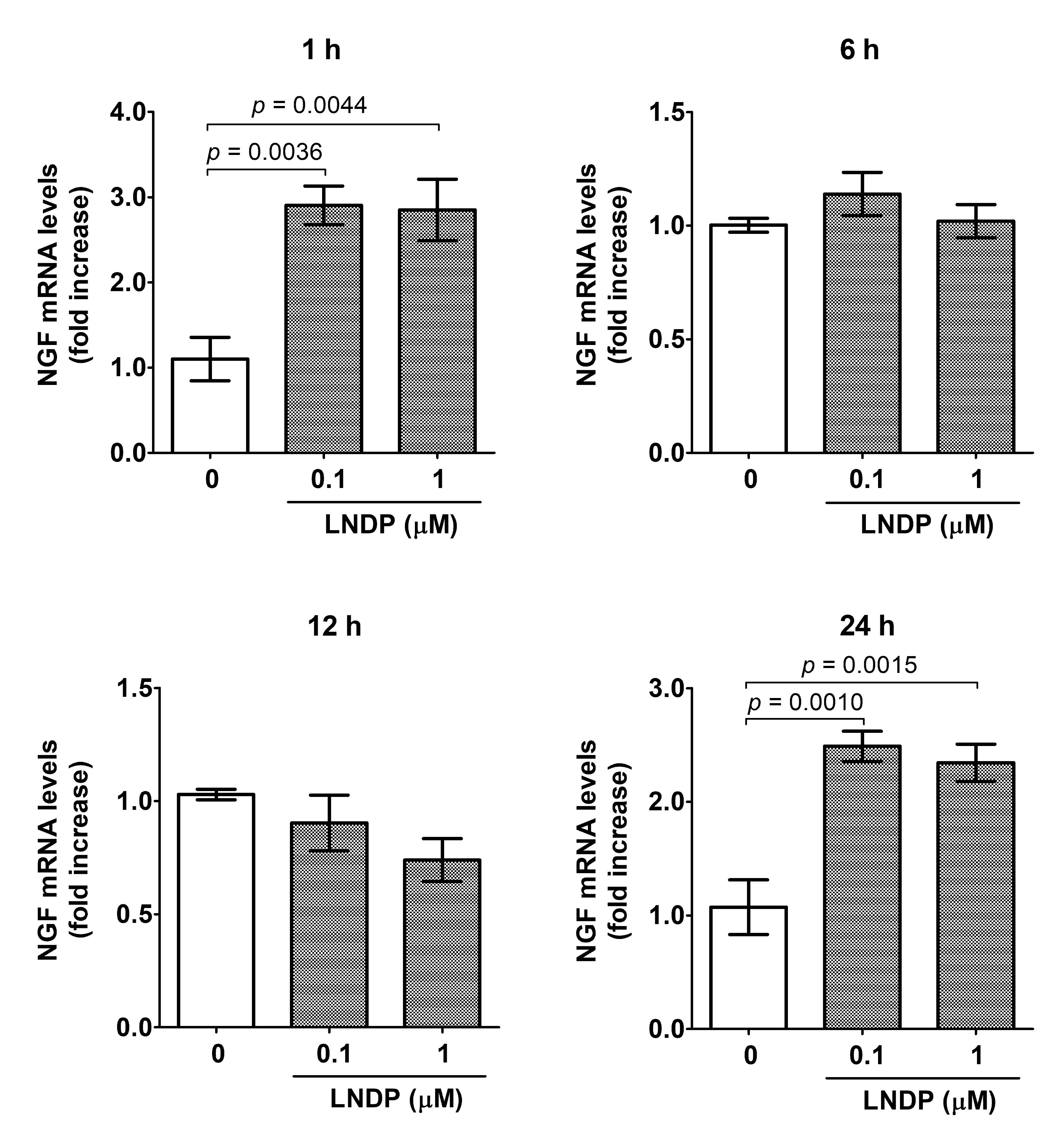
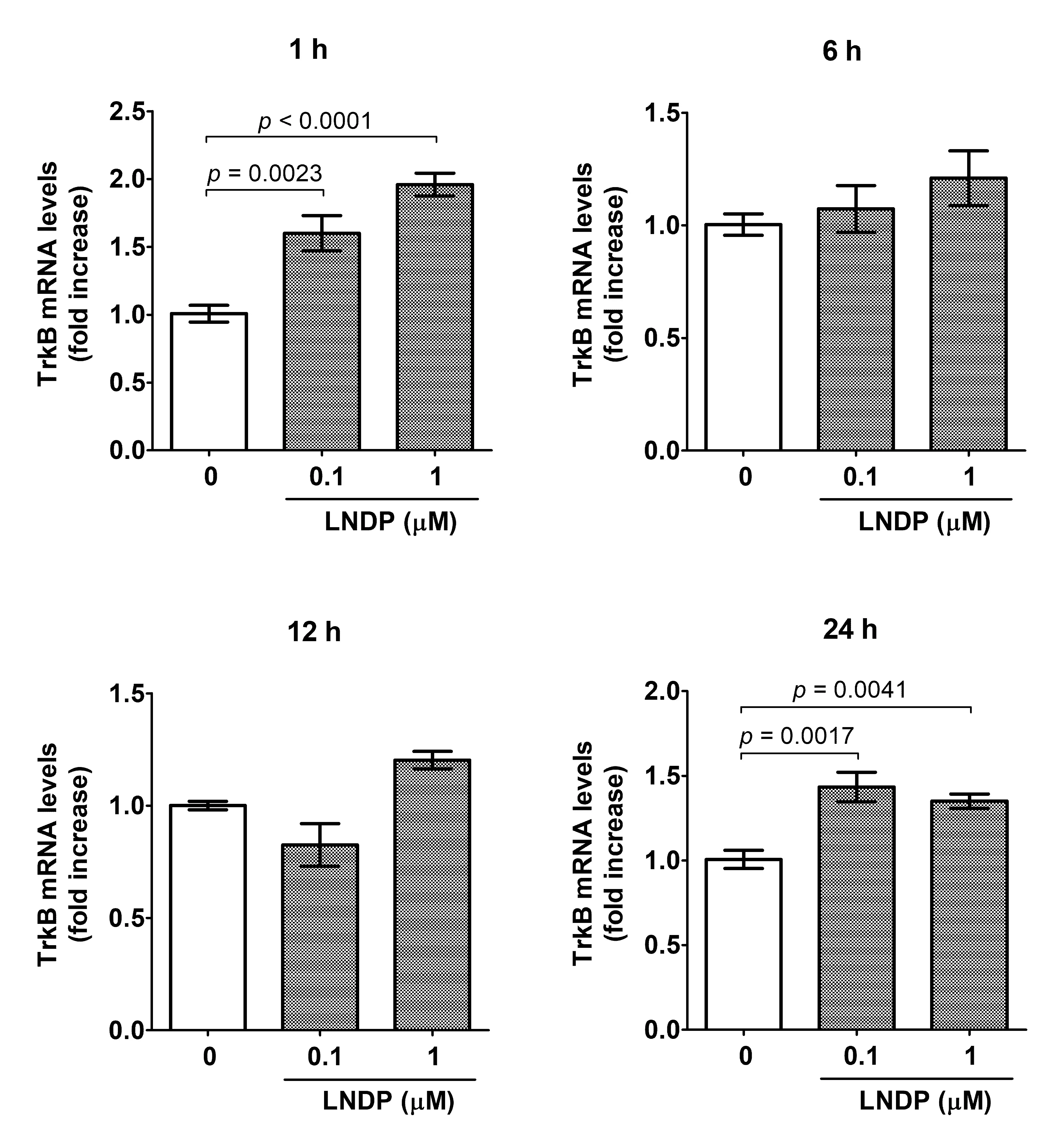
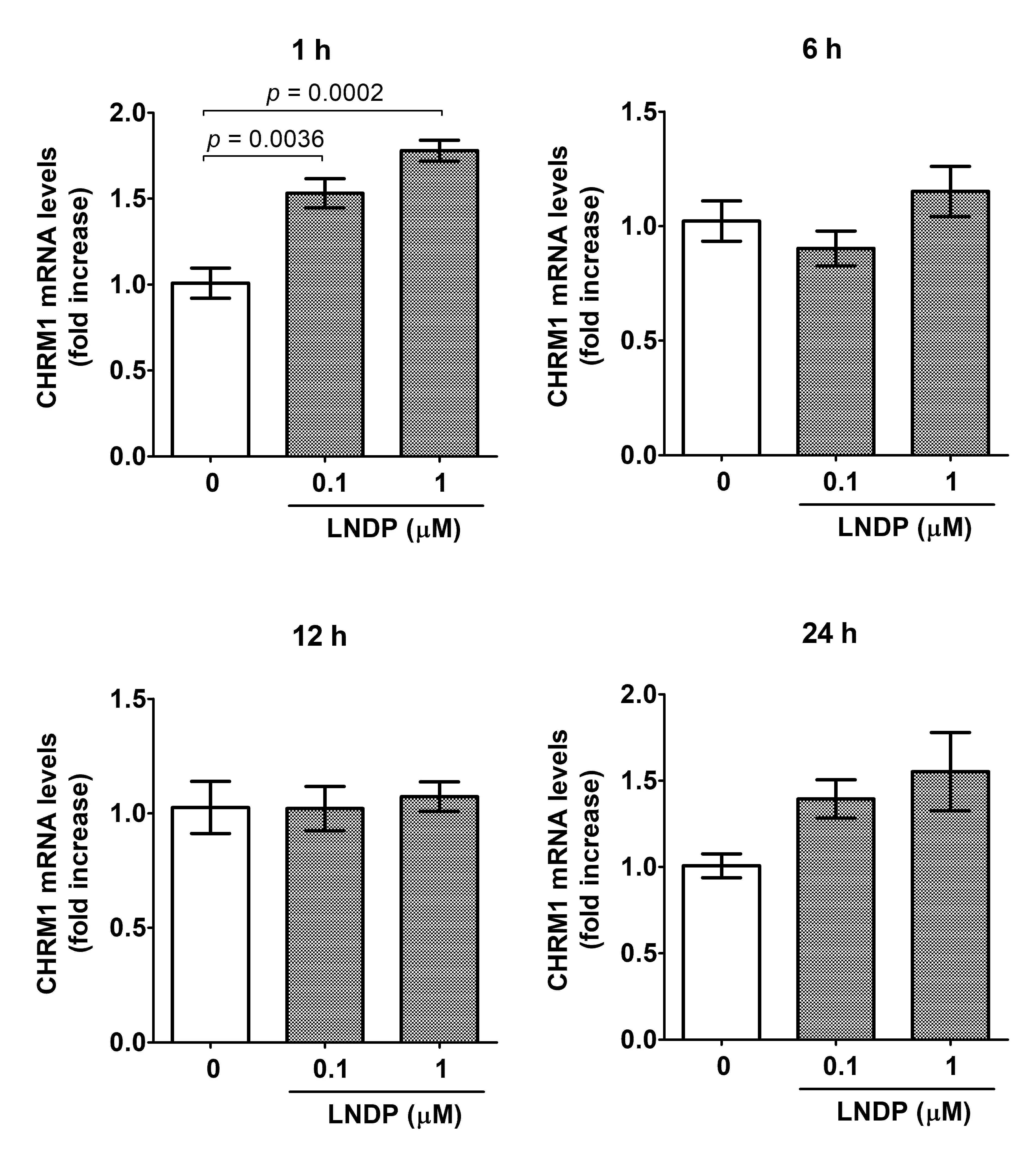
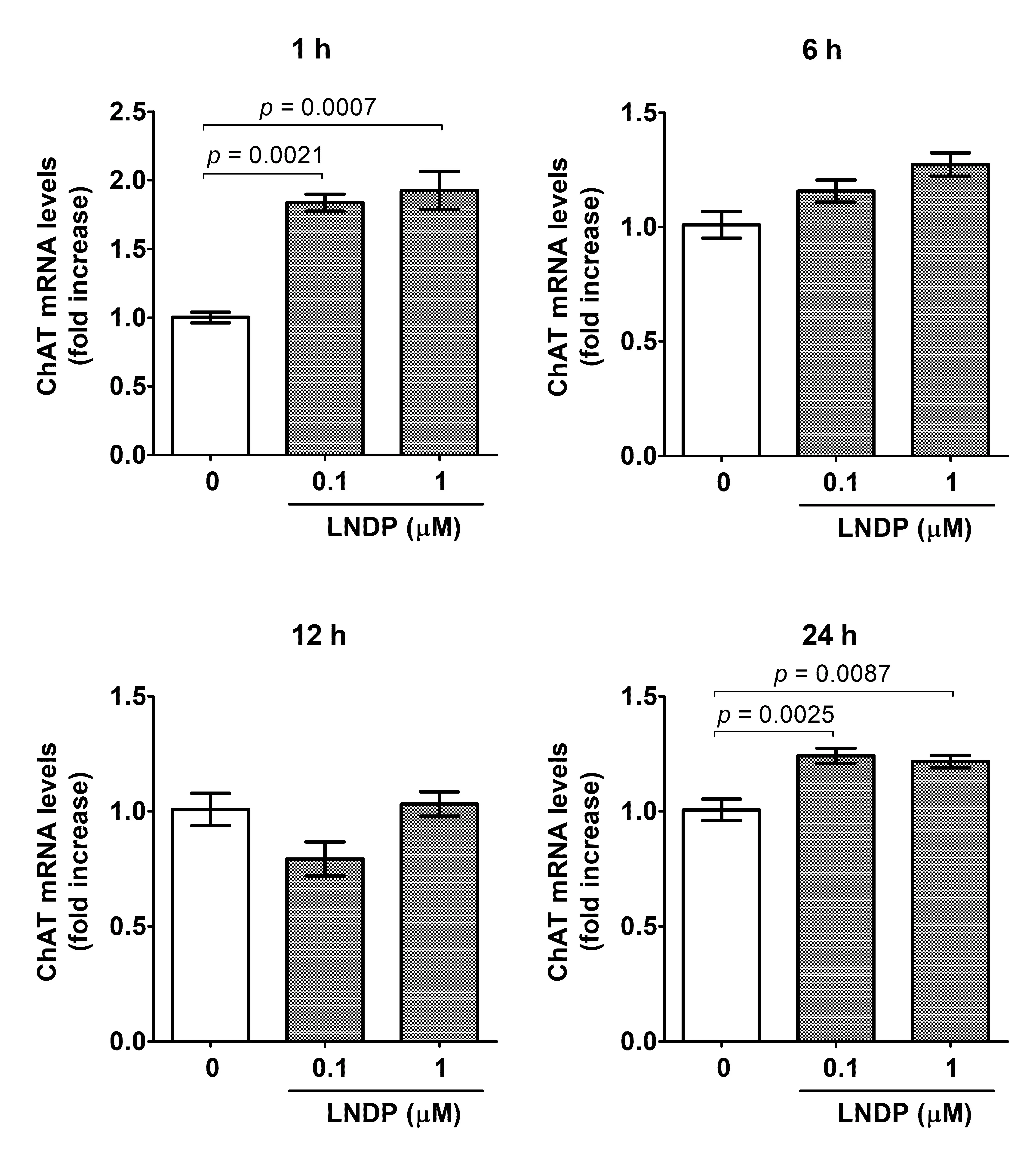
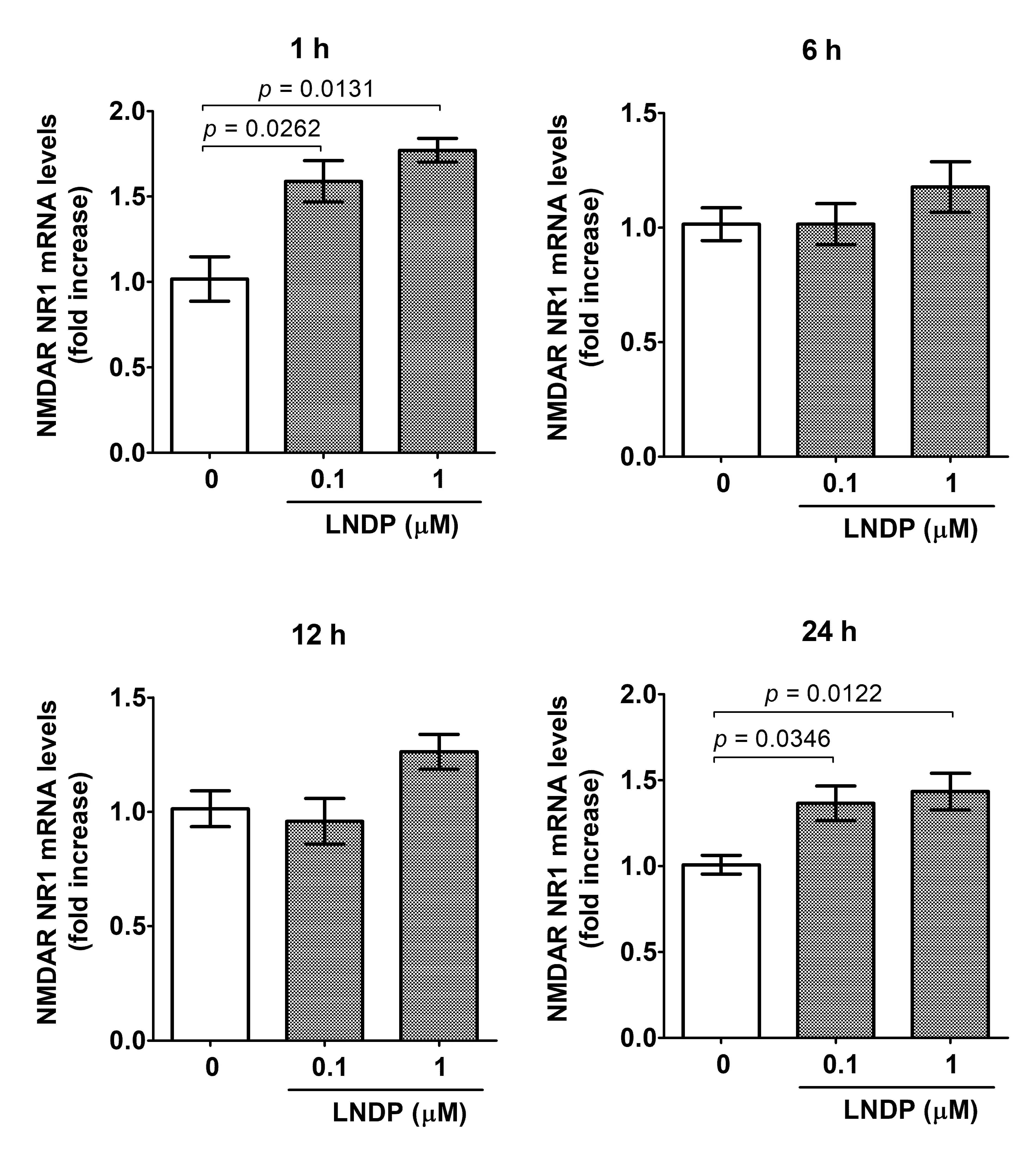
We treated hiPSC-derived neurons with 0.1 and 1 μM LNDP for 1, 6, 12, and 24 h, and then performed qRT-PCR to elucidate the effect of LNDP on the expression of memory-related genes in Table 1. The gene expression of neurotrophic factors, BDNF, NGF, and TrkB (receptor for BDNF) in hiPSC-derived neurons was analyzed (Figure 2). The BDNF expression levels began to increase at 1 h after LNDP administration compared to the vehicle control (Figure 2A). Then, the significant increases peaked at 24 h after the treatment with 0.1 and 1 μM LNDP, which were approximately 2.7- and 3.9-fold (p = 0.0038 and 0.0002), respectively, of the vehicle control. The NGF gene expression levels were also increased 2.9-fold (vs. vehicle control, p = 0.0036 and 0.0044) at 1 h by 0.1 and 1 μM LNDP, and then returned to baseline after 6–12 h (Figure 2B). Interestingly, after 24 h, the NGF gene expression again increased to 2.5- and 2.3-fold (p = 0.0010 and 0.0015), respectively, by 0.1 and 1 μM LNDP. The TrkB gene expression was also significantly upregulated by 1.6- and 2.0-fold (p = 0.0023 and <0.0001), respectively, at 1 h after 0.1 and 1 μM LNDP. At 24 h, both concentrations of LNDP induced a significant (1.4-fold, p = 0.0017 and 0.0041) increase in TrkB gene expression (Figure 2C). Next, we examined the mRNA expression of other memory-related factors, specifically CHRM1, ChAT, and NMDAR NR1 (Figure 3). LNDP treatment was found to upregulate these cholinergic and NMDAR-related genes at several time points tested. The CHRM1 gene expression was significantly increased 1.5- and 1.8-fold (p = 0.0036 and 0.0002), respectively, after 1 h of treatment with 0.1 and 1 μM LNDP (Figure 3A), and gradually returned to control levels at 6 and 12 h. Similarly, ChAT expression was significantly upregulated by 1.8- and 1.9-fold at 1 h (p = 0.0021 and 0.0007) and by 1.2- and 1.2-fold at 24 h (p = 0.0025 and 0.0087), respectively, after treatment with 0.1 and 1 μM LNDP (Figure 3B). The gene expression of NMDAR NR1 was also significantly increased by 1.6- and 1.8-fold at 1 h (p = 0.0262 and 0.0131) and by 1.4- and 1.4-fold at 24 h (p = 0.0346 and 0.0122), respectively, after treatment with 0.1 and 1 μM LNDP (Figure 3C).
 |
 |
|
(A) |
(B) |
 |
|
|
(C) |
|
Figure 2. Effects of LNDP on the mRNA expression of neurotrophic factors and their receptor. qRT-PCR was performed to analyze (A) BDNF, (B) NGF, and (C) TrkB mRNA levels in hiPSC-derived neurons after treatment with vehicle control, 0.1 μM LNDP, or 1 μM LNDP for 1, 6, 12, or 24 h. Data were normalized to ACTB mRNA levels and expressed as relative fold change compared to the vehicle control at each time point. Bars represent the mean ± S.E.M. (n = 3–6). Statistical significance was determined by one-way ANOVA followed by Tukey’s multiple comparison test. Exact p values were indicated above the brackets for relevant comparisons; non-significant comparisons (p ≥ 0.1) were omitted for clarity.
 |
 |
|
(A) |
(B) |
 |
|
|
(C) |
|
Figure 3. Effects of LNDP on the mRNA expression of cholinergic and NMDAR-related genes. qRT-PCR was performed to analyze (A) CHRM1, (B) ChAT, and (C) NMDAR NR1 mRNA levels in hiPSC-derived neurons after treatment with vehicle control, 0.1 μM LNDP, or 1 μM LNDP for 1, 6, 12, or 24 h. Data were normalized to ACTB mRNA levels and expressed as relative fold change compared to the vehicle control at each time point. Bars represent the mean ± S.E.M. (n = 3–6). Statistical significance was determined by one-way ANOVA followed by Tukey’s multiple comparison test. Exact P values were indicated above the brackets for relevant comparisons; non-significant comparisons (p ≥ 0.1) were omitted for clarity.
4. Discussion
The number of patients with neurodegenerative diseases, particularly AD, is rapidly increasing worldwide. Previous preclinical and clinical studies have revealed that LNDP-containing foods improve cognitive and memory functions [10,11,12,13,14], but the underlying neuronal mechanisms remain unclear. The present study has shown that LNDP significantly upregulates the gene expression of multiple factors critical for neuroplasticity and cholinergic signaling in a time-dependent manner, in hiPSC-derived neurons which reflect human neuronal characteristics. LNDP was shown to be non-cytotoxic in PC12 cells, indicating that significant upregulation of gene expression may reflect genuine neuronal activation rather than stress responses. A key finding was the robust induction of BDNF gene expression, which reached about four-fold at 24 h after the LNDP treatment (Figure 2A). Given the central role of BDNF in synaptic plasticity and memory consolidation [2], this result suggests that LNDP activates transcriptional pathways important for long-term neural adaptation. The NGF gene expression showed a transient rise at 1 h after the treatment and a second increase at 24 h (Figure 2B). This early transcriptional surge at 1 h, which was also observed for TrkB and CHRM1, suggests an immediate-early response to LNDP treatment that potentially primes neural networks for subsequent activation. On the other hand, the delayed but robust increases observed at 24 h for genes such as BDNF, NGF, TrkB, and ChAT point toward secondary or downstream regulatory pathways, which are characteristic of long-term neural adaptation and cumulative trophic support.
Notably, significant reductions in CHRM1 protein levels have been reported in the hippocampus and temporal cortex of patients with dementia [7]. LNDP also enhanced the gene expression of CHRM1 and ChAT, both of which are key components of the cholinergic system crucial for learning and memory functions (Figure 3A,B). In addition, LNDP upregulated gene expression of TrkB, the receptor for BDNF (Figure 2C), suggesting amplification of BDNF-TrkB signaling, a pathway essential for synaptic plasticity and neuronal survival. This LNDP-mediated transcriptional regulation likely involves the activation of upstream signaling pathways, such as the PKA/MEK/ERK/CREB cascade, which is well-known to drive neurotrophic factor expression. Interestingly, LNDP also increased the gene expression of NMDAR NR1 (Figure 3C), indicating the possible enhancement of NMDAR-mediated synaptic plasticity, which underlies learning and memory [5,6]. NR1 is a key subunit of the NMDAR, which is essential for synaptic plasticity and memory formation [5,6]. This finding suggests that LNDP may enhance not only the cholinergic system but also the glutamatergic system in human neurons. This provides a new insight into the molecular mechanisms of LNDP, as it may directly support the basic mechanisms of learning and memory.
Regarding the bioavailability of LNDP, Ohsawa et al. [11] previously suggested that a 19-mer peptide might be too long to be absorbed into the systemic circulation in large amounts. Remarkably, a recent study by Nakatani et al. [19] demonstrated that LNDP exhibits extraordinary resistance to gastrointestinal enzymes, retaining over 90% stability after exposure to pepsin and pancreatin. Furthermore, they revealed that LNDP successfully traverses the human intestinal epithelium in its intact form via an active transport pathway mediated by peptide transporter 1. These robust findings strongly support the physiological relevance of LNDP as an orally active functional food ingredient that reaches systemic circulation.
Provided that LNDP or its active forms circulate systemically, they possess the potential to directly modulate human neuronal gene expression upon gaining access to the central nervous system. While our current results clearly show that LNDP can directly act on hiPSC-derived neurons in vitro, future in vivo studies are required to fully elucidate the exact blood–brain barrier (BBB) penetration and transport mechanisms into the brain parenchyma.
We acknowledge that the present study primarily focuses on transcriptional modulation (mRNA expression) and lacks detailed protein-level validations or functional physiological assays. However, as demonstrated in our previous work profiling bioactive candidate compounds [17], profiling early transcriptional responses in hiPSC-derived neurons is a valuable, well-established screening approach for identifying the initial molecular targets of neuroprotective factors. Protein-level validations, including evaluation of synaptic plasticity markers such as PSD-95 and MAP2, as well as functional neuronal assays (e.g., calcium imaging or electrophysiological measurements), remain important limitations to be addressed in future investigations.
5. Conclusions
In conclusion, the current study provides the first in vitro molecular evidence that LNDP modulates the gene expression of critical factors related to neuroplasticity and cholinergic signaling in hiPSC-derived neurons. Although further in vivo validation and functional physiological assays are required, these findings suggest that LNDP exhibits a potential neuroprotective profile at the transcriptional level, supporting its further evaluation as a functional food ingredient for the prevention of cognitive decline. Therefore, our future research will focus on validating these transcriptional changes at the protein level, performing functional neuronal assays such as calcium imaging, and clarifying the in vivo transport kinetics across the BBB to fully elucidate the neuroprotective mechanisms of LNDP.
Statement of the Use of Generative AI and AI-Assisted Technologies in the Writing Process
During the preparatrion of this manuscdript, the authors used Gemini (Google) in order to refine the English phrasing and improve the overall clarity of the text and response letters. After using this tool/service, the authors reviewed and edited the content as needed and take full responsibility for the content of the published article.
Author Contributions
Study design and concept, J.K., F.N., K.O. and S.Y. Data collection, J.K., A.K., K.S. and S.Y. Data analysis, J.K., A.K., K.S. and Y.K. All authors contributed to preparing the draft and gave final approval of the manuscript for publication. All authors have read and agreed to the published version of the manuscript.
Ethics Statement
Not applicable.
Informed Consent Statement
Not applicable.
Data Availability Statement
The original contributions presented in this study are included in the article. Further inquiries can be directed to the corresponding author.
Funding
This study was supported by Asahi Group Holdings, Ltd. (Tokyo, Japan).
Declaration of Competing Interest
A.K., K.S., F.N. and K.O. are employed by Asahi Quality & Innovations, Ltd., a group company under Asahi Group Holdings, Ltd. This affiliation may be considered a potential conflict of interest. The company has no role in data collection or data analysis.
References
- Scheltens P, De Strooper B, Kivipelto M, Holstege H, Chételat G, Teunissen CE, et al. Alzheimer’s disease. Lancet 2021, 397, 1577–1590. DOI:10.1016/S0140-6736(20)32205-4 [Google Scholar]
- Kowiański P, Lietzau G, Czuba E, Waśkow M, Steliga A, Moryś J. BDNF: A key factor with multipotent impact on brain signaling and synaptic plasticity. Cell Mol. Neurobiol. 2018, 38, 579–593. DOI:10.1007/s10571-017-0510-4 [Google Scholar]
- De Sousa Fernandes MS, Ordônio TF, Santos GCJ, Santos LER, Calazans CT, Gomes DA, et al. Effects of physical exercise on neuroplasticity and brain function: a systematic review in human and animal studies. Neural Plast. 2020, 2020, 8856621. DOI:10.1155/2020/8856621 [Google Scholar]
- Hozumi S, Nakagawasai O, Tan-No K, Niijima F, Yamadera F, Murata A, et al. Characteristics of changes in cholinergic function and impairment of learning and memory-related behavior induced by olfactory bulbectomy. Behav. Brain Res. 2003, 138, 9–15. DOI:10.1016/S0166-4328(02)00183-3 [Google Scholar]
- Riedel G, Platt B, Micheau J. Glutamate receptor function in learning and memory. Behav. Brain Res. 2003, 140, 1–47. DOI:10.1016/S0166-4328(02)00272-3 [Google Scholar]
- Hansen KB, Yi F, Perszyk RE, Furukawa H, Wollmuth LP, Gibb AJ, et al. Structure, function, and allosteric modulation of NMDA receptors. J. Gen. Physiol. 2018, 150, 1081–1105. DOI:10.1085/jgp.201812032 [Google Scholar]
- Sabbir MG, Speth RC, Albensi BC. Loss of cholinergic receptor muscarinic 1 (CHRM1) protein in the hippocampus and temporal cortex of a subset of individuals with Alzheimer’s disease, Parkinson’s disease, or frontotemporal dementia: Implications for patient survival. J. Alzheimers Dis. 2022, 90, 727–747. DOI:10.3233/JAD-220766 [Google Scholar]
- Scott-Solomon E, Kuruvilla R. Mechanisms of neurotrophin trafficking via Trk receptors. Mol. Cell Neurosci. 2018, 91, 25–33. DOI:10.1016/j.mcn.2018.03.013 [Google Scholar]
- Tamime AY. Fermented milks: a historical food with modern applications—A review. Eur. J. Clin. Nutr. 2002, 56, S2–S15. DOI:10.1038/sj.ejcn.1601657 [Google Scholar]
- Ohsawa K, Uchida N, Ohki K, Nakamura Y, Yokogoshi H. Lactobacillus helveticus-fermented milk improves learning and memory in mice. Nutr. Neurosci. 2015, 18, 232–240. DOI:10.1179/1476830514Y.0000000122 [Google Scholar]
- Ohsawa K, Uchida N, Ohki K, Yokogoshi H. Identification of peptides present in sour milk whey that ameliorate scopolamine-induced memory impairment in mice. Int. J. Food Sci. Nutr. 2018, 69, 33–45. DOI:10.1080/09637486.2017.1324564 [Google Scholar]
- Ohsawa K, Nakamura F, Uchida N, Mizuno S, Yokogoshi H. Lactobacillus helveticus-fermented milk containing lactononadecapeptide (NIPPLTQTPVVVPPFLQPE) improves cognitive function in healthy middle-aged adults: A randomised, double-blind, placebo-controlled trial. Int. J. Food Sci. Nutr. 2018, 69, 369–376. DOI:10.1080/09637486.2017.1365824 [Google Scholar]
- Sasai M, Kato M, Ohsawa K, Sashihara K, Nakamura Y, Kaneko T. Effects of a single dose of tablets containing lactononadecapeptide on cognitive function in healthy adults: A randomized, double-blind, cross-over, placebo-controlled trial. Biosci. Biotechnol. Biochem. 2021, 85, 948–956. DOI:10.1093/bbb/zbaa117 [Google Scholar]
- Yamada S, Shirai M, Nakamura F, Ohsawa K, Watanabe M, Koga M. Beneficial effects of food containing lactononadecapeptide on memory function in elderly Japanese subjects–A randomized, double-blind, placebo-controlled study. Food Res. Suppl. 2026, 1, 10006. DOI:10.70322/frs.2026.10006 [Google Scholar]
- Benito E, Barco A. CREB’s control of intrinsic and synaptic plasticity: implications for CREB-dependent memory models. Trends Neurosci. 2010, 33, 230–240. DOI:10.1016/j.tins.2010.02.001 [Google Scholar]
- Kimura J, Shimizu K, Takito J, Nemoto K, Degawa M, Yokosuka A, et al. Nobiletin reduces intracellular and extracellular β-amyloid in iPS cell-derived Alzheimer’s disease model neurons. Biol. Pharm. Bull. 2018, 41, 451–457. DOI:10.1248/bpb.b17-00364 [Google Scholar]
- Kimura J, Shimizu K, Takito J, Nemoto K, Degawa M, Yokosuka A, et al. Upregulatory effects of nobiletin, a citrus flavonoid with anti-dementia activity, on the gene expression of mAChR, ChAT, and CBP. Planta Med. Lett. 2015, 2, e12–e14. DOI:10.1055/s-0035-1545937 [Google Scholar]
- Livak KJ, Schmittgen TD. Analysis of relative gene expression data using real-time quantitative PCR and the 2−ΔΔCT method. Methods 2001, 25, 402–408. DOI:10.1006/meth.2001.1262 [Google Scholar]
-
Nakatani E, Sasai M, Miyazaki H, Tanaka S, Hirota T, Okura T. Investigating the transepithelial transport and enzymatic stability of lactononadecapeptide (NIPPLTQTPVVVPPFLQPE), a 19-amino acid casein-derived peptide in Caco-2 cells. J. Agric. Food Chem. 2024, 72, 12719–12724. DOI:10.1021/acs.jafc.4c00974 [Google Scholar]