
Micropropagation by Axillary Budding of Ornamental Camellia Species: A Case Study of Camellia japonica and Camellia reticulata
Micropropagation by Axillary Budding of Ornamental Camellia Species: A Case Study of Camellia japonica and Camellia reticulata

Received: 18 April 2026 Revised: 09 June 2026 Accepted: 22 June 2026 Published: 26 June 2026

© 2026 The authors. This is an open access article under the Creative Commons Attribution 4.0 International License (https://creativecommons.org/licenses/by/4.0/).
1. Introduction
Camellia is the most economically important genus in the family Theaceae, which comprises numerous tropical and subtropical trees and shrubs. The genus Camellia is native to East Asia, but has been introduced to Europe, North America, Australia, and New Zealand. At present, the genus is believed to comprise more than 300 species [1], of which the most important are C. sinensis, C. japonica, C. sasanqua, C. reticulata, and C. oleifera.
The economic importance of the genus Camellia is largely attributed to C. sinensis, the young leaves of which are used to make tea. Among the wild species, C. japonica is economically the most important because of its attractive ornamental flowers, and more than 32,000 cultivars are now available worldwide. Other species used as ornamentals include C. reticulata, C. sasanqua, and C. saluensis [2].
In the last years, various diseases have affected camellia, among which the one caused by Ciborinia camelliae is considered particularly significant. This relatively recent disease targets only the flowers, causing petal spots, wilting, and ultimately flower drop. It poses one of the most serious threats to camellia cultivation because it damages the plant’s most distinctive feature, which underpins its ornamental value. Despite extensive efforts, no fully effective method has yet been developed to control this disease [3,4]. Consequently, methods for the propagation and conservation based on in vitro culture of the most valuable genotypes have become increasingly important.
We focus this report on the propagation of two important cultivars of Camellia: C. japonica ‘Alba Plena’ and C. reticulata ‘Captain Rawes’, both of great ornamental value [5]. Propagation of these cultivars and other species of Camellia by conventional methods is difficult and slow, and other means of propagation have been investigated. In the late 1970s, the use of biotechnological methods, specifically in vitro or tissue culture techniques, was suggested as a means of solving the constraints in propagation, mainly due to slow rooting of plant cuttings [6]. Micropropagation plays a key role in plant breeding by allowing the fast production of selected plant varieties. Among micropropagation techniques, axillary shoot proliferation from cultured meristems is most widely used, as it ensures genetic stability and can be readily applied to many plant species [7]. A successful tissue culture based in vitro propagation method would enable rapid, large-scale multiplication of selected, horticulturally superior cultivars and could also facilitate breeding programs. In addition, these protocols contribute to the conservation of valuable germplasm, the recovery of disease-free plant material, and the long-term sustainability of economically and ornamentally important species [8,9]. Furthermore, optimized micropropagation protocols are crucial for the success of cryopreservation and genetic transformation techniques, which complement conventional breeding strategies [10].
This review summarizes current protocols for in vitro propagation via axillary budding, aiming to support large-scale production, germplasm preservation, and horticultural improvement of these two valuable ornamental species.
2. Micropropagation of Camellia japonica
Camellia japonica was first cultivated in European gardens in the early eighteenth century. The earliest documented plants arrived in England around 1739, notably in the garden of Lord Petre in Essex. However, it was not until 1792 that Captain Connor, of the East India Company merchant vessel Carnatic, introduced some of the most successful cultivars of C. japonica, including ‘Alba Plena’ and ‘Variegata’, to England. The cultivar ‘Alba Plena’ is considered one of the original camellia varieties recognized in modern horticulture. It was first described in The Botanical Repository in 1797 and again in the 1812 edition of the same publication. This camellia produces a fully double, symmetrically imbricated (formal double), medium- to large-sized white flower lacking visible stamens and opening in a flat form (Figure 1A). The flower measures approximately 10 cm in diameter and 3.5 cm in depth. The petals gradually decrease in size toward the center. The plant is an evergreen shrub or small tree characterized by glossy, dark green, leathery leaves with serrated margins and a compact growth habit. Due to its ornamental value, adaptability, and wide range of flower forms and colors, C. japonica has become one of the most widely cultivated ornamental species in temperate regions worldwide.
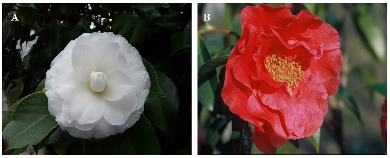
Figure 1. (A). Flower of Camellia japonica cv ‘Alba Plena’. (B) Flower of Camellia reticulata cv ‘Captain Rawes’.
2.1. Initiation
Camellia japonica ‘Alba Plena’ was established and multiplied in vitro by using material from a 50-year-old tree chosen for the attractiveness of its flowers [11]. The ability to propagate plantlets from mature trees is especially desirable, as elite traits are best assessed when the tree is at an advanced age [12]. One-year-old branches bearing vegetative buds were cut from the top of the tree and placed in a growth chamber to force development of new shoots. The new shoots (20–40 mm long) were sterilized with 5% calcium hypochlorite, and the terminal shoot tips and nodes (5 mm long) were excised and placed in test tubes containing 15 mL of culture medium. The basal medium used for initial cultures was Heller medium [13] with the concentration of all macronutrients increased by a factor of 1.25 and the inclusion of 1 mM (NH4)2SO4 (Hmod). The medium was also supplemented with 1 mg/L 6-benzyladenine (BA), 0.01 mg/L indole-3-butyric acid (IBA), 30 g/L sucrose, and 6 g/L agar. The pH of the medium was adjusted to 5.5–5.6 before autoclaving. The initial cultures were transferred to fresh medium every 4 weeks, and three such transfers to fresh medium of the same composition were necessary to obtain shoot cultures suitable for multiplication. In all experiments, the cultures were grown under a 16 h photoperiod (25 °C day/18 °C night).
2.2. Multiplication
To improve shoot proliferation of ‘Alba Plena’ cultivar, the effect of two factors was investigated during the shoot multiplication stage:
- (a)
-
Culture medium: Six macronutrient formulations were evaluated: Hmod, Gresshoff and Doy medium (GD) [14], Murashige and Skoog medium (MS) [15], half-strength MS (½ MS), Woody Plant Medium (WPM) [16], and Anderson medium [17]. All media were supplemented with 2 mg/L BA, 2 mg/L N6-(Δ2-isopentenyl)adenine (2iP), 2 mg/L zeatin (Z), and 0.01 mg/L IBA, as well as 30 g/L sucrose and 6 g/L agar. After four weeks, the cultures were transferred to fresh medium of the same composition, except that the concentrations of the BA, Z, and 2 iP were reduced to 1 mg/L. Transfer to fresh medium every four weeks was compared with replenishing each tube of the existing medium with 1.5 mL of fresh liquid medium of the same composition as the transfer medium.
- (b)
-
Type of explant: Three types of explant were distinguished: shoot tips of harvested shoots longer than 13–14 mm (ST1), nodal segments constituting the lower portions of the shoots from which the shoot tips were taken (NS), and whole harvested shoots measuring 5–10 mm in length (ST2).
Multiplication rates were highest for ST2 and NS explants, which reached lengths sufficient to allow direct use for rooting without further elongation (Figure 2A). The highest multiplication and elongation rates were obtained on WPM and Hmod media. Shoot length was generally improved by replenishing the medium after four weeks rather than transferring the cultures to fresh agar-solidified medium. Recent studies have expanded the micropropagation of C. japonica through axillary shoot culture in several ornamental cultivars, including ‘Reine des Beautés’, ‘Lelie’, and ‘David Bocchi’. A notable development in these protocols has been the successful application of thidiazuron (TDZ), a highly active cytokinin-like growth regulator that has significantly improved shoot proliferation compared with conventional cytokinin treatments. Malyarovskaya and Samarina [18] reported that the highest shoot proliferation rates in these cultivars were achieved on WPM medium supplemented with 2.0 mg/L TDZ, 0.5 mg/L kinetin, and 1.0 mg/L gibberellic acid (GA3). More recently, Kim et al. [19] established axillary shoot cultures from seedling-derived explants and demonstrated that MS medium containing only 0.5 mg/L TDZ produced the highest number of multiple shoots.
Table 1. Culture media used for in vitro establishment, shoot proliferation, and rooting of axillary buds from adult explants of Camellia japonica and Camellia reticulata.
|
Species/Cultivar |
Establishment Medium |
Proliferation Medium |
Rooting Medium |
|---|---|---|---|
|
Camellia japonica ‘Alba Plena’ |
Hmod with 1 mg/L BA + 0.01 mg/L IBA |
WPM or Hmod media with 2 mg/L BA + 2 mg/L 2iP + 2 mg/L Z + 0.01 mg/L IBA |
Dipping 1 g/L IBA solution for 15 min → ½WPM 12 days darkness |
|
Camellia reticulata ‘Captain Rawes’ |
Hmod with 2 mg/L BA + 2 mg/L 2iP + 2 mg/L Z + 0.01 mg/L IBA |
WPM with 2 mg/L BA + 2 mg/L 2iP + 2 mg L−1 Z + 0.01 mg/L IBA |
Dipping 1 g/L IBA solution for 30 min → ½WPM |
BA: 6-benzyladenine; 2iP: N6-(Δ2-isopentenyl)adenine; Z: zeatin; IBA: indole-3-butyric acid; WPM: Woody Plant Medium; Hmod: modified Heller medium; ½WPM: half-strength Woody Plant Medium.
2.3. Rooting
For rooting, dipping the base of the shoots Camellia japonica ‘Alba Plena’ in a 1 g/L IBA solution for 15 min, followed by incubation for 12 days in darkness in half-strength WPM (½ WPM), resulted in optimal rooting (Figure 2B), with rates exceeding 85% [11,20]. The type of support (agar or paper bridges) did not significantly affect the rooting percentage or the number of roots formed per rooted shoot; however, liquid medium enhanced root elongation. Although Kim et al. [19] reported rooting rates of up to 66% for shoots of Camellia japonica following pulse treatments with 500 or 1000 mg/L IBA, the shoots used for rooting were derived from seedlings. This is a significant limitation, as the rooting ability of juvenile material is generally much higher than that of mature selected genotypes.
For acclimatization, the highest survival rates were achieved when rooted shoots were transferred to a mix of perlite–peat substrate 12 days after auxin treatment, immediately following the dark incubation period (Figure 2C).
Table 1 presents the main culture media employed throughout the three stages of micropropagation via axillary budding of adult plant material of the cultivar ‘Alba Plena’.
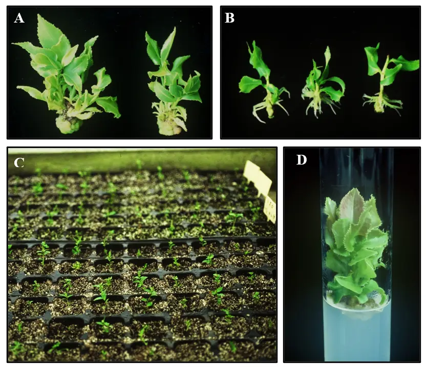
Figure 2. Micropropagation of Camellia japonica ‘Alba Plena’ and Camellia reticulata ‘Captain Rawes’ by axillary budding. (A) Shoot multiplication cultures of C. japonica ‘Alba Plena’. (B) In vitro rooting of C. japonica ‘Alba Plena’ microshoots. (C) Micropropagated camellia plants established under ex vitro conditions. (D) Shots of Camellia reticulata ‘Captain Rawes’ at multiplication stage.
3. Micropropagation of Camellia reticulata ‘Captain Rawes’
Camellia reticulata was recorded in China as early as the reign of the first T’ang emperors, with documented cultivation dating back many centuries. The species has long been valued in Chinese horticulture and culture, particularly in the southwestern province of Yunnan, where it has been cultivated in temple gardens and imperial estates for its striking ornamental qualities [21]. Over time, numerous cultivars have been selected for their large flowers, vibrant colours, and decorative foliage. Among these, several cultivars possess outstanding ornamental value, notably ‘Captain Rawes’, which produces large, semi-double flowers varying in colour from soft pink to deep red (Figure 1B). The blossoms are typically showy, with a well-formed structure and glossy evergreen leaves that enhance its aesthetic appeal in landscape and garden settings. This cultivar was first introduced to England by the sea merchant Captain Rawes, after whom it is named, during the nineteenth century, and it subsequently spread to other European countries, where it became popular among collectors and botanical gardens. Propagation of C. reticulata presents certain horticultural challenges. Many cultivars produce few or no viable seeds, limiting the use of sexual reproduction for commercial multiplication. In addition, vegetative propagation by cuttings is often difficult due to slow and inconsistent rooting responses.
3.1. Initiation
The first protocol for micropropagation of C. reticulata via axillary budding was established by San José and Vieitez [12] and San José et al. [22]. Terminal branches collected from the crown of a 90-year-old tree were placed in a growth chamber to induce bud development. The new shoots that developed were sterilized with hypochlorite as described before, and the apical and nodular segments were used as explants for initiating the cultures. The culture medium consisted of Hmod supplemented with 2 mg/L BA, 2 mg/L 2iP, 2 mg/L Z, 0.01 mg/L IBA, 30 g/L sucrose, and 6 g/L agar. The pH of the medium was adjusted to 5.0 before autoclaving. The initial cultures were transferred to fresh medium every 4 weeks. All explants responded well to culture, and the rates of establishment were higher than 50%. In all experiments, the cultures were grown under a 16 h photoperiod (25 °C day/18 °C night).
3.2. Multiplication
To optimize shoot proliferation, various factors potentially affecting the in vitro response were evaluated:
- (a)
-
Explant type: Three types of explants were used: shoot tips (ST1) from harvested shoots longer than 14–15 mm, nodal segments (NS) from the basal portions of the shoots from which ST1 were excised, and whole harvested axillary shoots 7–10 mm long (ST2).
- (b)
-
Culture medium: Multiplication rates were assessed on three mineral media: WPM, Anderson, and Hmod. All media were supplemented with the previously mentioned growth regulators during the induction stage. After 4 weeks, explants were transferred to fresh medium of the same composition, but with the growth regulator concentrations reduced by half, and cultured for an additional 4 weeks.
- (c)
-
Orientation and reculture: Shoots 15–20 mm in length were excised and placed either vertically or horizontally on the multiplication medium. Axillary shoots developing from horizontally arranged explants were collected after each subculture, and the same horizontal explants could be recultured up to four times on fresh medium, with newly developed shoots harvested before each transfer.
Regardless of the explant type, and similar to C. japonica ‘Alba Plena’, the best results were obtained with NS and ST2, the latter producing the highest multiplication rates. Among the media tested, WPM supported the best shoot growth. The highest multiplication rates were achieved through reculture of horizontally arranged shoots, as reflected by an increase in both the number and length of shoots, particularly during the first two recultures (Figure 2D).
3.3. Rooting
Shoots measuring 15–20 mm, obtained during the multiplication stage, were used to evaluate the effects of various factors on rooting success, including shoot orientation, successive reculturing, type of explant used during multiplication, vessel type, and the type and concentration of sugar (30–60 g/L sucrose or 10–70 g/L glucose) added to the rooting medium [12,22]. Rooting was induced by dipping the basal ends in a 1 g/L IBA solution for 30 min, after which the shoots were transferred to WPM with half-strength macronutrients and no growth regulators. The type of sugar had the most significant effect on both the number of roots formed and the overall rooting percentage. Optimal results were obtained with 30 g/L glucose or 60 g/L sucrose. Shoots derived from horizontally oriented explants exhibited successful rooting in 50% of cases, whereas only 9% of shoots from vertically oriented explants rooted successfully. Ultimately, higher rooting rates were achieved in jars compared to tubes. The in vitro–produced plants were readily acclimatized, with survival rates exceeding 50%. Table 1 presents the principal culture media employed across the three stages of micropropagation via axillary budding of adult plant material derived from the cultivar ‘Captain Rawes’.
4. Medium Term Conservation of Camellia Germplasm
Axillary shoot culture combined with reduced-growth strategies represents an effective approach for the medium-term conservation of plant germplasm [23,24]. In camellia, shoot cultures can be preserved under cold storage as a medium-term conservation strategy, as demonstrated by Ballester et al. [25]. They reported that in vitro shoot cultures from eight clones of C. japonica and C. reticulata could be maintained at 2–4 °C for up to 12 months, confirming the suitability of cold storage for medium-term in vitro conservation. Survival rates close to 100% were achieved during the first or second subculture following storage in seven of the eight clones tested, whereas the remaining clone could not be successfully stored for more than 6 months. Several of these clones have since been maintained in vitro using this cold storage approach for more than 25 years, with the storage interval extended to as long as 18 months. In vitro slow-growth techniques are routinely used for the medium-term conservation of numerous woody species, including chestnut [26], black alder [24], or different oak species [23,26,27].
5. Conclusions
This review paper focuses on the application of micropropagation by axillary budding for the rapid and efficient propagation of two ornamental Camellia cultivars, C. reticulata ‘Captain Rawes’ and C. japonica ‘Alba Plena’, both highly valued for their aesthetic qualities. Among the available biotechnological approaches, micropropagation has gained increasing importance due to its ability to clonally reproduce selected Camellia genotypes with high genetic fidelity. Furthermore, these techniques are essential for advanced applications in the conservation of elite germplasm, particularly in supporting long-term preservation, germplasm exchange, and the development of improved horticultural traits. Their relevance is especially significant in the current context, as Camellia populations are being severely affected by Ciborinia infections, which threaten plant health, flowering capacity, and the sustainability of valuable ornamental cultivars.
Statement of the Use of Generative AI and AI-Assisted Technologies in the Writing Process
Generative AI tools were used solely to assist in refining the academic language and improving the clarity of the manuscript. The authors independently conducted all research activities, including study design, data collection, analysis, and interpretation. The authors take full responsibility for the scientific integrity, validity, and originality of the work.
Acknowledgements
The authors sincerely thank Ana Mª Vieitez for her invaluable guidance and for sharing her enthusiasm for the micropropagation of Camellia.
Author Contributions
M.T.M. and J.L.C.: Investigation, Writing—review and editing; E.C.: Project administration, Supervision, Funding acquisition, Conceptualization and Writing—review.
Ethics Statement
Not applicable.
Informed Consent Statement
Not applicable.
Data Availability Statement
Not applicable.
Funding
This work was partially supported by the Ministerio de Ciencia, Innovación y Universidades (MICIN, Spain) through project PID2024-156422OB-C33.
Declaration of Competing Interest
The authors declare that they have no known competing financial interests or personal relationships that could have appeared to influence the work reported in this paper.
References
-
Izco J. Camellias: Diversity and globalization processes. In Proceedings of the International Camellia Congress, Pontevedra, Spain, 11–15 March 2014; pp. 9–16. [Google Scholar]
-
San José MC, Couselo JL, Martínez TM, Mansilla P, Corredoira E. Somatic embryogenesis in Camellia japonica L.: Challenges and future prospects. In Somatic Embryogenesis in Ornamentals and Its Applications; Mujib A, Ed.; Springer: Pontevedra, India; 2016; pp. 91–105. [Google Scholar]
-
Couselo JL, Vela P, Salinero C, Mansilla P. Limiting factors for the biological and chemical control of camellia flower blight. In Proceedings of the International Camellia Congress, Pontevedra, Spain, 11–15 March 2014; pp. 266–279. [Google Scholar]
-
Saracchi M, Locati D, Colombo EM, Pasquali M. Updates on Ciborinia camelliae, the causal agent of camellia flower blight. J. Plant Pathol. 2019, 101, 215–223. DOI:10.1007/s42161-018-0173-0 [Google Scholar]
-
Vieitez AM, Vieitez ML, Ballester A, Vieitez E. Micropropagation of Camellia spp. In High-Tech and Micropropagation III.; Bajaj YPS, Ed.; Springer: Berlin/Heidelberg, Germany, 1992; Volume 19, pp. 361–387. DOI:10.1007/978-3-662-07770-2_23 [Google Scholar]
-
Mondal TK. Camellia. In Wild Crop Relatives: Genomic and Breeding Resources; Kole C, Ed.; Springer: Berlin/Heidelberg, Germany, 2011; pp. 15–39. [Google Scholar]
-
Piñeiro P, Martínez MT, Sampedro J, Corredoira E. Efficient micropropagation of Argania spinosa through axillary budding. Ind. Crops. Prod. 2026, 241, 122815. DOI:10.1016/j.indcrop.2026.122815 [Google Scholar]
-
Ozudogru EA, Previati A, Lambardi M. In vitro conservation and cryopreservation of ornamental plants. In Protocols for In Vitro Propagation of Ornamental Plants; Jain S, Ochatt S, Eds.; Part of the book series: Methods in Molecular Biology ((MIMB,volume 589)); Humana Press: Totowa, NJ, USA, 2010; pp. 303–324. DOI:10.1007/978-1-60327-114-1_28 [Google Scholar]
-
Mehbub H, Akter A, Akter MA, Mandal MSH, Hoque MA, Tuleja M, et al. Tissue Culture in Ornamentals: Cultivation Factors, Propagation Techniques, and Its Application. Plants 2022, 11, 3208. DOI:10.3390/plants11233208 [Google Scholar]
-
George EF, Hall MA, De Klerk GJ. (Eds.) Micropropagation: Uses and methods. In Plant Propagation by Tissue Culture, 3rd ed.; Springer: Dordrecht, The Netherlands, 2008; pp. 29–64. DOI:10.1007/978-1-4020-5005-3_2 [Google Scholar]
-
Vieitez AM, Barciela J, Ballester A. Propagation of Camellia japonica cv. Alba Plena by tissue culture. J. Hortic. Sci. 1989, 64, 177–182. DOI:10.1080/14620316.1989.11515942 [Google Scholar]
-
San-José MC, Vieitez AM. In vitro regeneration of Camellia reticulata cultivar ‘Captain Rawes’ from adult material. Sci. Hortic. 1990, 43, 155–162. DOI:10.1016/0304-4238(90)90046-H [Google Scholar]
-
Heller R. Recherches sur la nutrition minérale des tissus végétaux cultivés in vitro. Ann. Sci. Nat. Bot. Biol. Veg. 1953, 14, 1–223. [Google Scholar]
-
Gresshoff PM, Doy CH. Development and differentiation of haploid Lycopersicon esculentum. Planta 1972, 107, 161–170. DOI:10.1007/BF00387721 [Google Scholar]
-
Murashige T, Skoog F. A revised medium for rapid growth and bioassays with tobacco tissue culture. Physiol. Plant. 1962, 15, 473–497. DOI:10.1111/j.1399-3054.1962.tb08052.x [Google Scholar]
-
Lloyd G, McCown B. Commercially feasible micropropagation of mountain laurel, Kalmia latifolia, by use of shoot tip cultures. Comb. Proc. Int. Plant. Propag. Soc. 1980, 30, 421–427. Available online: https://cir.nii.ac.jp/crid/1572543025024601088?lang=en&utm_source=chatgpt.com (accessed on 21 June 2026).
-
Anderson WC. A revised tissue culture medium for shoot multiplication of Rhododendron. J. Am. Soc. Hortic. Sci. 1984, 109, 343–347. DOI:10.21273/JASHS.109.3.343 [Google Scholar]
-
Malyarovskaya V, Samarina L. In vitro morphogenesis of ornamental shrubs Camellia japonica and Hydrangea macrophylla. Plant Tissue Cult. Biotechnol. 2017, 27, 181–187. DOI:10.3329/ptcb.v27i2.35023 [Google Scholar]
-
Kim DH, Park KB, Cha SA, Lee JH, Kim SA, Jeong J, et al. Optimization and integrated efficiency assessment of in vitro mass micropropagation for Camellia japonica L. J. Agric. Life Sci. 2025, 59, 49–57. DOI:10.14397/jals.2025.59.6.49 [Google Scholar]
-
Vieitez AM, San-José MC, Ballester A. Progress towards clonal propagation of Camellia japonica cv. Alba Plena by tissue culture techniques. J. Hortic. Sci. 1989, 64, 605–610. DOI:10.1080/14620316.1989.11515997 [Google Scholar]
-
Wu Z, Raven PH, Hong D. (Eds.) Flora of China; Science Press: Beijing, China; Missouri Botanical Garden Press: St. Louis, MO, USA. 2007; Volume 12, pp. 1–534. [Google Scholar]
-
San-José MC, Vidal N, Vieitez AM. Improved efficiency of in vitro propagation of Camellia reticulata cv. Captain Rawes. J. Hortic. Sci. 1991, 66, 755–762. DOI:10.1080/00221589.1991.11516208 [Google Scholar]
-
Corredoira E, Martínez MT, Cernadas MJ, San José MC. Application of Biotechnology in the Conservation of the Genus Castanea. Forests 2017, 8, 394. DOI:10.3390/f8100394 [Google Scholar]
-
San José MC, Janeiro LV, Corredoira E. Simple strategy for the in vitro conservation of Alnus glutinosa (L.) Gaertn. germplasm. Trees 2015, 29, 539–549. DOI:10.1007/s00468-014-1133-8 [Google Scholar]
-
Ballester LV, Janeiro LV, Vieitez AM. Cold storage of shoot cultures and alginate encapsulation of shoot tips of Camellia japonica L. and Camellia reticulata Lindley. Sci. Hortic. 1997, 71, 67–78. DOI:10.1016/S0304-4238(97)00074-5 [Google Scholar]
-
Janeiro LV, Vieitez AM, Ballester A. Cold storage of in vitro cultures of wild cherry, chestnut and oak. Ann. Sci. For. 1995, 52, 287–293. DOI:10.1051/forest:19950309 [Google Scholar]
-
Martínez MT, Vieitez FJ, Solla A, Tapias R, Ramírez-Martín N, Corredoira E. Vegetative Propagation of Phytophthora cinnamomi-Tolerant Holm Oak Genotypes by Axillary Budding and Somatic Embryogenesis. Forests 2020, 11, 841. DOI:10.3390/f11080841 [Google Scholar]