1. Introduction
Investigating the cause and mechanism of death in cases of suspected fatal poisoning with new psychoactive substances (“legal highs”) is no different from classic post-mortem diagnostics in forensic medicine [
1]. There is no characteristic autopsy appearance in individuals poisoned with “legal highs”, therefore, in practice, biological material is most often reserved for complementary histopathological and toxicological examinations.
2. Materials and Methods
This study aimed to assess the usefulness of microscopic examinations in assessing cases of fatal poisoning with new psychoactive substances by analyzing the scientific literature and the results of histopathological examinations of 30 cases of victims of poisoning with “legal highs” from the own practice of the Department of Forensic Medicine and Forensic Medical Toxicology of the Silesian Medical University in Katowice. The age of the autopsied individuals was 31 ± 9 (min. 19, max. 52). There were only 2 women. The detected “legal highs” were: 3-MMC, 4-CEC, 4-CMC, 4-MEAP, α-PVP, amphetamine, ethylbuphedrone, ethylfenidate, N-ethylpentylone, MDMA, methadone, methamphetamine, pentedrone, PMA, PMMA, pseudoephedrine, PV8, UR-144.
A database of histopathological results was searched, and cases in which toxicological examination revealed the presence of new psychoactive substances were selected. Inclusion criteria for cases included: histopathological and toxicological examination, the presence of new psychoactive substances, and the determination of poisoning as the cause of death. Exclusion criteria included the determination of another cause of death and the lack of a histopathological examination.
3. Results
The authors’ analysis of scarce literature data [
1,
2] and the results of histopathological examinations of victims of “legal high” poisoning revealed that the most common pathological or diagnostically questionable changes are observed in the heart (), kidneys (), and the liver ().
Case reports of poisonings usually focus on toxicological findings, with microscopic findings rarely presented. Noteworthy are reports describing liver changes after taking kratom (a psychoactive substance isolated from the plant
Mitragyna speciosa) [
3,
4]. Liver biopsy in these cases revealed changes mimicking primary biliary cholangitis.
The use of fentanyl, which is a worldwide problem, can cause a range of toxic effects in various organs, each with distinct histopathological characteristics. Histological analysis reveals critical evidence of fentanyl’s effects on the following target organs: brain: hypoxia in the hippocampus and cerebellum, neuronal apoptosis, microglia inflammation, altered distribution of microglia, vacuolization and gliosis of the affected regions, lungs: pulmonary edema, amorphous eosinophilic material in the alveolar spaces, intra-alveolar hemorrhage, inflammation of neutrophils, septic embolism, and heart: myocyte necrosis, endocarditis, fibrous connective tissue (myocardium) [
5].
.
Pathological or diagnostically questionable changes observed in the heart [n = 30].
| Type of Change |
Number of Cases [n = 30] |
| Cardiomyocyte fragmentation |
17 |
| Hyperemia |
15 |
| Selye stain positive |
9 |
| Autolysis |
7 |
| Effacement of striations |
7 |
| Increased eosinophilia |
6 |
| Cardiac fibrosis |
6 |
| Cardiomyocyte hypertrophy |
4 |
| Increased amount (proliferation) of fat tissue |
2 |
| Adiposis |
2 |
| Granulocyte inflammatory infiltrates |
2 |
| Lymphocyte inflammatory infiltrates |
2 |
| Large amount of fat tissue between muscle fibers in the right ventricle |
1 |
| Foci of early ischemic changes |
1 |
| Contraction nodes |
1 |
.
Pathological or diagnostically questionable changes observed in the kidneys [n = 30].
| Type of Change |
Number of Cases [n = 30] |
| Hyperemia |
22 |
| Autolysis |
17 |
| Features of acute tubular necrosis (ATN) |
11 |
| Glomerular hyalinization |
1 |
| Inflammatory interstitial infiltrates |
1 |
.
Pathological or diagnostically questionable changes observed in the liver [n = 30].
| Type of Change |
Number of Cases [n = 30] |
| Hyperemia |
15 |
| Autolysis |
9 |
| Mixed steatosis |
6 |
| Hypovolumic degeneration (edema) of hepatocytes |
4 |
| Macrovesicular steatosis |
3 |
| Cholestasis |
3 |
| Fibrosis |
3 |
| Inflammatory infiltrates |
3 |
| Glycogenic degeneration of nuclei |
2 |
| Microvesicular steatosis |
1 |
| Balloon degeneration |
1 |
| Lipofuscin deposits |
1 |
| Hepatocyte necrosis |
1 |
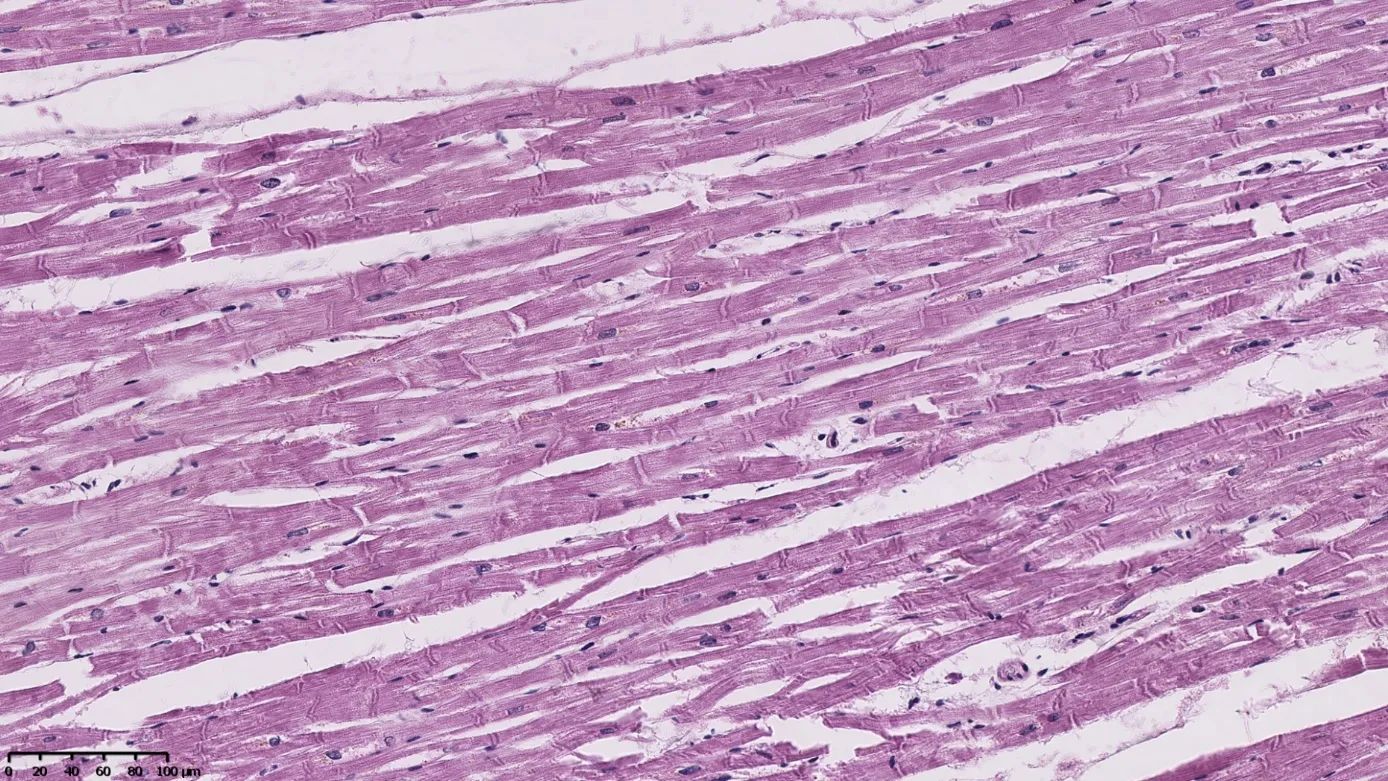
In the heart, signs of early myocardial ischemia are often observed in the absence of atherosclerotic changes in the coronary vessels or changes such as muscle bridging along these vessels (
). Considering the relatively young age of the deceased, it is highly probable that the pathological changes observed are related to the use of “legal highs”, especially given their known cardiotoxicity.
. Cardiac biopsies show extensive areas of prominent contractile nodes in the absence of other morphological features of ischemia. This is an example of excessive adrenergic stimulation due to psychoactive substances or catecholamine release. Hematoxilin & eosin stain, magnification ×200.
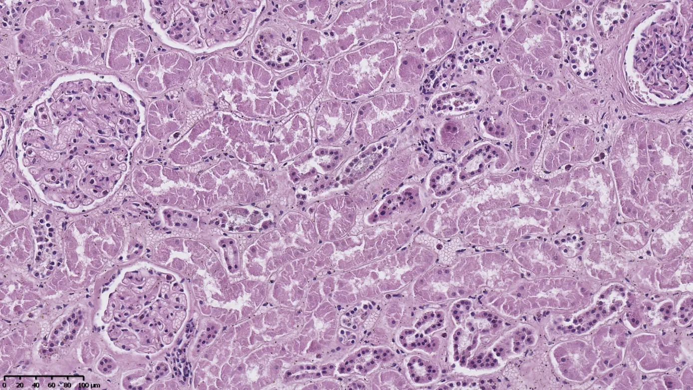
In the kidneys, signs of acute tubular necrosis (ATN) are most frequently seen (
). These signs are usually mild and overlap with autolytic changes, making their assessment difficult, especially since they may be periagonal (artifacts).
. Features of acute tubular necrosis (ATN). Most of the proximal convoluted tubules demonstrate epithelial cell necrosis with accumulation of debris within the tubular lumen. The epithelium of the distal convoluted tubules and glomeruli is largely intact. Autolytic changes are superimposed on the overall image, often complicating the unambiguous interpretation of the overall image. Hematoxilin & eosin stain, magnification ×200.
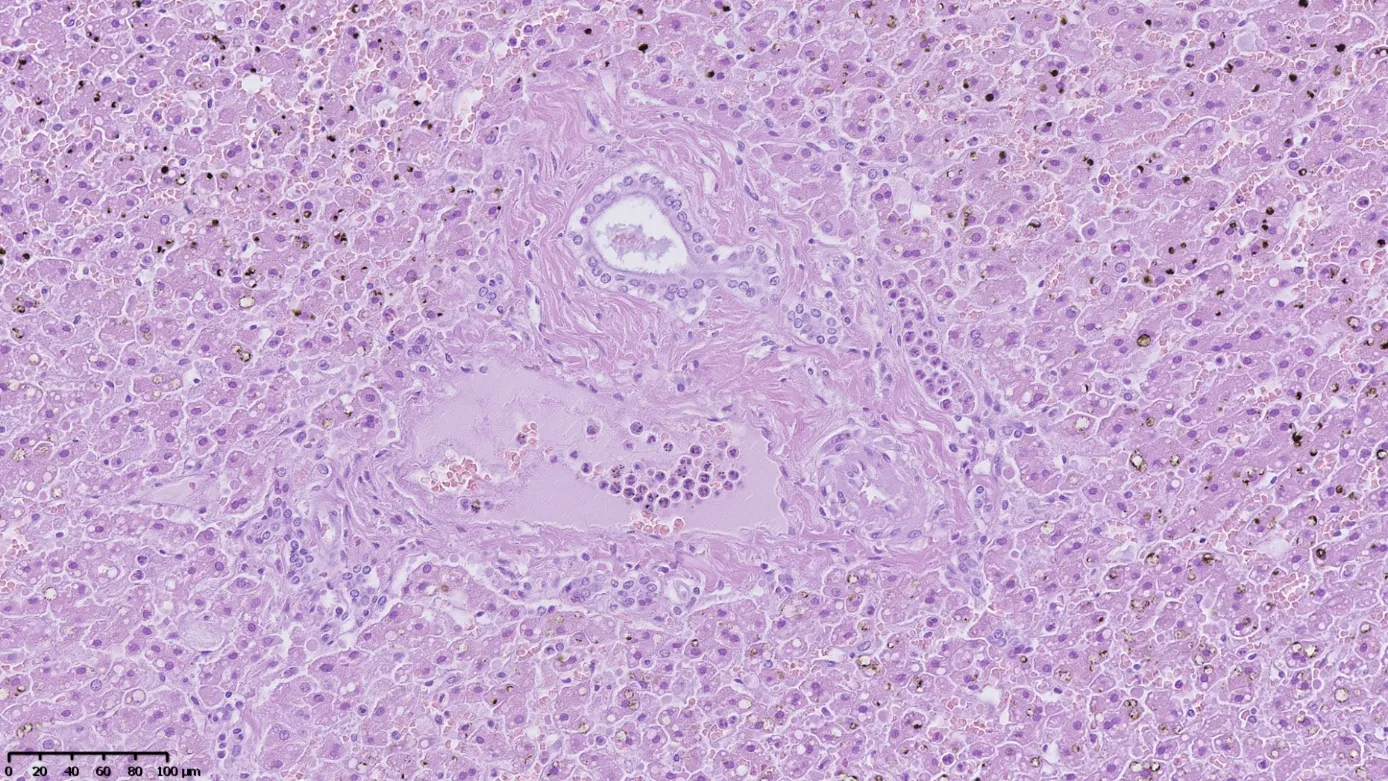
Morphological changes in the liver typically represent focal hepatocyte degeneration. Only in one case did they demonstrate signs of active inflammation and developing fibrosis (
). The nature of the observed changes does not allow for a clear connection with the use of “legal highs”, as the same changes may be associated with metabolic disorders, obesity, alcohol abuse, or viral hepatitis.
. Numerous neutrophils are visible in the lumen of the venous vessel of the biliary space and in the dilated sinusoidal vessel. There is evidence of mild fibrosis of the biliary space and microvesicular steatosis of the hepatocytes. Numerous yellowish-brown artifacts associated with autolysis are visible in the upper and lower portions of the image. Hematoxilin & eosin stain, magnification ×200.
4. Conclusions
In summary, microscopic examination of internal organ samples collected during autopsies and post-mortem examinations of individuals who died from legal highs is only supportive, as there is no characteristic microscopic image that would allow for a definitive diagnosis. The extent of the pathological changes observed depends primarily on age and whether the poisoned individual was hospitalized. Infectious complications are often observed in cases of long-term stays in intensive care units (e.g., pneumonia associated with respiratory therapy, signs of generalized infection).
Further future research should include a larger group of cases. It would be appropriate to statistically compare the histopathological findings in the studied group with those from a control group, for example, traumatic deaths (traffic accidents, falls from height, etc.).
Ethics Statement
Not applicable.
Informed Consent Statement
Not applicable.
Data Availability Statement
Research data is available from the author.
Funding
This research received no external funding.
Declaration of Competing Interest
The author declare that they have no known competing financial interests or personal relationships that could have appeared to influence the work reported in this paper.
References
-
1.
Ikematsu K, Fukunaga T, Kubo SI, Waters B, Hara K. A report of novel psychoactive substances in forensic autopsy cases and a review of fatal cases in the literature.
Leg. Med. 2017,
26, 79–85.
[Google Scholar]
-
2.
Logan BK, Mohr AL, Friscia M, Krotulski AJ, Papsun DM, Kacinko SL, et al. Reports of Adverse Events Associated with Use of Novel Psychoactive Substances, 2013–2016: A Review.
J. Anal. Toxicol. 2017,
41, 573–610.
[Google Scholar]
-
3.
Gandhi D, Ahuja K, Quade A, Batts KP, Patel L. Kratom induced severe cholestatic liver injury histologically mimicking primary biliary cholangitis: A case report.
World J. Hepatol. 2020,
12, 863–869.
[Google Scholar]
-
4.
Aldyab M, Ells PF, Bui R, Chapman TD, Lee H. Kratom-Induced Cholestatic Liver Injury Mimicking Anti-Mitochondrial Antibody-Negative Primary Biliary Cholangitis: A Case Report and Review of Literature.
Gastroenterol. Res. 2019,
12, 211–215.
[Google Scholar]
-
5.
Barletta C, Di Natale V, Esposito M, Chisari M, Cocimano G, Di Mauro L, et al. The Rise of Fentanyl: Molecular Aspects and Forensic Investigations.
Int. J. Mol. Sci. 2025,
7, 444.
[Google Scholar]