1. Introduction
Veterinary forensic pathology is an increasingly vital interdisciplinary field that bridges veterinary medicine, forensic science, and law. It is primarily concerned with systematically investigating animal deaths, particularly in unexplained, suspicious, or unlawful circumstances [
1]. This field is gaining prominence due to growing societal and legislative recognition of animal rights and welfare. Veterinary forensic pathologists play a crucial role in identifying causes of death, assessing trauma, and interpreting evidence for judicial proceedings, thereby contributing to both animal justice and public safety [
2]. Their work supports investigations in contexts such as animal abuse, neglect, poisoning, wildlife crime, insurance fraud, zoonotic disease outbreaks, and even cases linked to interpersonal violence, where animal harm may be a precursor or indicator of broader criminal activity [
3].
Among the array of causes of animal fatalities, drowning remains one of the most diagnostically challenging to confirm. Drowning is clinically defined as a form of asphyxia resulting from submersion (entire body below water) or immersion (airways only submerged) in a fluid medium, leading to primary respiratory failure [
4]. In animals, this process typically involves involuntary fluid aspiration into the lower respiratory tract, triggering a cascade of physiological disturbances, as shown in [
5]. The type of water, freshwater or saltwater, has profound implications for the pathophysiological outcome. Freshwater drowning causes rapid diffusion of hypotonic fluid into pulmonary circulation, leading to erythrocyte lysis, electrolyte imbalances such as hypercalcemia and hyponatremia, and ventricular fibrillation [
6]. Saltwater drowning, being hypertonic, draws fluid into the alveolar spaces, resulting in pulmonary oedema, haemoconcentration, and hypovolemic shock. In both scenarios, systemic hypoxia and metabolic acidosis precede cardiac arrest [
7].
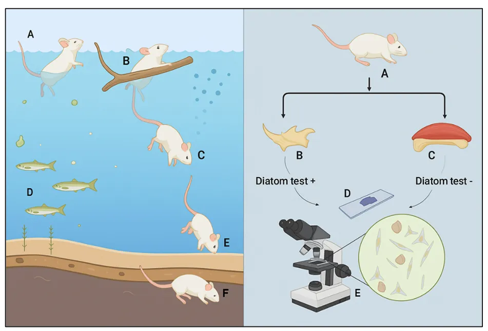
. This schematic depicts the sequence of drowning in a laboratory animal (mouse) model, illustrating stages from initial immersion (A), struggle (B), submersion (C–E), and post-mortem settlement (F). The right panel demonstrates the forensic workflow for determining antemortem drowning using the diatom test: tissue samples (B, C) are processed (D) and examined microscopically (E) for the presence of diatoms. A positive result supports a diagnosis of drowning, while a negative result suggests post-mortem immersion.
Diagnosing drowning post-mortem is inherently complex due to the absence of pathognomonic signs. Classical findings, such as overinflated, oedematous lungs; frothy exudate from the nose and trachea; and water in the stomach or sinuses, are often non-specific and can result from alternative causes like pulmonary oedema from cardiac failure or decomposition changes [
8]. Water immersion accelerates post-mortem autolysis and putrefaction, masking or mimicking drowning-associated pathology. Furthermore, the differentiation between antemortem drowning and post-mortem immersion is especially problematic. The concept of dry drowning, characterised by laryngospasm preventing water entry into the lungs, presents additional diagnostic ambiguity, with minimal or absent internal signs [
9,
10].
In this context, a comprehensive, multidisciplinary diagnostic approach becomes indispensable. It integrates findings from gross pathology, histopathology, radiology, toxicology, microbiology, and advanced forensic tests [
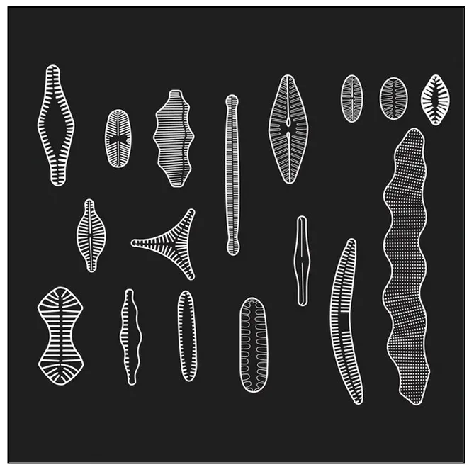
11]. A critical element in drowning investigations is the diatom test, based on the premise that if drowning occurred, diatoms (microscopic silica-shelled algae present in natural water bodies, some of which are shown in ) inhaled into the bloodstream may become distributed to internal organs such as the brain, bone marrow, and kidneys. The detection of these diatoms, matched with those from the suspected drowning site, can support the diagnosis () [
12]. However, the diatom test remains controversial due to concerns over contamination, diatom ubiquity, false positives, and variable analytical sensitivity. Alternative or supplementary techniques under research include molecular markers of hypoxia, electron microscopy, and elemental analysis of inhaled materials, which may provide more specific indicators of drowning [
13].
. Representative morphotypes of diatoms used in forensic drowning diagnosis.
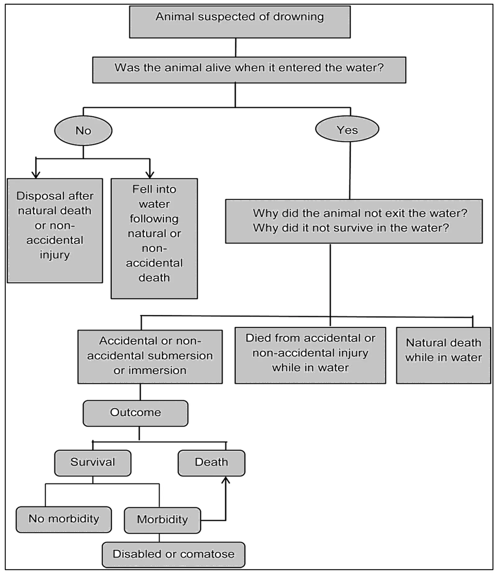
. Situational variations in bodies recovered from submerged settings.
This review presents a thorough and critical examination of the current forensic methodologies used to diagnose drowning in animals. It will assess the diagnostic value and limitations of macroscopic, microscopic, and ancillary approaches, focusing on the diatom test’s scientific validity and forensic applicability [
14,
15]. Additionally, the review will explore advancements in diagnostic technologies, the role of environmental and species-specific variables in interpretation, and propose a multi-modal framework to improve diagnostic precision in veterinary forensic drowning cases. By doing so, it seeks to advance veterinary forensic standards and support the broader goals of legal clarity, animal welfare, and scientific integrity [
16].
2. Review of Forensic Techniques
2.1. Extraction and Analysis of Diatoms
The extraction and analysis of diatoms have become a key technique in drowning investigations. The process begins with extracting these diatoms from tissue samples, a step that requires meticulous execution to ensure the integrity and reliability of the evidence.
- I.
-
Acid digestion: One of the most established methods of diatom extraction is acid digestion. In this technique, the tissue sample is digested in nitric acid, effectively dissolving organic matter, leaving a residue rich in diatoms. The residue is then centrifuged to concentrate the diatoms into a pellet prepared for microscopic examination. An advancement in this method is introducing a device known as a “can”, which optimises the process by combining tissue liquefaction with strong acid at high temperatures, increasing efficiency and reducing processing time [17].
- II.
-
Enzymatic digestion: Enzymatic digestion offers an alternative approach that uses proteinase K to selectively degrade proteinaceous material in the tissue sample while preserving the siliceous diatoms. After overnight incubation in Proteinase K and Tris HCl buffer, the sample is centrifuged to separate the diatoms for further microscopic analysis. This method is noted for its specificity and effectiveness in isolating diatoms from tissue samples. Another enzyme, papain. It is a protease enzyme from papaya that has been used in the past for meat tenderization and is now used in various industries such as food processing and pharmaceuticals. It can be used to extract diatoms from tissue samples at autopsy to aid in the diagnosis of drowning. The proposed papain method offers a simpler, safer, and less expensive alternative. It involves the enzymatic digestion of lung, kidney, and liver tissues to aid in the confirmation of drowning as the cause of death [18]. This technique is instrumental when traditional methods are impractical or when autopsy findings strongly suggest drowning.
- III.
-
Soluene-350: The Soluene-350 method is particularly effective for freshwater diatoms. This procedure involves multiple washes and centrifugations of the tissue sample, followed by incubation in Soluene-350 solution. The resulting pellet containing the extracted diatoms is then ready for microscopic observation. This method has seen limited adoption.
- IV.
-
Microwave digestion: A relatively recent innovation in diatom extraction is microwave digestion, in which the tissue sample is combined with an acid solution in a microwave digestion apparatus. This method may be characterised by high efficiency and reduced risk of contamination, with the digested solution suitable for analysis by advanced microscopy techniques such as scanning electron microscopy (SEM). While efficient, this method has seen limited application in the broader scientific community due to its reliance on specialised equipment.
Each of these methods aims to efficiently isolate diatoms from tissue samples, allowing subsequent microscopic examination to confirm the presence and identify the species of diatoms. Thus, the careful extraction and analysis of diatoms is a key component of forensic investigation in drowning cases. These methods, each with specific applications and advantages, provide a robust set of tools for determining the occurrence of drowning [
19].
2.2. Detection Methods
These techniques are applied after the extraction procedure. The diatoms are mounted in Naphrax on a glass slide and examined at 1000× magnification under oil immersion. Quantitative analysis and counting are done using phase contrast (100× objective, oil immersion, 10× eyepiece). Diatom identification is typically performed by referencing standard taxonomic works, such as the four volumes by Krammer & Lange-Bertalot. Various microscopic methods are described for observing diatoms, such as light microscopy, electron microscopy, or atomic force microscopy. Light microscopy is widely used in routine work. The observation is relatively easy if the background on the slides is not too pronounced due to residual particles, and it is less time-consuming than the other methods. However, distinguishing smaller species can be challenging with this technique. Scanning electron microscopy (SEM) provides a high-resolution 3D image of the diatom. A novel method, microwave digestion-vacuum filtration-automated SEM analysis, is highly sensitive and specific. One disadvantage of this technique is that it is time-consuming. A deep learning-based algorithm has been developed to automatically detect diatoms from SEM images [
20]. The limitation of this method is the availability of the required equipment, which is not accessible to all laboratories involved in diatom testing.
3. Macroscopic and Microscopic Post-Mortem Findings in Drowning
Post-mortem assessment in suspected drowning cases plays a central role in veterinary forensic pathology, offering essential information about the circumstances surrounding death. However, gross and histological findings are frequently non-specific and can be confounded by decomposition, traumatic injuries, or environmental factors. Therefore, a systematic, comparative approach is essential to interpret findings in context.
- I.
-
External Findings: Externally, drowned animals may exhibit wet and matted fur or feathers, often embedded with aquatic vegetation, mud, sand, or algae. These materials are commonly found in natural orifices such as the nostrils, oral cavity, and anogenital regions. In advanced putrefaction, gas accumulation may cause subcutaneous emphysema or bloating. Superficial injuries, including abrasions, bite wounds, or claw marks, may suggest pre-mortem struggle or entrapment, notably if correlated with environmental evidence. The death scene often provides contextual clues, such as disturbances in sediment, plant entanglement, or drag marks, which can aid in reconstructing the chain of events [21].
- II.
-
Respiratory System: The respiratory system typically shows the most characteristic changes. Lungs are distended, pale, heavy, and crepitant or doughy due to oedema and emphysema aquosum. A hallmark finding is abundant pinkish-white, fine frothy fluid within the trachea and bronchi, sometimes extending to the nostrils, resulting from the admixture of water, mucus, air, and surfactant during forced inhalation. Water and debris may also be found in the pleural spaces. Foreign material such as diatoms, sand grains, or plant fragments in bronchial or alveolar spaces provides strong evidence of ante-mortem aspiration, though decomposition or infections may obscure these findings.
- III.
-
Cardiovascular System: Cardiovascular examination may reveal right heart dilatation secondary to acute pulmonary hypertension. Blood viscosity may also provide indirect evidence of drowning type: freshwater immersion leads to haemodilution and haemolyzed blood, whereas saltwater immersion produces haemoconcentration and thick, viscous blood due to osmotic shifts. Petechiae on serosa and mucosal surfaces are sometimes observed but are non-specific indicators of terminal asphyxia.
- IV.
-
Gastrointestinal System: The gastrointestinal system may contain large volumes of fluid, sediment, algae, or aquatic organisms within the stomach, suggesting active swallowing and a vital response to submersion. Stomach content analysis may also help correlate findings with the suspected drowning medium. Other visceral organs, including the liver, kidneys, and spleen, often appear congested due to systemic hypoxia [22]. Cerebral oedema is another frequent finding, sometimes accompanied by raised intracranial pressure, especially in small animals or neonates, though not pathognomonic for drowning.
- V.
-
Microscopic Findings: Microscopically, the lungs show alveolar oedema, vascular congestion, intra-alveolar haemorrhage, and, in prolonged cases, eosinophilic hyaline membranes consistent with acute respiratory distress. Foreign material such as diatoms, pollen, or plant fragments within the terminal airways or alveoli strongly indicates active respiration during submersion. Special stains, polarised light microscopy, or SEM can assist in their identification.
The myocardium frequently exhibits vascular engorgement, interstitial oedema, and occasionally contraction band necrosis or ischemic changes, reflecting terminal hypoxia but not exclusive to drowning. Renal tissues, particularly in freshwater immersion, may show osmotic injury, tubular degeneration, hydropic change, and haemoglobin pigment casts due to intravascular haemolysis. While none of these features is individually diagnostic, their combined presence substantially strengthens the inference of drowning when correlated with environmental findings and ancillary tests [
23,
24].
4. Ancillary Diagnostic Tests for Drowning
Ancillary diagnostic tests provide valuable adjunctive evidence in the forensic evaluation of drowning, particularly in cases where classical autopsy findings are equivocal or confounded by decomposition. These tests encompass biochemical, radiological, and toxicological assessments that enhance diagnostic resolution by identifying physiological changes associated with the drowning process [
17]. Biochemical markers are among the most studied parameters. In freshwater drowning, the hypotonicity of the inhaled water leads to osmotic influx into the pulmonary circulation through alveolar membranes, causing haemodilution. This manifests as decreased packed cell volume (PCV), haemoglobin levels, and electrolyte imbalances such as hyponatremia and hypochloraemia. Additionally, the osmotic lysis of red blood cells may result in elevated potassium levels [
25]. In contrast, saltwater drowning induces osmotic withdrawal of plasma fluid into alveolar spaces, leading to haemoconcentration, reflected by increased PCV and elevated sodium and chloride levels. Despite these trends, the forensic utility of such markers is limited by post-mortem changes, interspecies differences, and the instability of biochemical parameters after death.
Pulmonary surfactant analysis has also been explored as a potential diagnostic tool. Surfactants, which maintain alveolar integrity, can be diminished or functionally impaired when drowning due to washout or biochemical alteration, resulting in alveolar collapse and atelectasis. Although bronchoalveolar lavage fluid has been used experimentally to assess surfactant phospholipids, rapid post-mortem degradation and individual variability restrict its routine use in veterinary forensics. Emerging research has turned to molecular biomarkers such as S100B protein, cardiac troponins, and cell-free nucleic acids, which may indicate hypoxic stress or cellular injury during the agonal phase of drowning. While these markers hold promise, none have been validated for consistent specificity, sensitivity, and post-mortem stability in animal models, limiting their practical application.
Radiological imaging techniques, especially post-mortem computed tomography (PMCT) and magnetic resonance imaging (PMMRI), offer non-invasive insights into drowning-related pathology. PMCT effectively visualises pulmonary oedema, fluid accumulation in airways, pleural effusions, and, in some cases, foreign material such as mud or aquatic organisms in the respiratory or digestive tracts. PMMRI may provide superior soft tissue resolution for evaluating subtle haemorrhages or parenchymal changes [
26]. However, these findings are not pathognomonic for drowning and require correlation with gross necropsy, histological, and biochemical evidence to avoid diagnostic overinterpretation. Toxicological analysis remains a critical component of forensic investigation in suspected drowning. It facilitates the detection of exogenous substances, such as sedatives, neurotoxins, or intoxicants, that may have contributed to incapacitation and subsequent immersion. Comprehensive sampling from multiple matrices, including liver, kidney, gastric contents, urine, and vitreous humour, ensures reliable detection. The presence of drugs or poisons may shift the interpretive focus toward non-accidental or secondary causes of death, thereby influencing legal and investigative conclusions. Thus, while ancillary tests cannot independently confirm drowning, they substantially enrich the evidentiary matrix when judiciously integrated into the broader forensic framework [
27].
Emerging Diagnostic Parameters
Recent advances in human forensic pathology have introduced novel quantitative indices that may offer greater diagnostic reliability than traditional ancillary methods such as diatom testing. Two promising parameters are the Drowning Index (DI) and the Pleural Effusion Ratio (PE ratio).
The DI, first evaluated in a comparative human autopsy study by Nagar et al., measures specific biochemical and physicochemical alterations in pulmonary and pleural fluids associated with freshwater drowning [
28]. Their findings highlighted that DI values in confirmed drowning cases differed significantly from non-drowning controls, providing an objective, reproducible metric that outperformed conventional gross and histological indicators. Importantly, DI showed greater consistency and reduced examiner variability, making it a robust potential supplement to classical methods. Building on these observations, Nagar et al. introduced another parameter, the PE ratio, derived from the analysis of pleural fluid and serum parameters, and compared its diagnostic performance with DI [
29]. Their study demonstrated that both indices not only distinguished drowning cases from other causes of death but also achieved higher diagnostic accuracy and reproducibility compared to morphological criteria alone. The PE ratio, in particular, demonstrated strong discriminatory power when used alongside DI, reinforcing the clinical value of combining complementary quantitative indices to enhance objectivity.
Although these parameters have been validated in a human forensic context, their underlying principles, quantitative analysis of pleural effusion dynamics and fluid biochemistry, are theoretically applicable to veterinary forensics. The adaptation of the DI and PE ratio to animal models could represent an essential translational advance, expanding the diagnostic arsenal available to veterinary pathologists investigating drowning deaths. Their adoption may reduce reliance on subjective morphological assessments and controversial ancillary methods, paving the way for a more standardised and objective approach in this challenging area of forensic pathology.
5. The Diatom Test in Veterinary Forensic Pathology
The diatom test holds significant forensic value in the post-mortem investigation of suspected drowning cases in animals, offering microscopic evidence that can support or refute the hypothesis of ante-mortem submersion. Diatoms are unicellular microalgae encased in durable silica frustules, widely distributed in natural and artificial aquatic environments. Their resistance to degradation makes them ideal markers for post-mortem detection. The foundational principle of the diatom test is based on the physiological process of drowning. During agonal respiratory efforts, water is forcibly aspirated into the lungs, allowing diatoms to traverse the compromised alveolar-capillary barrier and enter the bloodstream. They are disseminated via systemic circulation to distant, highly vascularized organs such as the liver, kidneys, brain, and bone marrow [
30]. The presence of diatoms in these extra-pulmonary sites, especially within the protected environment of bone marrow, strongly supports the notion of vital submersion as opposed to passive post-mortem immersion. Species-specific identification of diatoms can further corroborate findings by linking them to a specific drowning medium, providing ecological and geographical traceability.
Accurate implementation of the diatom test necessitates meticulous sample collection and preparation. Primary target organs include the lungs, brain, liver, kidneys, and bone marrow, with the latter considered the most diagnostically reliable due to its vascular richness and lower susceptibility to external contamination. Contamination control is paramount; sterilised dissection instruments, avoiding washing tissues, and including procedural negative controls are essential to prevent false-positive results arising from environmental or laboratory sources. For microscopic analysis, tissue samples undergo chemical digestion, typically with concentrated nitric or sulfuric acid, to dissolve organic matter and isolate the resilient diatom frustules. The processed material is then mounted on slides and examined under light, phase contrast, or differential interference contrast (DIC) microscopy at magnifications ranging from 400× to 1000×. Identification relies on the frustules’ unique morphological features and ornamentation [
31]. While some laboratories employ semi-quantitative methods, such as counting diatoms per high-power field or per unit of tissue mass, there remains a lack of international standardisation in protocol and threshold values.
Interpretation of diatom test results requires nuanced forensic judgment. Quantitative assessments may offer a degree of objectivity. Still, qualitative aspects, such as the diversity of diatom species and their correlation with those found in the suspected drowning site, are often more probative in legal and diagnostic contexts. Notably, the presence of diatoms limited only to pulmonary tissues may be inconclusive, as passive post-mortem infiltration can occur, especially in decomposed or submerged carcasses. In contrast, detecting diatoms in remote organs like bone marrow is robust evidence of ante-mortem aspiration and systemic circulation. Numerous veterinary case reports highlight the diagnostic utility of the diatom test, particularly when external findings or histological features of drowning are ambiguous or absent. In such cases, the identification of diatoms has frequently served as a pivotal factor in establishing drowning as the cause of death. Nevertheless, given its limitations, the diatom test should be employed as part of a broader forensic framework that includes scene investigation, necropsy, toxicology, and biochemical analysis to arrive at a definitive and legally defensible conclusion [
32].
6. Limitations and Challenges of the Diatom Test
Despite its historical role as an ancillary diagnostic tool, the diatom test in veterinary forensic pathology is increasingly recognised as a method with significant limitations that restrict its diagnostic reliability. One of the foremost concerns is the high risk of environmental contamination, which can result in false-positive outcomes. Diatoms are ubiquitous in aquatic and terrestrial settings, and their inadvertent introduction during sample collection, necropsy procedures, or laboratory processing, through sources such as tap water, airborne particles, or inadequately sterilised instruments, can compromise the integrity of results. Post-mortem immersion presents an additional challenge, as diatoms may enter the lungs or translocate into systemic circulation during decomposition, obscuring the distinction between ante-mortem aspiration and passive post-mortem exposure. Similarly, variability in diatom species distribution across aquatic environments complicates the correlation of tissue profiles with the putative drowning medium. False negatives are also possible in instances of dry drowning, where laryngospasm prevents diatom aspiration, or when ambient water contains insufficient diatom density for detection [
27].
Beyond these interpretative challenges, the absence of standardised protocols for digestion, extraction, quantification, and interpretation results in considerable inter-laboratory variability, undermining reproducibility and legal defensibility. In human forensic pathology, these limitations have led to a growing consensus that the diagnostic value of the diatom test is highly restricted, and this constraint is even more pronounced in veterinary contexts where environmental and procedural controls are challenging to enforce. These concerns underscore the necessity of treating the diatom test not as a cornerstone but as one component within a broader, multi-modal diagnostic framework.
7. Integration of Diagnostic Modalities
To overcome the limitations inherent in singular diagnostic techniques, a multi-modal and integrative approach is essential for the accurate forensic diagnosis of drowning in animals. This entails a comprehensive synthesis of all available investigative modalities, beginning with a meticulous scene investigation that documents environmental parameters, water characteristics, and any signs of perimortem trauma. A thorough post-mortem examination should assess external signs of immersion and internal findings such as pulmonary oedema, frothy fluid in the airways, and circulatory congestion, supplemented by histopathological evidence of alveolar damage. Toxicological screening is vital to exclude alternative causes of death, such as poisoning or drug-induced incapacitation, that may precede submersion. Biochemical markers like electrolyte imbalances and PCV changes may offer supportive evidence, though their post-mortem lability necessitates cautious interpretation. The diatom test plays a pivotal role, especially when diatoms are detected in multiple distant organs with species profiles matching those in the suspected water source [
24]. A systematic, algorithmic framework for integrating these findings enhances diagnostic clarity, guiding forensic practitioners toward a more robust and defensible conclusion. Contextual information from the scene and anamnestic data (including behavioural observations and pre-existing health conditions) further aid in excluding other plausible causes and reinforcing the diagnosis of drowning.
8. Conclusions and Future Directions
The diagnosis of drowning in veterinary forensic cases remains a complex and often ambiguous task. While gross and microscopic post-mortem findings may indicate immersion, their non-specific nature necessitates the application of additional ancillary tests. Among these, the diatom test has historically stood out for its ability to provide supportive evidence of a vital reaction, particularly when diatoms are identified in extra-pulmonary tissues and correlate with environmental samples. However, the test cannot be relied upon in isolation due to challenges such as contamination risk, inter-laboratory inconsistencies, and diagnostic ambiguity in dry drowning or putrefaction cases. Best practices, therefore, recommend a holistic forensic approach, integrating scene investigation, toxicological screening, detailed necropsy findings, biochemical assays, and diatom analysis under strict contamination control protocols.
Future research should prioritise the development of standardised methodologies for diatom detection, as well as the exploration of improved molecular, immunohistochemical, and imaging-based techniques to enhance diagnostic specificity. In addition, systematic comparative studies are needed to validate emerging biochemical and radiological indices, such as the Drowning Index (DI) and Pleural Effusion Ratio (PE ratio), in animal drowning cases. Establishing the applicability of these quantitative markers would help bridge the gap between veterinary and human forensic practice and provide a roadmap for advancing this subfield. Advancements in these areas will strengthen the forensic determination of drowning in animals and contribute to broader translational applications in veterinary and human forensic sciences.
Statement of the Use of Generative Al and Al-Assisted Technologies in the Writing Process
During the preparation of this manuscript, the authors used Google Gemini to assist with language editing and grammar refinement. All content was subsequently reviewed, revised, and approved by the authors, who take full responsibility for the final version of the manuscript.
Acknowledgments
This work would not have been possible without our colleagues’ invaluable discussions and paramount suggestions.
Author Contributions
Conceptualization, P.D., A.B. and S.K.; Investigation, P.D. and A.B.; Writing Original Draft, A.B.; Writing Review & Editing, P.D.; Supervision and Project Administration, S.K. All authors read and approved the final manuscript.
Ethics Statement
Not applicable.
Informed Consent Statement
Not applicable.
Data Availability Statement
No datasets were generated or analysed during the current study.
Funding
This work received no funding from internal or external sources.
Declaration of Competing Interest
The authors reported no potential competing interests. The authors declare no conflict of interest and confirm that all have contributed substantially to this work.
References
-
1.
Cooper JE, Cooper ME. (Eds) Special features of veterinary and comparative forensic medicine. In Introduction to Veterinary and Comparative Forensic Medicine; Blackwell Publishing Ltd.: Oxford, UK, 2007; pp. 168–224.
-
2.
Arkow P. Human–animal relationships and social work: Opportunities beyond the veterinary environment.
Child Adolesc. Soc. Work. J. 2020,
37, 573–588.
[Google Scholar]
-
3.
Golden FS, Tipton MJ, Scott RC. Immersion, near-drowning and drowning.
Br. J. Anaesth. 1997,
79, 214–225.
[Google Scholar]
-
4.
Armstrong EJ, Erskine KL. Investigation of drowning deaths: A practical review.
Acad. Forensic Pathol. 2018,
8, 8–43.
[Google Scholar]
-
5.
Modell JH, Gabrielli A. Cardiopulmonary Resuscitation Following Drowning. In Cardiopulmonary Resuscitation; Humana Press: Totowa, NJ, USA, 2005; pp. 407–424.
-
6.
Sarnaik AP, Vohra MP. Near-drowning: fresh, salt, and cold water immersion.
Clin. Sports Med. 1986,
5, 33–46.
[Google Scholar]
-
7.
Bierens JJ, Lunetta P, Tipton M, Warner DS. Physiology of drowning: A review.
Physiology 2016,
31, 147–166.
[Google Scholar]
-
8.
Grover IR. Drowning and Diving Accidents. Manual of Clinical ProblEMS in PulMonary MEdicinE. 498. Available online: http://ndl.ethernet.edu.et/bitstream/123456789/21724/1/1900.pdf#page=518 (accessed on 18 June 2025).
-
9.
McEwen BJ, Gerdin J. Veterinary forensic pathology: drowning and bodies recovered from water.
Vet. Pathol. 2016,
53, 1049–1056.
[Google Scholar]
-
10.
Denge K, Gatfane R. A Critical Study of Violent Asphyxial Deaths with Ancient Perspective.
J. Ayurveda Integr. Med. Sci. 2016,
1, 85–93.
[Google Scholar]
-
11.
Piegari G, De Biase D, d’Aquino I, Prisco F, Fico R, Ilsami R, et al. Diagnosis of drowning and the value of the diatom test in veterinary forensic pathology.
Front. Vet. Sci. 2019,
6, 404.
[Google Scholar]
-
12.
Ludes BP, Chambre A, Delabarde T. The diatom test in the field of forensic medicine: a review of a long-standing question.
Int. J. Leg. Med. 2025,
139, 597–605.
[Google Scholar]
-
13.
Mukhopadhyay P, Chowdhuri S. 16 Recent Advances in the Autopsy Diagnosis of Drowning.
Recent Adv. Forensic Med. Toxicol. -2 Good Pract. Guidel. Curr. Medicolegal Issues 2018,
2, 394.
[Google Scholar]
-
14.
Wu H, Liu R, Wang G, Shen C, Liang X, Chen R, et al. Accurate forensic identification of asphyxial deaths: Differentiating strangulation and drowning using ATR-FTIR spectroscopy.
Sci. Justice 2025,
65, 101257.
[Google Scholar]
-
15.
Beausoleil NJ, Mellor DJ, Baker L, Baker SE, Bellio M, Clarke AS, et al. “Feelings and fitness” not “feelings or fitness”–the raison d’être of conservation welfare, which aligns conservation and animal welfare objectives.
Front. Vet. Sci. 2018,
5, 296.
[Google Scholar]
-
16.
Robertson IA. Legally protecting and compelling veterinarians in issues of animal abuse and domestic violence.
New Zealand Vet. J. 2010,
58, 114–120.
[Google Scholar]
-
17.
Singh R, Singh R, Thakar MK. Extraction methods of diatoms—A review.
Indian Internet J. Forensic Med. Toxicol. 2006,
4, 1–6.
[Google Scholar]
-
18.
Tsuneya S, Nakajima M, Makino Y, Torimitsu S, Yamaguchi R, Iwase H. A quantitative comparison between using sodium hypochlorite as a digestion method for the diatom test and the conventional method using fuming nitric acid.
Forensic Sci. Int. 2021,
329, 111086.
[Google Scholar]
-
19.
Hu S, Liu C, Wen J, Dai W, Wang S, Su H, Zhao J. Detection of diatoms in water and tissues by combination of microwave digestion, vacuum filtration and scanning electron microscopy.
Forensic Sci. Int. 2013,
226, e48–e51.
[Google Scholar]
-
20.
Girela-Lopez E, Beltran-Aroca CM, García-Mozo H. Diatoms in forensic analysis. In Modern Trends in Diatom Identification: Fundamentals and Applications; Springer: Cham, Switzerland, 2020; pp. 239–256.
-
21.
Marshall M, Chraïbi V, Morgan R. A method for bone marrow extraction of diatoms for forensic science applications.
J. Forensic Sci. 2023,
68, 1343–1351.
[Google Scholar]
-
22.
Christe A, Aghayev E, Jackowski C, Thali MJ, Vock P. Drowning—post-mortem imaging findings by computed tomography.
Eur. Radiol. 2008,
18, 283–290.
[Google Scholar]
-
23.
Dahiya P, Kumar S, Shukla MA, Yadav CS. Forensic limnology: Diversity of diatom population in relation to environmental factors in Gujarat region, India.
Mater. Today Proc. 2022,
69, A14–A24.
[Google Scholar]
-
24.
Lockyer BE. The body recovered from water: considerations for an approach to the non-suspicious post-mortem examination.
Diagn. Histopathol. 2021,
27, 411–417.
[Google Scholar]
-
25.
Vinayak V, Gautam S. Diatoms in forensics: A molecular approach to diatom testing in forensic science. In Diatoms: Fundamentals and Applications; Wiley: Hoboken, NJ, USA, 2019; pp. 435–470. doi:10.1002/9781119370741.ch18.
-
26.
Krstic S, Duma A, Janevska B, Levkov Z, Nikolova K, Noveska M. Diatoms in forensic expertise of drowning—a Macedonian experience.
Forensic Sci. Int. 2002,
127, 198–203.
[Google Scholar]
-
27.
Dahiya P, Makwana MD, Chaniyara P, Bhatia A. A comprehensive review of forensic diatomology: contemporary developments and future trajectories.
Egypt. J. Forensic Sci. 2024,
14, 2.
[Google Scholar]
-
28.
Nagar N, Bastia BK, Singi Y, Dabhi D, Nagar K. Assessment of the drowning index in diagnosing freshwater drowning: A comparative study.
J. Forensic Sci. 2025,
advance online publication.
[Google Scholar]
-
29.
Nagar N, Bastia BK, Dabhi D, Singi Y, Jain J. Comparing the diagnostic utility of the Pleural Effusion Ratio and the Drowning Index in identifying freshwater drowning deaths.
J. Forensic Leg. Med. 2025,
115, 102950.
[Google Scholar]
-
30.
Bozarth A, Maier UG, Zauner S. Diatoms in biotechnology: modern tools and applications.
Appl. Microbiol. Biotechnol. 2009,
82, 195–201.
[Google Scholar]
-
31.
Sims PA, Mann DG, Medlin LK. Evolution of the diatoms: insights from fossil, biological and molecular data.
Phycologia 2006,
45, 361–402.
[Google Scholar]
-
32.
Nissan E, Keppens J, Akin LL. The forensic disciplines: Some areas of actual or potential application. In Computer Applications for Handling Legal Evidence, Police Investigation and Case Argumentation; Springer: Dordrecht, The Netherlands, 2011; pp. 841–989.
 Ashna Bhatia
1
Ashna Bhatia
1