1. Introduction
In response to epithelial (or muscle) damage, a dynamically complex but tightly regulated process of wound healing is orchestrated to successfully repair the tissue and restore its normal function. This process, that involves local inflammation and myofibroblast activation, proceeds with the deposition of collagens and other extracellular matrix (ECM) components that remodel the environment promoting the replacement of parenchymal cells [
1]. In physiological wound healing, a resolution step is established with the abatement of inflammation, the apoptosis of myofibroblasts and the resorption of ECM in excess. However, under certain circumstances, when tissues are subjected to persistent insult and injury, this wound repair process derails, leading to uncontrolled myofibroblast activation and thus to pathological accumulation of ECM, also known as fibrosis [
2]. A fibrotic process is intrinsic to a plethora of pathological conditions such as end-stage liver disease, kidney disease, idiopathic pulmonary fibrosis (IPF), heart failure, scleroderma, rheumatoid arthritis, Crohn’s disease, ulcerative colitis, myelofibrosis and systemic lupus erythematosus, among others, and is also germane to many forms of cancer, influencing tumor invasion and metastasis [
3]. Therefore, fibrotic disorders represent an increasing cause of morbidity and mortality worldwide. Intensive research in recent years has identified several pathways involved in the activation and persistence of fibroblasts typically found in fibrotic tissues, with transforming growth factor-β (TGF-β) signaling pathway playing a fundamental role [
4]. However, the treatment options are scarce and ineffective, Actually, considering IPF, the paradigmatic fibrotic disease, only two molecules are currently being used for treatment, pirfenidone (Esbriet®), a drug targeting TGF-β pathway, and nintedanib (Ofev®), a broad kinase inhibitor, both capable of slowing disease progression, but very far from providing effective healing [
5]. Therefore, there is an urgent need for new pharmacological approaches to combat fibrosis.
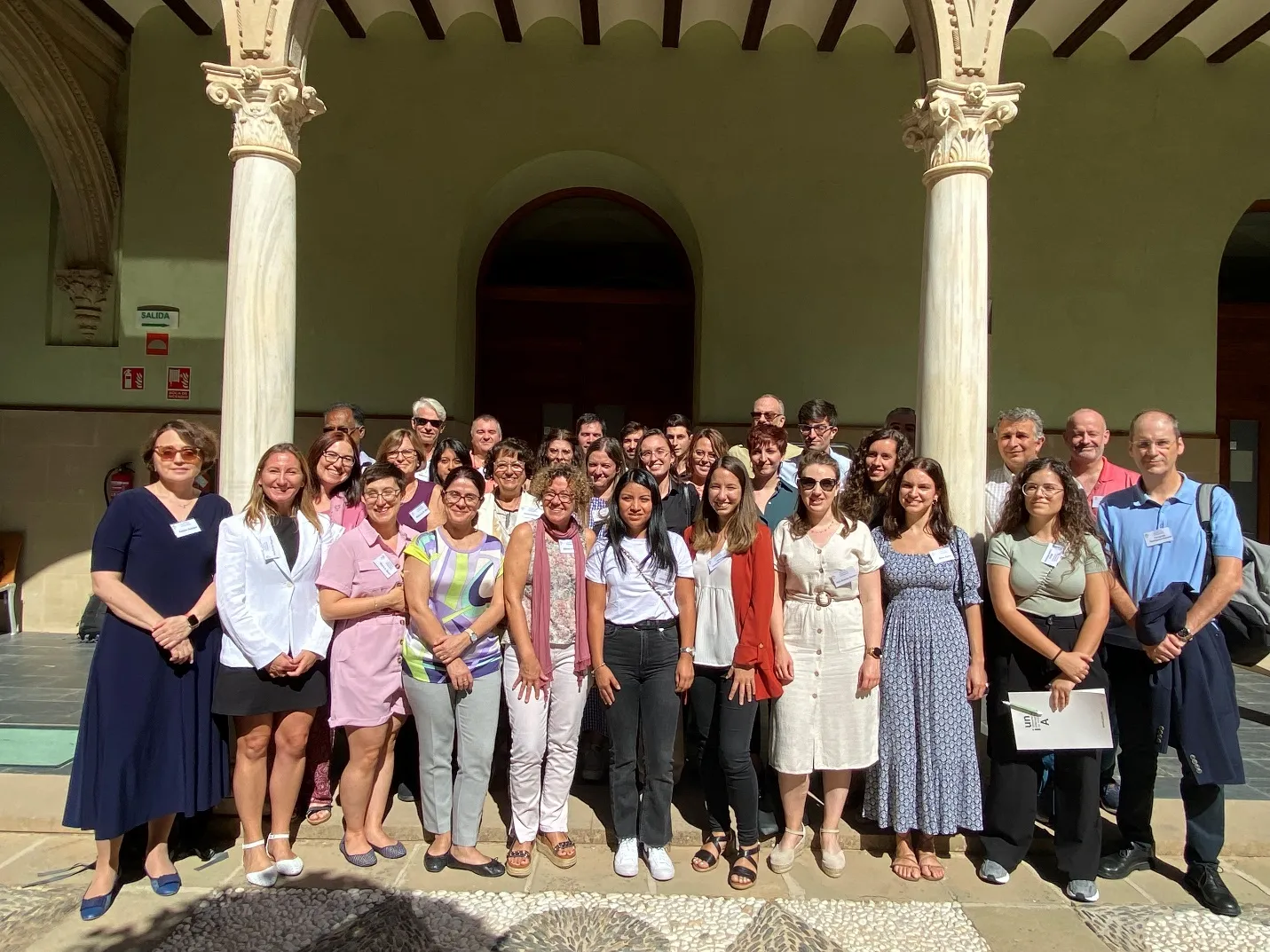
The International University of Andalusia Workshop “Current Trends in Biomedicine” on The Cellular and Metabolic Bases of Organ Fibrosis, was organized by Santiago Lamas, Katalin Susztak and Fernando Rodríguez-Pascual in Baeza, Spain, from October 8 to 11, 2023 with about 40 scientists from a broad spectrum of countries, disciplines and stages in their professional careers (). The meeting aimed to bring together some of the top-level, highly experienced investigators working on different aspects of the pathophysiology of fibrosis and nearby research areas. They were all keen to share and discuss with other participants their work and contributions to clarify the mechanisms contributing to the fibrotic process in different organs and tissues, with particular focus on its metabolic basis and the development of novel therapeutic strategies. Here, we briefly review the current knowledge of the metabolic control of fibrosis and provide an overview of the scientific content of the meeting.
. Speakers, participants and organizers of the International University of Andalusia Workshop “Current Trends in Biomedicine” on The Cellular and Metabolic Bases of Organ Fibrosis, Baeza, Spain, October 8-11, 2023. Photo courtesy of Katalin Susztak.
2. Metabolic Regulation of Tissue Fibrosis
While many aspects of the fibrotic conundrum are still to be deciphered, we know today that a pathological hallmark of the fibrotic process is a tight crosstalk among different cell types from epithelial (or muscle), mesenchymal and immune lineages [
6]. The first two types constitute the basic unit conforming the sensor, the epithelial (or muscle) cell that detects the damage or insult and initiates the healing response and the effector, the mesenchymal cell type that becomes activated at the site of damage and is ultimately responsible for tissue remodeling and fibrosis. A fundamental cell-cell communication between these lineages is observed in a variety of tissues undergoing fibrosis, such as the lung, with the alveolar epithelium and the pulmonary fibroblasts, the kidney with the tubular epithelial cells and the pericyte-myofibroblast lineage or the liver, with the hepatocytes and the hepatic stellate cells. At the crossroad of these cell types, immune cells are potent regulators of the tissue repair response and therefore implicated in the development of fibrosis. With this cellular landscape in mind, most of the investigation established so far has focused on the mediators governing the fibrotic process and their signaling pathways.
Unfortunately, while providing significant progress at the mechanistic level, this approach has yielded limited information of therapeutic value beyond the role of TGF-β [
4]. Upstream signaling pathways may converge at key metabolic alterations that ultimately can regulate the cell phenotype. Therefore, cell metabolism represents a very attractive target to identify novel points of intervention for the treatment of fibrosis [
7]. For instance, de novo lipid synthesis is required for alveolar type 2 epithelial (AE2) cells to generate lipid-rich surfactants, essential for reducing surface tension in the lungs during respiratory function. AE2 lipid metabolism deficiency secondary to the downregulation of the expression of AE2 specific fatty acid synthase (FASN) enzyme has been linked to IPF in patients and shown to aggravate pulmonary fibrosis in animal models [
8]. Not only do metabolic alterations promote fibrotic-prone transformations in sensor cells, but they are also key in the activation of effector mesenchymal cells. In fact, myofibroblast differentiation must come along with profound metabolic changes that fulfill the high consumption of energy and building blocks imposed by the activation of the profibrotic cellular program [
9,
10].
In terms of energy and biomass, the cost of the biosynthesis of fibrillary collagens, the main ECM component produced by myofibroblasts, is remarkable. The triple helix structure of the collagen domain is featured by a particularly unusual amino acid composition, containing approximately 30% glycine and 15–20% proline residues, and an extensive network of post-translational modifications, including hydroxylation, oxidation and glycosylation, let alone the large size of collagen polypeptides (typically > 300 kDa), which adds extra demands for correct folding, assembling and secretion from the proteostasis machinery [
11]. Regarding the supply of glycine and proline, it has been reported that myofibroblasts undergo specific metabolic reprogramming, including upregulation of glycolysis and glutaminolysis, to feed de novo biosynthetic pathways for these amino acids [
9,
12].
Other authors have also observed increased activity of the citric acid cycle and the oxidative phosphorylation associated to fibroblast activation and ECM biosynthesis [
13]. These observations uncover myofibroblast metabolic vulnerabilities that can eventually be exploited to revert fibrosis. In this line, a recent report has shown an increased metabolic demand for glycogen metabolism in activated fibroblasts during pulmonary fibrosis [
14]. Through the lysosomal salvage pathway, glycogen provides substrates for the synthesis of proteoglycans and N-linked glycans that are critical components of the fibrotic ECM. Based on these evidences, compounds targeting glycogen catabolism are promising candidates for the treatment of lung fibrosis patients. These studies and many more to come are possible thanks to the advent of dedicated techniques to detect and quantify tissue metabolites within their spatial context, the so-called spatial metabolomics. Conveniently combined with single-cell transcriptomics, this near-single-cell resolution approach is well-suited for interrogating human biopsies and organs from animal models to address metabolic alterations in diseases such as fibrosis. The application of these techniques as well as the complementary studies in cell and animal models will substantially advance our knowledge on the pathogenic mechanisms involved in the development of fibrosis. As appetizer for this exciting time about to come, in two main sessions, the meeting covered the role of metabolism, inflammation and hypoxia in fibrosis, their contribution to disease in major tissues developing fibrosis, such as lung, kidney, liver and muscle, as well as the potential therapeutic approaches thereby.
3. Role of Metabolism in Kidney Fibrosis
Kidney fibrosis is the common pathological outcome of nearly all chronic and progressive nephropathies [
15]. Morphological characteristics include glomerulosclerosis, tubule atrophy, interstitial chronic inflammation, and fibrogenesis. The kidney has one of the most diverse cellular populations and spatial organization in the human body, with more than 40 cell types based on cluster-specific marker gene expression [
16]. Despite this complexity, tubular epithelial, mesenchymal, and immune cells are the main components of the fibrotic niche in the kidney [
17,
18]. As a recurrent theme, persistent injury on tubular epithelial cells triggers an aberrant wound healing response with secretion of profibrotic factors which impact interstitial mesenchymal cells (pericytes and fibroblasts) to activate myofibroblast differentiation, proliferation, and ECM accumulation. While several signal transduction pathways have been reported to be involved in the development of kidney fibrosis, including Notch, Wnt, and Hedgehog, much less is known about the metabolic alterations intrinsic to the process. Using metabolomics, Katalin Susztak (Perelman School of Medicine, USA) showed that accumulation of lipid droplets is commonly observed in kidney fibrosis. The team has identified several mechanisms contributing to lipid accumulation, including lower fatty acid oxidation, increased fatty acid synthesis and mitochondrial defects such as nicotinamide adenine dinucleotide (NAD+) deficiency [
19,
20]. Dysregulated NAD+ homeostasis seems to drive mitochondrial dysfunction causing inflammation and kidney disease. From a therapeutic perspective, these observations support a beneficial effect for NAD+ precursor supplementation to protect against kidney injuries. In this line, Benjamin D. Humphreys (Washington University in St. Louis, USA) described the results of a comprehensive single-cell transcriptional profiling of mouse fibrotic kidneys, combined with a preliminary metabolomic approach [
21]. This study unveiled tubular epithelial cell responses featured by alterations in lipid metabolism. This metabolic signature is consistent with previous observations of defective fatty acid β-oxidation (FAO) metabolism in chronic kidney disease, which could be reversed by restoring the capacity of FAO via enhanced overexpression of carnitine palmitoyl-transferase 1A (CPT1A), the rate-limiting enzyme in this pathway [
22]. The enormous potential of high-spatial-resolution metabolomics approaches was further demonstrated by Ton J. Rabelink (Leiden University Medical Center, The Netherlands). The work by his group has delineated the cell-type specific changes in metabolism taking place in a bilateral renal ischemia–reperfusion injury experimental model [
23]. These studies have revealed that in the fibrotic kidney, tubular epithelial cells exhibit a metabolic phenotype characterized by an anaplerotic failure in the tricarboxylic acid cycle (TCA) with consequences in chromatin remodeling and cell signaling pathways.
Kidney disease is also characterized by a reduction in vascular density, a phenomenon also known as vascular rarefaction which arises by an inability of renal vascular endothelial cells to support a functional network. The kidney vasculature is highly sensitive to hypoxia and hypoxia-mediated signaling was described to be protective from acute kidney injury [
24]. Pinelopi P. Kapitsinou (Northwestern University Feinberg School of Medicine, USA) described the contribution of the prolyl-4 hydroxylase domain (PHD) proteins, the oxygen sensors of the hypoxia/hypoxia-inducible factor (HIF) pathway, to endothelial cell metabolism and function. Chemical inhibition of PHD proteins severely impaired the angiogenic potential (proliferation, migration and tube formation), responses that were associated with metabolic reprogramming (enhanced glycolysis and reduced mitochondrial activity) [
25]. Interestingly, endothelial inactivation of PHD2, the most important HIF regulating isoform, was related to more favorable outcomes following ischemic kidney injury with preserved kidney function and reduced fibrotic response [
26]. In the field of hypoxia research, Fernando Rodriguez-Pascual (Centro de Biología Molecular Severo Ochoa, Spain) reported the influence of hypoxia/HIF signaling in the accumulation of ECM components, mainly collagens, through transcriptional upregulation of genes encoding for various collagen modifying enzymes such as collagen hydroxylases or oxidases [
27]. Based on these evidences, targeting HIF factors may represent a promising approach for the treatment of fibrotic diseases.
Tubular cell cycle arrest and senescence is a feature of chronic kidney disease. Senescent cells are resistant to apoptosis and continually produce a complex secretome as senescence-associated secretory phenotype (SASP) composed by proinflammatory and profibrotic mediators [
28]. Marta Ruiz-Ortega (Universidad Autónoma de Madrid, Spain) revised the current knowledge about the role of cellular senescence and the secreted SASP in the development of chronic kidney disease [
29]. Recent studies targeting components of SASP have demonstrated renal protective effects in preclinical studies, as her group has recently described in the case of CCN2 (cellular communication network factor-2) [
30].
4. Pulmonary Fibrosis: Metabolism and Beyond
A number of diseases can affect the pulmonary interstitium, and many of them lead to fibrosis. These so-called interstitial lung diseases (ILD) are caused by different conditions, including infection, exposure to asbestos (e.g., silicosis), certain medications and radiation. They can also be of autoimmune origin (e.g., rheumatoid arthritis, systemic sclerosis, systemic lupus erythematosus, or Sjögren’s syndrome), albeit frequently, there is not a defined cause and are hence classified as idiopathic. The common histopathological feature of these diseases is the presence of fibroblastic foci, consisting of fibroblasts and myofibroblasts covered by hyperplastic alveolar epithelium. Here again, the current pathophysiological tenet is that repeated injury results in the activation of alveolar epithelial (AE) cells, that, in turn, by production of a variety of molecules, recruit fibroblasts and promote their differentiation into myofibroblasts, which synthesize ECM [
31]. The most potent profibrotic mediator produced by AE cells is TGF-β, a cytokine with many cell-type and context-dependent functions. Chronic TGF-β activation has been shown to induce fibrosis in animal models, an evidence supporting an anti-TFG-β therapy for IPF patients [
32]. Pirfenidone, one of the drugs currently used for treatment, exerts inhibitory effects on the TGF-β pathway [
33]. However, this does not unveil the whole story as pirfenidone can only slow the progressive decline in lung function, but it definitely cannot improve survival [
5].
IPF often debuts in middle-aged and mainly elderly adults. Therefore, aging has emerged as its strongest risk factor. Research on the connections between the pathogenesis of IPF and the biology of aging may lead to novel and more effective therapies. Mitochondrial dysfunction is found in the aging lung, with increased production of reactive oxygen species and altered bioenergetics [
34]. Victor J. Thannickal (Tulane University School of Medicine, USA) delved into the contribution of cellular metabolism to IPF pathogenesis. Work of his group has shown a reduced activity of the adenosine monophosphate (AMP)-activated protein kinase (AMPK), a master cellular bioenergetics sensor and metabolic regulator, in fibrotic regions of lungs from IPF patients and fibrosis mouse models, an observation correlating with mitochondrial dysfunction [
35]. Pharmacological activation of AMPK with the antidiabetic agent, metformin, proved to be effective in reverting established fibrosis by deactivation and apoptosis of myofibroblasts. Although still controversial, inflammation has been proposed as an important component in IPF etiology. Along the fibrotic process, monocytes infiltrate the tissue and differentiate into macrophages through multiple steps in an inflammation-dependent manner. Clinical and basic research studies have shown that macrophages may be pivotal regulators of lung fibrosis [
36]. David Sancho (Centro Nacional de Investigaciones Cardiovasculares, Spain) investigated the metabolic requirements of tissue macrophages and their functions in vivo. Mice deficient in mitochondrial transcription factor A (TFAM), a key mitochondrial transcription factor, showed a large decline in the number of alveolar macrophages [
37]. Further observations of his group revealed that decreased oxidative phosphorylation impaired the lipid-handling capacity of macrophages in the surfactant-rich lung alveoli, inducing endoplasmic reticulum (ER) stress that reduced proliferation and enhanced apoptosis. Whether this pathogenic mechanism contributes to the development of IPF in patients has not yet been investigated.
Due to the lack of effective therapies for lung fibrosis, a significant number of molecular mechanisms or targets have been explored. Daniel M. Greif (Yale University School of Medicine, USA) described the role of the pluripotency Kruppel-like factor 4 (KLF4) in the mesenchymal cell transitions leading to myofibroblasts during the pathogenesis of lung fibrosis [
38]. Using conditional genetic labeling in mouse fibrosis models, he showed that the majority of myofibroblasts arise from a specific mesenchymal population expressing platelet-derived growth factor receptor-β (PDGFR-β), a process that requires the commitment of KLF4. Ana Pardo-Saganta (Justus-Liebig University Giessen, Germany) discussed the potential role of clonal hematopoiesis of indeterminate potential (CHIP) in the development of lung fibrosis. CHIP is a form of clonal hematopoiesis, an age-related phenomenon by which hematopoietic stem cells acquire one or more somatic mutations that, in the case of CHIP, are not associated to hematologic malignancy. In spite of their apparent harmlessness, CHIP mutations have been recently linked to an increased risk of cardiovascular diseases, diabetes or premature menopause, among others [
39]. Interestingly, the work of Pardo-Saganta has shown a specific set of CHIP mutations specifically associated to IPF patients, and a mouse model to investigate this phenomenon is underway.
Not all human respiratory diseases can be modelled in mice. Chronic obstructive pulmonary disease (COPD) constitutes a group of diseases characterized by airflow blockage and breathing-related problems. COPD is a smoking-linked disease and, in the absence of effective therapies, incurable. Maria C. Basil (Perelman School of Medicine, USA) described the fundamental role of distal airways of the human lung, an anatomic structure absent in the mouse, in the development of COPD [
40]. The work of her group has shown that distal human airways contain a particular cell population, that they termed respiratory airway secretory (RAS) cells that act as progenitors for AE2 cells and are essential for lung repair and regeneration. By modeling this disease in ferrets, which also display distal airway structures, their experiments showed that this differentiation process is altered upon exposure to cigarette smoke, like in human COPD, laying the groundwork for the identification of new molecular targets for therapeutics in COPD.
5. Fibrosis in Other Territories
Fibrosis can arise in almost any tissue, and the workshop has saved some spots to discuss additional mechanisms and targets in the liver and heart. Fibrosis is a central phenomenon in the liver where it accompanies the progression of chronic diseases caused by different pathological conditions, such as hepatitis viral infection, alcohol consumption, and metabolic-associated fatty liver disease [
41]. Similar to kidney and lung, damage in a parenchymal cell, the hepatocyte, is humorally transmitted to the mesenchymal partner in the liver, the hepatic stellate cell, promoting its activation and the subsequent excessive accumulation of ECM proteins. Several pathogenic mechanisms have been proposed to contribute to liver fibrosis. However, beyond preventing the initial insult or injury, reverting the process is extremely difficult, when not impossible. Isabel Fabregat (Bellvitge Biomedical Research Institute-IDIBELL, Spain) invoked the involvement of the hepatocyte epidermal growth factor receptor (EGFR) pathway in supporting a pro-inflammatory and pro-fibrogenic niche during chronic liver damage [
42,
43]. By establishing a mouse model that expresses an inactive truncated form of the EGFR specifically in hepatocytes, the work of her group has reported that liver fibrosis was attenuated in transgenic mice compared to wild type animals where the absence of EGFR activity in hepatocytes induced notable changes in the pattern of immune cells and in the composition of the cell secretome. Timely connected with the cellular metabolism, Rebecca Wells has studied the contribution of lipid droplets, the main storage unit for lipid handling in the hepatocytes, as internal mechanical stressors, as opposed to conventional external stressors including the extracellular matrix or other external forces [
44]. Using cell culture models, she showed that these highly dynamic organelles exert a profound mechanical effect on hepatocytes that includes nuclear deformation, chromatin reorganization, hepatocyte dedifferentiation, and cytoskeletal disruption. Interestingly, some of these features are also observed in a murine model of steatosis, the pathological state that, in humans, can precede fibrosis and further lead to cirrhosis and hepatocellular carcinoma. These observations establish a clear link between hepatic fat accumulation and injury to the hepatocyte, a cell highly sensitive to mechanical stress.
Mesenchymal cells acquiring capabilities of myofibroblasts are the main effectors of fibrosis in different cells and tissues. In contrast to the liver, where the cells contributing to the myofibroblast population are reasonably well defined, in other tissues, including the heart, where the cardiomyocyte is the cell type receiving the injury, the origin of the myofibroblast is still a matter of intense investigation. José María Pérez-Pomares and Adrián Ruiz-Villalba (University of Málaga, Spain) described their major advances in the characterization of the embryonic origins of cardiac fibroblasts, their heterogeneity and involvement in responses to specific pathologic stimuli [
45]. As a result of their investigation, their group has identified reparative cardiac fibroblast subpopulations that express different extracellular matrix components, including periostin, collagen triple helix repeat containing 1 (CTHRC1) or asporin, where these proteins define potential targets for modulating cardiac fibrosis [
46].
6. Concluding Remarks
A highly interactive program of three days of duration, which included full communications, short talks and poster presentations, fostered a lively exchange of ideas among scientists working on fibrosis from different perspectives and approaches. It is expected that these research efforts will contribute significantly to our knowledge on the mechanisms underlying the fibrotic process, eventually leading to novel, more effective therapies for the treatment. A metabolic perspective is indeed a crucial asset in this respect, as was actually reflected in several communications presented in this meeting. To date, most of the studies have focused on the analysis of the metabolic alterations that occurred within the epithelial (or muscle) parenchyma, the site of the initial injury. However, it should be kept in mind that most patients suffering from fibrosis in the context of different diseases seek consultation with an already fully established fibrotic process, arguably irreversible. Therefore, while the benefit of avoiding the causative agent of the tissue damage and more immediate consequences is indisputable, the investigation on fibrosis needs to extend the focus on the myofibroblast and its metabolic phenotype in order to block or attenuate matrix production, thereby providing an effective pathway for reverting fibrosis.
Acknowledgments
We highly appreciate the contribution of the International University of Andalusia (UNIA) and the consortium INNOREN (New Diagnostic and Therapeutic Strategies in Chronic Kidney Disease, Comunidad Autónoma de Madrid, P2022/BMD-7221).
Author Contributions
F.R.-P. wrote the manuscript. K.S. and S.L. critically revised it. All authors read and approved the final manuscript.
Ethics Statement
Not applicable.
Informed Consent Statement
Not applicable.
Funding
This research received no external funding.
Declaration of Competing Interest
The authors declare that they have no competing interests.
References
-
1.
Moretti L, Stalfort J, Barker TH, Abebayehu D. The interplay of fibroblasts, the extracellular matrix, and inflammation in scar formation.
J. Biol. Chem. 2022,
298, 101530.
[Google Scholar]
-
2.
Henderson NC, Rieder F, Wynn TA. Fibrosis: from mechanisms to medicines.
Nature 2020,
587, 555–566.
[Google Scholar]
-
3.
Rosenbloom J, Macarak E, Piera-Velazquez S, Jimenez SA. Human Fibrotic Diseases: Current Challenges in Fibrosis Research. In Fibrosis: Methods and Protocols; Springer: New York, NY, USA; 2017; pp 1–23.
-
4.
Distler JHW, Györfi A-H, Ramanujam M, Whitfield ML, Königshoff M, Lafyatis R. Shared and distinct mechanisms of fibrosis.
Nat. Rev. Rheumatol. 2019,
15, 705–730.
[Google Scholar]
-
5.
Senior M. Fighting fibrosis.
Nat. Biotechnol. 2022,
40, 1169–1173.
[Google Scholar]
-
6.
Wynn T. Cellular and molecular mechanisms of fibrosis.
J. Pathol. 2008,
214, 199–210.
[Google Scholar]
-
7.
Zhao X, Kwan JYY, Yip K, Liu PP, Liu FF. Targeting metabolic dysregulation for fibrosis therapy.
Nat. Rev. Drug Discov. 2020,
19, 57–75.
[Google Scholar]
-
8.
Shin H, Park S, Hong J, Baek A-R, Lee J, Kim D-J, et al. Overexpression of fatty acid synthase attenuates bleomycin induced lung fibrosis by restoring mitochondrial dysfunction in mice.
Sci. Rep. 2023,
13, 9044.
[Google Scholar]
-
9.
Hamanaka RB, Mutlu GM. Metabolic requirements of pulmonary fibrosis: role of fibroblast metabolism.
FEBS J. 2021,
288, 6331–6352.
[Google Scholar]
-
10.
Kay EJ, Koulouras G, Zanivan S. Regulation of Extracellular Matrix Production in Activated Fibroblasts: Roles of Amino Acid Metabolism in Collagen Synthesis.
Front. Oncol. 2021,
11, 719922.
[Google Scholar]
-
11.
Staab-Weijnitz CA. Fighting the Fiber: Targeting Collagen in Lung Fibrosis.
Am. J. Respir. Cell Mol. Biol. 2022,
66, 363–381.
[Google Scholar]
-
12.
Hamanaka RB, O’Leary EM, Witt LJ, Tian Y, Gökalp GA, Meliton AY, et al. Glutamine Metabolism Is Required for Collagen Protein Synthesis in Lung Fibroblasts.
Am. J. Respir. Cell Mol. Biol. 2019,
61, 597–606.
[Google Scholar]
-
13.
Schwörer S, Berisa M, Violante S, Qin W, Zhu J, Hendrickson RC, et al. Proline biosynthesis is a vent for TGFβ‐induced mitochondrial redox stress.
EMBO J. 2020,
39, e103334.
[Google Scholar]
-
14.
Conroy LR, Clarke HA, Allison DB, Valenca SS, Sun Q, Hawkinson TR, et al. Spatial metabolomics reveals glycogen as an actionable target for pulmonary fibrosis.
Nat. Commun. 2023,
14, 2759.
[Google Scholar]
-
15.
Huang R, Fu P, Ma L. Kidney fibrosis: from mechanisms to therapeutic medicines.
Signal Transduct. Target. Ther. 2023,
8, 129.
[Google Scholar]
-
16.
Balzer MS, Rohacs T, Susztak K. How Many Cell Types Are in the Kidney and What Do They Do?
Ann. Rev. Physiol. 2022,
84, 507–531.
[Google Scholar]
-
17.
Li L, Fu H, Liu Y. The fibrogenic niche in kidney fibrosis: components and mechanisms.
Nat. Rev. Nephrol. 2022,
18, 545–557.
[Google Scholar]
-
18.
Abedini A, Ma Z, Frederick J, Dhillon P, Balzer MS, Shrestha R, et al. Spatially resolved human kidney multi-omics single cell atlas highlights the key role of the fibrotic microenvironment in kidney disease progression. bioRxiv 2022, 2022.10.24.513598.
-
19.
Doke T, Mukherjee S, Mukhi D, Dhillon P, Abedini A, Davis JG, et al. NAD
+ precursor supplementation prevents mtRNA/RIG-I-dependent inflammation during kidney injury.
Nat. Metabol. 2023,
5, 414–430.
[Google Scholar]
-
20.
Mukhi D, Li L, Liu H, Doke T, Kolligundla LP, Ha E, et al. ACSS2 gene variants determine kidney disease risk by controlling de novo lipogenesis in kidney tubules.
J. Clin. Invest. 2023,
134, e172963.
[Google Scholar]
-
21.
Li H, Dixon EE, Wu,H, Humphreys BD. Comprehensive single-cell transcriptional profiling defines shared and unique epithelial injury responses during kidney fibrosis. Cell Metabol. 2022, 34, 1977–1998.e9.
-
22.
Miguel V, Tituaña J, Herrero JI, Herrero L, Serra D, Cuevas P, et al. Renal tubule Cpt1a overexpression protects from kidney fibrosis by restoring mitochondrial homeostasis.
J. Clin. Invest. 2021,
131, e140695.
[Google Scholar]
-
23.
Wang G, Heijs B, Kostidis S, Mahfouz A, Rietjens RGJ, Bijkerk R, et al. Analyzing cell-type-specific dynamics of metabolism in kidney repair.
Nat. Metabol. 2022,
4, 1109–1118.
[Google Scholar]
-
24.
Tiwari R, Kapitsinou PP. Role of Endothelial Prolyl-4-Hydroxylase Domain Protein/Hypoxia-Inducible Factor Axis in Acute Kidney Injury.
Nephron 2021,
146, 243–248.
[Google Scholar]
-
25.
Tiwari R, Bommi PV, Gao P, Schipma MJ, Zhou Y, Quaggin SE, et al. Chemical inhibition of oxygen-sensing prolyl hydroxylases impairs angiogenic competence of human vascular endothelium through metabolic reprogramming.
iScience 2022,
25, 105086.
[Google Scholar]
-
26.
Rajendran G, Schonfeld MP, Tiwari R, Huang S, Torosyan R, Fields T, et al. Inhibition of Endothelial PHD2 Suppresses Post-Ischemic Kidney Inflammation through Hypoxia-Inducible Factor-1.
J. Am. Soc. Nephrol. 2020,
31, 501–516.
[Google Scholar]
-
27.
Rosell-Garcia T, Rivas-Muñoz S, Kin K, Romero-Albillo V, Alcaraz S, Fernandez-Tornero C, et al. Multimerization of HIF enhances transcription of target genes containing the hypoxia ancillary sequence.
Biochim. Biophys. Acta Gene Regul. Mech. 2023,
1866, 194963.
[Google Scholar]
-
28.
Zhao JL, Qiao XH, Mao JH, Liu F, Fu HD. The interaction between cellular senescence and chronic kidney disease as a therapeutic opportunity.
Front. Pharmacol. 2022,
13, 974361.
[Google Scholar]
-
29.
Marquez-Exposito L, Tejedor-Santamaria L, Valentijn FA, Tejera-Muñoz A, Rayego-Mateos S, Marchant V, et al. Oxidative Stress and Cellular Senescence Are Involved in the Aging Kidney.
Antioxidants 2022,
11, 301.
[Google Scholar]
-
30.
Valentijn FA, Knoppert SN, Marquez-Exposito L, Rodrigues-Diez RR, Pissas G, Tang J, et al. Cellular communication network 2 (connective tissue growth factor) aggravates acute DNA damage and subsequent DNA damage response-senescence-fibrosis following kidney ischemia reperfusion injury.
Kidney Int. 2022,
102, 1305–1319.
[Google Scholar]
-
31.
Wynn TA. Integrating mechanisms of pulmonary fibrosis.
J. Exp. Med. 2011,
208, 1339–1350.
[Google Scholar]
-
32.
Yue X, Shan B, Lasky JA. TGF-β: Titan of Lung Fibrogenesis.
Curr. Enzym. Inhib. 2010,
6, 67–77.
[Google Scholar]
-
33.
Jin J, Togo S, Kadoya K, Tulafu M, Namba Y, Iwai M, et al. Pirfenidone attenuates lung fibrotic fibroblast responses to transforming growth factor-β1.
Respir. Res. 2019,
20, 119.
[Google Scholar]
-
34.
Rangarajan S, Bernard K, Thannickal VJ. Mitochondrial Dysfunction in Pulmonary Fibrosis.
Ann. Am. Thorac. Soc. 2017,
14, s383–s388.
[Google Scholar]
-
35.
Rangarajan S, Bone NB, Zmijewska AA, Jiang S, Park DW, Bernard K, et al. Metformin reverses established lung fibrosis in a bleomycin model.
Nat. Med. 2018,
24, 1121–1127.
[Google Scholar]
-
36.
Ogawa T, Shichino S, Ueha S, Matsushima K. Macrophages in lung fibrosis.
Int. Immunol. 2021,
33, 665–671.
[Google Scholar]
-
37.
Wculek SK, Heras-Murillo I, Mastrangelo A, Mañanes D, Galán M, Miguel V, et al. Oxidative phosphorylation selectively orchestrates tissue macrophage homeostasis.
Immunity 2023,
56, 516–530.
[Google Scholar]
-
38.
Chandran RR, Xie Y, Gallardo-Vara E, Adams T, Garcia-Milian R, Kabir I, et al. Distinct roles of KLF4 in mesenchymal cell subtypes during lung fibrogenesis.
Nat. Commun. 2021,
12, 7179.
[Google Scholar]
-
39.
Marnell CS, Bick A, Natarajan P. Clonal hematopoiesis of indeterminate potential (CHIP): Linking somatic mutations, hematopoiesis, chronic inflammation and cardiovascular disease.
J. Mol. Cell. Cardiol. 2021,
161, 98–105.
[Google Scholar]
-
40.
Basil MC, Cardenas-Diaz FL, Kathiriya JJ, Morley MP, Carl J, Brumwell AN, et al. Human distal airways contain a multipotent secretory cell that can regenerate alveoli.
Nature 2022,
604, 120–126.
[Google Scholar]
-
41.
Roehlen N, Crouchet E, Baumert TF. Liver Fibrosis: Mechanistic Concepts and Therapeutic Perspectives.
Cells 2020,
9, 875.
[Google Scholar]
-
42.
Lazcanoiturburu N, García-Sáez J, González-Corralejo C, Roncero C, Sanz J, Martín-Rodríguez C, et al. Lack of EGFR catalytic activity in hepatocytes improves liver regeneration following DDC-induced cholestatic injury by promoting a pro-restorative inflammatory response.
J. Pathol. 2022,
258, 312–324.
[Google Scholar]
-
43.
López-Luque J, Caballero-Díaz D, Martinez-Palacián A, Roncero C, Moreno-Càceres J, García-Bravo M, et al. Dissecting the role of epidermal growth factor receptor catalytic activity during liver regeneration and hepatocarcinogenesis.
Hepatology 2016,
63, 604–619.
[Google Scholar]
-
44.
Loneker AE, Alisafaei F, Kant A, Li D, Janmey PA, Shenoy VB, et al. Lipid droplets are intracellular mechanical stressors that impair hepatocyte function.
Proc. Nat. Acad. Sci. 2023,
120, e2216811120.
[Google Scholar]
-
45.
Marín-Sedeño E, de Morentin XM, Pérez-Pomares JM, Gómez-Cabrero D, Ruiz-Villalba A. Understanding the Adult Mammalian Heart at Single-Cell RNA-Seq Resolution.
Front. Cell Dev. Biol. 2021,
9, 645276.
[Google Scholar]
-
46.
Ruiz-Villalba A, Romero JP, Hernández SC, Vilas-Zornoza A, Fortelny N, Castro-Labrador L, et al. Single-Cell RNA Sequencing Analysis Reveals a Crucial Role for CTHRC1 (Collagen Triple Helix Repeat Containing 1) Cardiac Fibroblasts After Myocardial Infarction.
Circulation 2020,
142, 1831–1847.
[Google Scholar]