
Current Status of Antibiotic Resistance in the Six Major Foodborne Bacteria
Current Status of Antibiotic Resistance in the Six Major Foodborne Bacteria

Received: 16 April 2026 Revised: 17 June 2026 Accepted: 25 August 2026 Published: 14 September 2026

© 2026 The authors. This is an open access article under the Creative Commons Attribution 4.0 International License (https://creativecommons.org/licenses/by/4.0/).
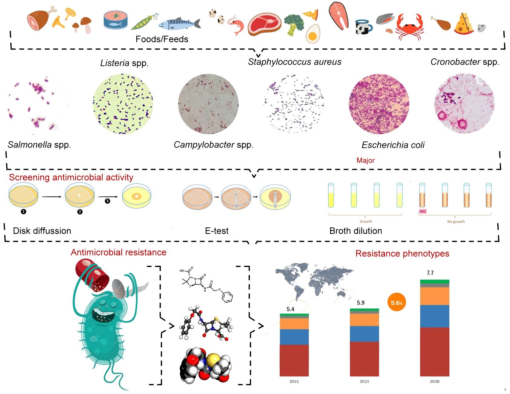
Graphical Abstract
1. Introduction
1.1. Foodborne Bacterial Pathogens
Foodborne pathogens have gained notoriety because, despite sustained quality practices and regulations regarding bacterial load in food production, foods are still found to be contaminated with bacteria. Pathogens are usually poor competitors and do not survive in the presence of existing food microflora [1] or after food processing or unit operations for food preservation (e.g., vacuum, pasteurization, nano/microfiltration) [2]. However, this ecological defense is not a guarantee of safety; food extended, erroneous manipulation, or bacterial survival mechanisms favor pathogen persistence. FDA food recalls are targeted actions to remove or correct food products that violate U.S. food safety laws. They are primarily driven by microbiological contamination, undeclared major allergens (such as milk or peanuts in mislabeled packages), and foreign objects. For example, of the 233 total food and beverage recalls from 2023, 70 (30.0%) were associated with possible or confirmed foodborne illness/bacterial contamination. Understanding the relevance of these recalls involves knowing how the FDA classifies and communicates the associated health risks. The most common contaminants were found to be 28 (41.2%), 32 (47.0%), 3 (4.4%), 4 (5.9%), and 1 (1.5%) for Salmonella spp., Listeria monocytogenes, E. coli, Cronobacter sakazakii, and Clostridium botulinum, respectively (Table 1). C. sakazakii has been the subject of contamination and product recalls of powdered and dairy formulas in Chile as well [3]. More recently, the most serious food recalls have involved Salmonella contamination in foods such as ice cream [4], cereals [5], peanuts [6], soft cheese [7], shelled walnuts [8], crispy fried onions [9], and hummus (Table 1) [10]. Recent French data indicate that, with some yearly decline in the number of cases, meats, dairy, and fishery products are among the most recalled products [11].
Table 1. List of Governmental food recalls requested within a two-year window 1.
|
Product Description 2 |
Recall Reason and Description |
||||
|---|---|---|---|---|---|
|
Year 01–12/2023 |
|||||
|
Salmonella |
Listeria monocytogenes |
Escherichia coli |
Cronobacter spp. |
Clostridium botulinum |
Summary |
|
28 (41.2%) |
32 (47.0%) |
3 (4.4%) |
4 (5.9%) |
1 (1.5%) |
68 |
|
Alfalfa Sprouts |
Contamination with Shiga toxin-producing E. coli |
||||
|
All Purpose Flour, bleached and unbleached |
Potential to be contaminated with Salmonella |
||||
|
Bagged Collard Greens |
Possible Listeria monocytogenes contamination |
||||
|
Bean Sprouts |
Listeria monocytogenes |
||||
|
Biltmore Smoked Wild Sockeye Salmon |
Potential Foodborne Illness/Listeria |
||||
|
Black Fungus (Nam Meo) |
Salmonella |
||||
|
Breeders Choice Raw Pet Food 2 lb |
Salmonella contamination |
||||
|
Cantaloupe Chunks and Cubes and Fruit Mixes and Medleys Containing Cantaloupes (×5) |
Potential Salmonella Contamination |
||||
|
Cheese products |
Potential Listeria monocytogenes contamination |
||||
|
Cocktail Shrimp |
Listeria monocytogenes |
||||
|
Dairy and Non-Dairy ice cream (parve), sorbet, cakes, and novelty items |
Potential to be contaminated with Listeria monocytogenes |
||||
|
Diced onions |
Salmonella |
||||
|
Diced Organic Butternut Squash |
Potential Escherichia coli O45 contamination |
||||
|
Dog and Cat food |
Potential to be contaminated with Salmonella |
||||
|
Dog food, Select Beef Meal & Brown Rice Formula |
Potential Salmonella contamination |
||||
|
Dry Dog, Dry Cat, and Catfish Food |
Potential Salmonella contamination |
||||
|
Enfamil Prosbee Simply Plant-Based Infant Formula in 12.9 oz containers |
Potential Cronobacter sakazakii contamination |
||||
|
Enoki Mushrooms (×4) |
Listeria monocytogenes |
||||
|
Fresh Cantaloupe (×2) |
Potential to be contaminated with Salmonella |
||||
|
Frozen Fruit |
Potential for Listeria monocytogenes contamination |
||||
|
Fruit cups and trays containing cantaloupe (×4) |
Potential to be contaminated with Salmonella |
||||
|
GEISHA Medium Shrimp, 4 oz can (×2) |
Potential contamination with Clostridium botulinum |
||||
|
Gift Baskets with Quaker Chewy Granola Bars |
Potential Salmonella contamination |
||||
|
Granola Bars and Granola Cereals |
Potential for Salmonella contamination |
||||
|
Ground Cumin |
Salmonella |
||||
|
Ice Cream, Yogurt, Ice Cream Bars, and Gelato in a Variety of Flavors |
Potential to be contaminated with Listeria monocytogenes |
||||
|
Kale, Spinach, Collard Green products (×2) |
Potential for Listeria monocytogenes |
||||
|
Lettuce & Salad Kits (×2) |
Listeria monocytogenes |
||||
|
Mini Chunk Chicken Recipe Dry Dog Food |
Potential Salmonella contamination |
||||
|
Multiple frozen fruits with mango products |
Potential Listeria monocytogenes contamination |
||||
|
Mung Bean Sprouts (×2) |
Listeria monocytogenes |
||||
|
Numerous human foods, animal (pet) food, medical devices, and drug products |
Potential Salmonella contamination and presence of rodent activity at the distribution center & temperature abuse |
||||
|
Nutramigen Powder infant formula in 12.6 and 19.8 oz cans |
Potential Cronobacter sakazakii contamination |
||||
|
Organic frozen Pineapple and Frozen Fruit Blend Containing Organic Frozen Pineapple |
Potential to be contaminated with Listeria monocytogenes |
||||
|
Organic green kiwifruit |
Possible Listeria monocytogenes contamination |
||||
|
Organic Tahini |
Potential Foodborne Illness/Salmonella |
||||
|
Pepper Jack Raw Milk Cheese |
Presence of Listeria monocytogenes |
||||
|
Powdered Infant Formula (×2) |
Potential Cronobacter sakazakii contamination |
||||
|
Premade Salads |
Potential Listeria monocytogenes contamination |
||||
|
Ready-to-Eat (RTE) Sandwiches, Salads, Yogurt, Wraps, and related products |
Potential Listeria monocytogenes contamination |
||||
|
Refrigerated and Frozen Cooked Lobster |
Possible Listeria monocytogenes contamination |
||||
|
Salad Greens |
Due to potential Escherichia coli O157:H7 contamination |
||||
|
Salads Kits |
Listeria monocytogenes |
||||
|
Sambhar Masala and Garam Masala Spices |
Potential Foodborne Illness-Salmonella |
||||
|
Soft Serve ice cream and sorbet cups |
Possible Listeria monocytogenes contamination |
||||
|
Sophelise, Tobasi, and Berkshire Bloom Cheeses (×2) |
Potential contamination with Listeria monocytogenes |
||||
|
Soybean Sprouts in 1 lb bags |
Potential contamination of Listeria monocytogenes |
||||
|
Spinach |
Listeria monocytogenes |
||||
|
Strawberry Granola & Greek Yogurt Parfait Bar |
Listeria monocytogenes |
||||
|
Various Deli Salads |
The product may be contaminated with Listeria monocytogenes |
||||
|
Whole Cantaloupe |
Potential to be contaminated with Salmonella |
||||
|
Whole Peaches, Plums, and Nectarines |
Potential to be contaminated with Listeria monocytogenes |
||||
|
Year 01–12/2024 |
|||||
|
Salmonella |
Listeria monocytogenes |
Escherichia coli |
Cronobacter spp. |
Clostridium botulinum |
Summary |
|
43 (38.7%) |
57 (51.3%) |
7 (6.3%) |
1 (0.9%) |
3 (2.7%) |
111 |
|
Falafel Bites |
Potential Shiga toxin-producing Escherichia coli O121:H19 contamination |
||||
|
Crab meat |
Potential foodborne illness—Listeria monocytogenes |
||||
|
Cashews |
Potential to be contaminated with Salmonella |
||||
|
Aged Cojita Mexican Cheese |
Potential to be contaminated with Listeria monocytogenes |
||||
|
Alfalfa and Onion Sprouts |
Listeria monocytogenes |
||||
|
Atovaquone oral suspension, USP, 750 mg/5 mL |
Potential Bacillus cereus contamination |
||||
|
Turkey Sandwiches |
Potential foodborne illness—Listeria monocytogenes |
||||
|
Baby Arugula |
Potential to be contaminated with Salmonella |
||||
|
Bacon Ranch Crunch Chopped Salad Kit (×2) |
Potential to be contaminated with Listeria monocytogenes |
||||
|
Beef and Chicken Dog Foods |
Due to Potential Salmonella and Listeria |
||||
|
Black bean six-layer dip |
Potential to be contaminated with Listeria monocytogenes |
||||
|
Southwest Chipotle Crunch Salad Kit |
Potential to be contaminated with Listeria monocytogenes |
||||
|
California Shelled Walnuts, Organic Light Halves and Pieces |
Due to potential Escherichia coli O157:H7 contamination |
||||
|
Chicken Chips Dog Treats (×2) |
Potential to be contaminated with Salmonella |
||||
|
Cereal, Bars, and Snacks |
Potential to be contaminated with Salmonella |
||||
|
Cheese, Yogurt, and Sour Cream |
Expanded recall for potential Listeria monocytogenes contamination |
||||
|
Chicken Enchiladas Verde, Cilantro Salad Dressing, Elote Chopped Salad Kit, and Southwest Salad |
Potential to be contaminated with Listeria monocytogenes |
||||
|
Chicken Street Taco Kit (×6) |
Potential to be contaminated with Listeria monocytogenes |
||||
|
Chillimami Sauce |
Potential foodborne illness—Clostridium botulinum |
||||
|
Chocolate Caramel Corn and Candy Tray |
Possible Salmonella contamination |
||||
|
Cilantro Line Crema, Everything Sauce Fiesta, Cilantro Cotija Dressing, Poblano Caesar Dressing, Cilantro Dressing, Street Taco Express Meal Kit |
Potential to be contaminated with Listeria monocytogenes |
||||
|
Coffee Products |
Potential to be contaminated with Clostridium botulinum |
||||
|
Cold Smoked Capelin |
Potential to be contaminated with Clostridium botulinum |
||||
|
Cucumbers and Salads with Kit |
Potential Foodborne Illness—Salmonella |
||||
|
Curly Mustards Greens |
Potential foodborne illness—Listeria monocytogenes |
||||
|
Cucumber Slices |
Potential Foodborne Illness—Salmonella |
||||
|
Eggs (×2) |
Potential to be contaminated with Salmonella |
||||
|
Enchiladas |
Potential to be contaminated with Listeria monocytogenes |
||||
|
Enoki Mushroom (×3) |
Potential to be contaminated with Listeria monocytogenes |
||||
|
Fresh Express Salsa Ensalada Kit, Southwest Chopped Salada Kit (×2) |
Potential to be contaminated with Listeria monocytogenes |
||||
|
Fresh Guacamole Products |
Potential to be contaminated with Listeria monocytogenes |
||||
|
Fresh Organic Basil (×2) |
Potential to be contaminated with Salmonella |
||||
|
Frozen Waffle Products (×2) |
Potential foodborne illness—Listeria monocytogenes |
||||
|
Chicken Caesar Salad Bowl |
Potential foodborne illness—Listeria monocytogenes |
||||
|
Green Onions |
Salmonella |
||||
|
Ground black pepper |
Potential Contamination with Salmonella |
||||
|
Gyro Family Kit |
Potential Foodborne Illness—Salmonella |
||||
|
Ham & Cotija Torta Sandwich on Telera Roll |
Potential to be contaminated with Listeria monocytogenes |
||||
|
Hedgehog food (×2) |
Potential Contamination with Salmonella |
||||
|
Honey Roasted Peanuts and Deluxe Lightly Salted Mixed Nuts |
Potential to be contaminated with Listeria monocytogenes |
||||
|
Ice Cream Products |
Listeria monocytogenes |
||||
|
Individually Wrapped Sandwiches |
Potential to be contaminated with Listeria monocytogenes |
||||
|
Jalapeños, Green Peppers, Green Beans |
Potential foodborne illness—Listeria monocytogenes |
||||
|
Smoked Salmon |
Potential foodborne illness—Listeria monocytogenes |
||||
|
Kitten Grind, Kitten Mix, Puppy Mix |
Potential Contamination with Salmonella and Listeria monocytogenes |
||||
|
Macadamia in the Raw |
Potential Contamination with Salmonella |
||||
|
Mango bars in the Variety Pack |
Potential Contamination with Salmonella |
||||
|
Meal Kits Containing Chicken |
Potential to be contaminated with Listeria monocytogenes |
||||
|
Mediterranean Inspired Party Tray |
|||||
|
Mexican Style Street Corn Bites |
Potential to be contaminated with Listeria monocytogenes |
||||
|
Moong Dal, Crunchy Green Gram |
Potential Contamination with Salmonella |
||||
|
Multiple Cucumber Products (×3) |
Potential Foodborne Illness—Salmonella |
||||
|
Thyroid Formula |
Potential to be contaminated with Salmonella |
||||
|
Organic Carrots and Celery |
Potential Foodborne Illness—Shiga toxin-producing Escherichia coli O121:H19 |
||||
|
Organic Chia Seeds |
Potential Presence of Salmonella |
||||
|
Organic Whole Carrots and Baby Carrots |
Potential Foodborne Illness—Shiga toxin-producing Escherichia coli O121:H19 |
||||
|
Parrot Food |
Due to Potential Salmonella contamination |
||||
|
Pepperjack Cheeseburger, Bacon Cheeseburger |
Potential foodborne illness—Listeria monocytogenes |
||||
|
Plain Whipped Cream Cheese, Plain Cream Cheese, and Cookies & Cream Mix |
Potential to be contaminated with Salmonella |
||||
|
Powdered Goat Milk Infant Formula |
Cronobacter spp. contamination |
||||
|
Puppy Mix |
Potential Foodborne Illness—Salmonella |
||||
|
Queso de Mano PAISA |
Potential to be contaminated with Listeria monocytogenes |
||||
|
Raw Cheddar Cheese |
Possible Contamination E. coli O157:H7 |
||||
|
Raw Dog and Cat Food |
Listeria monocytogenes |
||||
|
RTE Fruit and Vegetables |
Potential foodborne illness—Listeria monocytogenes |
||||
|
Shelled Walnuts |
Potential foodborne illness—Listeria monocytogenes |
||||
|
Smoked Norwegian Salmon Slices—Toast |
Potential to be contaminated with Listeria monocytogenes |
||||
|
Soft Ripened Cheese (×2) |
Potential to be contaminated with Listeria monocytogenes |
||||
|
Soybean Sprouts |
Potential to be contaminated with Listeria monocytogenes |
||||
|
Spinach and Salad Kits (×3) |
Potential to be contaminated with Listeria monocytogenes |
||||
|
Saint Jerome Cheese |
Potential foodborne illness—Listeria monocytogenes |
||||
|
Tahini (×2) |
Potential to be contaminated with Salmonella |
||||
|
Beef & Lamb Gyro Sandwich Express Meal Kits |
May be Contaminated with Salmonella |
||||
|
Torta Sandwiches |
Potential to be contaminated with Listeria monocytogenes |
||||
|
Various Confectionery Products (×2) |
Potential Contamination with Salmonella |
||||
|
Vegetable Medleys and Whole Organic Carrots |
Potential Foodborne Illness—Shiga toxin-producing Escherichia coli O121:H19 |
||||
|
Vegetable products (×4) |
Potential foodborne illness—Listeria monocytogenes |
||||
|
Whole Cucumbers (×5) |
Salmonella |
||||
|
Yogurt-covered pretzels |
Potential to be contaminated with Salmonella |
||||
1 Adapted from the United States Food and Drug Administration (US FDA) [12]. 2 Counters expressed as ×n represent the number of times a specific food item has been subjected to a safety callback.
1.2. Official Guidelines: Regulatory Documents, Methods, and Institutes
The presence of pathogens, especially in RTE foods, and the evidence of antibiotic resistance among these strains have evolved into a worldwide public health challenge [13,14]. Hence, detection and constant surveillance programs of these pathogens in foods are paramount. A primer on novel approaches to analyzing these bacteria in food can be found in recent work [15,16]. From the regulatory standpoint, the European Commission decision 2020/1729 includes the surveillance and antibiotic resistance report of the specific pathogens Salmonella spp., E. coli (especially those strains with extended spectrum β-lactamases (ESBL), AmpC β-lactamases, or carbapenemases), C. coli, C. jenuni, Enterobacter faecium, E. faecalis in foods such as broilers, laying hens, fattening turkeys and pigs, bovine animals under a year of age, fresh meat from broilers, turkeys, pigs, and bovine animals. Analogously, it has regulations regarding microbiological criteria for foodstuffs, specifically, 2073/2005/EC.
Official methods for detection of pathogens include ISO 6579-1:2017, ISO 11290-1:2017, ISO 10272-1:2017/Amd 1:2023, ISO 6888-1:2021, ISO 22964:2017 [17], ISO 16649-1:2018, DIS 13136-1, for Salmonella spp., and for Listeria spp., Campylobacter spp., Staphylococcus spp., Cronobacter spp., and E. coli, respectively. Relevant methods for food analysis also comprise ISO 20976-1:2019, 23418:2022, and 20976-2:2022. These methods have also evolved with the advent of new technologies for detecting and controlling foodborne pathogens [18,19,20,21,22,23,24]. Rapid, reliable, and cost-effective methods for bacteria detection are of paramount importance in food safety monitoring [25].
In the case of regulations for antimicrobial susceptibility testing, the European Committee on Antimicrobial Susceptibility Testing (EUCAST) sets epidemiological cut-off values (ECOFFs) and tentative ECOFFs (TECOFFs). For the USA, the Clinical & Laboratory Standards Institute CLSI sets these guidelines, and a workbook is available [26]. Additional guidelines for monitoring and reporting in zoonotic and commensal bacteria include 2013/652/EU (repealed by 2020/1729/EU) and 2003/99 [27]. Finally, ISO 20776-1/-2:2022 sets standards for micro broth dilution methods and other in vitro diagnostic tools for testing minimal inhibitory concentration (MIC).
Official methods for detecting pathogens and performing Antimicrobial Susceptibility Testing (AST) combine traditional phenotypic cultures with rapid molecular diagnostics. Standardized by global authorities, these tools are vital for guiding life-saving therapies, managing antimicrobial resistance (surveillance, policy, and stewardship), and ensuring optimal patient outcomes [28].
Only phenotypic results from recognized, standardized in vitro antimicrobial susceptibility testing of isolates, along with interpretive criteria, can successfully help classify multiple, extended, pan, and resistant bacteria [29].
Phenotypic testing is the recognized gold standard; it works by exposing isolated bacteria to specific antimicrobial concentrations and observing their actual growth response. While molecular/genotypic tests (such as PCR or sequencing) are excellent for identifying specific resistance genes, they cannot determine the cumulative effect of multiple resistance mechanisms. Because of this, standardizing definitions of drug resistance relies exclusively on phenotypic measurements [28].
1.3. Foodborne Bacterial Pathogens and Antibiotic Resistance
The long-term, overuse and misuse of antibiotics in food animals and humans create ideal conditions for the rising threat of antibiotic resistance. Resistant bacteria in animals may reach humans directly or indirectly through food, water, mud, and manure used as fertilizer [30]. Some types of bacteria (high-burden resistant pathogens such as Salmonella, Shigella, Neisseria gonorrhoeae, Pseudomonas aeruginosa, and Staphylococcus aureus) [31,32] that cause serious infections in humans have already developed resistance to most or all the available treatments, and there are very few promising options in the research pipeline [33,34].
Interventions that restrict antibiotic use in food-producing animals, such as biosecurity, improved animal management, vaccination, improved hygiene, and changes in animal housing and husbandry practices, have reduced antibiotic-resistant bacteria in these animals [35]. Hygiene practice alone can attenuate the effect of antibiotic selection pressure [36]. These practices, in conjunction with One Health approaches, should be implemented to counter antibiotic resistance [37,38]. In this regard, the One Health approach is a collaborative strategy recognizing that human, animal, plant, and environmental health are deeply interconnected. Because antibiotic resistance easily spreads across these domains, containing it requires integrated monitoring, responsible prescribing, and environmental stewardship across all sectors [39].
The overall reduction in the use of antibiotics in food-producing animals, including the complete restriction of these substances for growth promotion and disease prevention, without proper diagnosis. Malaise animals should be assessed to determine the most effective and prudent antibiotic to treat their specific infection. Meanwhile, healthy individuals should only receive antibiotics to prevent disease if, within a particular population, an animal has already been diagnosed [40].
Antibiotic use is further exemplified by the application of streptomycin (STR), oxytetracycline (OXY), copper-based products, and certain fungicides (e.g., triazoles) in plant agriculture. Their use is correlated with increased resistance among plant pathogens [41,42,43]. Horizontal gene transfer is responsible for genes being exchanged among a variety of bacteria in the plant production setting, including phytopathogens, soil bacteria, and zoonotic bacteria that are occasionally present in that environment and the food chain [30,42]. Through gene transfer, co-resistance, cross-resistance, and gene up-regulation, resistance to one compound may confer resistance and multi-drug resistance to other similar, or even very dissimilar, compounds [42].
Genetic information encoding bacterial resistance mechanisms can be chromosomal or carried by mobile elements called plasmids. Some bacteria are also inherently resistant to a particular antibiotic if they lack the structure on which the antibiotic acts. Several mechanisms of resistance have continuously evolved, including target modification or mutation, permeability reduction, expression of efflux pump genes, production of hydrolases or inactivating enzymes, metabolic enhancement or auxotrophy, synthesis of target-protective proteins, self-repair systems, changes in cell morphology, and biofilm protection (Figure 1) [44,45]. For a thorough primer on specific mechanisms of resistance by antibiotic class, the work by van Hoeck et al. [46] is suggested.
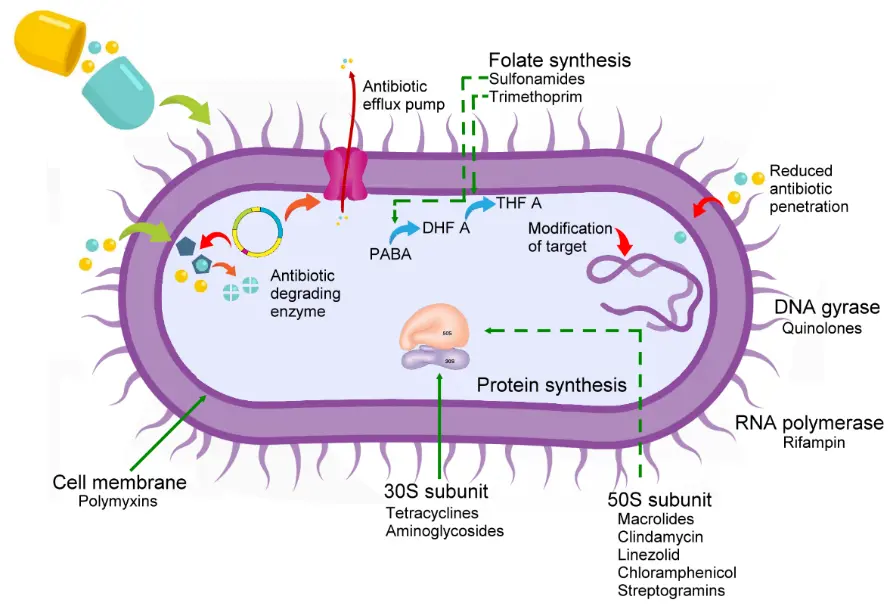
Figure 1. Schematic representation of common mechanisms of antibiotic resistance exhibited by bacteria. Yellow/Light blue spheres: Antimicrobial/active ingredient. Green arrows: antimicrobial introduction routes or target cell structures. Red arrows: antimicrobial export or target inhibition (e.g., outward export from the cell). Figure prepared using CorelDRAW® Graphics Suite 2026 (Corel Corporation, Ottawa, ON, Canada).
Plasmids are major vectors of bacterial antibiotic resistance [47]. Recent evidence demonstrates that antibiotic resistance-related plasmids are differentially dispersed among humans and livestock, reflecting known antibiotic prescribing practices [48]. For example, using a large-scale multivariate approach, the authors found that only 0.42% of livestock plasmids carried carbapenem resistance (compared with 12% of human plasmids); conversely, tetracycline resistance was enriched in livestock vs. human plasmids [48]. However, they also found that the prevalence of carbapenem and colistin plasmid-related resistant genes is on the rise.
Despite the long list of antibiotics commercially and clinically available (Table 2), resistance has made some of these drugs therapeutically useless against multidrug-resistant bacteria. Hence, the discussion of alternatives to antibiotics has resulted in natural product-led research [49,50,51]. Several primers regarding natural products with antimicrobial capabilities are advised [52,53,54,55,56,57]. Interestingly, recent research suggests that constant bacterial exposure to even these alternatives can affect their resistance profile (that is, decreased sensitivity). Exposure to biocides, pesticides, common home cleaners, heavy metals, food preservatives, and even natural products can lead to a cross-adaptation to antibiotics induced by exposure to non-antibiotic antimicrobials [58,59,60]. This co-resistance occurs when bacterial exposure to sub-lethal concentrations of non-antibiotic agents, used within the clinical, agricultural, and domestic settings, inadvertently selects strains that are resistant to therapeutic antibiotics (i.e., the physiological changes often double as defense mechanisms against antibiotics) [61]. More worrisomely, evidence of this behavior in pathogenic foodborne bacteria such as S. aureus, E. coli, S. typhimurium, P. aeruginosa, K. pneumoniae, L. monocytogenes, A. baumanii, C. sakazakii, and other species is clear [59].
Table 2. The ninety-three antimicrobials mentioned during this survey, their mechanisms of action, and their abbreviations.
|
Drug Class |
||
|---|---|---|
|
Action Mechanism 1 |
Antibiotics |
Abbreviations |
|
Aminoglycosides |
||
|
Inhibition of protein synthesis. |
Amikacin |
AMK |
|
Apramycin |
APM |
|
|
Gentamycin |
GEN |
|
|
Kanamycin |
KAN |
|
|
Neomycin |
NEO |
|
|
Netilmicin |
NTM |
|
|
Spectinomycin |
SPC |
|
|
Streptomycin |
STR |
|
|
Tobramycin |
TMP |
|
|
Ansamycins/Rifamycins |
||
|
Inhibits bacterial DNA-dependent RNA synthesis (RNA polymerase). |
Rifampicin |
RIF |
|
β-lactam/β-lactamase inhibitor |
||
|
Bind to plasmid-mediated β-lactamases and inactivate them. |
Amoxicillin-clavulanic acid |
AMC |
|
Ampicillin-sulbactam |
SAM |
|
|
Cefotaxime-clavulanic acid |
CTC |
|
|
Ceftazidime-avibactam |
CVI |
|
|
Ceftazidime-clavulanic acid |
FOX |
|
|
Piperacillin-tazobactam |
TZP |
|
|
Carbapenems/β-lactams |
||
|
Fix to PBPs 2, inhibiting bacterial cell wall synthesis. |
Ertapenem |
ETP |
|
Imipenem |
IMP |
|
|
Meropenem |
MEM |
|
|
Carboxypenicillins/β-lactams |
||
|
Inhibit cell-wall synthesis. |
Carbenicillin |
CAB |
|
Ticarcillin |
TIC |
|
|
Carboxylic acids |
||
|
Pseudomonic acid inhibits isoleucine—tRNA ligase in bacteria, leading to depletion of isoleucyl-tRNA & accumulation of the corresponding uncharged tRNA. Depletion of isoleucyl-tRNA results in inhibition of protein synthesis. |
Mupirocin (Bactroban) |
MCP |
|
Cephalosporins/β-lactams |
||
|
Inhibit cell-wall synthesis. |
Cefalexin (1st class) |
CPX |
|
Cefazolin (1st class) |
CEZ |
|
|
Cefepime (4th class) |
CFP |
|
|
Cefixime (3rd class) |
CFM |
|
|
Cefotaxime (3rd class) |
CFX |
|
|
Cefotetan (2nd class) |
CTT |
|
|
Ceftiofur (3rd class) |
CFT |
|
|
Cefovecin |
CFO |
|
|
Cefoxitin (2nd class) |
CEX |
|
|
Ceftachlor (2nd class) |
CFC |
|
|
Ceftazidime (3rd class) |
CZA |
|
|
Cefpodoxime (3rd class) |
CPD |
|
|
Cefoperazone (3rd class) |
CPZ |
|
|
Cefquinome (4th class) |
CFQ |
|
|
Ceftaroline (5th Class) |
CRN |
|
|
Ceftriaxone (3rd class) |
CTX |
|
|
Cefuroxime (2nd class) |
CXM |
|
|
Cephalothin (1st class) |
CEP |
|
|
Diaminopyrimidines/Sulfonamides |
||
|
Dihydrofolate reductase inhibitor & Competitive inhibitors of the enzyme dihydropteroate (folate) synthase. |
Trimethoprim/sulfamethoxazole (Bactrim® or Septra®) |
STX |
|
Sulfadiazine |
SDZ |
|
|
Sulfamethaxole |
SMZ |
|
|
Sulfafurazole/Sulfisoxazole |
INN |
|
|
Trimethoprim |
TMP |
|
|
(Fluoro)quinolones |
||
|
Interferes with DNA replication by preventing bacterial DNA from unwinding and duplicating. Specifically, they inhibit the ligase activity of the type II topoisomerases, DNA gyrase, and topoisomerase IV, which cut DNA to introduce supercoiling, while leaving nuclease activity unaffected. |
Ciprofloxacin |
CIP |
|
Enrofloxacin |
ENR |
|
|
Flumenique |
FMQ |
|
|
Gamifloxacin (4th generation) |
GMX |
|
|
Gatifloxacin (4th generation) |
GTX |
|
|
Levofloxacin |
LFX |
|
|
Nalidixic acid |
NAL |
|
|
Marbofloxacin |
MBF |
|
|
Moxifloxacin |
MFX |
|
|
Norfloxacin |
NOR |
|
|
Ofloxacin |
OFX |
|
|
Oxolinic acid |
OXO |
|
|
Pefloxacin |
PFX |
|
|
Phosphonic acid derivatives |
||
|
Bactericidal inhibits bacterial cell wall biogenesis by inactivating the enzyme UDP-N-acetylglucosamine-3-enolpyruvyltransferase (MurA) |
Fosfomycin (Monurol) |
FOS |
|
Glycopeptides |
||
|
Bind to acyl-D-alanyl-D-alanine in lipid II, preventing the addition of new units to the peptidoglycan. |
Vancomycin (1st gen) |
VAN |
|
Teicoplanin (Targocid) |
TEI |
|
|
Macrolides/Polyketide/Ketolide |
||
|
Bacteriostatic, protein synthesis inhibitors. Prevent peptidyltransferase from adding the growing peptide attached to tRNA to the next amino acid. Inhibit bacterial ribosomal (bind reversibly to the P site on the 50S) translation. Dissociate the peptidyl-tRNA from the ribosome prematurely. |
Azitromycin |
AZM |
|
Erythromycin |
ERY |
|
|
Clarithromycin |
CAM |
|
|
Midecamycin |
MDY |
|
|
Telithromycin |
TYM |
|
|
Tylosin |
TYL |
|
|
Nitrofurans |
||
|
Rapid reduction inside the bacterial cell by flavoproteins to multiple reactive intermediates that attack ribosomal proteins, DNA, respiration, pyruvate metabolism, and other macromolecules within the cell. |
Furazolidone |
FZD |
|
Nitrofurantoin |
NFT |
|
|
Lincosamides |
||
|
Bacteriostatic, interferes with the synthesis of proteins. Bind close to the peptidyl transferase center on the 23S portion of the 50S subunit of bacterial ribosomes. |
Lincomycin |
LIN |
|
Clindamycin |
CDA |
|
|
Oxazolidinones |
||
|
Blocks the initiation of protein production |
Linezolid |
LZD |
|
Penicillins/β-lactams |
||
|
Inhibit the completion of the synthesis of peptidoglycans, the structural component of the bacterial cell wall. Inhibits the activity of enzymes that are needed for the cross-linking of peptidoglycans during the final step in cell wall biosynthesis. |
Amoxicillin |
AMO |
|
Ampicillin |
AMP |
|
|
Aztreonam (monobactam) |
ATM |
|
|
Cloxacillin |
CLX |
|
|
Methicillin |
MET |
|
|
Oxacillin |
OXA |
|
|
Penicillin G |
PEN |
|
|
Temocillin |
TMC |
|
|
Phenicols |
||
|
Bacteriostatic, inhibits protein synthesis. Prevents protein chain elongation by inhibiting the peptidyl transferase activity of the bacterial ribosome. It specifically binds to A2451 and A2452 residues in the 23S rRNA of the 50S ribosomal subunit, preventing peptide bond formation. |
Chloramphenicol |
CHL |
|
Florfenicol |
FLO |
|
|
Polymyxins |
||
|
Bind to lipopolysaccharide in the outer membrane of Gram-negative bacteria, polymyxins disrupt both the outer and inner membranes. The hydrophobic tail is important in causing membrane damage, suggesting a detergent-like mode of action (increase membrane permeability/disorganization). |
Colistin (Polymyxin E) |
COL |
|
Polymyxin B |
PMB |
|
|
Steroids |
||
|
Binds to EF-G after translocation and GTP (guanosine-5′-triphosphate) hydrolysis. |
Fusidic acid (Fucidin) |
FAS |
|
Streptogramins |
||
|
Bacteriostatic, binds to the bacterial 50S ribosomal subunit and inhibits the elongation process of protein synthesis. |
Pristinamycin |
PRI |
|
Tetracyclines |
||
|
Protein synthesis inhibitors. Prevent the initiation of translation in various ways by binding to the 30S ribosomal subunit. Hinder the binding of aminoacyl-tRNA to the mRNA translation complex. |
Doxycycline (Semi-synthetic) |
DOX |
|
Minocycline (Semi-synthetic) |
MIN |
|
|
Oxytetracycline (Naturally occurring) |
OXY |
|
|
Tetracycline (Naturally occurring) |
TET |
|
|
Tigecycline (Glycecyclines) |
TIG |
|
|
Ureidopenicillins/β-lactams |
||
|
Inhibit cell-wall synthesis |
Mezlocillin |
MEZ |
|
Piperacillin |
PIP |
|
1 Mechanisms of action for antimicrobials adapted from Singh et al. [62]. 2 PBPs, Penicillin-binding proteins.
2. Salmonella spp.
2.1. Foods for Human Consumption
Salmonella is a genus of rod-shaped (bacillus), motile, non-spore-forming, chemotrophic, facultative anaerobe, and Gram-negative bacteria of the family Enterobacteriaceae. It is known for its two species, S. enterica and S. bongori. S. enterica is further divided into six subspecies that include over 2600 serotypes [63,64].
Nontyphoidal Salmonella serotypes are zoonotic and can be transferred from animals to humans. Infection by this pathogen is usually due to ingestion of contaminated, undercooked food. Salmonella is one of the most common causative agents of foodborne illness in humans, causing approximately 1.35 million infections, 26,500 hospitalizations, and 420 deaths in the United States alone every year [65].
Treatment of salmonellosis is further complicated by the fact that isolates may also carry resistance genes conferring resistance to antimicrobials. For example, Salmonella species carrying antimicrobial resistance genes have been isolated from animal- and plant-derived foods, including poultry, eggs, fish, pork, beef, vegetables, fruits, nuts, and flour [56]. Outbreak studies have recognized food commodities, including fresh produce, vegetables, cantaloupes, cereals, alfalfa sprouts, fruit/fruit pulp, pistachios, poultry meat, turkeys, tuna, ground beef, pork, dried/shredded coconut, tomatoes, and mangos, as sources of foodborne Salmonella-related outbreaks in the past decade [66,67,68,69].
Soubeiga et al. [70] found Salmonella-related virulence genes associated with vegetable/fruit sources. Multidrug resistance profiles were consistent determinants in the 148 isolates, as genes blaTEM, temAB (β-lactams), StrA, aadA (aminoglycosides), sul1, sul2 (sulfonamides), and tetAB (tetracyclines) were consistently present (Figure 2).
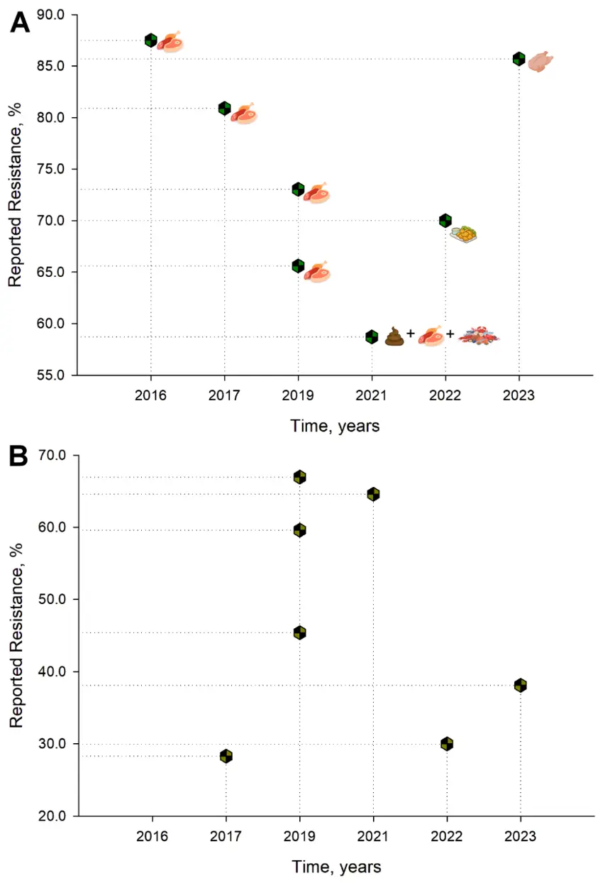
Figure 2. Reported resistance profiles for (A) Tetracycline and (B) Ampicillin for foodstuffs and related matrices (assorted meats, RTE, feces, aquatic products, and poultry) from China from 2016–2024. China was selected as it is the country with the most entries (nine) as per Table 3. Figure elaborated using Sigma Plot 16 (Grafiti LLC, Palo Alto, CA, USA).
Similar gene profiles [e.g., aadA, aac, aph, blaCARB, blaPSE, blaTEM, blaCTX-M, blaCMY, floR (phenicols), folate pathway inhibitors sul and dfrA (folate pathway inhibitors), qnrAB, oqxAB (fluoroquinolones), tetABM, and mph(A) and lnu(F) (macrolides)] were found recently, but in retail American poultry [71], Mexican ground beef samples [72], and Chilean poultry production line samples [45]. Analogous gene profiles were also found in food chain links in Poland [73]. On the other hand, recent Italian isolates from swine mostly exhibited resistance determinants parC, gyrA, catA, nfsAB, blaTEM, and tetAB. For a detailed primer regarding mechanisms of resistance for Salmonella, the work by Monte et al. [74] and Wang et al. [75] is recommended.
Furthermore, as global trends indicate that more people are basing their diets on fresh produce and vegetables [76], Salmonella contamination is relevant, as an increase in produce-related outbreaks has been documented [77]. Additionally, multidrug-resistant bacteria can disseminate more quickly due to an ever-expanding regional and international food trade [77]. More problematic still, it is easy to forget that foods, in general, and especially fruit and vegetables [78], can carry more than one pathogen, and these pathogens can carry resistance genes and even share them amongst themselves and other bacteria. In a very thorough Cuban study, 14 different food groups were analyzed for the presence of Salmonella spp., E. coli, Staphylococcus spp., and V. cholerae [79]. Meat and dairy products and RTE foods showed not only multiple pathogen contamination but also multiple drug resistance profiles [79]. A recent study saw lettuce, cabbage, spinach, carrot, tomato, and green peppers contaminated with other antibiotic-resistant species alongside Salmonella (e.g., E. coli, S. aureus, S. epidermis) [78].
On the other hand, Cao et al. [80] found a high prevalence of Salmonella (isolating the bacteria at a rate of 29.4% per sample tested) contamination in cow’s milk. A similar account was reported by Ejo et al. [81], who found both a higher prevalence of contamination and a drug resistance profile in raw meat, eggs, and milk.
Quesada et al. [82] reviewed Salmonella reports from 2003–2014 from Latin America, as well as the antibiotic-resistant profiles of these isolates (especially recovered from the Brazilian and Mexican poultry industry). Among their findings, S. Enteritidis, S. Typhimurium, S. Paratyphi, and S. Heidelberg were the most commonly encountered serovars. Similar to other reports [83,84] most strains were multidrug-resistant and showed a high tolerance to STR, NAL, AMP, TET, CHL, and STX (Table 3). Relatedly, a 22-year historical analysis of antibiotic-resistant Salmonella from Bangladesh found 81% tetracycline resistance [85], rendering this antibiotic effectively without any therapeutic or clinical use. However, the continued use of tetracyclines in other agricultural settings (e.g., crop production, apiculture, and aquaculture) still provides a selective pressure on bacteria [40].
As macrolides (the first-choice drugs) for Salmonella infections in humans have failed, cephalosporins are now the paramount therapeutics. However, ESBL enzymes encoded by blaCMY2, blaSHV, and blaTEM emerged, thus reducing the efficacy of cephalosporins. Also, generalized mcr genes have been described in Salmonella and encode colistin resistance, an antibiotic regarded as critical. In both cases, resistance genes were carried by conjugative plasmids (e.g., IncI2, IncHI2, IncX3, IncX4) [86].
Interestingly, Siddique et al. [87] not only found Salmonella strains carrying β-lactam, cephalosporins, and carbapenems resistance genes (e.g., blaCMY-2, blaOXA-1, blaPSE-1, blaTEM-1, blaNDM-1, and ampC), and the zoonotic potential virulence gene sopE, but also noted that the strains recovered had moderate-to-high abilities to aggregate into biofilms at 30 and 37 °C. Other virulence factors (e.g., fimA, invA, spvC) co-occurring with similar resistance gene patterns have been recently reported in Poland [73], Italy [88] and China [89,90,91,92].
For Brazilian poultry farms, Rau et al. [93] found, in two different sampling moments (i.e., 3 years apart), that traditional Salmonella serovars Typhimurium and Enteritidis have been supplanted by other serovars like Heidelberg or Kentucky. In the same period, Brazil saw an increase in the number of isolates exhibiting antimicrobial resistance by almost double. Similar phenomena seem to be occurring of late in countries like the USA and Costa Rica [94,95].
As the debate about rendering animals without antibiotics rages on, evidence suggests that alternative meat production practices reduce the frequency of bacteria exhibiting antibiotic resistance, which favors “organic” practices [71]
2.2. The Relation Between the Poultry Industry and the Emerging Salmonella Infantis (pESI)-Plasmid
Salmonella enterica serovar Infantis (or S. Infantis) is an emergent serotype worldwide (Table 3) [96]. This serotype has been reported as one of the most prevalent non-typhoidal Salmonella (NTS) in humans in Europe and the USA [97,98,99]. Several studies describe S. Infantis as the most prevalent serotype in poultry [98,99]. Recent data from over 10,000 isolates demonstrated that S. Infantis and S. Seftenberg with pESI plasmid carrying a blaCTX-M-65 gene constitute one of the biggest clusters of Salmonella along the United States, both originating from turkeys [100].
Moreover, multidrug-resistant phenotypes of this serotype are of public health concern [101]. For example, Mattock et al. [102] recently related the presence of pESI megaplasmid with 73% of the poultry isolates from 74 different countries showing multidrug resistance. Hence, the presence of Salmonella in poultry production has attracted serious attention, and several studies have found in poultry a vector for several species of this bacterium. Whole genome sequencing from broiler integrations determined that the most prevalent non-typhoid Salmonella serotype was S. Infantis [103]. Not only do most isolates show potential for resistance against medically important antibiotics [especially aminoglycosides (mutations in genes aac, ant, aph) and quinolones (parC, gyrA)] and disinfectants, but also almost all isolates harbored the pESI-like megaplasmid, which plays an important role in the global dissemination of antimicrobial resistance. Among analogous Latin American studies, the blaCTX-M-65 ESBL antibiotic resistance gene is also prevalent in isolates. This was more recently confirmed by Costa Rican-led research, in which ampicillin resistance in the more prevalent S. Infantis reached 81.6% [95]. In terms of plasmid prevalence, a similar trait has been observed in S. Muenchen, S. Schwarzengrund, S. Agona, and S. Senftenberg in a large-scale broiler and chicken isolate study [104]. Previous studies, again in the Andean Region (mostly Ecuador, Chile, and Peru), seem to support similar findings [105,106,107,108,109]. A more recent Chilean study [47] has reported mobile elements carrying multiple drug resistance determinants in the poultry production line, predominantly related to S. Infantis and S. Heidelberg (IncFIB and Incl1-Iα plasmids, respectively).
Furthermore, the ESI clone carrying an extended spectrum β-lactamase blaCTX-M-65 on a pESI-like plasmid and a mutation in the gyrA gene has also been found in poultry meat in regions such as Russia, USA, Italy, Germany, Hungary, Poland, and Slovenia [110,111,112,113,114]. All these data suggest that pESI is not only widespread but is acquired by Salmonella spp. with relative ease. Finally, recent evidence has described pESI-like megaplasmids as carriers of additional antimicrobial resistance genes, such as tet, dfr, and sul, and has demonstrated that these structures are genetically plastic and can also harbor virulence and fitness genes, as well as toxin/antitoxin systems that enhance their persistence in the S. Infantis host [97].
2.3. Animal Feeds and Related Matrices
Salmonella monitoring and surveillance in animal feed are not only related to production costs and animal welfare but are also instrumental in understanding the behavior of foodborne pathogens at the start of the food chain and how this relates (if any) to prevalence, drug-resistant profiles, and specific species found downstream in human or clinical isolates.
Relatively few studies have tackled feed contamination despite being considered a main source of contamination on farm grounds (Table 3) [65]. In Thailand, the prevalence of Salmonella contamination in compound feeds (a specially formulated, balanced mixture of feed components designed for feeding livestock and poultry) was registered at 12.5% [115]. Similar data have been recovered from isolates from American and Australian feeds [71,116,117,118]. The authors found more prevalence in the compound feed than in any of its respective ingredients. However, Parker et al. [118] recently related contamination hotspots with the milling equipment during feed manufacturing. Hsieh et al. [117] found an 84% contamination in fish meals and 48% in animal products. From these samples, the acquisition of the blaCMY-2 gene, which confers resistance to cephalosporins, was most commonly observed. Interestingly, multidrug-resistant isolates were recovered from monensin (an ionophore/Rumensin®)-medicated dry beef cattle feeds. A possible association between the use of ionophores in livestock and antibiotic resistance has recently been raised [119].
Of all the bacterial pathogens discussed, Salmonella seems to have received the most attention in the feed sector (both in surveillance and research). Antibiotic resistance in Salmonella often stems from agricultural practices, where antimicrobial usage in animals selects resistant strains that spread into the human food supply. This poses a major public health issue, as these pathogens can trigger severe infections that are difficult to treat with standard medications. The cycle of how antibiotic resistance moves from animal feed to human consumers operates as follows: i. Contaminated Feed, ii. Livestock Exposure, iii. Indirect Transmission to Humans [120].
As stated, however, there is a very close link between livestock and human pathogen contamination. For instance, the main source of Salmonella infections in humans is contaminated animal-derived foodstuffs, with pork products being among the most important contributors [65].
Table 3. Summary of bacterial isolates from foodstuffs and other food-related matrices for Salmonella.
|
Country |
Isolates |
Source |
Antibiotics Tested |
Resistance to 1 |
Testing Method |
References |
|---|---|---|---|---|---|---|
|
Feeds |
||||||
|
USA |
S. Senftenberg, S. Montevideo, S. Mbandaka |
Animal feeds (finished feeds), feed ingredients, pet foods, treats, & supplements (n = 2058) |
AMC, AMP, CEX, CHL, CIP, GEN, KAN, NAL, NOR, STX, INN, STR, TET |
TET, INN, AMP, NAL |
Sensititre system (micro version of the broth dilution method) |
[116] |
|
Costa Rica |
S. Give, S. Rissen, S. Havana, S. Soerenga, S. Schwarzengrund |
Feed & ingredients (n = 1725; meat and bone meal, poultry feed, pet foods) |
TET |
TET (7.4%) |
E-test |
[121] |
|
USA |
S. Newport, S. Typhimurium var. O 5-(Copenhagen), & S. Lexington var. 15+ (Manila) |
Dry beef (n = 3) & dry cattle feeds (n = 1), cabbage culls (n = 2), animal protein products (n = 2), & fish meal (n = 1) |
AMC, AMP, CEX, CFT, CHL, KAN, STR, INN, TET, SXT |
AMC, AMP, CEX, CFT, CHL, STR, INN, TET |
Sensititre automated antimicrobial susceptibility system |
[117] |
|
Tanzania |
Salmonella spp. |
Commercial chicken feed samples (n = 384; Broiler starter mash (n = 121), Broiler grower mash (n = 47), Broiler finisher mash (n = 102) & Layers mash (n = 114) |
STR, AMO, AMK, CIP, TET, KAN, GEN, STX |
TET, AMK, KAN, GEN, STX |
Kirby-Bauer disk-diffusion |
[122] |
|
Kenya |
Salmonella spp. |
Poultry feed (n = 150) |
AMP, CTX, STX, TET, CHL, CIP |
AMP (41%), STX, CTX |
Kirby-Bauer disk-diffusion |
[123] |
|
Colombia |
Salmonella spp. |
Feed for swine, poultry, canine, feline, leporine, & equine species (n = 1758) |
AMP, SAM, CEZ, CFP, CEX, CTX, CIP, IMP, MEM, TZP |
AMP (44.5%), SAM, CEZ |
Becton Dickinson PhoenixTM Automated Dilution System |
[124] |
|
Costa Rica |
S. Typhimurium, S. Braenderup, S. Newport, S. Anatum, S. Westhampton |
Non-human primate feces (n = 180) & feed (n = 43) |
AMP, AMC, TPZ, CFX, CZA, CFP, IMP, MEM, AMK, GEN, NAL, CIP, NFT, COL, STX |
CIP (9.8%) |
VITEK®2 AST-N279 cards |
[125] |
|
Thailand |
S. Rissen, S. Mbandaka, & S. Livingstone |
Compound feed (n = 60), Soybean (n = 10), pork (n = 5), fish (n = 3) & poultry meal (n = 2) |
AMP, AMC, PIP, CPX, CFP, CFT, CFO, AMK, GEN, TMP, IPM, STX, CHL, ENR, MBF, TET, DOX, MIN, NFT |
TMP (100%), CPX, AMK, GEN |
VITEK®2 Card type AST-GN65 |
[115] |
|
Foods and related matrices |
||||||
|
Thailand |
S. Rissen, S. Weltevreden & S. Bredeney for pork; S. Albany & Typhimurium for chicken; S. Typhimurium & S. Hvittingfoss for vegetables |
Pork (n = 41) & chicken meat (n = 40) & fresh vegetables (n = 80) |
TET, CIP, CHL, AMP, NOR, NAL, STR, STX, CEP, GEN |
STR (92%), TET, NAL, CHL, AMP, CEP |
Kirby-Bauer disk-diffusion |
[126] |
|
Türkiye |
Salmonella spp. |
RTE salads & vegetables (n = 81) |
KAN, NEO, NAL, TET, SPC, STX, AMP, AMC, CHL, FLO, GEN, CIP, STR, CFT |
NEO, NAL |
Micro dilution method |
[127] |
|
Nigeria |
Salmonella spp. |
Fecal samples of cattle, pig, sheep, goat, & chicken (n = 200 each) |
AMP, AMO, AMC, CHL, STR, TET, CEP, GEN, CIP, SMZ, NAL |
Cattle [TET (85%), AMP, CIP, NAL], Sheep [AMP (25%), STR, CHL, TET] Goat [TET (44%), AMP, CHL, STR, CEP, SMZ, NAL], Chicken [TET (90%), NAL, SMZ, AMP, CHL, STR, CEP], Pig [TET (75%), AMP, CHL, CEP, SMZ, NAL] |
Kirby-Bauer disk-diffusion |
[128] |
|
Nigeria |
S. Typhi, S. Paratyphi & S. Typhimurium |
Cabbage, lettuce, cucumber, tomatoes, green pepper & spinach |
AMC, STX, CIP, CTX, GEN, TET, KAN, CEP |
CEP, STX, TET, KAN, multidrug-resistant profile |
Kirby-Bauer disk-diffusion |
[129] |
|
Poland |
S. Enteritidis |
Foods other than meat: confectionery products, eggs, fruits, vegetables, spices |
ATM, AMC, AMP, CFP, CFX, CEX, CZA, CTX, CHL, CIP, ETP, GEN, IMP, NAL, STR, TMP, TET, STX |
NAL (35.2%) |
Kirby-Bauer disk-diffusion |
[130] |
|
Malaysia |
Salmonella spp., S. Enteritidis & S. Typhimurium |
Ulam (salad, fresh leaves, vegetables, or fruits; n = 96) |
CHL, CIP, AMP, ERY, CEP, STR, AMC, NAL, GEN, STX, TET |
AMP (100%), ERY (100%), AMC, CEP, STR, CIP |
Kirby-Bauer disk-diffusion |
[131] |
|
Ghana |
S. Kentucky, S. Nima, S. Muenster, S. Enteritidis |
Laying hens & broilers (feces n = 75, dust n = 75, feed n = 10 & drinking water n = 10) |
AMP, CFX, CEX, GEN, CZA, TET, CHL, TMP, SMZ, NAL |
NAL, TET, CIP |
Kirby-Bauer disk-diffusion |
[132] |
|
Ethiopia |
Salmonella spp. |
Animal-origin food samples (n = 384: egg sandwich, minced & raw meat, burger patties, cottage cheese, cream cake, beef pizza, raw egg & pasteurized & raw milk from restaurants, hotels, cafeterias, & retail shops) |
AMO, AMP, CEP, CTX, GEN, NAL, NFT, STX, TET |
TET (42.6%), STX, AMP |
Kirby-Bauer disk-diffusion |
[81] |
|
Nigeria |
Salmonella spp. |
Raw chicken (retail table) eggs (n = 340) |
NEO, DOX, STX, AMO, VAN, NFT, PEN, CIP, ERY, GEN, STR, TET, OXA, AMC |
STR (85%), AMC, GEN, VAN, DOX |
Kirby-Bauer disk-diffusion |
[133] |
|
China |
S. Typhi, S. Enteritidis, S. Typhimurium |
Pork (n = 85) & chicken (n = 200) at retail stores |
CFX, GEN, LFX, TET, DOX |
TET |
Kirby-Bauer disk-diffusion |
[89] |
|
South Africa and Brazil |
Salmonella spp. |
Broiler chicken caecum samples (n = 200) |
GEN, AMO, ERY, CHL, TET, TMP, AMP, STR, STX |
TET (93%), STX, TMP, KAN, GEN, AMP |
Kirby-Bauer disk-diffusion |
[134] |
|
China |
Salmonella spp. |
Pork (n = 137), chicken (n = 91), & beef (n = 99) from supermarkets |
AMO, AMC, AMP, CFT, CIP, OXY, LFX, TMP, GEN |
OXY (80.9%), TMP, AMP, LFX |
Agar dilution method |
[58] |
|
Malaysia |
S. Corvallis, S. Typhimurium, S. Braenderup, S. Albany |
Spent hens (n = 470) |
AMP, CTX, CHL, CIP, ERY, GEN, KAN, NAL, STR, STX, TET |
ERY (97%), TET, STR, STX, AMP, NAL |
Kirby-Bauer disk-diffusion |
[135] |
|
Latvia |
S. Typhimurium, S. Derby, S. Enteritidis |
Meat and meat products (n = 3152) |
AMP, ATM, CFX, CZA, CHL, MEM, CIP, TET, TIG, NAL, COL, GEN, TMP, SMX. |
SMX (38%), NAL, CIP, AMP, TET |
Broth microdilution (Sensititre® EUVSEC3® & EUVSEC2® plates) |
[136] |
|
Ghana |
Salmonella spp. |
Cabbage and lettuce samples |
AMP, PEN, CHL, CIP, CTX, GEN, ERY, OFX, STX, TET |
AMP (72.2%), ERY, OFX |
Kirby-Bauer disk-diffusion |
[137] |
|
Vietnam |
Salmonella spp. |
Fecal samples (n = 240) & Environmental samples (feed, water, barn floor & wastewater; n = 640) from household and pig farms |
AMC, CZA, CXM, COL, GEN, AMK, STR, TET, DOX, CHL, STX, AMP, OFX, LFX |
STX (64.5%), CHL, AMP |
Kirby-Bauer disk-diffusion |
[138] |
|
Bangladesh |
Salmonella spp. |
Fuska (n = 55), sugarcane juice (n = 58), Borhani (n = 30) |
AMP, AMO, CIP, ENR, COL, OXY, TET, AZM, ERY, PFX, CTX |
High level of resistance to all antibiotics tested, especially TET, AZM, ERY, AMP, AMO |
Kirby-Bauer disk-diffusion |
[139] |
|
Ethiopia |
S. Dublin, S. Virchow |
Fecal samples of slaughtered cattle (n = 720) |
AMP, AMC, CHL, CTX, CEP, CIP, CEX, GEN, KAN, STX, TMP, TET, SMZ, STR, NFT, NAL |
STR (89.3%), CEP, AMP, AMC |
Kirby-Bauer disk-diffusion (BD BBLTM Sensi-DiscTM) |
[140] |
|
USA |
S. Newport |
Humans (n = 2763), animals (n = 901), & retail-meats (n = 64) |
AMK, APM, GEN, KAN, STR, AMC, TZP, CEP, CEX, CTX, CFT, CZA, CFX, CFQ, CFP, INN, STX, AZM, ATM, IMP, AMP, CHL, CIP, NAL, TET |
Multidrug resistance-AMP & single drug resistance-TET |
Agar dilution Method |
[141] |
|
South Africa |
S. bongori, S. Pullorum, S. Typhimurium, S. Weltevreden |
Fecal samples (poultry pens of broilers, layers & indigenous chickens, n = 20 each) |
AMP, OXY, CIP, STR, GEN, STX, CHL, ERY, NOR, CEP, NAL |
ERY (100%), STR, STX, OXY, AMP |
Kirby-Bauer disk-diffusion |
[142] |
|
Nigeria |
S. Enteritidis, S. Typhimurium |
Ready to eat shrimp (n = 1440) |
KAN, GEN, STR, ERY, TOB, AMP, AMO, IMP, SAM, MEM, CEP, TMP, AMC, COL, CHL, CHL, PEN, PMB, OXY, DOX, TET, OFX, CIP |
ERY, PEN (100%), AMO (92.9%), AMC, AMP (85.7%) |
Kirby-Bauer disk-diffusion |
[143] |
|
China |
S. Enteritidis, S. Typhimurium, S. Kentucky |
Feces, cecum, & milk (n = 1578) from chicken, pig, duck, & cows |
AMC, AMP, CFT, CZA, GEN, ENR, OFX, MEME, STX, SPC, APM, FLO, INN, TET, COL |
AMP (59.6%) |
Not indicated |
[80] |
|
China |
S. Derby, S. London, S. Rissen |
Pork, chicken & the Environment (n = 1112) |
AMP, TET, PIP, CZA, DOX, CTX, MIN, NOR, CHL, SMX, OFX, CIP |
TET (73.04%), AMP, DOX |
Kirby-Bauer disk-diffusion |
[144] |
|
Thailand |
S. Brunel & S. Hvittingfoss for vegetable, S. Stanley for fermented pork, S. Tennessee for fermented fish |
Vegetables (n = 100) & 72 Thai fermented foods (n = 72, pla-som & nham) |
GEN, TET, CIP, STX, NAL, SMZ, CEP, NEO, NFT |
SMZ (20%), TET, STX, NAL, GEN |
Kirby-Bauer disk-diffusion |
[145] |
|
USA |
S. Typhimurium |
Humans (n = 6381), animals (n = 2940), & retail meats (n = 2126) |
AMK, APM, GEN, KAN, STR, AMC, TZP, CEP, CEX, CTX, CFT, CZA, CFX, CFQ, CFP, INN, STX, AZM, ATM, IMP, AMP, CHL, CIP, NAL, TET |
AMP, CHL, STR, STX, TET, AMC, CTX, CFT |
Agar dilution Method |
[146] |
|
China |
S. Derby, S. Typhimurium, S. London, S. Rissen |
Pork (n = 287), beef (n = 161), mutton (n = 92), dumplings (n = 212), & smoked pork (n = 55). |
AMP, AMC, CEZ, CEX, CTX, CZA, CFX, CFT, CFP, ATM, IMP, GEN, KAN, AMK, STR, TET, CIP, ENR, NAL, STX, CHL, FLO |
TET (65.6%), AMP, STX, STR, NAL |
Kirby-Bauer disk-diffusion |
[147] |
|
Brazil |
S. Mbandaka, S. Braenderup |
Layer hens’ fecal samples (n = 2008) |
NAL, ENR, NOR, CIP, CHL, KAN, STR, GEN, AMK, STX, CFX, CFP, CFT, AMP, AMO, AMC, IMP, ATM, TET, OXY, PMB |
STX, STR, CIP |
Kirby-Bauer disk-diffusion & dilution method for PMB |
[148] |
|
Zambia |
Salmonella spp. |
Raw retail table eggs (n = 1080) |
AMC, AMP, CTX, IMP, CHL, NAL, CIP, COL, TET |
TET, COL (83.3%) |
Kirby-Bauer disk-diffusion |
[149] |
|
Ecuador |
S. Infantis, S. Gallinarum |
Retail fresh meats (n = 1095, chicken, pork, veal, lamb, beef, and turkey) |
STR, SXT, CTX, AMC, CIP, NAL, TET, CHL, GEN, AMP, AZM, KAN |
TET (97.1%), NAL, STR, CIP, AMP, CTX (all > 90% resistance) |
Kirby-Bauer disk-diffusion |
[150] |
|
Cuba |
Salmonella spp. |
Meat, dairy, eggs & seafood, RTE foods |
NAL, AMK, AMP, AZM, CAB, CFX, CZA, CTX, CIP, CHL, DOX, ERY, STR, GEN, KAN, OXA, PEN, STX, TET |
TET (59.3%), NAL, AMP, CAB |
Kirby-Bauer disk-diffusion |
[79] |
|
China |
Salmonella spp. |
Fecal samples patients with diarrhea (n = 14,579), chicken meat (n = 1237), pork (n = 1354), aquatic products (n = 814) |
AMP, SAM, CZA, CFX, IMP, MEM, STX, GEN, TET, CIP, NAL, CHL |
Human: AMP (68%), TET (68%), NAL (56%); Aquatic food products: TET (56%), CHL (48%), AMP (42%); Chicken meat: NAL (82%), AMP (53%); Pork: TET (85%), AMP (68%), NAL (68%), STX (53%) |
Broth dilution method and Biofosun Gram-negative bacterial panels |
[90] |
|
Rwanda |
Salmonella spp. |
Pig fecal samples (n = 180) |
AMC, AMP, AZM, CEX, CTX, CHL, CIP, COL, MEM, NAL, STR, TET |
All pan-susceptible |
Kirby-Bauer disk-diffusion |
[151] |
|
Vietnam |
Salmonella spp. |
Chicken feces (n = 417), Environmental samples [bedding (n = 170), feed (n = 219), & drinking water (n = 142)], & Pest animals [rats (n = 32), geckos (n = 808), flies (n = 450), ants (n = 530), & cockroaches (n = 287) |
AMP, AMK, AMC, CZA, CHL, COL, CXM, DOX, GEN, OFX, LFX, STR, STX |
CHL (63.0%), TET, AMP, STX |
Kirby-Bauer disk-diffusion |
[152] |
|
Brazil |
S. Heidelberg, S. Minnesota & S. Saintpaul |
Poultry meat |
AMP, AZM, CZA, CHL, CIP, COL, CFX, GEN, MEM, NAL, STX, TET, TIG |
CIP (91.3%), NAL, AMP, CZA, CFX, TET |
Broth microdilution |
[93] |
|
Pakistan |
S. Typhimurium, S. Enteritidis |
Poultry feces (n = 180), organs (n = 70), meat (n = 60), & eggs (n = 60) |
AMK, CHL, TET, AMC, CIP, GEN, NAL, STX, AMP, IMP, MEM, STR, ERY, MIN, KAN, CFM, CFP, VAN, LZD, RIF, ENR, OXA, CDA |
All isolates are resistant to OXA, CDA, ERY, NAL, LZD, RIF, TET, MIN & VAN. ENR (95%), GEN (93%), KAN (91%), STX (91%), AMP & AMC (81%), CFP (76%) |
Kirby-Bauer disk-diffusion |
[87] |
|
USA |
Salmonella spp. |
Poultry (Chicken, breasts with bone-in & skin on, legs, wings, & thighs, n = 27,591 & ground turkey products, n = 19,346). Conventional & “organic” |
GEN, STR, AMP, AMC, CEX, CFT, CTX, AZM, CHL, NAL, CIP, INN, STX, TET |
TET (57.6–75.0%), GEN/STR (27.0–52.9%), AMP (17.7–44.3%) |
Broth microdilution |
[71] |
|
Italy |
S. Infantis, Monophasic Variant of S. Typhimurium 4 [5], 12:I (MVST), S. Typhimurium, S. Kentucky |
Meat (broiler, swine & cattle), mixed meat, mussels, sheep milk & cheese & feed. Carcasses, feces, swabs from poultry (broiler and laying hens), cattle, sheep & pigs. |
AMP, AZM, AMK, GEN, TIG, CZA, CFX, COL, NAL, TET, TMP, SMZ, CHL, MEM, CIP, IMP, CFP, |
NAL (63.6%), CIP, TET, TMP, AMP, CFX, CZA |
Broth microdilution (Sensititre® EUVSEC3® & EUVSEC2® plates) |
[153] |
|
Italy |
S. Typhimurium, S. Enteritidis, S. Rissen |
Pig slaughterhouses (n = 150) carcasses, cecal samples (n = 30), environmental samples (n = 180) & traditional pork dry sausages (n = 75). |
AMP, AMC, CPD, CFT, TMP, MBF, PIP, GEN, AMK, ENR, CHL, TET, NTF, STX |
AMK, TMP, GEN (86.1%), TET, STX |
VITEK® 2 AST GN-65 |
[154] |
|
Iran |
Salmonella spp. |
Chicken meat samples (i.e., breast muscle, n = 150) |
PEN, TYL, TET, ERY, TIA, FOS, SXT, DFX, LIN/SPC, FLM, FLO |
TET, PEN, TYL, ERY, TIA (100%) |
Kirby-Bauer disk-diffusion |
[155] |
|
Russia |
S. Infantis, S. Enteritidis, S. Typhimurium |
Poultry (n = 112), pork (n = 91), beef (n = 103), & minced meat (n = 168) |
AMP, IMP, AMK, STR, TOB, CFX, CEZ, STX, CHL, ATM, AMC, NFT, TET |
STR (75%), STX, TET, ATM, CEZ |
Kirby-Bauer disk-diffusion |
[156] |
|
Chile |
S. Typhimurium, S. Infantis, S. Enteritidis, S. Derby |
Stool samples from pigs (n = 500), from chickens (n = 300) |
AMP, AMC, CZA, CFT, CTX, CIP, GEN, NAL, STX, TET, STR, ENR, ATM, TMP, SMZ, CHL |
Chickens: NAL (94.7%), Pigs: TET (53.3%) |
Kirby-Bauer disk-diffusion |
[157] |
|
Italy |
S. Derby, S. Typhimurium, MVST, & S. Infantis |
Pigs (n = 5), broiler (n = 8), pigeon, turkey, and laying hen (n = 1 each). Pork (n = 20), bovine (n = 4), & chicken meat (n = 7), mollusks (n = 4). Food processing (n = 2) & slaughter rooms (n = 4) & poultry farms (n = 3) |
AMP, CFX, CZA, AMC, CEX, MEM, TET, CHL, CIP, NAL, PFX, GEN, STR, STX, SMZ, TMP |
INN (86%), TET, AMP, STR, NAL |
Kirby-Bauer disk-diffusion |
[158] |
|
Burkina Faso |
Salmonella spp. |
Sesames (n = 480), mango juices (n = 156), lettuces (n = 92), salads (n = 324) |
AMC, AMP, CHL, STX, NAL, TET, IMP, MEM, GEN, AMK, CIP, CFP, CZA, CFX, ATM |
GEN (26.8%), AMP, CEX, NAL |
Kirby-Bauer disk-diffusion |
[70] |
|
Bangladesh |
S. Enteritidis, S. Typhimurium |
Carcass dressing water, chopping board, & knife swabs (n = 290) |
AMK, GEN, STR, MEM, CTX, CFX, CZA, ATM, AMC, AMP, AZM, CIP, NAL, STX, TET, CHL |
CIP (68.9%), NAL, TET, AMP, STR |
Kirby-Bauer disk-diffusion |
[159] |
|
Poland |
Salmonella spp. |
Food chain llinks |
AMP, SAM, AMC, PIP, TZP, CTX, CFP, CZA, IMP, MEM, ATM, CIP, PFX, LFX, OFX, NOR, AMK, GEN, TOB, CHL, STX |
AMK (58.5%), GEN, TOB |
Kirby-Bauer disk-diffusion |
[73] |
|
China |
S. Derby, S. Meleagridis, S. Enteritidis, S. Senftenberg |
RTE food samples (n = 74 cooked meat, n = 175 cooked poultry, n = 30 cold vegetable dishes, n = 21 fried rice/noodles) |
AMC, AMP, CEZ, CEX, CZA, CTX, CFX, CFT, CFP, IMP, ATM, SRT, GEN, AMK, KAN, TET, NAL, CIP, ENR, STX, FLO, CHL |
TET (70%), AMP, STR, STX |
Kirby-Bauer disk-diffusion |
[91] |
|
Ethiopia |
Salmonella spp. |
Lettuce, cabbage, spinach, carrots, tomatoes, & green peppers (n = 30 each). |
AMC, AMP, PEN, STX, CIP, CHL, GEN, ERY, TET, DOX, CEX, CTX, IMP, MEM, CFX, CZA |
AMC & AMP (87.5%), STX, GEN, ERY, TET, DOX, CEX |
Kirby-Bauer disk-diffusion |
[78] |
|
Mexico |
S. Montevideo, S. Meleagridis |
Calves, periparturient cow feces, & maternity beds (n = 377) |
AMP, CIP, FLX, AMK, GEN |
AMP, GEN (10%) |
Kirby-Bauer disk-diffusion |
[160] |
|
Mexico |
S. Aantum, S. Adelaide, S. Newport, S. Infantis |
Retail ground beef (n = 115) |
AMP, AMC, CTX, CFP, MEM, AMK, STR, CIP, AZM, TET, CHL, STX |
TET (39.7%), CHL, STR, STX, AMP |
Kirby-Bauer disk-diffusion |
[72] |
|
India |
Salmonella spp. |
Fresh eggs (backyard & commercial chicken & duck eggs, n = 60 each) |
AMP, CFX, CIP, GEN, OXY |
CFX (50%), AMP (39.3%) |
Kirby-Bauer disk-diffusion |
[161] |
|
Italy |
S. Infantis, S. Typhimurium |
Poultry (n = 145) & pig meat (n = 106), beef (n = 54), bivalve molluscs (n = 109), eggs (n = 43) & sprouted seeds (n = 36). |
KAN, GEN, STR, TOB, AMP, AMC, CFX, IMP, NAL, CIP, ENR, CTX, CZA, LFX, STX, TET, CHL |
TET (50.7%), NAL, STX, KAN |
Kirby-Bauer disk-diffusion |
[88] |
|
Vietnam |
Salmonella spp. |
Poultry fecal samples (n = 200), house floors (n = 150), drinking water (n = 45), feed (n = 45), sewages (n = 45) & tools (n = 30) |
AMP, CIP, CZA, GEN, NAL, NFT, NOR, STR, TMP, TET |
STR (89.2%), TET, AMP, NAL |
Kirby-Bauer disk-diffusion |
[162] |
|
Chile |
S. Infantis & S. Heidelberg |
Poultry meat from the production line |
AMP, CFP, CZA, CTX, CIP, GEN, AMK, IMP, MEM, ETP, TET, CVI, TZP, STX, SAM, NFT, CHL, ATM |
GEN, KAN, STR, AMP, CEX, CTX, CHL, TET, STX |
Kirby-Bauer disk-diffusion |
[47] |
|
Costa Rica |
S. Infantis, S. Anatum, & S. Kentucky |
Poultry meat (n = 38), Broiler caecum (n = 63) |
AMK, GEN, AMP, CEX, CFX, CFP, ATM, AMP, AMC, TZP, CHL, AZM, NAL, CIP |
NAL (92.1%), CHL, AMP, CFX |
Kirby-Bauer disk-diffusion |
[95] |
|
Kenya |
Salmonella spp. |
Fresh Nile tilapia (Oreochromis niloticus) fish |
PEN, VAN, RIF, AMP, CFP, CPD, CHL, NFT, CTX, MEM, STR |
PEN (22.2%) |
Kirby-Bauer disk-diffusion |
[163] |
|
Bhutan |
S. Typhimurium, S. Paratyphi |
Healthy broiler carcasses (n = 180) from commercial farms |
AMP, AMO, STR, STX, GEN |
TET (95.7), STX, AMO, AMP |
Kirby-Bauer disk-diffusion |
[164] |
|
China |
S. Enteritidis & S. Typhimurium |
Pork (n = 140), chicken (n = 128), vegetables (n = 232) |
CIP, GEN, STR, ERY, CFX, AMP, AMO, ENR, KAN, SMZ, CEP, TET |
TET (85.7%), CIP (71.4%) |
Kirby-Bauer disk-diffusion |
[92] |
|
Morocco |
S. Kentucky, S. Muenster, S. Typhimurium, S. Menston |
Miscellaneous foods |
AMP, CFX, CEX, CZA, CTX, CHL, CIP, GEN, ERT, NAL, TET, TMP, STX |
TET (46.7%) |
Kirby-Bauer disk-diffusion |
[165] |
|
Spain |
S. Derby |
Human clinical samples (n = 20), pig carcasses, pork- or pork & beef-derived products, or wild boar (n = 16) |
AMP, AMC, CFP, CFX, CEX, ERT, AMK, GEN, STR, NAL, CIP, KAN, ATM, NFT, TET, TOB, CHL, TMP |
TET (66.7%), STR, NAL, AMP |
Kirby-Bauer disk-diffusion |
[166] |
|
China |
S. Enteritidis, S. Corvallis, S. Indiana, S. Typhimurium, & S. Kentucky |
Poultry meat |
TET, CHL, SMZ, AMP, CTX, CZA, IMP, GEN, NAL, CIP, ATM, COL, CEX, CEZ |
TET (59.2%), NAL, CHL |
Broth microdilution |
[167] |
1 Numbers in round brackets next to the first antibiotic represent the percentage of resistance reported for the antibiotic for which the strains analyzed showed the most tolerance. If no number is indicated, please assume 100% resistance.
3. Listeria spp.
Listeria is a genus of Gram-positive, rod-shaped, facultatively anaerobic, and non-endospore-producing bacteria that acts as an intracellular parasite in mammals. Although in food analysis L. monocytogenes is the most targeted species, early reports have focused on other strains, such as L. innocua, L. seeligeri, and L. welshimeri. Though no resistance was found in the latter, 19.3% of the L. innocua isolates, mostly from frozen food items, were resistant to one or more antibiotics [168]. A more recent study showed that 29% of samples from butcher shops were positive for Listeria spp. (48, 28, and 7% for L. innocua, L. seelligeri, and L. monocytogenes, respectively) [169].
L. monocytogenes resistance has been attributed to conjugation (e.g., plasmid horizontal transfer from other Gram-positive bacteria such as Enterococcus, Streptococcus, and Staphylococcus), efflux pumps [170,171] genetic alteration, and biofilm formation (especially contamination during food packaging) [172]. An excellent primer on Listeria-specific mechanisms of resistance can be found in the work of Luque-Sastre and collaborators [173].
Listeria isolates are differentiated by probing for immunoreactivity to two cell-surface structures, i.e., the somatic O and flagellar H antigens. For L. monocytogenes, four O-serogroups (1/2, 3, 4, 7) have been recognized, which are further differentiated into serotypes 1/2a, 1/2b, 1/2c, 3a, 3b, 3c, 4a, 4b, 4c, 4d, 4e, and 7. Three of these (1/2a, 1/2b, and 4b) cause most of the human illness cases. Specifically, serotype 4b is most associated with outbreaks [174,175]. For example, in Mexico, from 300 food samples, 5.6% of recovered strains were identified as L. monocytogenes, of which 68.4% belonged to serogroups 4b, 4d, and 4e, and 92.1% exhibited at least four virulence genes (i.e., actA, hly, and plcB) [176]. A similar case was found in Romania, where L. monocytogenes isolates from RTE, meat, and dairy foods included 47.1 and 29.4% of the 1/2a-3a and 4b-4d-4c serotypes, respectively [177]. An additional study in this country found that 75% of L. monocytogenes strains recovered from RTE foods were serotype 1/2a (with regulatory factor prfA present in all isolates). Analysis of antimicrobial resistant genes identified tetracycline-resistant genes, particularly tet(C), tet(M), and tet(K). The presence of ampC and dfrD was also noticeable [178].
From a total of 50 Listeria strains isolated from meat and dairy products, consisting of seven L. monocytogenes and 43 L. innocua strains, which showed the presence of antimicrobial resistance genes: tet M, tet L, mef A, msr A, erm A, erm B, lnu A, and lnu B. Multidrug resistance was identified in 27 Listeria strains, four belonging to L. monocytogenes. Resistance to CDA (a lincosamide) was the most common resistance phenotype, identified in 45 Listeria strains. Hence, the preferred first-line therapy remains AMP or PEN, often combined with GEN or RIF for synergistic effects [170]. Worryingly, recent data suggest L. monocytogenes strains recovered from foods exhibited 87.3% of multidrug resistance and 92.4% to β-lactams [179]. Similarly, South African isolates exhibit resistance determinants for sulfonamides (sul1 and sul2, 64.4%), aminoglycosides [strA, aadA, aph (3)-IIA, 47.9%], and β-lactams (blaTEM, and blaz, 44.3%). For patients allergic to β-lactams, STX is the standard alternative, as Listeria remains broadly susceptible to it (Table 4) [170]. Additionally, in some cases, high resistance to FOS has been documented as a natural feature of the pathogen (together with NAL, FMQ, and cephalosporins) (Table 4) [179,180,181].
Interestingly, a longitudinal study found that significant resistance genes in L. monocytogenes included fosX, lin, abc-f (macrolide), and tet(M). No increase in antimicrobial resistance determinants was observed over 11 years [182]. There is additional evidence suggesting co-selection of resistance traits in food production systems and bacterial responses to stress and disinfectants (e.g., bcrABC) [181]. Over eight years, acquired antimicrobial phenotypes were shown to confer resistance to tetracyclines (tetM), trimethoprim (dfrD), lincosamides (lnuG), macrolides (ermB and mphB), and phenicols (fexA). A similar trend was recently described in isolates recovered from Chile, in which 100% of the L. monocytogenes strains carried the fosX, lin, norB (fluoroquinolones), mprF (lipopeptides), tetA, and tetC resistance genes. In addition, the arsBC, bcrBC, and clpL genes were detected, which conferred resistance to stress and disinfectants [183]. Recent research also supports these findings, with next-generation sequencing of L. monocytogenes showing that fosX determinants are ubiquitous across 58 studies and, in up to 60.38% of reports, the presence of the lin and norB genes [184].
Since L. monocytogenes is ubiquitous in agricultural environments, the cultivation of fresh produce predisposes it to contamination. Cold-stored dairy, meat products, fresh produce, and novel foods made thereof (e.g., hummus or guacamole) are considered high-risk foods [185].
In this scenario, the contamination of RTE foods originates either from raw ingredients (of both plant and animal origin) or, more commonly, from cross-contamination within food-processing facilities [163]. Interestingly, recent research has also demonstrated that stressors in food-processing environments (e.g., heat, acidic and alkaline pH, UV irradiation, and high osmolarity) can induce tolerance that may contribute to the predominance of Listeria and the development of antimicrobial resistance, even without antibiotic selection pressure [186]. Said stressors induce bacterial tolerance by triggering global stress responses that alter cellular physiology. When bacteria encounter stressors such as sublethal antimicrobials, temperature shifts, or nutrient starvation, they temporarily slow their metabolism, deploy protective chaperones, and alter membrane permeability, thereby effectively blocking antibiotics from reaching their targets [187].
For example, in carcasses obtained from poultry slaughterhouses in Northern Greece, an 80 and 99% of samples were positive for Listeria spp. and L. monocytogenes, respectively [166]. All isolates were resistant to NAL (a naphthyridone) and OXO (both used to treat Gram-negative bacteria), most were resistant to CDA, and only a few were resistant to OXY and TET [188].
Finally, although relatively recent attention has been drawn to the resistance of Listeria species in food, some countries, possibly compelled by listeriosis outbreaks, have already amassed considerable research on the matter [189,190]. Notably, some other African countries have also identified human breast milk as a reservoir for Listeria [191].
Table 4. Summary of bacterial isolates from foodstuffs and other food-related matrices for Listeria species.
|
Country |
Isolates |
Source |
Antibiotics Tested |
Resistance to 1 |
Testing Method |
References |
|---|---|---|---|---|---|---|
|
United Kingdom |
L. monocytogenes |
Foods |
AMP, CHL, COL, ERY, GEN, KAN, STR, SPC, SMZ, TET, TMP, VAN, FZD, NAL, CIP |
TET (2–5%), CIP (2.2%), Listeria spp. are constitutively resistant to NAL, COL & cephalosporins |
Agar dilution Breakpoint |
[192] |
|
Ireland |
L. monocytogenes, L. innocua, L. seeligeri, L. welshimeri. |
n = 67 retail food samples |
AMP, CHL, ERY, GEN, PEN, STR, TET, VAN |
TET (6.3%), PEN (3.7%), AMP (2.0%) |
Kirby-Bauer disk-diffusion |
[168] |
|
Greece |
L. monocytogenes |
Poultry slaughterhouses (n = 4 establishments; 100 samples) |
AMO, AMP, CFX, CEP, CHL, CIP, CDA, ENR, ERY, GEN, KAN, NAL, NEO, OXO, OXY, PEN |
OXO & NAL (100%), CDA (83.6%), OXY (12.7%) |
Kirby-Bauer disk diffusion |
[188] |
|
Türkiye |
L. monocytogenes |
Raw or cooked foods, including sausage, raw minced beef, chicken breast, chicken gizzard, raw meat, raw milk, cooked turkey döner, anchovy, and mussel |
PEN, VAN, TET, CHL, RIF, ERY, GEN, TMP, FOS, STR |
FOS (92.9%), STR (7.1%) |
Kirby-Bauer disk diffusion |
[180] |
|
Spain |
L. monocytogenes, L. innocua |
RTE products of animal origin (meat and dairy products) |
AMP, CIP, CDA, ERY, GEN, PEN, RIF, TET, VAN |
CDA (100%), CIP (52%), AMP (32%) |
Kirby-Bauer disk-diffusion |
[193] |
|
South Africa |
L. monocytogenes |
Environmental, raw seafood, raw meats, & RTE foods (e.g, fresh produce, dairy, deli meats), n = 177 isolates |
AMP, CHL, ERY, GEN, TET |
RTE foods 25% at least to n = 2 (CHL, ERY, TET) antibiotics, raw meats 50% at least to n = 4 antibiotics |
Kirby-Bauer disk-diffusion |
[189] |
|
South Africa |
L. monocytogenes |
RTE foods (n = 194 isolates) |
FOS, CHL, OXY, CIP, VAN, STR, AMK, GEN, STX, TRP, CAM, CTT, CTX, SMX, ERT, MEM, ERY, IMP, AMO, SAM, AMP, PEN |
CTX (53.1%), TMP (56.2%), SMZ & CTT (61.9%), VAN & OXY (62.9%) |
Kirby-Bauer disk diffusion |
[194] |
|
Chile |
L. monocytogenes |
RTE foods (n = 436). Cheeses (n = 161), cooked meats (artisanal ham, pâté, sausages, & blood sausage) (n = 186), pre-processed fruit & vegetables (chopped fruit, fruit salads with strawberries, melon, & peaches, & leafy vegetable salads) (n = 22), & meals and mixed dishes with raw & cooked ingredients (n = 67) |
AMP, PEN, STX, ERY, VAN, TET, CIP, CHL |
AMP (78% sensitivity) |
Kirby-Bauer disk diffusion |
[183] |
|
Romania |
L. monocytogenes |
n = 130 RTE products (ham sausages, smoked specialties), n = 47 raw meat samples (minced pork & pork organs, snails), n = 44 dairy samples (assorted cheese) |
PEN, IMP, GEN, CIP, MFX, FOS, TEI, TIG, VAN, LIN, CDA, ERY, LZD, FAS, RIF, STX, TET |
PEN & FAS (100%), MFX (88.2%), FOS (82.4%), CDA (76.5%), IMP (52.9%) |
Vitek 2 Compact automated system |
[177] |
|
France |
L. monocytogenes |
Food isolates (n = 2424) |
AMO, AMP, CTX, CIP, CDA, CHL, ERY, FOS, FAS, GEN, IMP, TET, LVX, MXF, NAL, KAN, PEN, RIF, STR, TMP, VAN |
NAL (100%), CTX (99%), FOS (96%) |
E-test |
[181] |
|
South Africa |
Listeria spp. (L. monocytogenes, L. innocua, & L. welshimeri) |
AMC, PEN, AMP, CFX, STR, GEN, KAN, TET, DOX, NAL, CIP, ENR, AZM, CDA, STX |
NAL 100%, CFX & CDA 66.7%, TET 33.3%, AMC 22.2% |
NAL (100%), CFX (80.4%) |
Kirby-Bauer disk-diffusion |
[195] |
|
Mexico |
L. monocytogenes |
Raw poultry (n = 60), ground beef (n = 60), seafood (n = 20 fish, n = 20 shrimp, & n = 20 crabs), cheese (n = 30 fresh cheese & n = 30 mature cheese), & milk (n = 30 pasteurized milk and n = 30 raw milk) |
AMP, CHL, CIP, ERY, GEN, LFX, MEM, STR, STX/TMP, TET, VAN |
STX (88.4%), STR & MEM (30.7%) |
Kirby-Bauer disk-diffusion |
[176] |
|
Ethiopia |
L. monocytogenes |
Bovine milk from dairy farms (n = 166), shops (n = 120), & cafeterias (n = 98). Bovine meat from abattoirs (n = 240), butcher shops (n = 84), & restaurants (n = 60) |
PEN, AMO, AMP, VAN, CIP, STR, NAL, GEN, ERY, CDA, CLX, TET |
NAL (96.1%), TET (53.8%), STR (15.4%), PEN (11.5%) |
Kirby-Bauer disk-diffusion |
[196] |
|
Romania |
L. monocytogenes |
RTE foods (n = 8151) |
AMP, CIP, CDA, CHL, GEN, OXA, CEP, LVX, MXF, PEN, RIF, STM, TET, VAN |
STM (26.9%), OXA (23.1%) |
Kirby-Bauer disk diffusion |
[178] |
|
Spain |
L. monocytogenes |
n = 104, n = 52 carcasses & n = 52 chicken cuts |
AMP, OXA, CEX, CFX, CFP, GEN, ERY, VAN, SXT, RIF, TET, CHL, CIP, ENR, NFT |
OXA + CEX, CTX + CEP (48.7%) |
Kirby-Bauer disk diffusion |
[197] |
|
Italy |
L. monocytogenes |
Meat products (n = 173), seafood (n = 54), dairy products (n = 19), sauces (n = 2), confectionery products (n = 1), RTE rice dishes (n = 1), & food-processing environments (n = 19). |
AMP, LIN, OXA, FMQ, FOS, GEN, MER, ERY, STX, TET |
OXA (88.48%), FOS (85.87%) & FMQ (78.44%). |
Kirby-Bauer disk diffusion |
[179] |
|
Ethiopia |
L. monocytogene, L. innocua, & L. seeligeri |
Raw meat from abattoirs & butcher shops |
AMO, CHL, CLX, PEN, TET, VAN, SMZ, GEN, NAL, AMP |
CLX & NAL (100%), PEN (76%), TET (59%), CHL (24%) |
Kirby-Bauer disk diffusion |
[169] |
|
South Africa |
L. monocytegenes (n = 24) & Listeria spp. (n = 122) |
Beef and beef products |
TET, STX, STR, PEN, NAL, KAN, ERN, DOX, CDA, CEX, GEN, CIP, AZM, AMP, AMC |
NAL (100%), STR (45.8%), ENR (41.7%), DOX (37.5%), AMP & ATM (33.3%) |
Kirby-Bauer disk diffusion |
[198] |
|
India |
L. monocytogenes |
Local ethnic foods (n = 2829). Raw meats, cooked foods, fermented products, dairy, street foods, & beverages |
AMP, PEN, SXT, MEM, CHL, CIP, GEN, TET, ERY, CDA, VAN |
CDA (48%), ERY (45%), AMP (42%), PEN (38%) |
Microbroth dilution technique |
[171] |
|
Romania |
L. monocytogenes |
n = 10,306 samples (i.e., milk, milk-based beverages, cheese, cream, butter, yogurt, ice cream, dairy desserts, milk, and whey powders |
AMP, PEN, OXA, MET, CIP, LFX, MFX, CDA, TET, GEN, CHL, RIF, SMZ |
OXA & SMZ (13.95%), AMP, TET (11.62%) |
Kirby-Bauer disk diffusion |
[199] |
|
Poland |
Listeria spp. |
n = 750 raw fish samples (Crucian carp, Sturgeon, Trout, Herring) |
OXA, PEN, MER, ERY, RIF, VAN, TET, SMZ, CHL, CIP, ENR, NIT, CDA |
OXA, CEF, MER, RIF, SMZ (100%), CIP (91.7%), TET (75.0%) |
Kirby-Bauer disk diffusion |
[200] |
1 Numbers in round brackets next to the first antibiotic represent the percentage of resistance reported for the antibiotic for which the strains analyzed showed the most tolerance. If no number is indicated, please assume 100% resistance.
4. Campylobacter spp.
Campylobacter spp. are Gram-negative spiral, non-spore-forming, chemoorganotrophic, rod-shaped, or curved bacteria. These bacteria are a leading cause of foodborne gastroenteritis [201,202]. The advent of molecular detection of genes responsible for antibiotic resistance (e.g., gyr for fluoroquinolones, erm for macrolides, tet for tetracyclines, or aph for aminoglycosides) has facilitated clinical therapy and surveillance of the emergence and dissemination of antibiotic-resistant bacteria [203]. For an excellent primer regarding Campylobacter resistance mechanisms (including fluoroquinolones, macrolides, FLO, β-lactams, aminoglycosides, and tetracyclines, the work by Iovine, Shen et al., Tang et al., and, more recently, Aleksic et al., are proposed [204,205,206,207].
In a very thorough work regarding contamination in a slaughterhouse, Schreyer et al. [208] found not only Campylobacter spp. in chicken cecum but also evisceration knives and workers’ hands, and samples before and after the chiller. Interestingly, C. coli contamination was significantly higher than that of C. jenuni. This trait has been observed in analogous (poultry-related) studies in other latitudes [209,210]. Not only was the contamination within the meat chain relatively high, but the strains also exhibited several genes responsible for resistance, such as cmeA/B, tet(O), and blaOXA-6.
Efflux pump CmeABC expels substances such as antimicrobials, toxic heavy metals, dyes, and detergents from the interior of the bacterial cell [211]. Interestingly, it has been correlated with surface polysaccharides, which serve as a resistance mechanism against phenolic compounds [212]. It is also largely responsible for macrolide [213] and multidrug resistance in Gram-negative bacteria [211,214], and its role in the clinical context is well defined, as it contributes significantly to high-level resistance to CIP and TET in C. jejuni and C. coli [215].
Additionally, the tet(O) determinant is a common trait and has been described in similar studies [216,217]. Such evidence adds to the previous body of research, which heavily linked fluoroquinolone-resistant Campylobacter spread from poultry (since Campylobacter is a normal enteric flora in many avian species) to humans [209,218,219]. However, as a zoonotic infection, it can be present in domestic and wild birds and other animals [220]. Hence, campylobacteriosis warrants a One Health approach to tackling this problem [221].
Alike results have also been obtained from pig slaughterhouses. Campylobacter spp. have been isolated from pig guts (200 samples, 47.6% contaminated), feces, and surfaces. 90.8% of strains exhibited multidrug resistance [222]. A general increase in Campylobacter resistance toward fluoroquinolones (e.g., NAL/CIP) and TET has been reported worldwide [220,223,224,225]. EFSA reported that rates of resistance to CIP were very high in C. jejuni and C. coli isolates from humans and very high to extremely high in C. jejuni and C. coli isolates from poultry, pigs, and calves [224]. A recent review that reported South American Campylobacter isolates indicated that although the bacteria showed an overall low resistance to ERY (a drug of choice to treat campylobacteriosis), it was widespread [225], a trait that persists in Europe [224]. On the other hand, in general, cattle and chickens seem to be important hosts in the Sub-Saharan African region, and may play an important role in transmitting to humans. Most Campylobacter isolates were resistant to ERY (44%), AMP (39%), TET (33%), NAL (31%), and CIP (30%) [226].
A recent study [227] found multidrug resistance in almost all isolates from cattle, chicken, and water samples, and also determined that the most resistance determinant within the strains was the tet(O) gene. Despite several restrictions in some countries, tetracyclines are still among the most systematically used as growth promoters in animal feed, which maintains selective pressure over gut microbiota [228,229,230,231]. A recent Canadian study [232] found that, even in 2021, the most used antibiotic in turkey farming includes bacitracin (at 472.7 mg·kg−1 per biomass), followed by β-lactams, orthomycin (a combination of isoniazid and a streptomycin derivative) [233], and tetracyclines (at 58.8 mg·kg−1 per biomass). Other genes coding for resistance were those for β-lactams (blaOXA-61), aminoglycosides (aph-3-1), (fluoro)quinolones (gyrA), and multidrug efflux pump (cmeB). Tunisian laying hens and eggs have been recently found to harbor similar resistance determinants [234]. The authors also found the erm(B) gene in almost all the isolates from these samples. Byambajav et al. also found the erm(B) gene or a nucleotide substitution at nt 2075 of 23S rDNA in all C. coli isolates from Mongolia [235].
An interesting finding that supports the (over)use of antibiotics in livestock as a major driver of the selection of resistant bacteria is reported by Paintsil et al. [210]. The authors found that fecal samples from animals on smallholder farms exhibited not only significantly lower Campylobacter contamination but also reduced antibiotic resistance.
Notably, a Belgian study [236] showed that human isolates (from diarrheal cases) had resistance profiles similar to those found in animals, with a tendency toward increased resistance to NAL, CIP, and TET. Whole genome sequencing of human isolates from the USA [237] found antimicrobial resistance genes encoding mostly resistance to the β-lactam (blaOXA-193, blaOXA-460), tetracyclines [tet(O), tet(O/32/O)], fluoroquinolones (gyrA), and aminoglycosides (aph(3′)-III). Another African study reports similar results in pediatric cases with clinical diarrhea, supporting the transfer of genetic determinants of antibiotic resistance from animals to humans [238].
In conclusion, evidence indicates that the relatively high rates of fluoroquinolone resistance (Table 5) have limited their usefulness in treating Campylobacter infections. Macrolides like azithromycin are the current drugs of choice when antibiotic treatment is indicated. Resistance to macrolides in Campylobacter has remained stable. The increased prevalence of antibiotic-resistant Campylobacter cannot be solely attributed to antibiotic overuse. Interspecies transmission and subsequent clonal expansion also contribute to the dissemination [221].
There seems to be a certain plasticity in Campylobacter species, and genetic material from C. jejuni to C. coli is a possibility, which then increases virulence [234]. The result is a higher number of antibiotic-resistant infections, which could become a health burden. Furthermore, genetic exchange between Campylobacter and Gram-positive bacteria occurs in the natural environment and is more frequent than previously realized. These antibiotic-resistance genes present in Campylobacter species can further disseminate by clonal expansion or horizontal gene transfer [221].
Table 5. Summary of Campylobacter bacterial isolates from foodstuffs and other food-related matrices.
|
Country |
Isolates |
Source |
Antibiotics Tested |
Resistance to 1 |
Testing Method |
References |
|---|---|---|---|---|---|---|
|
Poland |
C. jejuni & C. coli |
Chicken meat and giblets (n = 218) |
CIP, ERY, TET, GEN |
CIP (97.9%) |
Kirby-Bauer disk-diffusion |
[209] |
|
Poland |
C. jejuni (n = 132) & C. coli (n = 189) |
Retail raw meat beef (n = 105), pork (n = 85), poultry (n = 368) |
CIP, NAL, STR, TET, ERY |
CIP (91% C. jenuni; 86.1% C. coli) & NAL (89.3% C. jenuni; % C. coli) |
Kirby-Bauer disk-diffusion |
[239] |
|
France |
C. jejuni (n = 175) |
Chicken meat products: carcasses (n = 120), legs (n = 121) & fillets (n = 120) |
GEN, STR, CIP, NAL, TET, ERY, CHL |
Broth microdilution |
[240] |
|
|
Nigeria |
C. jejuni (n = 242) |
Retail chicken (n = 252) |
NAL, GEN, ERY, ENR, CHL, STR, TET |
NAL (100%), GEN, ERY, ENR, CHL, STR, TET |
Kirby-Bauer disk-diffusion |
[241] |
|
Canada |
C. jejuni (n = 53) & C. coli (n = 3); (23.5 & 14.2% from chicken and turkey simples) |
Retail chicken (n = 204), turkey (n = 110), ground beef (n = 145) & pork (n = 147) |
TMY, AZM, CDA, ERY, GEN, NAL, FLO, TET |
TET (48.1%) |
Broth microdilution |
[216] |
|
United Kingdom |
C. jejuni & C. coli (n = 393) |
Chilled chickens at retail (skin samples) |
CIP, ERY, TET, GEN, STR |
CIP (52% C. jenuni; 48% C. coli) & TET (52% C. jenuni; 60% C. coli) |
Broth microdilution |
[242] |
|
Egypt |
Campylobacter spp. (n = 98)/C. jejuni (n = 65) |
Chicken (n = 315), Animal Products (n = 122) & Human Stool Swabs (n = 128) |
AMP, CEP, ERY, GEN, NAL, OXY |
CEP (94.7%), TET, ERY, NAL |
Kirby-Bauer disk-diffusion |
[243] |
|
Canada |
C. jejuni & C. coli (n = 178) |
Chicken, Turkey, Duck, Game Birds |
GEN, TMY, CDA, AZM, ERY, FLO, CIP, NAL, TET |
TET (56.25%) |
Broth microdilution |
[244] |
|
Mongolia |
C. jejuni (n = 25) & C. coli (n = 6) |
Cloacal Swabs of Chickens (Broiler/Laying, n = 71) |
CIP, ERY, NAL, NOR, TET |
NAL (51.6%) |
E-Tests |
[235] |
|
Russia |
C. jenuni (n = 40) |
Raw milk, poultry products, and washings from processing equipment |
ERY, AZM, CDA, CAM |
ERY (27.5%) |
Kirby-Bauer disk-diffusion |
[213] |
|
Brazil |
C. jejuni (n = 54) |
Broiler carcass/Poultry slaughterhouses |
CHL, CIP, ERY, GEN, NAL, TET |
TET (51.8%), ERY |
Broth microdilution |
[217] |
|
Japan |
C. jejuni (n = 13) & C. coli (n = 3) |
Pork (n = 14) & chicken (n = 53) |
AMP, CEZ, STX, IPM, MIN, GEN, KAN, ERY, AZM, NAL, CIP, LFX |
CEZ, STX, NAL, CIP, LFX |
Kirby-Bauer disk-diffusion |
[245] |
|
Benin |
C. jejuni & C. coli |
Cutting Tables at Swine Slaughterhouse and Taverns |
AMP, GEN, ERY, CIP, TET, AMC |
CIP (44%), AMP, ERY, GEN, AMC |
Kirby-Bauer disk-diffusion |
[222] |
|
Italy |
C. jejuni (n = 2734) |
Domestic/wild animals & humans |
NAL, CIP, CHL, ERY, GEN, STR, TET |
NAL (67.39%), CIP, TET |
Broth microdilution |
[246] |
|
Tunisia |
C. jejuni & C. coli (n = 177) |
Layer hens and eggs |
AMP, AMC, GEN, TET, ERY, NAL, CIP, CHL |
ERY (98.87%) & TET |
Kirby-Bauer disk-diffusion |
[234] |
|
Malaysia |
C. jejuni & E. coli |
Wild birds (n = 38), chickens (n = 71), humans (n = 47), & environment (n = 153). |
CIP, ENR, SAM, TET, GEN, STR, ERY, NAL, SXT, CPD |
CPD & TET |
Kirby-Bauer disk-diffusion |
[247] |
|
Ghana |
C. jejuni & C. coli |
Livestock fecal samples from commercial & smallholder farms |
CIP, TET, STR, AMP, KAN, ERY, CHL |
CIP (41.3% C. jenuni; 58.7% C. coli) & TET (52.9% C. jenuni; 43.6% C. coli) |
Kirby-Bauer disk-diffusion |
[236] |
|
Argentina |
C. jejuni & C. coli |
Poultry slaughterhouse (n = 333) |
CIP, ERY, TET, ENR, GEN, AMP, CHL, STR |
C. jenuni: CIP, TET, ENR. C. coli: CIP, ERY, TET, ENR, AMP |
Kirby-Bauer disk-diffusion |
[208] |
|
Estonia/Latvia/Lithuania |
C. jejuni (n = 46) & C. coli (n = 15) |
Fresh broiler chicken meat and human patients |
ERY, CIP, STR, NAL, GEN |
NAL (100–86.7%), CIP, TET, STR (68.8–26.7%) |
Broth microdilution |
[248] |
|
Pakistan |
Campylobacter spp. (n = 38/6) |
Chicken, beef & mutton (n = 300) |
NAL, CFX, CDA, TYL, ENR, STR, DOX, TET |
TET (100%), CFX, TYL, ENR, CDA, NAL |
Kirby-Bauer disk-diffusion |
[249] |
|
Canada |
C. jejuni (n = 871) & C. coli (n = 383) |
Turkeys (fecal samples, n = 293 flocks) |
CIP, AZM, ERY, GEN, NAL, TET, TYM, CDA, FLO |
CIP, NAL, TET |
Broth microdilution |
[232] |
|
Kenya |
C. jejuni (n = 74) & C. coli (n = 29) |
Chicken, cattle, and cattle-trough water samples |
AMP, TET, GEN, ERY, CIP, NAL |
AMP (100%), TET, ERY, CIP |
Kirby-Bauer disk-diffusion |
[227] |
1 Numbers in round brackets next to the first antibiotic represent the percentage of resistance reported for the antibiotic for which the strains analyzed showed the most tolerance. If no number is indicated, please assume 100% resistance.
5. Staphylococcus spp.
Staphylococci are clump-forming Gram-positive cocci of 1 μm in diameter. They are salt-tolerant and often hemolytic. S. aureus and S. intermedius are coagulase-positive. All other staphylococci are coagulase negative. Staphylococcal food poisoning is a rapid-onset illness caused by eating foods contaminated with S. aureus, often transmitted by food handlers or through dairy/meat products.
An early report on whey and cheese showed that staphylococci coagulase-negative (CoNS, 54% of the isolates) included species such as S. lentus. Coagulase-positive staphylococci identified were divided into 3 groups: S. aureus (40%), S. intermedius (2%), and S. hyicus (4%), indicating a high incidence of PEN resistance. S. lentus isolates exhibited resistance to OXA, ERY, and LIN [250].
More recently, Osman et al. [251] found S. hyicus, S. aureus, S. schleiferi subsp. coagulans, S. intermedius, and S. lentus in beef meat. These strains were characterized by possessing genes gyrA, gyrB, and grlA (encoding essential type II/IV topoisomerases in bacteria that regulate DNA supercoiling during replication and transcription). Though phenotypically already resistant to OXA, only 40.7 and 7.4% carried the mecA and cfr genes, respectively. The chloramphenicol-florfenicol resistance gene is a mobile genetic element that encodes a 23S rRNA methyltransferase, conferring broad-spectrum, multidrug resistance to Phenicols, Lincosamides, Oxazolidinones, Pleuromutilins, and Streptogramin A [251].
Although S. aureus is mostly considered a clinically relevant pathogen [252], several reports have contributed to establishing its incidence in food samples, food handlers, food-producing animals, and food processing environments worldwide (e.g., Brazil [253,254], Portugal [255], China [256], Italy[257]). Of special interest are reports that include the presence of Methicillin-Resistant Staphylococcus aureus (or MRSA) in foods. MRSA (resistance primarily driven by the mecA gene, which encodes a low-affinity penicillin-binding protein [PBP2a]) is one of the leading causes of infections and a major threat to human health worldwide due to its numerous toxins [252,258].
One particularity among food Staphylococcal contamination is the specific attention given to direct handling (Table 6) [259]. S. aureus is known to easily colonize the hands and nose. In fact, among health professionals, carriage of S. aureus on hands and nose was found to be 8.9 and 39.6% (4.7 and 17.2% for MRSA), respectively. In addition to β-lactams, the majority of nasal MRSA were also resistant to ERY, and CIP [260].
Mekhloufi et al. [261] found that the S. aureus strains recovered from foods belonged to seven different staphylococcal enterotoxin gene (SEg)-types harboring the following combination of se and sel genes: (1) selW, selX; (2) egc (seG, seI, seM, seN, seO), selW, selX; (3) seA, seH, seK, seQ, selW, selX; (4) seB, selW, selX; (5) seD, selJ, seR, selW, selX; (6) seH, selW, selX, selY; and (7) seA, egc (seG, seI, seM, seN, seO), selW, selX. Meanwhile, 2.1% and 4.2% tested positive for the tst- (toxic shock syndrome toxin-1) and mecA- (staphylococcal chromosomal cassette mec-type IV) genes, respectively.
Interestingly, Elsalkh et al. [262] found that 48.6 and 8.6% of isolates recovered from pizza were identified as methicillin- and vancomycin-resistant S. aureus through detection of mecA and vanA genes, respectively.
In yet another report [263], the hla (encoding an α-hemolysin) gene was detected in 87.1% of the isolated strains. Antimicrobial treatments heavily influence the hla gene; while some antibiotics (like β-lactams) can inadvertently trigger its expression, others (like CDA) actively suppress toxin production to help limit tissue damage [263].
Analogously, do Nascimento Pereira et al. [264] evaluated S. aureus strains isolated from food handlers of pilot kitchens. In this case, 83.8 and 18.9% of isolates exhibited seA and tst genes, respectively. Also, agr (the accessory gene regulator locus is a critical quorum-sensing system in S. aureus that controls virulence factor expression based on cell density). Typing classified 58.3% of the strains as type I, and 18.9% were classified as MRSA. The SCCmec typing characterized strains as type II, III, IV, and V. For a thorough description of the staphylococcal cassette chromosome mec (SCCmec) typing, please refer to Uehara [265].
Achek et al. [266] found in both food and clinical isolates, the tetM/K, blaZ, aacA-aphD, ermC, and mecA genes. More recently, Anas et al. [267] found that the prevalence of Staphylococcus was very high (75 and 50%) in street kebab and raw buffalo meat, respectively. These isolates (82%) showed not only multidrug resistance (MDR) patterns but also produced β-lactamases or extended-spectrum β-lactamases, at rates of 48% and 24%, respectively. The most detected resistance genes were mecA, sul1, and qnrS/qnrB (29.2, 25, 20.8%, respectively). Similarly, Urimi et al. [268] found that 75% of S. aureus isolates from fast food restaurants were MDR, but their indices of multiple antibiotic resistance (MAR) ranged from 0.2 to 0.6. For a comprehensive list of resistance genes found in Staphylococcus, food, and food-related environments, the work by Chieffi et al. [257] is suggested.
Some researchers have used specific antibiotics against Staphylococci species, such as MCP (effective against topical infections caused by MRSA and S. pyogenes) [268], PRI (to treat severe Gram-positive infections, including MRSA) [250,261], or CRN (used to treat acute MRSA skin and skin structure infections) to test sensitivity [267]. Notably, from these three antimicrobials, resistance has already been proven towards MCP [269,270,271].
Finally, several approaches have been suggested as alternatives to antimicrobials for controlling S. aureus disease (e.g., bacteriophages, plant-based antimicrobials, nanoparticle- or light-based therapeutics, and probiotics) [272].
Table 6. Summary of Staphylococcus bacterial isolates from foodstuffs and other food-related matrices.
|
Country |
Isolates |
Source |
Antibiotics Tested |
Resistance to 1 |
Testing Method |
References |
|---|---|---|---|---|---|---|
|
Spain |
Staphylococcus aureus |
Foods (n = 50 isolates) & Food handlers (n = 14) |
AMP, PEN, OXA, MET, GEN, AMK, KAN, MCP, TMP, TET, ERY, CDA, CHL, CIP, MFX, RIF, LZD, VAN, TIG, TMP, STX |
AMP & PEN (61.3%), ERY (25.8%) MCP (19.4%), OXA & MET (6.5%) |
Kirby-Bauer disk-diffusion |
[269] |
|
Egypt |
Staphylococcus spp. & coagulase-negative staphylococci |
n = 30 each, fresh normal cow milk, pasteurized milk, pasteurized yogurt, beef burger & minced beef meat |
CIP, ERY, TET, MET, RIF, PEN, VAN |
PEN, MET, & TET (33.3%) |
Kirby-Bauer disk-diffusion |
[273] |
|
Egypt |
S. hyicus, S. aureus, S. schleiferi subsp. coagulans, S. intermedius, & S. lentus |
n = 100 retail whole beef |
MET, PEN, SAM, OXA, CHL, CIP, CDA, GEN, ERY, STX, TET, VAN |
ERY (48.1%), CIP (59.3%), MET (63%), TET (66.7%), VAN (74.1%), OXA (77.8%), PEN (92.6%), CDA (96.3%), GEN (96.3%), STX (100%) |
Kirby-Bauer disk-diffusion |
[251] |
|
Algeria |
Staphylococcus aureus & MRSA |
Raw milk (n = 30), minced beef meat (n = 25), chicken meat (n = 18), creamy cake (n = 14), pizza (n = 10), beef meat (n = 10) & sausages (n = 5) |
PEN, OXA, CEX, AMC, GEN, ERY, KAN, TET, VAN, CDA, RIF, STX |
PEN (100%), TET (45%) |
Kirby-Bauer disk-diffusion/E-test |
[266] |
|
Türkiye |
Staphylococcus aureus |
n = 387 Cheese samples |
PEN, OXA, AMP, MET, LZD, KAN, STR, GEN, NTM, VAN, TEI, ERY, ATM, RIF, CDA, LIN, TET, DOX, CEX, CEZ, CFP, CHL, TMP, FZD, SMZ, CIP, ENR, NOR, OFX |
PEN (84.7%), AMP (72.9%), OXA (63.5%), LIN (57.7%), CEX (51.8%), CDA (50.6%), GEN (31.8%), MET (30.6%) |
Kirby-Bauer disk-diffusion |
[274] |
|
Morocco |
Staphylococcus spp. |
Hospital food, work surfaces & food handlers. n = 608 samples: 300 food, 238 food contact surfaces & 70 nasal & hand |
PEN, CEX, OXA, GEN, TMP, CIP, NOR, OFX, STX, FAS |
OXA & PEN (100%) |
Kirby-Bauer disk-diffusion |
[275] |
|
Algeria |
Staphylococcus aureus & MRSA |
n = 207 RTE foods (e.g., pastries n = 43 & cereals n = 17) |
PEN, OXA, CEX, GEN, KAN, TMP, OFX, ERY, LIN, CDA, PRI, LZD, TEI, VAN, TET, FOS, NTF, FAS, RIF, STX |
PEN (83.3%), TET & KAN (16.7%), OFX, ERY & LIN (8.3%) |
Vitek 2 |
[261] |
|
Malasya |
Staphylococcus aureus |
Food handlers & cooked food |
PEN, CEX, ERY, CDA, MCP |
PEN (58%) |
Kirby-Bauer disk-diffusion |
[271] |
|
Bangladesh |
Staphylococcus spp. |
n = 60 samples of fast foods sold in different restaurants |
ATM, ERY, PEN, AMO, AMP, CIP, DOX, GEN, CHL, CFX, CFM |
ERY & ATM (95%), CFM (75%), AMP (50%), AMO (37.5%) |
Kirby-Bauer disk-diffusion |
[268] |
|
Brazil |
Staphylococcus aureus |
n = 41 food handlers |
PEN, OXA, CAM, ERY, CEX, TET, LFX, AMO, CDA, LZD, VAN |
CAM & ERY (67.6%), OXA (64.9%, PEN (59.5%) |
Kirby-Bauer disk-diffusion |
[264] |
|
Ethiopia |
Staphylococcus aureus & E. coli |
n = 525 Street Foods & n = 175 water samples |
AMC, CHL, CIP, ERY, STR, SXT |
ERY (78.6%), AMC (36.4%), STR (36.4%)—ERY (67.9% for E. coli strains) |
Kirby-Bauer disk-diffusion |
[276] |
|
Ethiopia |
Staphylococcus aureus |
Milk, beef, eggs, and cheese, n = 96 each |
CIP, ERY, TET, PEN, KAN, CEX, TMP, GEN, IMP, CRN |
KAN (39%), TET (36%) |
Kirby-Bauer disk-diffusion |
[270] |
|
India |
Staphylococcus spp. |
Raw buffalo minced meat (n = 70) & street meat kebabs (n = 20) |
MET, CFC, SDZ, GTX, GMX, CFP, CZA, CTC, ATM, VAN, NAL, ERT, MEM, IMP, CFP, CTX, TET, OXA, CHL, CIP, KAN, ERY |
NAL (80%), MET (68%), OXA (54%), CFP (36%) |
Kirby-Bauer disk-diffusion |
[267] |
|
Poland |
Staphylococcus epidermis |
RTE foods including cheese, sushi, salad, & juice (n = 26 isolates) |
AMK, GEN, OXA, MXF, VAN, CDA, ATM, CAM, ERY, NFT, LZD, FAS, TMP, STZ, TET |
ERY (80.8%), CDA (73.1%), AZM (65.4%), CAM (61.5%) |
Broth microdilution |
[277] |
|
Egypt |
Staphylococcus aureus |
n = 100 each, meat, chicken, & canned tuna pizzas |
RIF, LZD, CIP, GEN, ATM, TET, AMP, PEN, KAN, CTX, CDA, CXM, STX, NFT |
AMP, OXA & PEN (100%), CXM (87%), CTX (77%), KAN (76%) |
Kirby-Bauer disk-diffusion |
[262] |
|
South Africa |
Staphylococcus aureus |
n = 168 samples, n = 42 each, maize meal/pap, chicken, pork & salad |
TET, PEN, CEX, ERY, GEN, CIP |
PEN (52%), CEX (46%), CIP (44%), GEN (24%), TET (14%) |
Kirby-Bauer disk-diffusion |
[278] |
1 Numbers in round brackets next to the first antibiotic represent the percentage of resistance reported for the antibiotic for which the strains analyzed showed the most tolerance. If no number is indicated, please assume 100% resistance.
6. Escherichia coli
E. coli is a gram-negative, facultative anaerobic, rod-shaped, coliform bacterium. As a fecal indicator, it may suggest fecal contamination or poor, unhygienic food handling. As an indicator organism of contamination, E. coli’s resistance profile is very relevant and has been documented (see discussion below and Table 7).
Babines-Orozco et al. [279] concluded that in Latin America, most antibiotic resistance genes present in E. coli strains are carried through IncF-type plasmids or class 1 integrons, including qnr (fluoroquinolones), dfrA1 (diaminopyrimidines), blaSHV, blaTEM-1, blaCTX-M (β-lactams), tetA, tetB (tetracyclines), aac (6)-Ib, aadA1 (aminoglycosides), sul (sulfonamides), and cat-1, cmlA (phenicols), being the most commonly found in diarrheagenic strains, food, water, and some livestock animals [279,280,281,282]. Similar findings have been documented elsewhere, e.g., China, Italy, Korea, or Nigeria in foods (including rabbits, poultry, pigs, dairy/beef cattle) [280].
Pathogenic E. coli utilizes a combination of specific virulence factors (molecules that enable it to cause disease) and antimicrobial resistance mechanisms to evade host immune responses and survive antibiotic treatments. These traits allow the bacteria to colonize tissues and cause severe intestinal or extraintestinal infections [281]. In this regard, at least 11 distinct pathotypes have been described in regional research [279].
For example, a recent study found that E. coli was present in 100% of food samples, with higher contamination rates in vegetables and fruits. Enterotoxigenic (ETEC: elt) and Uropathogenic E. coli (UPEC: vat, cnf1, hylA) genes were also identified in all samples; meanwhile, Diffusely Adhering E. coli (DAEC: afa) was found in 27% of the assay objects. The most effective antibiotics against the recovered strains were TET, STX, polymyxins, and quinolones [282]. Other virulence factors of pathogenic E coli, tested elsewhere, include Enteropathogenic (EPEC: eaeA, bfpA), Enterohemorrhagic (EHEC: hylA), and Enteroinvasive E. coli (EIEC: ial) [283].
Li et al. [284] found a detection rate of E. coli isolated from foods as follows: pork (59.6%), mutton (52.6%), retail fresh milk (52.4%), duck (36.4%), beef (35.3%), chicken (33.3%), RTE food (12.9%), and fish and vegetables were <11%. The detection rate of resistance genes was as follows: tetA (38%), tetB (27%), blaOXA (40%), blaTEM (20%), floR (20%), sul1 (16%), sul2 (27%), aadAla (19%), aadB (11%), strA (28%), and strB (24%); tetC and blaPSE were not detected. Virulence genes fimC, agg, stx2, fimA, fyuA, papA, stx1, and eaeA were found. Finally, they also found an interesting association between fimC and resistance toward CIP, GEN, AMK, LFX, and STR.
Likewise, Ronald et al. [285] isolated 30 E. coli with multiple antibiotic resistance (MAR) index of greater than 0.2 from meat samples; 88.9% of the samples tested positive. Isolates showed that the tetA (for TET resistance) and sul1 (for STX resistance) genes were present in 100.0% of isolates, while the blaTEM (for AMP resistance) and strA (for STR) genes were present in 2 isolates. O157 lipopolysaccharide O-antigen synthesis gene rfbE-O157 was absent in all strains. Similar findings (i.e., high resistance to TET and STX and high homology in genes blaTEM and gryA) have been further corroborated in food/clinical E. coli isolates from China [286].
A recent review of 61 studies from 18 countries showed that most food samples (i.e., chicken meat and eggs) had prevalence rates of 8.9% and 80.5% for Shiga toxin-producing E. coli (STEC) and serotype O157, respectively. Additionally, the isolates were resistant to the following antibiotics: AMC, CHL, TET, CIP, GEN, AMP, NEO, and AMO [287]. An independent study found similar trends in eggs and poultry sausage, with prevalence of 59.3% and 14.4% for E. coli and 2.2% and 5.6% for Salmonella, respectively [288].
Finally, some reports demonstrate a close link between agriculture [289] and aquaculture [290] based human activities and the dissemination of bacteria with antimicrobial resistance traits. For example, van Hamelsveld et al. [291] showed that polluted water (with normal contamination levels of E. coli) in continuous contact with shellfish magnifies their microbial contamination levels, rendering the food above the threshold apt for human consumption. The authors also demonstrated that the bacteria found within the shellfish show a regular and increased rate of plasmid horizontal transfer. Osman et al. [292] demonstrated that β-lactam resistance genes (blaOXA, blaMOX, blaCIT-like, blaSHV, blaFOX) in E. coli were abundant both in poultry hatchlings and in the hatchery environment. Isolates were also able to form biofilms, thereby increasing their tolerance to antibiotics [293].
Furthermore, at least eight studies, published from 1940–2016, representing 18% of all revised literature, could directly link the transmission of antimicrobial resistance in E. coli from food animals to humans, 25 studies (56%) suggested transmission between animals and humans with no direction specified, and 12 studies (26%) did not support transmission [294].
Table 7. Summary of Escherichia coli bacterial isolates from foodstuffs and other food-related matrices.
|
Country |
Isolates |
Source |
Antibiotics Tested |
Resistance to 1 |
Testing Method |
References |
|---|---|---|---|---|---|---|
|
China |
Escherichia coli |
RTE food samples (supermarkets & street markets, n = 150, from each) |
AMP, AMC, CFX, CPZ, CTX, CHL, CIP, GTX, GEN, KAN, NAL, STR, AMK, TET, STX |
AMC (80.9%), STX (79.0%), NAL (77.8%), AMP (75.6%), TET (73.1) |
Agar diffusion |
[295] |
|
USA |
Escherichia coli |
all available brands of retail chicken (n = 1367, 88% prevalence) & turkey (n = 546, 91% prevalence) |
AMK, AMP, SAM, CEZ, CEX, CTX, CIP, GEN, IMP, NAL, TET, STX |
AMP (62%), SAM (51%), CEZ (52%), TET (76%). Prevalence is highest in isolates from turkey meat |
Kirby-Bauer disk-diffusion |
[296] |
|
Qatar |
ESBL-Escherichia coli |
n = 456 fecal samples from food handlers |
AMP, AMC, GEN, CIP, CHL, TET, TMP, SMZ, CFX |
SMZ (33%), AMP (32%), TMP (31%), TET (26%) |
E-test & Double Disk Sinergy (for ESBL) |
[297] |
|
Egypt |
Escherichia coli & hemolytic E. coli Serotypes: O1, O2, O8, O78, O119, & O126 |
n = 450 hatchlings & n = 24 environmental samples per hatchery. Water, broken eggshells, infertile eggs, swabs from the workers’ hands, air tunnels, floors, incubators, hatchery machines & egg refrigerators |
COL, FOS, GEN, NEO, STR, CHL, ENR, CIP, NOR, FMQ, CDA, AMO, AMP, STX, ERY, SPC, RIF, OXY, DOX |
CIP (62.9% environment & 30% hatchlings) |
Kirby-Bauer disk-diffusion |
[294] |
|
China |
Escherichia coli |
n = 431 retail foods. Pork, mutton, fresh milk, duck, beef, chicken, RTE foods, fish & vegetables |
AMP, CFX, CZA, GEN, IMP, CIP, LFX, TET, CHL, AMK, PIP, STX, ERY, STR, NAL, PMB |
TET (52%), AMP (42%), STX (37%), AMO (33%), NAL (32%) |
Kirby-Bauer disk-diffusion |
[284] |
|
Iran |
Escherichia coli |
n = 350 Pastry cream samples |
GEN, STR, NAL, CFM, CHL, CIP, TET, OXA, VAN, PEN |
TET, VAN, OXA (100%) |
Kirby-Bauer disk-diffusion |
[298] |
|
Guadaloupe |
ESBL-Escherichia coli |
n = 216 fecal samples from food-producing animals. n = 124 pigs, n = 75 beef cattle, & n = 17 poultry litter. |
AMP, AMC, TMC, CFX, CZA, CEX, ERT, GEN, AMK, STR, ENR, NAL, CIP, TET, STX, FOS |
Usage in farm: Tetracyclines 100–33%, β-lactams ≤ 54.5, aminoglycosides: ≤45.5%. Resistance: TET (50–82%), AMP (61–100%), STR (46–73%) |
Kirby-Bauer disk-diffusion |
[289] |
|
Poland |
Escherichia coli & Klebsiella spp. |
n = 4496 E. coli isolates from liver, spleen, heart, and lungs of chickens from commercial farms |
AMO, AMC, COL, DOX, ENR, FLO, NEO, NOR, SPC, STX |
AMC (73%), DOX (52%), ENR (49%), NOR (45%), FLO (38%) |
Broth microdilution |
[299] |
|
Emirate of Abu Dhabi |
Escherichia coli |
Isolated from different animal species (camel n = 42, sheep n = 42, goat n = 54, & poultry n = 27) |
TET, OXY, PEN, AMP, AMO, OXA, CEX, GEN, STX, ENR, CFT |
AMP, TET, STX, GEN, ENR (95–83%) |
Kirby-Bauer disk-diffusion |
[300] |
|
Ethiopia |
Escherichia coli (54.7% prevalence) & Escherichia coli O157:H7 (6.5% prevalence) |
n = 384 different samples of foods of bovine origin. Carcass swabs (n = 162), udder milk (n = 146) & milk tank (n = 6), yoghurt (n = 36) & cottage cheese (n = 9), & beef swabs (n = 25) from abattoirs, butcher shops & restaurants |
AMO, AMP, PEN, OXA, CIP, NOR, ERY, AMK, GEN, KAN, NAL, TET, DOX, STX |
PEN, OXA, & VAN (100%), ERY (92%), AMO (40%), TET & AMK (4%) |
Kirby-Bauer disk-diffusion |
[301] |
|
Sierra Leone |
E. coli & Salmonella spp. |
n = 100 Samples Fresh Poultry Excreta Used for Vegetable Farming |
AMP, CHL, ERY, CEX, PEN, STR, INN, TET |
TET, ERY, CEX (100%), AMP (12%) |
Kirby-Bauer disk-diffusion |
[302] |
|
Bangladesh |
Pathogenic Escherichia coli |
Potato smash & chicken curry, 25 samples each |
AMP, ATM, CIP, COL, CHL, GEN, LFX, TET |
AMP (91%), COL (52%) |
Kirby-Bauer disk-diffusion |
[283] |
|
Kenya |
Escherichia coli |
n = 105 RTE meat products |
AMK, AMP, TET, NFT, NAL, STX, CIP |
STX (80%), TET (73%), STR & AMP (67%) |
Kirby-Bauer disk-diffusion |
[285] |
|
New Zealand |
Escherichia coli |
Aquatic wild foods/“mahinga kai”: water, watercress, Austrovenus stutchburyi, Nasturtium officinale & Perna canaliculus |
CHL, TET, CIP, TMP, GEN, KAN, AMP, CFX, CZA |
AMP, CIP, CHL |
Combination disk method |
[291] |
|
Bangladesh |
ESBL-producing E. coli |
n = 400 meat samples (beef & sheep, 200 each) |
STX, CIP, COL, ERY, STR, GEN |
STR, ERY, AMP (100%) ESBL (21%) resistant to CFX & CTX |
Kirby-Bauer disk-diffusion Double Disk Sinergy (for ESBL) |
[303] |
|
Burundi |
Escherichia coli |
n = 30 heat-treated fresh milk samples |
TET, CIP, AMP, CHL, STX, GEN |
Amp (100%), TET (75%), STX (33%) |
Kirby-Bauer disk-diffusion |
[304] |
|
Mexico |
ETEC/UEPC/DAEC |
n = 189 street foods |
TET, CPX, STX, AMP, AMK, PMB, LFX, AMC, GEN |
AMC (100%), CPX (45%) |
Kirby-Bauer disk-diffusion |
[282] |
|
Benin |
E. coli & Salmonella spp. |
n = 135 table egg pools, n = 90 poultry sausages n = 81 clinical isolates |
CTX, CXM, GEN, STR, TET, NAL, CIP, AMC, CHL, NFT |
TET (88%), STR (65%), AMC (55%) |
Kirby-Bauer disk-diffusion |
[288] |
|
Ethiopia |
E. coli O157:H7 |
Fish (n = 120) and fish handling equipment |
KAN, TET, SMZ, AMP, ERY, STR |
AMP, PEN |
Kirby-Bauer disk-diffusion |
[305] |
1 Numbers in round brackets next to the first antibiotic represent the percentage of resistance reported for the antibiotic for which the strains analyzed showed the most tolerance. If no number is indicated, please assume 100% resistance.
7. Cronobacter spp.
The genus Cronobacter comprises seven species that are opportunistic pathogenic, Gram-negative, facultative anaerobic, non-spore-forming, bacillus-shaped bacteria. They can cause serious and even fatal infections, affecting infants, immunocompromised patients, and the elderly. In infants, the typical infection can cause meningitis, necrotizing enterocolitis, and bacteremia, and is usually foodborne, whereas in adults, bacteremia and urosepsis are typical and are normally nosocomial [306].
Foodwise C. sakazakii has been isolated from various plant- and animal-derived matrices, including confectionery, dried herbs and spices, teas, nuts, pasta, rice, oats, linseed, and cereal products, fresh/frozen produce [306,307], fruits, and powdered infant, follow-up formulas and milk, cheese, and meats (usually powdered foods with low moisture and water activity values). Table 8 provides a detailed account of the prevalence of the bacteria. The work of Sani and Odeyemi, and, more recently, that of Kumar et al., is recommended [308,309].
A pivotal study on C. sakazakii in households was conducted in 2012 and demonstrated that the bacterium is not only a relevant foodborne pathogen but is also present in food-related domestic environments. From this study, 76.1% of isolates were resistant to penicillin. Most of the research regarding C. sakazakii in foods and its antibiotic resistance profile has been focused on infant formulae. Overall, strains isolated from foods have shown special resistance to β-lactams. For example, Fei et al. [310] found resistance to AMC/AMP with MIC ≤ 8 µg·mL−1. Similar values were found for isolates from a milk processing facility in Serbia [311]. The EUCAST threshold for assessing AMC is 8 µg·mL−1. They also found that some strains were tolerant to dryness and osmotic pressure, which has a profound impact on food processing and storage [312,313]. More worrisomely, at least one study has positively identified C. sakazakii as an ESBL-producer (e.g., class C β-lactamases) [312,314]. On the other hand, a Brazilian study demonstrated that 94.4% of isolates from infant foods were tested resistant to cefazolin. Additionally, six antibiotic-resistance genes, particularly those encoding efflux pumps, were identified. Pakbin et al. [315] found that isolates from powdered infant formula milk were highly resistant to AMC, AMP, CEX, CFP, ERY, CTX, CIP, and CHL and susceptible to GEN, TET, NOR, and ATM antibiotics. Meanwhile, the blaCTX-M-1 gene was detected in 96% of the isolates. Previously, the same research group [316] also found a higher prevalence of C. sakazakii in bulky imported domestically packaged powdered milk infant formula samples.
Although isolated strains do seem to possess relevant resistant phenotypes, the overall prevalence is relatively low. For example, in an Iranian study of 200 samples (100 infant formula and 100 baby foods), only eight (4%) samples tested positive [317]. However, the sole presence of a few bacteria and their multidrug resistance profile suggests that this pathogen can disseminate resistance genes with relative ease within these food samples [318]. Worrisomely, most reports of C. sakazakii have demonstrated multiple drug resistance profiles and biofilm formation in isolates from infant contaminated foods; both bacterial mechanisms result in therapy failure [319]. Biofilms and drug resistance profiles are the primary drivers of chronic, recurrent, and untreatable infections. Together, they cause therapy failure by creating physical, chemical, and genetic barriers that prevent antibiotics from either reaching or eliminating target pathogens [320].
Holý et al. [321] found in their C. sakazakii isolates a type IV secretion system-related gene (found on hybrid or cointegrate plasmids, incFIB/incH1 are central components of bacterial conjugation machineries), which plays a pivotal role in bacterial evolution by facilitating the horizontal transfer of genetic material—such as antimicrobial resistance and virulence factors—between bacteria [307,318]. These genes have also been described in C. malonaticus, C. turicensis, C. muytjensii, and C. universalis [322]. Several antibiotic target site modification and efflux-related genes have also been identified in C. sakazakii isolates (e.g., mdf(A), which encodes a multidrug efflux transporter, emrR, emrB, rsmA, cRP, adeF, and marR for fluoroquinolones or fos genes encoding resistance for fosfomycin) [323,324,325]. Recently, in a large Chinese study performed by Gan et al. [318], Strains isolated from powdered infant formula and supplementary food collected from 29 provinces exhibited at least 7 antimicrobial resistance genes, which corroborates other findings supporting multidrug resistance. Similarly, Fei et al. [325] also isolated C. sakazakii from goat milk-based infant formula from Shaanxi Province, China. From the serotypes found, O2 was the most abundant (n = 15/32, 46.88%) and showed, as well, mostly resistance to antimicrobials such as CEP (87.5%) and AMO (25%). Resistance phenotyping for other Enterobacteriaceae can be found in the work by Mardaneh et al. [326]. Recently, Song et al. [327] found, using a proteomics approach, that there is a certain relation between antibiotic tolerance and the production of enterotoxin and hemolysin.
Antibiotic resistance and the production of enterotoxins and hemolysins are critical virulence characteristics in pathogenic bacteria like S. aureus, E. coli, and B. cereus. Bacteria often acquire genes for drug resistance and virulence simultaneously and therefore are often located together on the same genetic elements (e.g., plasmids, transposons, or pathogenicity islands). When bacteria share genetic material, they can spread both traits to neighboring cells [328].
When antibiotics are used, bacteria with resistance genes survive. Because these genes are linked to toxin-producing genes, antibiotic use promotes the survival and proliferation of the most virulent strains. Hence, it appears to be a correlation between multidrug resistance and an increased presence of hemolysin and enterotoxin genes. Strains exhibiting this combination are associated with severe complications and a higher mortality rate in clinical settings [328].
On the other hand, Oloninefa et al. [329] demonstrated that strains with antibiotic sensitivity (to CIP and GEN) were eliminated in culture conditions after 240 min of exposure to the antimicrobials. Hence, those sensitive strains still seem to respond quickly to antibiosis. Interestingly, Capita et al. [330] described that C. sakazakii strains increased their resistance profile after treatment with several biocides (e.g., sodium hypochlorite, peracetic acid, benzalkonium chloride) commonly used in food processing equipment cleaning procedures [331,332]. There is also compelling evidence that suggests that antibiotic resistance increases virulence in Cronobacter spp. isolates from food [321,333].
Finally, resistance in isolates from clinical cases of neonatal hemorrhagic diarrhea and meningitis has been reported in countries like Mexico and China [322,324,334,335,336].
Table 8. Summary of Cronobacter bacterial isolates from foodstuffs and other food-related matrices.
|
Country |
Isolates |
Source |
Antibiotics Tested |
Resistance to 1 |
Testing Method |
References |
|---|---|---|---|---|---|---|
|
China |
C. sakazakii (n = 9) |
Infant Formula Milk |
AMP, TET, VAN, CHL, NOR, CEP, GEN, ERY |
VAN (100%), CEP |
Kirby-Bauer disk-diffusion |
[337] |
|
USA |
C. sakazakii |
Kitchens Surfaces (n = 234/78) |
TET, GEN, STR, AMP, KAN, PEN, CHL, CIP, CEF, NAL |
PEN (76.1%), TET, CIP, NAL |
Kirby-Bauer disk-diffusion |
[338] |
|
Egypt |
C. sakazakii (n = 11) |
Powdered Infant Formula Milk (n = 173), Powdered Infant Foods (n = 61), Blood (n = 7) & Environmt (n = 3) |
LFX, NOR, OFX, CIP, NAL, GEN, IMP, CAZ, SXT, ATM, STR, CTX, AMC, CPX, RIF, AMP |
RIF (100%), AMP, CPX |
Kirby-Bauer disk-diffusion |
[339] |
|
Bangladesh |
C. sakazakii (n = 6) |
Assorted foods (n = 54) |
VAN, CIP, NFT, CHL, PEN, TET, AMP, IMP, DOX, NEO, AMK, GEN |
PEN (100%), AMP, IMP, NEO |
Kirby-Bauer disk-diffusion |
[312] |
|
Iran |
C. sakazakii |
Infant Formula & Baby Foods (n = 200) |
TIG, TET, TIC, CHL, CZA, CIP, CFP, IMP, LFX, MIN, PIP, TZP, CAB, TMP, STX, MFX, GEN, COL, AMP, AMO, ATM, CFX, AMK, STR, MEM, MEZ, NAL |
AMO, TIG, TIC, ATM, CZA |
Kirby-Bauer disk-diffusion |
[317] |
|
China |
C. sakazakii (n = 66) & C. malonaticus (n = 4) |
Powered Infant Formula & Processing Environment |
AMK, AMC, AMP, ATM, CFX, CZA, CFP, CHL, CIP, COL, GEN, IMP, LFX, MEM, MFX, PIP, TZP, TET, STX |
AMC (100%), AMP, CEZ |
BD PhoenixTM 100 Automated Microbiology System |
[310] |
|
Iran |
C. sakazakii (n = 9) |
Powdered Infant Formula Milk (n = 125) |
AMO, AMP, ATM, CFX, CZA, IMP, MEM, MEZ, TIC, MFX, NAL, STR, GEN, AMK, TET, CHL, LFX, CIP, CFP, MIN, PIP, TZP, CAB, TMP, STX, TIG, COL |
CFX (77.8%), MEZ, STR, MIN, TMP, TIG |
Kirby-Bauer disk-diffusion |
[326] |
|
Brazil |
C. sakazakii (n = 44) |
Powdered Infant Cereals (n = 11) |
AMO, COL, CIP, CHL, ENR, GEN, STR, MEM, STX, TET, CEZ, CPD |
CEZ (6.81%), AMO, STX, CFP |
Kirby-Bauer disk-diffusion |
[340] |
|
Brazil |
Cronobacter spp. (n = 72) |
Infant Formulas (n = 77) & Cereals (n = 75) |
AMO, ENR, CIP, CHL, GEN, STR, MEM, STX, TET, CEZ, CPD |
CEZ (94.4%), AMO, CED, STR, STX |
Kirby-Bauer disk-diffusion |
[323] |
|
Nigeria |
C. sakazakii |
Powdered Infant Formula |
AMO, AMC, ERY, TET, GEN, CLX, STX, CHL, CIP, STR |
CHL, AMO, AMC, ERY, TET, CLX, STX |
Kirby-Bauer disk-diffusion |
[329] |
|
Iran |
C. sakazakii (n = 6) |
Imported and Domestic Powdered Milk Infant Formula (n = 118) |
TET, LFX, AMC, AMO, CHL, IMP, CIP, CFP |
AMP & AMO (100%), CIP & TET (intermediate) |
Kirby-Bauer disk-diffusion |
[316] |
|
Iraq |
C. sakazakii, C. malonaticus, C. Dublinensis |
Powdered Infant Formula (n = 130), Spices & Herbs (n = 55) & Bouillon Flavoured Powder (n = 50) |
AMP, CHL, CEP, ERY, GEN, KAN, NAL, STR, TET, NOR |
CEP (66%), KAN, STR, ERY, NOR |
Kirby-Bauer disk-diffusion |
[341] |
|
Serbia |
C. sakazakii (n = 15) |
Milk Powder Processing Facility (n = 100) |
TZP, AMC, SAM, MEM, CFX |
AMC (7%) |
Epsilometer test (Etest) |
[311] |
|
China |
C. sakazakii (n = 1048 infant supplementary food & n = 7 powdered infant formula |
Powdered infant formula and supplementary food (n = 12,105) |
PEN, AMP, SAM, CTX, CAZ, IMP, GEN, KAN, TET, CIP, NAL, SXT, CHL |
Biofosun Gram-negative panels by the broth dilution method |
[318] |
|
|
Iran |
C. sakazakii (n = 25) |
Powdered Infant Formula Milk (n = 364) |
NAL, CEX, AZM, STR, AMC, IMP, KAN, AMO, NOR, AMP, TET, CFP, GEN, COL, CHL, ERY, CTX, CIP |
AMC, AMO, AMP, CEX, CFP, ERY, CTX, CIP, CHL |
Kirby–Bauer disk diffusion |
[315] |
|
Mexico |
C. sakazakii (n = 7), F. helveticus |
Powdered and dairy formulas |
CAZ, CTX, AMO, AMC, CIP, CEP, NAL, GEN, TET, CHL, AMP |
CEP (71.4%), NAL, AMP |
Kirby–Bauer disk diffusion |
[324] |
|
Czech Republic |
C. sakazakii (n = 15) |
Food (n = 997), milk-producing environments (n = 855), & feces from live poultry (n = 148) |
AMP, AMC, CZA, CIP, CHL, CFP, GEN, CEP, TET, NAL |
CEP (93%), AMP, TET, CFP, AMC |
Kirby–Bauer disk diffusion |
[321] |
|
China |
C. sakazakii (n = 35) |
Powdered Infant Formula and Processing Environment |
AMP, PIP, CEZ, CMX, CZA, CTX, CPZ, AMK, GEN, KAN, NEO, TET, DOX, ERY, NOR, OFX, CIP, PMB, STX, CHL |
ERY (100%), STX, NEO, CEZ, KAN, CTX, AMK, AMP, CZA, CXM, PMB, GEN, NOR, PIP |
Kirby–Bauer disk diffusion |
[342] |
|
China |
C. sakazakii (n = 96) |
Infant Formula Powder & Processing Environment |
AMP, PIP, CAB, CEZ, CXM, CZA, CTX, CPZ, AMK, GEN, KAN, NEO, TET, DOX, ERY, MDY, NOR, OFX, CIP, CHL, PMB, VAN, STX, LIN |
ERY (100%), MDY, VAN, LIN |
Kirby–Bauer disk diffusion |
[327] |
|
Iran |
C. sakazakii (n = 2/14 suspected) |
Raw milk and Instant Formula (n = 120 raw, 60 powdered milk samples) |
ERY, GEN, CHL, AMP, NEO, AMO, TET, CIP, SMZ |
ERY (100%), AMP, AMO, TET |
Kirby–Bauer disk diffusion |
[343] |
1 Numbers in round brackets next to the first antibiotic represent the percentage of resistance reported for the antibiotic for which the strains analyzed showed the most tolerance.
8. Conclusions
Continuous use of an antibiotic that has reached the end of therapeutic usefulness, due to propagated or widespread resistance among bacteria, will only conserve the selective pressure and favor further phenotypic resistance. Consequences of the overuse of antibiotics, even after years of regulatory restrictions and efforts to reduce their application, are still visible today (e.g., overall resistance toward tetracyclines in Campylobacter). Recent cumulative findings reinforce that, despite some efforts [344], antibiotic resistance within pathogen species is increasing. The data herein are in line with current predictions, which set E. coli and nontyphoidal Salmonella resistance for β-lactams (often producing Extended-Spectrum Beta-Lactamases, which are resistant to critical, last-resort antibiotics), sulfonamides, and tetracyclines above 50% in most African, Asian (e.g., China), and American (e.g., Brazil, Chile) territories [345]. Foodborne pathogen contamination per se is a worrisome health issue that requires constant surveillance programs and allocation of resources, but disseminated antibiotic resistance among these bacteria exacerbates the problem as therapeutic options to fight infections caused by these microorganisms become more limited. This is especially true since a significant percentage of isolates are resistant to three or more antibiotic classes. Some crucial food industries deserve a closer look in terms of pathogen monitoring and inspection (e.g., Infant powdered milk products for Cronobacter, minced or ground bovine beef for Staphylococcus, raw milk/dairy for Listeria, or the poultry industry for Campylobacter and Salmonella) as they seem to be hotspots for multidrug-resistant bacterial proliferation and transmission. Resistant strains often originate in animal gut microbiota (often due to farm antibiotic usage) and transfer to humans through contamination of meat, vegetables, or water. These foodborne pathogens can cause severe, antibiotic-resistant infections, limiting therapeutic options in clinical settings. Nevertheless, despite its relevance, we see few surveillance efforts regarding bacterial contamination and drug resistance within animal feed, an issue that should be addressed, especially in pet foods, considering the proximity of pets to their owners. In this respect, the One Health approach should be the go-to tool for preventing bacterial food contamination throughout the food chain. Food safety-wise, transmission pathways of antimicrobial-resistant bacteria can be diminished by proper handling and cooking of raw meat (especially poultry) to prevent cross-contamination. Also, for some particular foods, ensuring a continuous cold chain and proper refrigeration is a must.
Ethics Statement
Not applicable.
Informed Consent Statement
Not applicable.
Data Availability Statement
Not applicable.
Funding
This research received no external funding.
Declaration of Competing Interest
The author declares that he has no known competing financial interests or personal relationships that could have appeared to influence the work reported in this paper.
References
-
Lisboa HM, Pasquali MB, dos Anjos AI, Sarinho AM, de Melo ED, Andrade R, et al. Innovative and Sustainable Food Preservation Techniques: Enhancing Food Quality, Safety, and Environmental Sustainability. Sustainability 2024, 16, 8223. DOI:10.3390/su16188223 [Google Scholar]
-
Jordan K, Dalmasso M, Zentek J, Mader A, Bruggeman G, Wallace J, et al. Microbes versus microbes: Control of pathogens in the food chain. J. Sci. Food Agric. 2014, 94, 3079–3089. DOI:10.1002/jsfa.6735 [Google Scholar]
-
Parra-Flores J, Holý O, Riffo F, Lepuschitz S, Maury-Sintjago E, Rodríguez-Fernández A, et al. Profiling the Virulence and Antibiotic Resistance Genes of Cronobacter sakazakii Strains Isolated From Powdered and Dairy Formulas by Whole-Genome Sequencing. Front. Microbiol. 2021, 12, 694922. DOI:10.3389/fmicb.2021.694922 [Google Scholar]
-
FSN Food Safety News. Ice Cream Bars Recalled After Testing Finds Salmonella. Available online: https://www.foodsafetynews.com/2024/04/ice-cream-bars-recalled-after-testing-finds-salmonella/ (accessed on 18 June 2026).
-
FSN Food Safety News. Officials Confirm Planned Closure of Quaker Oats Plant Behind Recall of Dozens of Products. Available online: https://www.foodsafetynews.com/2024/04/officials-confirm-planned-closure-of-quaker-oats-plant-behind-recall-of-dozens-of-products/ (accessed on 19 June 2026).
-
U.S. Food and Drug Administration [US FDA]. Hormel Foods Sales, LLC Recalls a Limited Number of Planters® Honey Roasted Peanuts 4 Oz. and Planters® Deluxe Lightly Salted Mixed Nuts 8.75 Oz. Because of Possible Health Risk. Available online: https://www.fda.gov/safety/recalls-market-withdrawals-safety-alerts/hormel-foods-sales-llc-recalls-limited-number-plantersr-honey-roasted-peanuts-4-oz-and-plantersr (accessed on 19 June 2026).
-
U.S. Food and Drug Administration [US FDA]. Outbreak Investigation of Listeria monocytogenes: Queso Fresco and Cotija Cheese (February 2024). Available online: https://www.fda.gov/food/outbreaks-foodborne-illness/outbreak-investigation-listeria-monocytogenes-queso-fresco-and-cotija-cheese-february-2024 (accessed on 19 June 2026).
-
FSN Food Safety News. California Company Recalls Shelled Walnuts Linked to Outbreak of E. coli Infections. Available online: https://www.foodsafetynews.com/2024/04/california-company-recalls-shelled-walnuts-linked-to-outbreak-of-e-coli-infections/ (accessed on 19 June 2026).
-
FSN Food Safety News. French’s Original Crispy Fried Onions Recalled Over Staphylococcus aureus Concerns. Available online: https://www.foodsafetynews.com/2024/05/frenchs-original-crispy-fried-onions-recalled-over-staphylococcus-aureus-concerns/#google_vignette (accessed on 19 June 2026).
-
FSN Food Safety News. Hummus Recalled in Canada Over Yeast Contamination. Available online: https://www.foodsafetynews.com/2024/04/hummus-recalled-in-canada-over-yeast-contamination/ (accessed on 19 June 2026).
-
FSN Food Safety News. Recalls Down But Still More than 2000 in France for 2023. Available online: https://www.foodsafetynews.com/2024/04/recalls-down-but-still-more-than-2000-in-france-for-2023/ (accessed on 19 June 2026).
-
U.S. Food and Drug Administration [US FDA]. Recalls, Market Withdrawals, & Safety Alerts. Available online: https://www.fda.gov/safety/recalls-market-withdrawals-safety-alerts (accessed on 19 June 2026).
-
Tropea A. Microbial Contamination and Public Health: An Overview. Int. J. Environ. Res. Public Health 2022, 19, 7441. DOI:10.3390/ijerph19127441 [Google Scholar]
-
Abalkhail A. Frequency and Antimicrobial Resistance Patterns of Foodborne Pathogens in Ready-to-Eat Foods: An Evolving Public Health Challenge. Appl. Sci. 2023, 13, 12846. DOI:10.3390/app132312846 [Google Scholar]
-
Aladhadh M. A Review of Modern Methods for the Detection of Foodborne Pathogens. Microorganisms 2023, 11, 1111. DOI:10.3390/microorganisms11051111 [Google Scholar]
-
Paramithiotis S. Molecular Targets for Foodborne Pathogenic Bacteria Detection. Pathogens 2023, 12, 104. DOI:10.3390/pathogens12010104 [Google Scholar]
-
de Benito A, Besse NG, Desforges I, Gerten B, Ruiz B, Tomás D. Validation of standard method EN ISO 22964:2017—Microbiology of the food chain—Horizontal method for the detection of Cronobacter spp. Int. J. Food Microbiol. 2019, 288, 47–52. DOI:10.1016/j.ijfoodmicro.2018.03.025 [Google Scholar]
-
Bramwell P, Clarke D, de Silva T. The food microbiological analyst: Pairing tradition with the future. Microbiol. Australia 2022, 43, 57–60. DOI:10.1071/MA22024 [Google Scholar]
-
Hadi J, Rapp D, Dhawan S, Gupta SK, Gupta TB, Brightwell G. Molecular detection and characterization of foodborne bacteria: Recent progresses and remaining challenges. Compr. Rev. Food Sci. Food Saf. 2023, 22, 2433–2464. DOI:10.1111/1541-4337.13153 [Google Scholar]
-
Liu S, Zhao K, Huang M, Zeng M, Deng Y, Li S, et al. Research progress on detection techniques for point-of-care testing of foodborne pathogens. Front. Bioeng. Biotechnol. 2022, 10, 958134. DOI:10.3389/fbioe.2022.958134 [Google Scholar]
-
Truchado P, Randazzo W. New Challenges for Detection and Control of Foodborne Pathogens: From Tools to People. Foods 2022, 11, 1788. DOI:10.3390/foods11121788 [Google Scholar]
-
Quintela IA, Vasse T, Lin C-S, Wu VCH. Advances, applications, and limitations of portable and rapid detection technologies for routinely encountered foodborne pathogens. Front. Microbiol. 2022, 13, 1054782. DOI:10.3389/fmicb.2022.1054782 [Google Scholar]
-
Paparella A, Maggio F. Detection and Control of Foodborne Pathogens. Foods 2023, 12, 3521. DOI:10.3390/foods12193521 [Google Scholar]
-
Wang B, Wang H, Lu X, Zheng X, Yang Z. Recent Advances in Electrochemical Biosensors for the Detection of Foodborne Pathogens: Current Perspective and Challenges. Foods 2023, 12, 2795. DOI:10.3390/foods12142795 [Google Scholar]
-
Zhao X, Bhat A, O’Connor C, Curtin J, Singh B, Tian F. Limits of Detection Analysis of Advanced Technologies for Bacterial Detection in Food Samples: Review & Future Perspective. 2024, preprints. DOI:10.20944/preprints202401.1268.v1 [Google Scholar]
-
CLSI VET01S Ed7. Performance Standards for Antimicrobial Disk and Dilution Susceptibility Tests for Bacteria Isolated From Animals. Available online: https://clsi.org/shop/standards/vet01s/ (accessed on 19 June 2026).
-
European Food Safety Authority (EFSA), Amore G, Beloeil P-A, Ferrer-Bustins N, Garcia Fierro R, Rizzi V, et al. Manual for reporting 2025 antimicrobial resistancedata under Directive 2003/99/EC and CommissionImplementing Decision (EU) 2020/172. EFSA supporting publication 2026:EN-9909. EFSA 2026, 23, 9909E. DOI:10.2903/sp.efsa.2026.EN-9909 [Google Scholar]
-
Gajic I, Kabic J, Kekic D, Jovicevic M, Milenkovic M, Mitic Culafic D, et al. Antimicrobial Susceptibility Testing: A Comprehensive Review of Currently Used Methods. Antibiotics 2022, 11, 427. DOI:10.3390/antibiotics11040427 [Google Scholar]
-
Sweeney MT, Lubbers BV, Schwarz S, Watts JL. Applying definitions for multidrug resistance, extensive drug resistance and pan drug resistance to clinically significant livestock and companion animal bacterial pathogens. J. Antimicrob. Chemother. 2018, 73, 1460–1463. DOI:10.1093/jac/dky043 [Google Scholar]
-
Ma F, Xu S, Tang Z, Li Z, Zhang L. Use of antimicrobials in food animals and impact of transmission of antimicrobial resistance on humans. Biosaf. Health 2021, 3, 32–38. DOI:10.1016/j.bsheal.2020.09.004 [Google Scholar]
-
Sati H, Carrara E, Savoldi A, Hansen P, Garlasco J, Campagnaro E, et al. The WHO Bacterial Priority Pathogens List 2024: A prioritisation study to guide research, development, and public health strategies against antimicrobial resistance. Lancet Infect. Dis. 2025, 25, 1033–1043. DOI:10.1016/S1473-3099(25)00118-5 [Google Scholar]
-
Muntean M, Muntean AA, Preda M, Manolescu L, Dragomirescu C, Popa MI, et al. Phenotypic and genotypic detection methods for antimicrobial resistance in ESKAPE pathogens (Review). Exp. Ther. Med. 2022, 24, 508. DOI:10.3892/etm.2022.11435 [Google Scholar]
-
Butler MS, Henderson IR, Capon RJ, Blaskovich MA. Antibiotics in the clinical pipeline as of December 2022. J. Antibiotics 2023, 76, 431–473. DOI:10.1038/s41429-023-00629-8 [Google Scholar]
-
Huseby DL, Cao S, Zamaratski E, Sooriyaarachchi S, Ahmad S, Bergfors T, et al. Antibiotic class with potent in vivo activity targeting lipopolysaccharide synthesis in Gram-negative bacteria. Proc. Natl. Acad. Sci. USA 2024, 121, e2317274121. DOI:10.1073/pnas.2317274121 [Google Scholar]
-
Ghimpețeanu OM, Pogurschi EN, Popa DC, Dragomir N, Drăgotoiu T, Mihai OD, et al. Antibiotic Use in Livestock and Residues in Food—A Public Health Threat: A Review. Foods 2022, 11, 1430. DOI:10.3390/foods11101430 [Google Scholar]
-
Aspenberg M, Sasane SM, Nilsson F, Brown SP, Waldetoft KW. Hygiene may attenuate selection for antibiotic resistance by changing microbial community structure. Evol. Med. Public Health 2023, 11, 1–7. DOI:10.1093/emph/eoac038 [Google Scholar]
-
Kasimanickam V, Kasimanickam M, Kasimanickam R. Antibiotics Use in Food Animal Production: Escalation of Antimicrobial Resistance: Where Are We Now in Combating AMR? Med. Sci. 2021, 9, 14. DOI:10.3390/medsci9010014 [Google Scholar]
-
Ye J, Chen X. Current Promising Strategies against Antibiotic-Resistant Bacterial Infections. Antibiotics 2023, 12, 67. DOI:10.3390/antibiotics12010067 [Google Scholar]
-
Velazquez-Meza ME, Galarde-Lóóez M, Carrillo-Quiróz B, Alpuche-Aranda CM. Antimicrobial resistance: One Health approach. Vet. World 2022, 15, 743–749. DOI:10.14202/vetworld.2022.743-749 [Google Scholar]
-
Okaiyeto SA, Sutar PP, Chen C, Ni JB, Wang J, Mujumdar AS, et al. Antibiotic resistant bacteria in food systems: Current status, resistance mechanisms, and mitigation strategies. Agric. Commun. 2024, 2, 100027. DOI:10.1016/j.agrcom.2024.100027 [Google Scholar]
-
Taylor P, Reeder R. Antibiotic use on crops in low and middle-income countries based on recommendations made by agricultural advisors. CABI Agric. Biosci. 2020, 1, 1. DOI:10.1186/s43170-020-00001-y [Google Scholar]
-
Miller SA, Ferreira JP, LeJeune JT. Antimicrobial Use and Resistance in Plant Agriculture: A One Health Perspective. Agriculture 2022, 12, 289. DOI:10.3390/agriculture12020289 [Google Scholar]
-
Verhaegen M, Bergot T, Liebana E, Stancanelli G, Streissl F, Mingeot-Leclercq MP, et al. On the use of antibiotics to control plant pathogenic bacteria: A genetic and genomic perspective. Front. Microbiol. 2023, 14, 1221478. DOI:10.3389/fmicb.2023.1221478 [Google Scholar]
-
Zhang F, Cheng W. The Mechanism of Bacterial Resistance and Potential Bacteriostatic Strategies. Antibiotics 2022, 11, 1215. DOI:10.3390/antibiotics11091215 [Google Scholar]
-
Okpashi VE. Pathogenic Speciation and Phylogenicity: Mechanisms of Antimicrobial Resistance in Food Quality. 2023, preprints. DOI:10.20944/preprints202307.0568.v1 [Google Scholar]
-
van Hoeck AHAM, Mevius D, Guerra B, Mullany P, Roberts AP, Aarts HJM. Acquired antibiotic resistance genes: An overview. Front. Microbiol. 2011, 2, 203. DOI:10.3389/fmicb.2011.00203 [Google Scholar]
-
Krüger GI, Pardo-Esté C, Zepeda P, Olivares-Pacheco J, Galleguillos N, Suarez M, et al. Mobile genetic elements drive the multidrug resistance and spread of Salmonella serotypes along a poultry meat production line. Front. Microbiol. 2023, 14, 1072793. DOI:10.3389/fmicb.2023.1072793 [Google Scholar]
-
Orlek A, Anjum MF, Mather AE, Stoesser N, Walker AS. Factors associated with plasmid antibiotic resistance gene carriage revealed using large-scale multivariable analysis. Sci. Rep. 2023, 13, 2500. DOI:10.1038/s41598-023-29530-y [Google Scholar]
-
Łojewska E, Sakowicz T. An Alternative to Antibiotics: Selected Methods to Combat Zoonotic Foodborne Bacterial Infections. Curr. Microbiol. 2021, 78, 4037–4049. DOI:10.1007/s00284-021-02665-9 [Google Scholar]
-
Gupta R, Sharma S. Role of alternatives to antibiotics in mitigating the antimicrobial resistance crisis. Indian J. Med. Res. 2022, 156, 464–477. DOI:10.4103/ijmr.IJMR_3514_20 [Google Scholar]
-
Helmy YA, Taha-Abdelaziz K, Hawwas HAEH, Ghosh S, AlKafaas SS, Moawad MMM, et al. Antimicrobial Resistance and Recent Alternatives to Antibiotics for the Control of Bacterial Pathogens with an Emphasis on Foodborne Pathogens. Antibiotics 2023, 12, 274. DOI:10.3390/antibiotics12020274 [Google Scholar]
-
Calo JR, Crandall PG, O’Bryan CA, Ricke S. Essential oils as antimicrobials in food systems—A review. Food Control 2015, 54, 111–119. DOI:10.1016/j.foodcont.2014.12.040 [Google Scholar]
-
Friedman M. Antibiotic-Resistant Bacteria: Prevalence in Food and Inactivation by Food-Compatible Compounds and Plant Extracts. J. Agric. Food Chem. 2015, 63, 3805–3822. DOI:10.1021/acs.jafc.5b00778 [Google Scholar]
-
Hintz T, Matthews KK, Di R. The Use of Plant Antimicrobial Compounds for Food Preservation. Biomed. Res. Int. 2015, 2015, 246264. DOI:10.1155/2015/246264 [Google Scholar]
-
Fan X, Ngo H, Wu C. Natural and Bio-Based Antimicrobials for Food Applications; ACS Symposium Series; American Chemical Society: Washington, DC, USA, 2018; Volume 1287. DOI:10.1021/bk-2018-1287 [Google Scholar]
-
Nair DVT, Venkitanarayanan K, Johny AK. Antibiotic-Resistant Salmonella in the Food Supply and the Potential Role of Antibiotic Alternatives for Control. Foods 2018, 7, 167. DOI:10.3390/foods7100167 [Google Scholar]
-
Álvarez-Martínez FJ, Barrajón-Catalán E, Herranz-López M, Micol V. Antibacterial plant compounds, extracts and essential oils: An updated review on their effects and putative mechanisms of action. Phytomedicine 2021, 91, 153626. DOI:10.1016/j.phymed.2021.153626 [Google Scholar]
-
Deng W, Quan Y, Yang S, Guo L, Zhang X, Liu S, et al. Antibiotic Resistance in Salmonella from Retail Foods of Animal Origin and Its Association with Disinfectant and Heavy Metal Resistance. Microb. Drug Resist. 2018, 24, 782–791. DOI:10.1089/mdr.2017.0127 [Google Scholar]
-
Giacometti F, Shirzad-Aski H, Ferrerira S. Antimicrobials and Food-Related Stresses as Selective Factors for Antibiotic Resistance along the Farm to Fork Continuum. Antibiotics 2021, 10, 671. DOI:10.3390/antibiotics10060671 [Google Scholar]
-
Mataragka A, Anthi R, Christodouli ZE, Malisova O, Andritsos ND. Presence of Major Bacterial Foodborne Pathogens in the Domestic Environment and Hygienic Status of Food Cleaning Utensils: A Narrative Review. Hygiene 2025, 5, 60. DOI:10.3390/hygiene5040060 [Google Scholar]
-
Murray LM, Hayes A, Snape J, Kasprzyk-Hordern B, Gaze WH, Murray AK. Co-selection for antibiotic resistance by environmental contaminants. NPJ Antimicrob. Resist. 2024, 2, 9. DOI:10.1038/s44259-024-00026-7 [Google Scholar]
-
Singh SP, Qureshi A, Hassan W. Mechanisms of Action by Antimicrobial Agents: A Review. McGill J. Med. 2021, 19, 4. DOI:10.26443/mjm.v19i1.217 [Google Scholar]
-
Diep B, Barretto C, Portmann AC, Fournier C, Karczmarek A, Voets G, et al. Salmonella Serotyping; Comparison of the Traditional Method to a Microarray-Based Method and an in silico Platform Using Whole Genome Sequencing Data. Front. Microbiol. 2019, 10, 2554. DOI:10.3389/fmicb.2019.02554 [Google Scholar]
-
Banerji S, Simon S, Tille A, Fruth A, Flieger A. Genome-based Salmonella serotyping as the new gold standard. Sci. Rep. 2020, 10, 4333. DOI:10.1038/s41598-020-61254-1 [Google Scholar]
-
Soliani L, Rugna G, Prosperi A, Chiapponi C, Luppi A. Salmonella Infection in Pigs: Disease, Prevalence, and a Link between Swine and Human Health. Pathogens 2023, 12, 1267. DOI:10.3390/pathogens12101267 [Google Scholar]
-
Jackson BR, Griffin PM, Cole D, Walsh KA, Chai SJ. Outbreak-associated Salmonella enterica serotypes and food Commodities, United States, 1998–2008. Emerg. Infect. Dis. 2013, 19, 1239–1244. DOI:10.3201/eid1908.121511 [Google Scholar]
-
Nadi ZR, Salehi TZ, Tamai IA, Foroushani AR, Silanpaa M, Soltan Dallal MM. Evaluation of antibiotic resistance and prevalence of common Salmonella enterica serovars isolated from foodborne outbreaks. Microchem. J. 2020, 155, 104660. DOI:10.1016/j.microc.2020.104660 [Google Scholar]
-
Dallal MMS, Ahmadi E, Sefidan AM, Zarebavani M, Yazdi SS, Nasser A, et al. Prevalence of Antibiotic Resistance Genes in Different Salmonella enterica Serovars Isolated from National Food Outbreaks. 2023, preprint. DOI:10.21203/rs.3.rs-7052748/v1 [Google Scholar]
-
Teklemariam AD, Al-Hindi RR, Albiheyri RS, Alharbi MG, Alghamdi MA, Filimban AAR, et al. Human Salmonellosis: A Continuous Global Threat in the Farm-to-Fork Food Safety Continuum. Foods 2023, 12, 1756. DOI:10.3390/ foods12091756 [Google Scholar]
-
Soubeiga AP, Kpoda DS, Compaoré MKA, Somda-Belemlougri A, Kaseko N, Rouamba SS, et al. Molecular Characterization and the Antimicrobial Resistance Profile of Salmonella spp. Isolated from Ready-to-Eat Foods in Ouagadougou, Burkina Faso. Int. J. Microbiol. 2022, 2022, 9640828. DOI:10.1155/2022/9640828 [Google Scholar]
-
Yin X, M’ikanatha NM, Nyirabahizi E, McDermott PF, Tate H. Antimicrobial resistance in non-Typhoidal Salmonella from retail poultry meat by antibiotic usage-related production claims—United States, 2008–2017. Int. J. Food Microbiol. 2021, 342, 109044. DOI:10.1016/j.ijfoodmicro.2021.109044 [Google Scholar]
-
Campos-Granados CM, Gómez-Pedroso LdCS, Hernández-Pérez CF, Ballesteros-Nova NE, Rubio-Lozano MS, Sánchez-Zamorano LM, et al. Strong antibiotic resistance profiles in Salmonella spp. isolated from ground beef in Central Mexico. Vet. México OA 2023, 10, 23. DOI:10.22201/fmvz.24486760e.2023.1215 [Google Scholar]
-
Wójcicki M, Chmielarczyk A, Świder O, Średnicka P, Strus M, Kasperski T, et al. Bacterial Pathogens in the Food Industry: Antibiotic Resistance and Virulence Factors of Salmonella enterica Strains Isolated from Food Chain Links. Pathogens 2022, 11, 1323. DOI:10.3390/pathogens11111323 [Google Scholar]
-
Monte DF, Lincopan N, Fedorka-Cray PJ, Landgraf M. Current insights on high priority antibiotic-resistant Salmonella enterica in food and foodstuffs: A review. Curr. Opinion Food Sci. 2019, 26, 35–46. DOI:10.1016/j.cofs.2019.03.004 [Google Scholar]
-
Wang Y, Ge H, Wei X, Zhao X. Research progress on antibiotic resistance of Salmonella. Food Qual. Saf. 2022, 6, fyac035. DOI:10.1093/fqsafe/fyac035 [Google Scholar]
-
Micha R, Khatibzadeh S, Shi P, Andrews KG, Engell RE, Mozaffarian D. Global, regional and national consumption of major food groups in 1990 and 2010: A systematic analysis including 266 country-specific nutrition surveys worldwide. BMJ Open 2015, 5, e008705. DOI:10.1136/bmjopen-2015-008705 [Google Scholar]
-
Kilonzo-Nthenge A, Mukuna W. Salmonella and Antimicrobial Resistance in Fresh Produce. In Salmonella—A Re-Emerging Pathogen, 1st ed.; Mascellino MT, Ed.; IntechOpen: London, UK, 2018; pp. 75–82. DOI:10.5772/intechopen.72894 [Google Scholar]
-
Asfaw T, Genetu D, Shenkute D, Shenkute TT, Yitayew B. Commonly Consumed Vegetables as a Potential Source of Multidrug-Resistant and β-Lactamase-Producing Bacteria in Debre Berhan Town, Ethiopia. Infect. Drug Resist. 2023, 16, 3693–3705. DOI:10.2147/IDR.S412126 [Google Scholar]
-
Puig-Peña Y, Leyva-Castillo V, Tejedor-Arias R, Illnait-Zaragozi MT, Aportela-López N, Camejo-Jardines A, et al. Antimicrobial Resistance in Bacteria Isolated from Foods in Cuba. MEDICC Rev. 2020, 22, 40–45. DOI:10.37757/MR2020.V22.N3.9 [Google Scholar]
-
Cao T, Liu P, Li Y, Cui M, Zhang C, Wang Y, et al. Prevalence of Salmonella and Antimicrobial Resistance in Isolates from Food Animals—Six PLADs, China, 2019. China CDC Wkly. 2021, 3, 514–517. DOI:10.46234/ccdcw2021.132 [Google Scholar]
-
Ejo M, Garedew L, Alebachew Z, Worku W. Prevalence and Antimicrobial Resistance of Salmonella Isolated from Animal-Origin Food Items in Gondar, Ethiopia. BioMed Res. Int. 2016, 2016, 4290506. DOI:10.1155/2016/4290506 [Google Scholar]
-
Quesada A, Reginatto GA, Ruiz Español A, Colantonio LD, Burrone MS. Resistencia antimicrobiana de Salmonella spp. aislada de alimentos de origen animal para consumo humano. Rev. Peru. Med. Exp. Salud Publica 2016, 33, 32–44. DOI:10.17843/rpmesp.2016.331.1899 [Google Scholar]
-
de Jesús Cortés-Sánchez A, Garcia-Barrientos R, Minor-Pérez H, Dublán-García O, Martin-Azocar ALS. Food Safety and Antimicrobial Resistance an Approach to the Genus Salmonella spp. J. Biosci. Med. 2017, 5, 55–71. DOI:10.4236/jbm.2017.52006 [Google Scholar]
-
Rincón-Gamboa SM, Poutou-Piñales RA, Carrascal-Camacho AK. Antimicrobial Resistance of Non-Typhoid Salmonella in Meat and Meat Products. Foods 2021, 10, 1731. DOI:10.3390/foods10081731 [Google Scholar]
-
Ripon RK, Motahara U, Ahmed A, Devnath N, Mahua FA, Hashem RB, et al. Exploring the prevalence of antibiotic resistance patterns and drivers of antibiotics resistance of Salmonella in livestock and poultry-derived foods: A systematic review and meta-analysis in Bangladesh from 2000 to 2022. JAC Antimicrob. Resist. 2023, 5, dlad059. DOI:10.1093/jacamr/dlad059 [Google Scholar]
-
Mthembu TP, Zishiri OT, El Zowalaty ME. Genomic Characterization of Antimicrobial Resistance in Food Chain and Livestock-Associated Salmonella Species. Animals 2021, 11, 872. DOI:10.3390/ani11030872 [Google Scholar]
-
Siddique A, Azim S, Ali A, Andleeb S, Ahsan A, Imran M, et al. Antimicrobial Resistance Profiling of Biofilm Forming Non Typhoidal Salmonella enterica Isolates from Poultry and Its Associated Food Products from Pakistan. Antibiotics 2021, 10, 785. DOI:10.3390/antibiotics10070785 [Google Scholar]
-
Gambino D, Gargano V, Butera G, Sciortino S, Pizzo M, Oliveri G, et al. Food Is Reservoir of MDR Salmonella: Prevalence of ESBLs Profiles and Resistance Genes in Strains Isolated from Food. Microorganisms 2022, 10, 780. DOI:10.3390/microorganisms10040780 [Google Scholar]
-
Ren D, Chen P, Wang Y, Wang J, Liu H, Liu H. Phenotypes and antimicrobial resistance genes in Salmonella isolated from retail chicken and pork in Changchun, China. J. Food Saf. 2017, 37, e12314. DOI:10.1111/jfs.12314 [Google Scholar]
-
Wang W, Zhao L, Hu Y, Dottorini T, Fanning S, Xu J, et al. Epidemiological study on prevalence, serovar diversity, multidrug resistance, and CTX-M-type extended-spectrum β-lactamases of Salmonella spp. from patients with diarrhea, food of animal origin, and pets in several provinces of China. Antimicrob. Agents Chemother. 2020, 64, e00092-20. DOI:10.1128/AAC.00092-20 [Google Scholar]
-
Yang X, Huang J, Wu Q, Zhang J, Yang S, Wang J, et al. Occurrence, serovars and antibiotic resistance of Salmonella spp. in retail ready-to-eat food products in some Chinese provinces. LWT-Food Sci. Technol. 2022, 154, 112699. DOI:10.1016/j.lwt.2021.112699 [Google Scholar]
-
Qian M, Xu D, Wang J, Zaeim D, Han J, Qu D. Isolation, antimicrobial resistance, and virulence characterization of Salmonella spp. from fresh foods in retail markets in Hangzhou, China. PLoS ONE 2023, 18, e0292621. DOI:10.1371/journal.pone.0292621 [Google Scholar]
-
Rau RB, Ribeiro AR, dos Santos A, Barth AL. Antimicrobial resistance of Salmonella from poultry meat in Brazil: Results of a nationwide survey. Epidemiol. Infect. 2021, 49, e228. DOI:10.1017/S0950268821002156 [Google Scholar]
-
Foley SL, Nayak R, Hanning IB, Johnson TJ, Han J, Ricke SC. Population dynamics of Salmonella enterica serotypes in commercial egg and poultry production. Appl. Environ. Microbiol. 2011, 77, 4273–4279. DOI:10.1128/AEM.00598-11 [Google Scholar]
-
Molina A, Thye T, Muñoz-Vargas L, Zamora-Sanabria R, Chercos DH, Hernández-Rojas R, et al. Molecular characterization of antibiotic resistant Salmonella entérica across the poultry production chain in Costa Rica: A cross-sectional study. Int. J. Food Microbiol. 2024, 416, 110663. DOI:10.1016/j.ijfoodmicro.2024.110663 [Google Scholar]
-
Alvarez DM, Barrón-Montenegro R, Conejeros J, Rivera D, Undurraga EA, Moreno-Switt AI. A review of the global emergence of multidrug-resistant Salmonella enterica subsp. enterica Serovar Infantis. Int. J. Food Microbiol. 2023, 403, 110297. DOI:10.1016/j.ijfoodmicro.2023.110297 [Google Scholar]
-
Alba P, Carfora V, Feltrin F, Diaconu EL, Sorbara L, Dell’Aira E, et al. Evidence of structural rearrangements in ESBL-positive pESI(like) megaplasmids of S. Infantis. FEMS Microbiol. Lett. 2023, 370, fnad014. DOI:10.1093/femsle/fnad014 [Google Scholar]
-
U.S. Department of Health and Human Services. Centers for Disease Control and Prevention (CDC). Antibiotic Resistance Threats in the United States. 2019. Available online: https://ndc.services.cdc.gov/wp-content/uploads/Antibiotic-Resistance-Threats-in-the-United-States-2019.pdf (accessed on 28 May 2024).
-
EFSA (European Food Safety Authority), Beloeil P-A, Guerra B, Stoicescu AV. Manual for reporting on antimicrobial resistance within the framework of Directive 2003/99/EC and Decision 2013/652/EU for information derived from the year 2017. EFSA J. 2018, 15, 1369E. DOI:10.2903/sp.efsa.2018.EN-1369 [Google Scholar]
-
Li C, Tate H, Huang X, Hsu CH, Harrison LB, Zhao S, et al. The spread of pESI mediated extended-spectrum cephalosporin resistance in Salmonella serovars—Infantis, Senftenberg, and Alachua isolated from food animal sources in the United States. PLoS ONE 2024, 19, e0299354. DOI:10.1371/journal. pone.0299354 [Google Scholar]
-
EFSA (European Food Safety Authority), ECDC (European Centre for Disease Prevention and Control). The European Union Summary Report on Antimicrobial Resistance in zoonotic and indicator bacteria from humans, animals and food in 2019–2020. EFSA J. 2022, 20, 7209. DOI:10.2903/j.efsa.2022.7209 [Google Scholar]
-
Mattock J, Chattaway MA, Hartman H, Dallman TJ, Smith AM, Keddy K, et al. A One Health Perspective on Salmonella enterica Serovar Infantis, an Emerging Human Multidrug-Resistant Pathogen. Emerg. Infect. Dis. 2024, 30, 701–710. DOI:10.3201/eid3004.231031 [Google Scholar]
-
Medina-Santana JL, Ortega-Paredes D, de Janon S, Burnett E, Ishida M, Sauders B, et al. Investigating the dynamics of Salmonella contamination in integrated poultry companies using a whole genome sequencing approach. Poultry Sci. 2022, 101, 101611. DOI:10.1016/j.psj.2021.101611 [Google Scholar]
-
dos Santos AMP, Panzenhagen P, Ferrari RG, Conte-Junior CA. Large-scale genomic analysis reveals the pESI-like megaplasmid presence in Salmonella Agona, Muenchen, Schwarzengrund, and Seftenberg. Food Microbiol. 2022, 108, 104112. DOI:10.1016/j.fm.2022.104112 [Google Scholar]
-
Valderrama M, Quevedo M, Pastor J, Mantilla Y, Ortiz M. Estudio de la prevalencia de serovares de Salmonella en granjas avícolas tecnificadas de Perú. SENASA 2014, 31. Available online: https://www.scribd.com/document/264956147/Estudio-sobre-la-salmonella-en-el-Peru (accessed on 10 August 2026).
-
Vinueza-Burgos C, Cevallos M, Ron-Garrido L, Bertrand S, De Zutter L. Prevalence and Diversity of Salmonella Serotypes in Ecuadorian Broilers at Slaughter Age. PLoS ONE 2016, 11, e0159567. DOI:10.1371/journal.pone.0159567 [Google Scholar]
-
Quino W, Hurtado CV, Escalante-Maldonado O, Flores-León D, Mestanza O, Vences-Rosales F, et al. Multidrug resistance of Salmonella Infantis in Peru: A study through next generation sequencing. Rev. Peru. Med. Exp. Salud Publica 2019, 36, 37–45. DOI:10.17843/rpmesp.2019.361.3934 [Google Scholar]
-
Mejía L, Medina JL, Bayas R, Salazar CS, Villavicencio F, Zapata S, et al. Genomic Epidemiology of Salmonella Infantis in Ecuador: From Poultry Farms to Human Infections. Front. Vet. Sci. 2020, 7, 547891. DOI:10.3389/fvets.2020.547891 [Google Scholar]
-
Pardo-Esté C, Lorca D, Castro-Severyn J, Krüger G, Alvarez-Thon L, Zepeda P, et al. Genetic Characterization of Salmonella Infantis with Multiple Drug Resistance Profiles Isolated from a Poultry-Farm in Chile. Microorganisms 2021, 9, 2370. DOI:10.3390/ microorganisms9112370 [Google Scholar]
-
Bogomazova AN, Gordeeva VD, Krylova EV, Soltynskaya IV, Davydova EE, Ivanova OE, et al. Mega-plasmid found worldwide confers multiple antimicrobial resistance in Salmonella Infantis of broiler origin in Russia. Int. J. Food Microbiol. 2020, 319, 108497. DOI:10.1016/j.ijfoodmicro.2019.108497 [Google Scholar]
-
Di Marcantonio L, Romantini R, Marotta F, Chiaverini A, Zilli K, Abass A, et al. The Current Landscape of Antibiotic Resistance of Salmonella Infantis in Italy: The Expansion of Extended-Spectrum Beta-Lactamase Producers on a Local Scale. Front. Microbiol. 2022, 13, 812481. DOI:10.3389/fmicb.2022.812481 [Google Scholar]
-
Papić B, Kušar D, Mićunović J, Pirš M, Ocepek M, Avberšek J. Clonal Spread of pESI-Positive Multidrug-Resistant ST32 Salmonella enterica Serovar Infantis Isolates among Broilers and Humans in Slovenia. Microbiol. Spectr. 2022, 10, e02481-22. DOI:10.1128/spectrum.02481-22 [Google Scholar]
-
Srednik ME, Morningstar-Shaw BR, Hicks JA, Tong C, Mackie TA, Schlater LK. Whole-genome sequencing and phylogenetic analysis capture the emergence of a multi-drug resistant Salmonella enterica serovar Infantis clone from diagnostic animal samples in the United States. Front. Microbiol. 2023, 14, 1166908. DOI:10.3389/fmicb.2023.1166908 [Google Scholar]
-
Guzinski J, Potter J, Tang Y, Davies R, Teale C, Petrovska L. Geographical and temporal distribution of multidrug resistant Salmonella Infantis in Europe and the Americas. Front. Microbiol. 2024, 14, 1244533. DOI:10.3389/fmicb.2023.1244533 [Google Scholar]
-
Sanguankiat A, Pinniam N, Tulayakul P. Surveillance of antimicrobial resistance, phenotypic, and genotypic patterns of Salmonella enterica isolated from animal feedstuffs: Annual Study. Vet. World 2023, 16, 939–945. DOI:10.14202/vetworld.2023.939-945 [Google Scholar]
-
Li X, Bethune LA, Jia Y, Lovell RA, Proescholdt TA, Benz SA, et al. Surveillance of Salmonella Prevalence in Animal Feeds and Characterization of the Salmonella Isolates by Serotyping and Antimicrobial Susceptibility. Foodborne Pathog. Dis. 2012, 9, 692–698. DOI:10.1089/fpd.2011.1083 [Google Scholar]
-
Hsieh Y-C, Poole TL, Runyon M, Hume M, Herrman TJ. Prevalence of Nontyphoidal Salmonella and Salmonella Strains with Conjugative Antimicrobial-Resistant Serovars Contaminating Animal Feed in Texas. J. Food Protect. 2016, 79, 194–204. DOI:10.4315/0362-028X.JFP-15-163 [Google Scholar]
-
Parker EM, Valcanis M, Edwards LJ, Anderson P, Mollenkopf DF, Wittum TE. Antimicrobial-resistant Salmonella is detected more frequently in feed milling equipment than in raw feed components or processed animal feed. Aust. Vet. J. 2022, 100, 213–219. DOI:10.1111/avj.13146 [Google Scholar]
-
Carresi C, Marabelli R, Roncada P, Britti D. Is the Use of Monensin Another Trojan Horse for the Spread of Antimicrobial Resistance? Antibiotics 2024, 13, 129. DOI:10.3390/antibiotics13020129 [Google Scholar]
-
Oh H, Choi Y, Lee J. Antibiotic-Resistant Salmonella in Animal Products Jeopardize Human Health. Food Sci. Anim. Resour. 2025, 45, 409–428. DOI:10.5851/kosfa.2025.e4 [Google Scholar]
-
Molina A, Granados-Chinchilla F, Jiménez M, Acuña-Calvo MT, Alfaro M, Chavarría G. Vigilance for Salmonella in Feedstuffs Available in Costa Rica: Prevalence, Serotyping and Tetracycline Resistance of Isolates Obtained from 2009 to 2014. Foodborne Pathog. Dis. 2016, 13, 119–127. DOI:10.1089/fpd.2015.2050 [Google Scholar]
-
Mdemu S, Mathara JM, Makondo ZE. Antibiotic resistance of Salmonella isolated from commercial chicken feeds in Dar es Salaam, Tanzania. Tanzania Vet. Assoc. Proceed. 2018, 35, 63–69. Available online: http://tva.or.tz/images/THE%2035%20TVA%20PROCEEDINGS%202017%20PUBLISHED.pdf#page=64 (accessed on 10 August 2026).
-
Ngai DG, Nyamache AK, Ombori O. Prevalence and antimicrobial resistance profiles of Salmonella species and Escherichia coli isolates from poultry feeds in Ruiru Sub-County, Kenya. BMC Res. Notes 2021, 14, 41. DOI:10.1186/s13104-021-05456-4 [Google Scholar]
-
Paredes R, Damme M, Mantilla J, Castellanos LR, Clavijo V, Celis Y, et al. Prevalence and antimicrobial resistance of Escherichia coli and Salmonella spp. in animal feed in Colombia. Rev. Panam. Salud Publica 2023, 47, e57. DOI:10.26633/RPSP.2023.57 [Google Scholar]
-
Rojas-Sánchez E, Jiménez-Soto M, Barquero-Calvo E, Duarte-Martínez F, Mollenkopf DF, Wittum TE, et al. Prevalence Estimation, Antimicrobial Susceptibility, and Serotyping of Salmonella enterica Recovered from New World Non-Human Primates (Platyrrhini), Feed, and Environmental Surfaces from Wildlife Centers in Costa Rica. Antibiotics 2023, 12, 844. DOI:10.3390/antibiotics12050844 [Google Scholar]
-
Lertworapreecha M, Sutthimusik S, Tontikapong K. Antimicrobial Resistance in Salmonella enterica Isolated From Pork, Chicken, and Vegetables in Southern Thailand. Jundishapur J. Microbiol. 2013, 6, 36–41. DOI:10.5812/jjm.4312 [Google Scholar]
-
Taban BM, Aytac SA, Akkoc N, Akcelik M. Characterization of antibiotic resistance in Salmonella enterica isolates determined from ready-to-eat (RTE) salad vegetables. Braz. J. Microbiol. 2013, 44, 385–391. DOI:10.1590/S1517-83822013005000047 [Google Scholar]
-
Umeh SI, Enwuru CP. Antimicrobial Resistance Profile of Salmonella Isolates from Livestock. Open J. Med. Microbiol. 2014, 4, 242–248. DOI:10.4236/ojmm.2014.44027 [Google Scholar]
-
Abakpa GO, Umoh VJ, Ameh JB, Yakubu SE, Kwaga JKP, Kamaruzaman S. Diversity and antimicrobial resistance of Salmonella enterica isolated from fresh produce and environmental samples. Environ. Nanotechnol. Monitor. Manag. 2015, 3, 38–46. DOI:10.1016/j.enmm.2014.11.004 [Google Scholar]
-
Mąka Ł, Maćkiw E, Ścieżyńska H, Popowska M. Occurrence and antimicrobial resistance of Salmonella spp. isolated from food other than meat in Poland. Ann. Agric. Environ. Med. 2015, 22, 405–408. DOI:10.5604/12321966.1167701 [Google Scholar]
-
Najwa MS, Rukayadi Y, Ubong A, Loo YY, Chang WS, Lye YL, et al. Quantification and antibiotic susceptibility of Salmonella spp., Salmonella Enteritidis and Salmonella Typhimurium in raw vegetables (ulam). Int. Food Res. J. 2015, 22, 1761–1769. Available online: https://research.monash.edu/en/publications/quantification-and-antibiotic-susceptibility-of-salmonella-spp-sa/ (accessed on 10 August 2026).
-
Andoh LA, Dalsgaard A, Obiri-Danso K, Newman MJ, Barco L, Olsen JE. Prevalence and antimicrobial resistance of Salmonella serovars isolated from poultry in Ghana. Epidemiol. Infect. 2016, 144, 3288–3299. DOI:10.1017/S0950268816001126 [Google Scholar]
-
Okorie-Kanu OJ, Ezenduka EV, Okorie-Kanu CO, Ugwu LC, Nnamani UJ. Occurrence and antimicrobial resistance of pathogenic Escherichia coli and Salmonella spp. in retail raw table eggs sold for human consumption in Enugu state, Nigeria. Vet. World 2016, 9, 1312–1319. DOI:10.14202/vetworld.2016.1312-1319 [Google Scholar]
-
Zishiri OT, Mkhize N, Mukaratirwa S. Prevalence of virulence and antimicrobial resistance genes in Salmonella spp. isolated from commercial chickens and human clinical isolates from South Africa and Brazil. Onderstepoort J. Vet. Res. 2016, 83, a1067. DOI:10.4102/ ojvr.v83i1.1067 [Google Scholar]
-
Deli RA, Adzitey F. Prevalence and antibiotic resistance of Salmonella serovars isolated from spent hens and its environmental samples in Penang and Kedah, Malaysia. J. Trop. Agric. Food Sci. 2017, 45, 37–50. DOI:10.5555/20173282271 [Google Scholar]
-
Terentjeva M, Avsejenko J, Streikiša M, Utināne A, Kovaļenko K, Bērziņš A. Prevalence and antimicrobial resistance of Salmonella in meat and meat products in Latvia. Ann. Agric. Environ. Med. 2017, 24, 317–321. DOI:10.5604/12321966.1235180 [Google Scholar]
-
Adzitey F. Antibiotic resistance of Escherichia coli and Salmonella enterica isolated from cabbage and lettuce samples in Tamale metropolis of Ghana. Int. J. Food Contam. 2018, 5, 7. DOI:10.1186/s40550-018-0068-z [Google Scholar]
-
An HTT, Khai LTL. The prevalence and antibiotic resistance of Salmonella spp. isolated from pigs and farm environments in Vinh Long province. CTU J. Innov. Sustain. Dev. 2018, 54, 26–33. DOI:10.22144/ctu.jsi.2018.091 [Google Scholar]
-
Hassan MM, Begum S, Al Faruq A, Alam M, Mahmud T, Islam A. Multidrug Resistant Salmonella Isolated from Street Foods in Chittagong, Bangladesh. Microb. Res. J. Int. 2018, 26, 1–8. DOI:10.9734/MRJI/2018/v26i630083 [Google Scholar]
-
Ketema L, Ketema Z, Kiflu B, Alemayehu H, Terefe Y, Ibrahim M, et al. Prevalence and Antimicrobial Susceptibility Profile of Salmonella Serovars Isolated from Slaughtered Cattle in Addis Ababa, Ethiopia. BioMed Res. Int. 2018, 2018, 9794869. DOI:10.1155/2018/9794869 [Google Scholar]
-
Pan H, Paudyal N, Li X, Fang W, Yue M. Multiple Food-Animal-Borne Route in Transmission of Antibiotic-Resistant Salmonella Newport to Humans. Front. Microbiol. 2018, 9, 23. DOI:10.3389/fmicb.2018.00023 [Google Scholar]
-
Akinola SA, Mwanza M, Ateba CN. Occurrence, Genetic Diversities And Antibiotic Resistance Profiles of Salmonella Serovars Isolated From Chickens. Infect. Drug Resist. 2019, 12, 3327–3342. DOI:10.2147/IDR.S217421 [Google Scholar]
-
Beshiru A, Igbinosa IH, Igbinosa EO. Prevalence of Antimicrobial Resistance and Virulence Gene Elements of Salmonella Serovars From Ready-to-Eat (RTE) Shrimps. Front. Microbiol. 2019, 10, 1613. DOI:10.3389/fmicb.2019.01613 [Google Scholar]
-
Chen T, Jiang J, Ye C, Xie J, Chen X, Xu D, et al. Genotypic characterization and antimicrobial resistance profile of Salmonella isolated from chicken, pork and the environment at abattoirs and supermarkets in Chongqing, China. BMC Vet. Res. 2019, 15, 456. DOI:10.1186/s12917-019-2202-4 [Google Scholar]
-
Rattanadilok na Phuket N, Siripornadulsil S, Siripornadulsil W. Prevalence of Antibiotic-resistant Salmonella in Vegetables and Fermented Foods and their Control by Lactic Acid Bacteria. J. Pure Appl. Microbiol. 2019, 13, 1929–1939. DOI:10.22207/JPAM.13.4.05 [Google Scholar]
-
Wang X, Biswas S, Paudyal N, Pan H, Li X, Fang W, et al. Antibiotic Resistance in Salmonella Typhimurium Isolates Recovered From the Food Chain Through National Antimicrobial Resistance Monitoring System Between 1996 and 2016. Front. Microbiol. 2019, 10, 985. DOI:10.3389/fmicb.2019.00985 [Google Scholar]
-
Yang X, Wu Q, Zhang J, Huang J, Chen L, Wu S, et al. Prevalence, Bacterial Load, and Antimicrobial Resistance of Salmonella Serovars Isolated From Retail Meat and Meat Products in China. Front. Microbiol. 2019, 10, 2121. DOI:10.3389/fmicb.2019.02121 [Google Scholar]
-
Benevides V, Rubio M, Alves L, Barbosa F, Souza A, Almeida A, et al. Antimicrobial Resistance in Salmonella Serovars Isolated From an Egg-Producing Region in Brazil. Braz. J. Poult. Sci. 2020, 22, eRBCA-2020. DOI:10.1590/1806-9061-2020-1259 [Google Scholar]
-
Kapena MS, Muma JB, Mubita CM, Munyeme M. Antimicrobial resistance of Escherichia coli and Salmonella in raw retail table eggs in Lusaka, Zambia. Vet. World 2020, 13, 2528–2533. DOI:10.14202/vetworld.2020.2528-2533 [Google Scholar]
-
Mejía L, Vela G, Zapata S. High Occurrence of Multiresistant Salmonella Infantis in Retail Meat in Ecuador. Foodborne Pathog. Dis. 2021, 18, 41–48. DOI:10.1089/fpd.2020.2808 [Google Scholar]
-
Manishimwe R, Moncada PM, Musanayire V, Shyaka A, Scott HM, Loneragan GH. Antibiotic-Resistant Escherichia coli and Salmonella from the Feces of Food Animals in the East Province of Rwanda. Animals 2021, 11, 1013. DOI:10.3390/ani11041013 [Google Scholar]
-
Nguyen TK, Nguyen LT, Chau TTH, Nguyen TT, Tran BN, Taniguchi T, et al. Prevalence and antibiotic resistance of Salmonella isolated from poultry and its environment in the Mekong Delta, Vietnam. Vet. World 2021, 14, 3216–3223. DOI:10.14202/vetworld.2021.3216-3223 [Google Scholar]
-
Alessiani A, Goffredo E, Mancini M, Occhiochiuso G, Faleo S, Didonna A, et al. Evaluation of Antimicrobial Resistance in Salmonella Strains Isolated from Food, Animal and Human Samples between 2017 and 2021 in Southern Italy. Microorganisms 2022, 10, 812. DOI:10.3390/microorganisms10040812 [Google Scholar]
-
Lauteri C, Festino AR, Conter C, Vergara A. Prevalence and antimicrobial resistance profile in Salmonella spp. isolates from swine food chain. Ital. J. Food Saf. 2022, 11, 9980. DOI:10.4081/ijfs.2022.9980 [Google Scholar]
-
Mir R, Salari S, Najimi M, Rashki A. Determination of frequency, multiple antibiotic resistance index and resistotype of Salmonella spp. in chicken meat collected from southeast of Iran. Vet. Med. Sci. 2022, 8, 229–236. DOI:10.1002/vms3.647 [Google Scholar]
-
Rakitin AL, Yushina YK, Zaiko EV, Bataeva DS, Kuznetsova OA, Semenova AA, et al. Evaluation of Antibiotic Resistance of Salmonella Serotypes and Whole-Genome Sequencing of Multiresistant Strains Isolated from Food Products in Russia. Antibiotics 2022, 11, 1. DOI:10.3390/antibiotics11010001 [Google Scholar]
-
Retamal P, Gaspar J, Benavides MB, Saenz L, Galarce N, Aravena T, et al. Virulence and antimicrobial resistance factors in Salmonella enterica serotypes isolated from pigs and chickens in central Chile. Front. Vet. Sci. 2022, 9, 971246. DOI:10.3389/fvets.2022.971246 [Google Scholar]
-
Russo I, Bencardino D, Napoleoni M, Andreoni F, Schiavano GF, Baldelli G, et al. Prevalence, Antibiotic-Resistance, and Replicon-Typing of Salmonella Strains among Serovars Mainly Isolated from Food Chain in Marche Region, Italy. Antibiotics 2022, 11, 725. DOI:10.3390/antibiotics11060725 [Google Scholar]
-
Siddiky NA, Sarker S, Khan SR, Rahman T, Kafi A, Samad MA. Virulence and antimicrobial resistance profile of non-typhoidal Salmonella enterica serovars recovered from poultry processing environments at wet markets in Dhaka, Bangladesh. PLoS ONE 2022, 17, e0254465. DOI:10.1371/journal.pone.0254465 [Google Scholar]
-
Barrera S, Vázquez-Flores S, Needle D, Rodríguez-Medina N, Iglesias D, Sevigny JL, et al. Serovars, Virulence and Antimicrobial Resistance Genes of Non-Typhoidal Salmonella Strains from Dairy Systems in Mexico. Antibiotics 2023, 12, 1662. DOI:10.3390/antibiotics12121662 [Google Scholar]
-
Ganjeer T, Patyal A, Shakya S, Chandrakar C, Verma SK, Gade NE, et al. Antibiotic Resistance Pattern and Distribution of Resistance Genes in Salmonella Isolated from Chicken and Duck Eggs in Chhattisgarh, India. Asian J. Dairy Food Res. 2023, 1, 1–8. DOI:10.18805/ajdfr.DR-2033 [Google Scholar]
-
Huong CTT, Ngoc PT, Thai TH. Prevalence and antibiotic resistance of Salmonella spp. and Escherichia coli isolated from poultry farms in the North Vietnam. J. Dairy Vet. Anim. Res. 2023, 12, 70–75. DOI:10.15406/jdvar.2023.12.00326 [Google Scholar]
-
Mumbo MT, Nyaboga EN, Kinyua JK, Muge EK, Mathenge SGK, Rotich H, et al. Antimicrobial resistance profiles of Salmonella spp. and Escherichia coli isolated from fresh nile tilapia (Oreochromis niloticus) fish marketed for human consumption. BMC Microbiol. 2023, 23, 306. DOI:10.1186/s12866-023-03049-8 [Google Scholar]
-
Penjor K, Guring M, Islam K, Magalhaes RJS. Prevalence and Antibiotic Resistance Profile of Salmonella spp. in Broiler Carcasses from Dominant Poultry Production Areas in Bhutan. J. Infect. Dis. Microbiol. 2023, 1, 1–11. DOI:10.37191/Mapsci-JIDM-1(3)-013 [Google Scholar]
-
Sabri C, Karraouan B, Berradi T, Bouchrif B, El Mellouli F, Ziyate N, et al. Prevalence and antimicrobial susceptibility profile of Salmonella isolated from food products in the region of Casablanca, Morocco. J. Infect. Dev. Ctries. 2023, 17, 388–396. DOI:10.3855/jidc.17236 [Google Scholar]
-
Vázquez X, García-Fierro R, Fernández J, Bances M, Herrero-Fresno A, Olsen JE, et al. Incidence and Genomic Background of Antibiotic Resistance in Food-Borne and Clinical Isolates of Salmonella enterica Serovar Derby from Spain. Antibiotics 2023, 12, 1204. DOI:10.3390/antibiotics12071204 [Google Scholar]
-
Xiao Q, Yu Y, Xu B, Fang Z, Chen W, Feng J, et al. Serotype and antimicrobial resistance of Salmonella from poultry meats in 2021 in Shanghai, China. Food Agric. Immunol. 2023, 34, 2220568. DOI:10.1080/09540105.2023.2220568 [Google Scholar]
-
Walsh D, Duffy G, Sheridan JJ, Blair IS, McDowell DA. Antibiotic resistance among Listeria, including Listeria monocytogenes, in retail foods. J. Appl. Microbiol. 2001, 90, 517–522. DOI:10.1046/j.1365-2672.2001.01273.x [Google Scholar]
-
Getaneh A, Berhanu L, Gume B, Deneke Y, Kassa T, Dadi LS, et al. Occurrences and antibiotic susceptibility patterns of Listeria monocytogenes in raw meat samples from abattoir and butcher shops in Jimma Town, Southwest Ethiopia. Heliyon 2025, 11, e42589. DOI:10.1016/j.heliyon.2025.e42589 [Google Scholar]
-
Lungu B, O'Bryan CA, Muthaiyan A, Milillo SR, Johnson MG, Crandall PG, et al. Listeria monocytogenes: Antibiotic Resistance in Food Production. Foodborne Pathog. Dis. 2011, 8, 569–578. DOI:10.1089/fpd.2010.0718 [Google Scholar]
-
Byahut A, Das M, Chowdhury G, Mukhopadhyay AK, Khati R, Das S, et al. Exploring the prevalence and antibiotic resistance of Listeria monocytogenes in diverse food commodities across Sikkim, India. Braz. J. Microbiol. 2025, 57, 23. DOI:10.1007/s42770-025-01853-0 [Google Scholar]
-
Anupama A, Pattapulavar V, Christopher JG. The past, present, future of Listeria monocytogenes: Understanding the molecular pathways, antibiotic resistance, and public health implications. Med. Macroecol. 2025, 25, 100127. DOI:10.1016/j.medmic.2025.100127 [Google Scholar]
-
Luque-Sastre L, Arroyo C, Fox EM, McMahon BJ, Bai L, Li F, et al. Antimicrobial Resistance in Listeria species. Microbiol. Spectr. 2018, 6, ARBA-0031-2017. DOI:10.1128/microbiolspec.ARBA-0031-2017 [Google Scholar]
-
Hyden P, Pietzka A, Lennkh A, Murer A, Springer B, Blaschitz M, et al. Whole genome sequence based serogrouping of Listeria monocytogenes isolates. J. Biotechnol. 2016, 235, 181–186. DOI:10.1016/j.jbiotec.2016.06.005 [Google Scholar]
-
Gorski L, Aviles Noriega A, Carter MQ. Listeria monocytogenes serotype 4b strains demonstrate a fitness advantage over strains of serotypes 1/2a and 4b-v1 on cantaloupe rind and flesh at different temperatures. Microbiol. Spectr. 2025, 13, e01252-25. DOI:10.1128/spectrum.01252-25 [Google Scholar]
-
Guel-García P, García De León FJ, Aguilera-Arreola G, Mandujano A, Mireles-Martínez M, Oliva-Hernández A, et al. Prevalence and Antimicrobial Resistance of Listeria monocytogenes in Different Raw Food from Reynosa, Tamaulipas, Mexico. Foods 2024, 13, 1656. DOI:10.3390/foods13111656 [Google Scholar]
-
TîRziu E, Herman V, Nichita I, Morar A, Imre M, Ban-Cucerzan A, et al. Diversity and Antibiotic Resistance Profiles of Listeria monocytogenes Serogroups in Different Food Products from the Transylvania Region of Central Romania. J. Food Prot. 2022, 85, 54–59. DOI:10.4315/JFP-21-172 [Google Scholar]
-
Duma MN, Ciupescu LM, Dan SD, Crisan-Reget OL, Tabaran A. Virulence and Antimicrobial Resistance of Listeria monocytogenes Isolated from Ready-to-Eat Food Products in Romania. Microorganisms 2024, 12, 954. DOI:10.3390/microorganisms12050954 [Google Scholar]
-
Rippa A, Bilei S, Peruzy MF, Marrocco MG, Leggeri P, Bossù T, et al. Antimicrobial Resistance of Listeria monocytogenes Strains Isolated in Food and Food-Processing Environments in Italy. Antibiotics 2024, 13, 525. DOI:10.3390/antibiotics13060525 [Google Scholar]
-
Altuntas EG, Kocan D, Cosansu S, Ayhan K, Juneja VK, Materon L. Antibiotic and Bacteriocin Sensitivity of Listeria monocytogenes Strains Isolated from Different Foods. Food Nutr. Sci. 2012, 3, 363–368. DOI:10.4236/fns.2012.33052 [Google Scholar]
-
Moura A, Leclercq A, Vales G, Tessaud-Rita N, Bracq-Dieye H, Thouvenot P, et al. Phenotyoic and genotypic antimicrobial resistance of Listeria monocytogenes: An observational study in France. Lancet Reg. Health Eur. 2024, 37, 100800. DOI:10.1016/j.lanepe.2023.100800 [Google Scholar]
-
Hanes RM, Huang Z. Investigation of Antimicrobial Resistance Genes in Listeria monocytogenes from 2010 through to 2021. Int. J. Environ. Res. Public Health 2022, 19, 5506. DOI:10.3390/ijerph19095506 [Google Scholar]
-
Parra-Flores J, Holý O, Bustamante F, Lepuschitz S, Pietzka A, Contreras-Fernández A, et al. Virulence and Antibiotic Resistance Genes in Listeria monocytogenes Strains Isolated From Ready-to-Eat Foods in Chile. Front. Microbiol. 2022, 12, 796040. DOI:10.3389/fmicb.2021.796040 [Google Scholar]
-
Wiśniewski P, Adamski P, Trymers M, Chajęcka-Wierzchowska W, Zadernowska A. Predicting Antibiotic Resistance in Listeria monocytogenes from Food and Food-Processing Environments Using Next-Generation Sequencing: A Systematic Review. Int. J. Mol. Sci. 2025, 26, 10112. DOI:10.3390/ijms262010112 [Google Scholar]
-
Anne du Toit S, Rip D. Exploring the genetic variability, virulence factors, and antibiotic resistance of Listeria monocytogenes from fresh produce, ready-to-eat hummus, and food-processing environments. J. Food Sci. 2024, 89, 6916–6945. DOI:10.1111/1750-3841.17399 [Google Scholar]
-
Wang H, Feng M, Anwar TM, Chai W, Ed-Dra A, Kang X, et al. Change in antimicrobial susceptibility of Listeria spp. in response to stress conditions. Front. Sustain. Food Syst. 2023, 7, 1179835. DOI:10.3389/fsufs.2023.1179835 [Google Scholar]
-
Marmion M, Macori G, Ferone M, Whyte P, Scannell AGM. Survive and thrive: Control mechanisms that facilitate bacterial adaptation to survive manufacturing-related stress. Int. J. Food Microbiol. 2022, 368, 109612. DOI:10.1016/j.ijfoodmicro.2022.109612 [Google Scholar]
-
Sakaridis I, Soultos N, Iossifidou E, Papa A, Ambrosiadis I, Koidis P. Prevalence and antimicrobial resistance of Listeria monocytogenes isolated in chicken slaughterhouses in Northern Greece. J. Food Prot. 2011, 74, 1017–1021. DOI:10.4315/0362-028X.JFP-10-545 [Google Scholar]
-
Keet R, Rip D. Listeria monocytogenes isolates from Western Cape, South Africa exhibit resistance to multiple antibiotics and contradicts certain global resistance patterns. AIMS Microbiol. 2021, 7, 40–58. DOI:10.3934/microbiol.2021004 [Google Scholar]
-
Ramatla T, Khasapane G, Achilonu CC, Tshikantwa TS, Nkhebenyane J, Lekota KE, et al. “One Health” perspective on the prevalence of Listeria monocytogenes and its antibiotic resistance profiles in South Africa: A comprehensive systematic review and meta-analysis. Sci. Rep. 2026, 16, 7680. DOI:10.1038/s41598-025-34005-3 [Google Scholar]
-
Sarr M, Alou MT, Padane A, Diouf FS, Beye M, Sokhna C, et al. A review of the literature of Listeria monocytogenes in Africa highlights breast milk as an overlooked human source. Front. Microbiol. 2023, 14, 1213953. DOI:10.3389/fmicb.2023.1213953 [Google Scholar]
-
Threlfall EJ, Skinner JA, McLauchlin J. Antimicrobial resistance in Listeria monocytogenes from humans and food in the UK, 1967–1996. Clin. Microbiol. Infect. 1998, 4, 410–412. DOI:10.1111/j.1469-0691.1998.tb00087.x [Google Scholar]
-
Escolar C, Gómez D, del Carmen Rota García M, Conchello P, Herrera A. Antimicrobial Resistance Profiles of Listeria monocytogenes and Listeria innocua Isolated from Ready-to-Eat Products of Animal Orgin in Spain. Foodborne Pathog. Dis. 2017, 14, 357–363. DOI:10.1089/fpd.2016.2248 [Google Scholar]
-
Kayode AJ, Okoh AI. Antibiotic Resistance Profile of Listeria monocytogenes Recovered from Ready-to-Eat Foods Surveyed in South Africa. J. Food Prot. 2022, 85, 1807–1814. DOI:10.4315/JFP-22-090 [Google Scholar]
-
Gana J, Gcebe N, Moerane R, Ngoshe Y, Tshuma T, Moabelo K, et al. Antimicrobial Resistance Profiles of Listeria Species Recovered from Retail Outlets in Gauteng Province, South Africa. J. Food Prot. 2024, 87, 100322. DOI:10.1016/j.jfp.2024.100322 [Google Scholar]
-
Hailu T, Gugsu G, Tsegaye Y, Ahmed M, Awol N. Occurrence and Antimicrobial Resistance Pattern of Listeria monocytogenes Isolated From Bovine’s Milk and Meat in Mekelle City, Ethiopia. Adv. Public Health 2024, 2024, 8813787. DOI:10.1155/2024/8813787 [Google Scholar]
-
Panera-Martínez S, Capita R, Pedriza-González Á, Díez-Moura M, Riesco-Peláez F, Alonso-Calleja C. Occurrence, Antibiotic Resistance and Biofilm-Forming Ability of Listeria monocytogenes in Chicken Carcasses and Cuts. Foods 2024, 13, 3822. DOI:10.3390/foods13233822 [Google Scholar]
-
Mtshali NC, Gcebe N, Moerane R, Adesiyun AA. Prevalence, Molecular Profile and Antibiotic Resistance of Listeria Species in Retail Beef Products in North-West Province, South Africa: A Cross-Sectional Analysis. Vet. Med. Sci. 2025, 11, e70680. DOI:10.1002/vms3.70680 [Google Scholar]
-
Nikolaou FG, Colobatiu LM, Ciupescu LM, Tabaran A, Hategan AR, Mihaiu R, et al. Prevalence and Antimicrobial Resistance of Listeria monocytogenes Isolated from Dairy Products in Romania. Antibiotics 2025, 14, 482. DOI:10.3390/antibiotics14050482 [Google Scholar]
-
Sołtysiuk M, Przyborowska P, Wiszniewska-Łaszczych A. Tobolski D. Prevalence and antimicrobial resistance profile of Listeria spp. isolated from raw fish. BMC Vet. Res. 2025, 21, 333. DOI:10.1186/s12917-025-04792-y [Google Scholar]
-
Kaakoush NO, Castaño-Rodríguez N, Mitchell HM, Man SM. Global Epidemiology of Campylobacter Infection. Clin. Microbiol. Rev. 2015, 28, 687–704. DOI:10.1128/CMR.00006-15 [Google Scholar]
-
Igwaran A, Okoh AI. Human campylobacteriosis: A public health concern of global importance. Heliyon 2019, 5, e02814. DOI:10.1016/j.heliyon.2019.e02814 [Google Scholar]
-
Sahin O, Shen Z, Zhang Q. Methods to Study Antimicrobial Resistance in Campylobacter jejuni. In Methods in Molecular Biology; Butcher J, Stintzi A, Eds.; Humana Press: New York, NY, USA, 2017; Volume 1512; pp. 29–42. DOI:10.1007/978-1-4939-6536-6_4 [Google Scholar]
-
Iovine NM. Resistance Mechanisms in Campylobacter jenuni. Virulence 2013, 4, 230–240. DOI:10.4161/viru.23753 [Google Scholar]
-
Shen Z, Wang Y, Zhang Q, Shen J. Antimicrobial Resistance in Campylobacter spp. Microbiol. Spectr. 2017, 6, 10-1128. DOI:10.1128/microbiolspec.ARBA-0013-2017 [Google Scholar]
-
Tang Y, Fang L, Xu C, Zhang Q. Antibiotic resistance trends and mechanisms in the foodborne pathogen, Campylobacter. Anim. Health Res. Rev. 2017, 18, 87–98. DOI:10.1017/S1466252317000135 [Google Scholar]
-
Aleksić E, Miljković-Selimović B, Tambur Z, Aleksić N, Biočanin V, Avramov S. Resistance to Antibiotics in Thermophilic Campylobacters. Front. Med. 2021, 8, 763434. DOI:10.3389/fmed.2021.763434 [Google Scholar]
-
Schreyer ME, Olivero CR, Rossler E, Soto LP, Frizzo LS, Zimmermann JA, et al. Prevalence and antimicrobial resistance of Campylobacter jejuni and C. coli identified in a slaughterhouse in Argentina. Curr. Res. Food Sci. 2022, 5, 590–597. DOI:10.1016/j.crfs.2022.03.005 [Google Scholar]
-
Maćkiw E, Korsak D, Rzewuska K, Tomczuk K, Rożynek E. Antibiotic resistance in Campylobacter jejuni and Campylobacter coli isolated from food in Poland. Food Control 2012, 23, 297–301. DOI:10.1016/j.foodcont.2011.08.022 [Google Scholar]
-
Paintsil EK, Ofori LA, Akenten CW, Zautner AE, Mbwana J, Jaeger A, et al. Antibiotic-resistant Campylobacter coli and Campylobacter jejuni in commercial and smallholder farm animals in the Asante Akim North Municipality of Ghana. Front. Microbiol. 2022, 13, 983047. DOI:10.3389/fmicb.2022.983047 [Google Scholar]
-
Chen S. The culmination of multidrug-resistant efflux pumps vs. meager antibiotic arsenal era: Urgent need for an improved new generation of EPIs. Front. Microbiol. 2023, 14, 1149418. DOI:10.3389/fmicb.2023.1149418 [Google Scholar]
-
Oh E, Jeon B. Contribution of surface polysaccharides to the resistance of Campylobacter jejuni to antimicrobial phenolic compounds. J. Antibiot. 2015, 68, 591–593. DOI:10.1038/ja.2015.26 [Google Scholar]
-
Efimochkina NR, Stetsenko VV, Sheveleva SA. Formation of the Resistance of Campylobacter jejuni to Macrolide Antibiotics. Bull. Exp. Biol. Med. 2020, 169, 351–356. DOI:10.1007/s10517-020-04885-8 [Google Scholar]
-
Davin-Regli A, Pages JM, Ferrand A. Clinical Status of Efflux Resistance Mechanisms in Gram-Negative Bacteria. Antibiotics 2021, 10, 1117. DOI:10.3390/antibiotics10091117 [Google Scholar]
-
Shrifi S, Bakhshi B, Najar-Peerayeh S. Significant contribution of the CmeABC Efflux pump in high-level resistance to ciprofloxacin and tetracycline in Campylobacter jejuni and Campylobacter coli clinical isolates. Ann. Clin. Microbiol. Antimicrob. 2021, 20, 36. DOI:10.1186/s12941-021-00439-6 [Google Scholar]
-
Narvaez-Bravo C, Taboada EN, Mutschall SK, Aslam M. Epidemiology of antimicrobial resistant Campylobacter spp. isolated from retail meats in Canada. Int. J. Food Microbiol. 2017, 253, 43–47. DOI:10.1016/j.ijfoodmicro.2017.04.019 [Google Scholar]
-
Paravisi M, Laviniki V, Bassani J, Kunert Filho H, Carvalho D, Wilsmann D, et al. Antimicrobial Resistance in Campylobacter jejuni Isolated from Brazilian Poultry Slaughterhouses. Braz. J. Poultry Sci. 2020, 22, eRBCA-2020. DOI:10.1590/1806-9061-2020-1262 [Google Scholar]
-
Iovine NM, Blasser MJ. Antibiotics in Animal Feed and Spread of Resistant Campylobacter from Poultry to Humans. Emerg. Infect. Dis. 2004, 10, 1158–1159. DOI:10.3201/eid1006.040403 [Google Scholar]
-
Yang Y, Feye KM, Shi Z, Pavlidis HO, Kogut M, J. Ashworth A, et al. A Historical Review on Antibiotic Resistance of Foodborne Campylobacter. Front. Microbiol. 2019, 10, 1509. DOI:10.3389/fmicb.2019.01509 [Google Scholar]
-
Kiran A, Alvarez MJR, Han KSS, Kumar A, Chakraborty D, Magisha L, et al. The notorious case of Campylobacter spp.: An increasingly antimicrobial resistant foodborne pathogen. AFFIDIA J. Food Diagn. 2021, 3, 32–41. Available online: https://www.amr-insights.eu/wp-content/uploads/2021/10/AFFIDIA-Vol3-Nr2-AMRITANJALI-et-al.pdf (accessed on 10 August 2026).
-
Zhang Q, Beyi AF, Yin Y. Zoonotic and antibiotic-resistant Campylobacter: A view through the One Health lens. One Health Adv. 2023, 1, 4. DOI:10.1186/s44280-023-00003-1 [Google Scholar]
-
Agbankpe AJ, Kougblenou SD, Dougnon TV, Oussou A, Gbotche E, Koudokpon CH, et al. Prevalence and Antimicrobial Resistance of Campylobacter coli and Campylobacter jejuni Isolated from Pig Guts, Pig Feces, and Surface Swabs from the Cutting Tables at Slaughterhouse and Taverns in Southern Benin. Int. J. Microbiol. 2022, 2022, 5120678. DOI:10.1155/2022/5120678 [Google Scholar]
-
Moradi F, Akbari M, Zandi H, Jahromi RR. Prevalence and Antimicrobial Resistance of Campylobacter coli and Campylobacter jejuni in the Animals, Food Products, and Human Clinical Specimens in Iran During 2004—2017: A Review Study. Jundishapur J. Health Sci. 2020, 12, e108609. DOI:10.5812/jjhs.108609 [Google Scholar]
-
European Food Safety Authority [EFSA]. The European Union summary report on antimicrobial resistance in zoonotic and indicator bacteria from humans, animals and food in 2022–2023. EFSA J. 2025, 23, e9237. DOI:10.2903/j.efsa.2025.9237 [Google Scholar]
-
Portes AB, Panzenhagen P, Pereira dos Santos AM, Conte Junior CA. Antibiotic Resistance in Campylobacter: A Systematic Review of South American Isolates. Antibiotics 2023, 12, 548. DOI:10.3390/antibiotics12030548 [Google Scholar]
-
Hlashwayo DF, Sigaúque B, Bila CG. Epidemiology and antimicrobial resistance of Campylobacter spp. in animals in Sub-Saharan Africa: A systematic review. Heliyon 2020, 6, e03537. DOI:10.1016/j.heliyon.2020.e03537 [Google Scholar]
-
Wanja DW, Mbuthia PG, Bebora LC, Aboge GO, Ogoti B. Antimicrobial Usage, Susceptibility Profiles, and Resistance Genes in Campylobacter Isolated from Cattle, Chicken, and Water Samples in Kajiado County, Kenya. Int. J. Microbiol. 2023, 2023, 8394605. DOI:10.1155/2023/8394605 [Google Scholar]
-
Granados-Chinchilla F, Rodríguez C. Tetracyclines in Food and Feedingstuffs: From Regulation to Analytical Methods, Bacterial Resistance, and Environmental and Health Implications. J. Anal. Methods Chem. 2017, 2017, 1315497. DOI:10.1155/2017/1315497 [Google Scholar]
-
di Cerbo A, Pezzuto F, Guindetti G, Canello S, Corsi L. Tetracyclines: Insights and Updates of their Use in Human and Animal Pathology and their Potential Toxicity. Open Biochem. J. 2019, 13, 1–12. DOI:10.2174/1874091X01913010001 [Google Scholar]
-
Przeniosło-Siwczyńska M, Patyra E, Grelik A, Chyłek-Purchała M, Kozak B, Kwiatek K. Contamination of Animal Feed with Undeclared Tetracyclines—Confirmatory Analysis by Liquid Chromatography–Mass Spectrometry after Microbiological Plate Test. Molecules 2020, 25, 2162. DOI:10.3390/molecules25092162 [Google Scholar]
-
Morello S, Pederiva S, Avolio R, Amato G, Zoppi S, Di Blasio A, et al. Tetracyclines in Processed Animal Proteins: A Monitoring Study on Their Occurrence and Antimicrobial Activity. Foods 2021, 10, 696. DOI:10.3390/foods10040696 [Google Scholar]
-
Shrestha RD, Agunos A, Gow SP, Varga C. Assessing antimicrobial resistance in Campylobacter jejuni and Campylobacter coli and its association with antimicrobial use in Canadian turkey flocks. Epidemiol. Infect. 2023, 151, e152. DOI:10.1017/S0950268823001462 [Google Scholar]
-
Gradmann C. Re-Inventing Infectious Disease: Antibiotic Resistance and Drug Development at the Bayer Company 1945–1980. Med. His. 2016, 60, 155–180. DOI:10.1017/mdh.2016.2 [Google Scholar]
-
Gharbi M, Kamoun S, Hkimi C, Ghedira K, Béjaoui A, Maarroufi A. Relationships between Virulence Genes and Antibiotic Resistance Phenotypes/Genotypes in Campylobacter spp. Isolated from Layer Hens and Eggs in the North of Tunisia: Statistical and Computational Insights. Foods 2022, 11, 3554. DOI:10.3390/foods11223554 [Google Scholar]
-
Byambajav Z, Bulgan E, Hirai Y, Nakayama M, Tanaka M, Nitta Y, et al. Research Note: Antimicrobial resistance of Campylobacter species isolated from chickens near Ulaanbaatar city, Mongolia. Poultry Sci. 2021, 100, 100916. DOI:10.1016/j.psj.2020.11.079 [Google Scholar]
-
Elhadidy M, Ali MM, El-Shibiny A, Miller WG, Elkatib WF, Botteldoorn N, et al. Antimicrobial resistance patterns and molecular resistance markers of Campylobacter jejuni isolates from human diarrheal cases. PLoS ONE 2020, 15, e0227833. DOI:10.1371/journal.pone.0227833 [Google Scholar]
-
Rodrigues JA, Blankenship HM, Cha W, Mukherjee S, Sloup RE, Rudrik JT, et al. Pangenomic analyses of antibiotic-resistant Campylobacter jejuni reveal unique lineage distributions and epidemiological associations. Microb. Genom. 2023, 9, 001073. DOI:10.1099/mgen.0.001073 [Google Scholar]
-
Zachariah OH, Lizzy MA, Rose K, Angela MM. Multiple drug resistance of Campylobacter jejuni and Shigella isolated from diarrhoeic children at Kapsabet County referral hospital, Kenya. BMC Infect. Dis. 2021, 21, 109. DOI:10.1186/s12879-021-05788-3 [Google Scholar]
-
Wieczorek K, Szewczyk R, Osek J. Prevalence, antimicrobial resistance, and molecular characterization of Campylobacter jejuni and C. coli isolated from retail raw meat in Poland. Vet. Med. 2012, 57, 293–299. DOI:10.17221/6016-VETMED [Google Scholar]
-
Guyard-Nicodème M, Rivoal K, Houard E, Rose V, Quesne S, Mourand G, et al. Prevalence and characterization of Campylobacter jejuni from chicken meat sold in French retail outlets. Int. J. Food Microbiol. 2015, 203, 8–14. DOI:10.1016/j.ijfoodmicro.2015.02.013 [Google Scholar]
-
Olatoye O, Ogunsemoyin O. Prevalence and Antibiotics Resistance of Campylobacter jejuni in Retail Chickens in Oyo State, Nigeria. Food Sci. Qual. Manag. 2016, 48, 7–11. Available online: https://www.iiste.org/Journals/index.php/FSQM/article/download/28672/29434 (accesed on 10 August 2026). [Google Scholar]
-
Jorgensen F, Kesby M, Aranega-Bou P, Willis C, Airo H, Lai S, et al. A Survey of Microbiological and Antimicrobial Resistance (AMR) Contamination Found in Raw Dog and Cat Food on Retail Sale in the UK. Available online: https://science.food.gov.uk/article/155340-a-survey-of-microbiological-and-antimicrobial-resistance-amr-contamination-found-in-raw-dog-and-cat-food-on-retail-sale-in-the-uk (accessed on 10 February 2026).
-
Salem LMA, Nashwa OK, Barakat AMA, Mona SA. Antimicrobial resistance of Campylobacter jejuni isolated from chicken, some animal products and human in Kalyoubia, Egypt, with special reference to its viability. Benha Vet. Med. J. 2019, 36, 156–163. DOI:10.21608/bvmj.2019.103407 [Google Scholar]
-
Varga C, Guerin MT, Brash ML, Slavic D, Boerlin P, Susta L. Antimicrobial resistance in Campylobacter jejuni and Campylobacter coli isolated from small poultry flocks in Ontario, Canada: A two-year surveillance study. PLoS ONE 2019, 14, e221429. DOI:10.1371/journal.pone.0221429 [Google Scholar]
-
Linn KZ, Furuta M, Nakayama M, Masuda Y, Honjoh K-I, Miyamoto T. Characterization and antimicrobial resistance of Campylobacter jejuni and Campylobacter coli isolated from chicken and pork. Int. J. Food Microbiol. 2021, 360, 109440. DOI:10.1016/j.ijfoodmicro.2021.109440 [Google Scholar]
-
Conesa A, Garofolo G, Di Pasquale A, Camma C. Monitoring AMR in Campylobacter jejuni from Italy in the last 10 years (2011–2021): Microbiological and WGS data risk assessment. EFSA J. 2022, 20, e200406. DOI:10.2903/j.efsa.2022.e20 [Google Scholar]
-
Mohamed M-YI, Abu J, Aziz SA, Zakaria Z, Khan AR, Habib I. Occurrence of antibiotic resistant C. jejuni and E. coli in wild birds, chickens, humans, and the environment in Malay villages, Kedah, Malaysia. Vet. Med. 2022, 67, 298–308. DOI:10.17221/102/2021-VETMED [Google Scholar]
-
Tedersoo T, Roasto M, Mäesaar M, Häkkinen L, Kisand V, Ivanova M, et al. Antibiotic Resistance in Campylobacter spp. Isolated from Broiler Chicken Meat and Human Patients in Estonia. Microorganisms 2022, 10, 1067. DOI:10.3390/microorganisms10051067 [Google Scholar]
-
Amjad M, Hassan MM, Raza S, Nisar M, Ahmad M, Zia U-U-R. Prevalence, Risk Factors Analysis and Antimicrobial Resistance Profile of Campylobacter Species Isolated from Food Animals in District Okara, Pakistan. Eur. J. Agric Food Sci. 2023, 5, 61–65. DOI:10.24018/ejfood.2023.5.1.643 [Google Scholar]
-
Bendahou A, Lebbadi M, Ennanei L, Essadqui FZ, Abid M. Characterization of Staphylococcus species isolated from raw milk and milk products (lben and jben) in North Morocco. J. Infect. Dev. Ctries. 2008, 2, 218–225. DOI:10.3855/jidc.266 [Google Scholar]
-
Osman KM, Amer AM, Badr JM, Helmy NM, Elhelw RA, Orabi A, et al. Antimicrobial Resistance, Biofilm Formation and mecA Characterization of Methicillin-Susceptible S. aureus and Non-S. aureus of Beef Meat Origin in Egypt. Front. Microbiol. 2016, 7, 222. DOI:10.3389/fmicb.2016.00222 [Google Scholar]
-
Ahmad-Mansour N, Loubet P, Pouget C, Dunyach-Remy C, Sotto A, Lavigne J-P, et al. Staphylococcus aureus Toxins: An Update on Their Pathogenic Properties and Potential Treatments. Toxins 2021, 13, 677. DOI:10.3390/toxins13100677 [Google Scholar]
-
da Silva AC, Rodrigues MX, Silva NCC. Methicillin-resistant Staphylococcus aureus in food and the prevalence in Brazil: A review. Braz. J. Microbiol. 2020, 51, 347–356. DOI:10.1007/s42770-019-00168-1 [Google Scholar]
-
Nunes Nascimento JC, Alvarenga VO, Paulino BN, de Cerqueira e Silva AB, Matos JR, Rosário ILDS, et al. Staphylococcus aureus in food safety: Antimicrobial resistance, detection technologies, and future perspectives. Crit. Rev. Food Sci. Nutr. 2025, 66, 607–630. DOI:10.1080/10408398.2025.2528165 [Google Scholar]
-
Castro A, Silva J, Teixeira P. Staphylococcus aureus, a Food Pathogen: Virulence Factors and Antibiotic Resistance. In Foodborne Diseases; Academic Press: Cambridge, MA, USA, 2018. DOI:10.1016/B978-0-12-811444-5.00008-7 [Google Scholar]
-
Jia K, Fang T, Wang X, Liu Y, Sun W, Wang Y, et al. Antibiotic Resistance Patterns of Staphylococcus aureus Isolates from Retail Foods in Mainland China: A Meta-Analysis. Foodborne Pathog. Dis. 2020, 17, 296–307. DOI:10.1089/fpd.2019.2686 [Google Scholar]
-
Chieffi D, Fanelli F, Fusco V. Antimicrobial and biocide resistance in Staphylococcus aureus: Genomic features, decontamination strategies, and the role of S. aureus complex-related species, with a focus on ready-to-eat food and food-contact surfaces. Front. Food. Sci. Technol. 2023, 3, 1165871. DOI:10.3389/frfst.2023.1165871 [Google Scholar]
-
Di Bella S, Marini B, Stroffolini G, Geremia N, Giacobbe DR, Campanile F, et al. The virulence toolkit of Staphylococcus aureus: A comprehensive review of toxin diversity, molecular mechanisms, and clinical implications. Eur. J. Clin. Microbiol. Infect. Dis. 2025, 44, 1797–1816. DOI:10.1007/s10096-025-05148-y [Google Scholar]
-
Viana GGF, Cardozo MV, Pereira JG, Rossi GAM. Antimicrobial Resistant Staphylococcus spp., Escherichia coli, and Salmonella spp. in Food Handlers: A Global Review of Persistence, Transmission, and Mitigation Challenges. Pathogens 2025, 14, 496. DOI:10.3390/pathogens14050496 [Google Scholar]
-
Castro A, Komora N, Ferreira V, Mota M, Silva J, Teixeira P. Prevalence of Staphylococcus aureus from nares and hands on health care professionals in a Portuguese Hospital. J. Appl. Microbiol. 2016, 121, 831–839. DOI:10.1111/jam.13186 [Google Scholar]
-
Mekhloufi OA, Chieffi D, Hammoudi A, Bensefia SA, Fanelli F, Fusco V. Prevalence, Enterotoxigenic Potential and Antimicrobial Resistance of Staphylococcus aureus and Methicillin-Resistant Staphylococcus aureus (MRSA) Isolated from Algerian Ready to Eat Foods. Toxins 2021, 13, 835. DOI:10.3390/toxins13120835 [Google Scholar]
-
Elsalkh SA, Zakaria AI, Abd-Elghany SM, Imre K, Morar A, Sallam KI. Prevalence and Characterization of the Antimicrobial Resistance and Virulence Profiles of Staphylococcus aureus in Ready-to-Eat (Meat, Chicken, and Tuna) Pizzas in Mansoura City, Egypt. Antibiotics 2025, 14, 817. DOI:10.3390/ antibiotics14080817 [Google Scholar]
-
Ohlsen K, Ziebuhr W, Koller KP, Hell W, Wichelhaus TA, Hacker J. Effects of subinhibitory concentrations of antibiotics on alpha-toxin (hla) gene expression of methicillin-sensitive and methicillin-resistant Staphylococcus aureus isolates. Antimicrob. Agents Chemother. 1998, 42, 2817–2823. DOI:10.1128/AAC.42.11.2817 [Google Scholar]
-
Pereira GDN, Rosa RDS, Dias AA, Gonçalves DJS, Seribelli AA, Pinheiro-Hubinger L, et al. Characterization of the virulence, agr typing and antimicrobial resistance profile of Staphylococcus aureus strains isolated from food handlers in Brazil. Braz. J. Infect. Dis. 2022, 26, 102698. DOI:10.1016/j.bjid.2022.102698 [Google Scholar]
-
Uehara Y. Current Status of Staphylococcal Cassette Chromosome mec (SCCmec). Antibiotics 2022, 11, 86. DOI:10.3390/antibiotics11010086 [Google Scholar]
-
Achek R, Hotzel H, Cantekin Z, Nabi I, Hamdi TM, Neubauer H, et al. Emerging of antimicrobial resistance in staphylococci isolated from clinical and food samples in Algeria. BMC Res. Notes 2018, 11, 663. DOI:10.1186/s13104-018-3762-2 [Google Scholar]
-
Anas M, Lone SA, Malik A, Ahmad J. Antimicrobial Resistance and Public Health Risks Associated with Staphylococci Isolated from Raw and Processed Meat Products. Foodborne Pathog. Dis. 2025, 22, 39–50. DOI:10.1089/fpd.2024.0081 [Google Scholar]
-
Urimi MR, Ansari WK, Islam MS, Sobur MA, Rahman M, Rahman MT. Antibiotic resistance patterns of Staphylococcus spp. isolated from fast foods sold in different restaurants of Mymensingh, Bangladesh. J. Adv. Vet. Anim. Res. 2021, 8, 274–281. DOI:10.5455/javar.2021.h512 [Google Scholar]
-
Argudín MA, Mendoza MC, González-Hevia MA, Bances M, Guerra B, Rodicio MR. Genotypes, Exotoxin Gene Content, and Antimicrobial Resistance of Staphylococcus aureus Strains Recovered from Foods and Food Handlers. Appl. Environ. Microbiol. 2012, 78, 2930–2935. DOI:10.1128/AEM.07487-11 [Google Scholar]
-
Taddese D, Abdurahaman M, Debelo M, Shumi E, Urgessa G, Kefyalew D, et al. Antimicrobial resistance characterization of Staphylococcus aureus from different animal food origins in Jimma, South Western Ethiopia. Discov. Food 2025, 5, 27. DOI:10.1007/s44187-025-00300-1 [Google Scholar]
-
Seow WL, Mahyudin NA, Amin-Nordin S, Radu S, Abdul-Mutalib NA. Antimicrobial resistance of Staphylococcus aureus among cooked food and food handlers associated with their occupational information in Klang Valley, Malaysia. Food Control 2021, 124, 107872. DOI:10.1016/j.foodcont.2021.107872 [Google Scholar]
-
Phan A, Mijar S, Harvey C, Biswas D. Staphylococcus aureus in Foodborne Diseases and Alternative Intervention Strategies to Overcome Antibiotic Resistance by Using Natural Antimicrobials. Microorganisms 2025, 13, 1732. DOI:10.3390/microorganisms13081732 [Google Scholar]
-
Arafa AA, Ibrahim ES, Fourad EA, Garber ES. Antibiotic Resistance of Staphylococci Concerning Strains Included in Food Industry in Egypt. Int. J. Pharm. Clin. Res. 2016, 8, 1583–1589. Available online: https://impactfactor.org/PDF/IJPCR/8/IJPCR,Vol8,Issue12,Article6.pdf (accessed on 10 August 2026).
-
Kayili E, Sanlibaba P. Prevalence, characterization and antibiotic resistance of Staphylococcus aureus isolated from traditional cheeses in Turkey. Int. J. Food Prop. 2020, 23, 1441–1451. DOI:10.1080/10942912.2020.1814323 [Google Scholar]
-
Touimi GB, Bennani L, Berrada S, Mousa B, Bennani B. Prevalence and antibiotic resistance profiles of Staphylococcus sp. isolated from food, food contact surfaces and food handlers in a Moroccan hospital kitchen. Lett. Appl. Microbiol. 2020, 70, 241–251. DOI:10.1111/lam.13278 [Google Scholar]
-
Moges M, Rodland EK, Legesse T, Argaw A. Antibiotic resistance patterns of Staphylococcus aureus and Enterobacteriaceae isolated from street foods in selected towns of Ethiopia. BMC Infect. Dis. 2024, 24, 267. DOI:10.1186/s12879-024-09266-4 [Google Scholar]
-
Byczkowska-Rostkowska Z, Gajewska J, Chajęcka-Wierzchowska W. Whole genome analysis and antimicrobial resistance assessment of Staphylococcus epidermidis isolated from food sources. Sci. Total Environ. 2025, 993, 179999. DOI:10.1016/j.scitotenv.2025.179999 [Google Scholar]
-
Letuka P, Nkhebenyane SJ, Ramatla T, Zikhona T-N, Lekota KE, Khasapane NG. Antimicrobial resistance and genomic characterization of Staphylococcus aureus in ready-to-eat foods from Mangaung Metro Municipality. Front. Microbiol. 2025, 16, 1669035. DOI:10.3389/fmicb.2025.1669035 [Google Scholar]
-
Babines-Orozco L, Balbuena-Alonso MG, Barrios-Villa E, Lozano-Zarain P, Martínez-Laguna Y, Rocha-Gracia RD, et al. Antimicrobial resistance in food-associated Escherichia coli in Mexico and Latin America. Biosci. Microbiota Food Health 2024, 43, 4–12. DOI:10.12938/bmfh.2023-022 [Google Scholar]
-
Silva A, Silva V, Pereira JE, Maltez L, Igrejas G, Valentão P, et al. Antimicrobial Resistance and Clonal Lineages of Escherichia coli from Food-Producing Animals. Antibiotics 2023, 12, 1061. DOI:10.3390/antibiotics12061061 [Google Scholar]
-
Naidoo N, Zishiri OT. Presence, Pathogenicity, Antibiotic Resistance, and Virulence Factors of Escherichia coli: A Review. Bacteria 2025, 4, 16. DOI:10.3390/bacteria4010016 [Google Scholar]
-
Mora-Coto D, Moreno-Vélez P, Luna-Muñoz J, Moreno-Campuzano S, Ontiveros-Torres MA. Intestinal and Extraintestinal Pathotypes of Escherichia coli Are Prevalent in Food Prepared and Marketed on the Streets from the Central Zone of Mexico and Exhibit a Differential Phenotype of Resistance Against Antibiotics. Antibiotics 2025, 14, 406. DOI:10.3390/antibiotics14040406 [Google Scholar]
-
Bakshi P, Bhowmik A, Ahsan S, Alim SR. Identification of antibiotic-resistant pathogens and virulence genes in Escherichia coli isolates from food samples in the Dhaka University campus of Bangladesh. Food Sci. Nutr. 2024, 12, 1995–2002. DOI:10.1002/fsn3.3896 [Google Scholar]
-
Li Y, Zhang M, Luo J, Chen J, Wang Q, Lu S, et al. Antimicrobial resistance of Escherichia coli isolated from retail foods in northern Xinjiang, China. Food Sci. Nutr. 2020, 8, 2035–2051. DOI:10.1002/fsn3.1491 [Google Scholar]
-
Ronald C, Matofari JW, Nduko JM. Antimicrobial resistance of E. coli strains in ready-to-eat red meat products in Nakuru County, Kenya. Microbe 2023, 1, 100022. DOI:10.1016/j.microb.2023.100022 [Google Scholar]
-
Liu C, Sun S, Sun Y, Li X, Gu W, Luo Y, et al. Antibiotic resistance of Escherichia coli isolated from food and clinical environment in China from 2001 to 2020. Sci. Total Environ. 2024, 939, 173498. DOI:10.1016/j.scitotenv.2024.173498 [Google Scholar]
-
Ramatla T, Jane N, Dineo M, Mpho T, Tshegofatso M, Khasapane NG. The Global Prevalence of Antibiotic Resistance and Shiga Toxin-Producing Escherichia coli in Chickens: A Systematic Review and Meta-Analysis (2011–2024). Antibiotics 2025, 14, 568. DOI:10.3390/antibiotics14060568 [Google Scholar]
-
Hinson C, Sessou P, Azokpota P, Daube G, Korsak N. Antibiotic Resistance of Escherichia coli and Salmonella Isolates from Table Eggs, Poultry Sausages, and Clinical Samples in Southwest Benin. Hygiene 2026, 6, 9. DOI:10.3390/hygiene6010009 [Google Scholar]
-
Gruel G, Sellin A, Riveiro H, Pot M, Breurec S, Guyomard-Rabenirina S, et al. Antimicrobial use and resistance in Escherichia coli from healthy food-producing animals in Guadeloupe. BMC Vet. Res. 2021, 17, 116. DOI:10.1186/s12917-021-02810-3 [Google Scholar]
-
Alexandre LA, da Silva AC, Nascimento FLDA, de Melo APZ, Verruck S. Antimicrobial resistance of Escherichia coli isolated from fish and aquaculture water: An emerging concern for consumers. Lett. Appl. Microbiol. 2025, 78, ovaf099. DOI:10.1093/lambio/ovaf099 [Google Scholar]
-
van Hamerlsveld S, Kurenbach B, Paull DJ, Godsoe WA, Ferguson GC, Heinemann JA. Indigenous food sources as vectors of Escherichia coli and antibiotic resistance. Environ. Pollut. 2023, 334, 122155. DOI:10.1016/j.envpol.2023.122155 [Google Scholar]
-
Osman KM, Kappell AD, Elhadidy M, ElMougy F, Abd El-Ghany WA, Orabi A, et al. Poultry hatcheries as potential reservoirs for antimicrobial resistant Escherichia coli: A risk to public health and food safety. Sci. Rep. 2018, 8, 5859. DOI:10.1038/s41598-018-23962-7 [Google Scholar]
-
Nesse LL, Osland AM, Asal B, Mo SS. Evolution of antimicrobial resistance in E. coli biofilm treated with high doses of ciprofloxacin. Front. Microbiol. 2023, 14, 1246895. DOI:10.3389/fmicb.2023.1246895 [Google Scholar]
-
Muloi D, Ward MJ, Pedersen AB, Fèvre EM, Woolhouse MEJ, van Bunnik BAD. Are Food Animals Responsible for Transfer of Antimicrobial-Resistant Escherichia coli or Their Resistance Determinants to Human Populations? A Systematic Review. Foodborne Pathog. Dis. 2018, 15, 467–474. DOI:10.1089/fpd.2017.2411 [Google Scholar]
-
Baloch AB, Yang H, Feng Y, Xi M, Wu Q, Yang Q, et al. Presence and Antimicrobial Resistance of Escherichia coli in Ready-to-Eat Foods in Shaanxi, China. J. Food Prot. 2017, 80, 420–424. DOI:10.4315/0362-028X.JFP-16-175 [Google Scholar]
-
Davis GS, Waits K, Nordstrom L, Grande H, Weaver B, Papp K, et al. Antibiotic-resistant Escherichia coli from retail poultry meat with different antibiotic use claims. BMC Microbiol. 2018, 18, 174. DOI:10.1186/s12866-018-1322-5 [Google Scholar]
-
Eltai NO, Yassine HM, Al Thani AA, Madi MAA, Ismail A, Ibrahim E, et al. Prevalence of antibiotic resistant Escherichia coli isolates from fecal samples of food handlers in Qatar. Antimicrob. Resist. Infect. Control. 2018, 7, 78. DOI:10.1186/s13756-018-0369-2 [Google Scholar]
-
Rezzaei N, Rezzaei A, Shahmoradi S, Bashtin A. Evaluation of Antibiotic Resistance in Escherichia coli Strains Isolated from Pastry Cream in Hamadan, Iran. Int. J. Health Life Sci. 2019, 5, e87103. DOI:10.5812/ijhls.87103 [Google Scholar]
-
Majewski M, Józefiak A, Kimsa-Furdzik M, Dziubdziela L, Hudak-Nowak M, Wilczyński J, et al. Antimicrobial resistance of Escherichia coli and Klebsiella spp. conventionally sampled from factory-farmed chickens—clinical submissions. Ann. Agric. Environ. Med. 2021, 28, 271–276. DOI:10.26444/aaem/120927 [Google Scholar]
-
Abdelwahab GE, Ishag HZA, Al Hammadi ZM, Al Yammahi SMS, Mohd Yusof MFB, Al Yassi MSY, et al. Antibiotics Resistance in Escherichia coli Isolated from Livestock in the Emirate of Abu Dhabi, UAE, 2014–2019. Int. J. Microbiol. 2022, 2022, 3411560. DOI:10.1155/2022/3411560 [Google Scholar]
-
Abebe E, Gugsa G, Ahmed M, Awol N, Tefera Y, Abegaz S, et al. Occurrence and antimicrobial resistance pattern of E. coli O157:H7 isolated from foods of Bovine origin in Dessie and Kombolcha towns, Ethiopia. PLoS Negl. Trop. Dis. 2023, 17, e0010706. DOI:10.1371/journal.pntd.0010706 [Google Scholar]
-
Mansaray AHD, Yankson DPY, Johnson RAB, Moses FL, Kanu JS, Kamara IF, et al. Bacterial Isolates and Antibiotic Resistance of Escherichia coli Isolated from Fresh Poultry Excreta Used for Vegetable Farming in Freetown, Sierra Leone. Int. J. Environ. Res. Public Health 2022, 19, 5405. DOI:10.3390/ijerph19095405 [Google Scholar]
-
Runa N, Yesmin S, Husna A, Runa N, Islam M, Lovelu M, et al. Prevalence of multidrug-resistant ESBL-producing Escherichia coli isolated from beef and sheep meat in Sylhet, Bangladesh. J. Adv. Biotechnol. Exp. Ther. 2024, 7, 520–529. DOI:10.5455/jabet.2024.d45 [Google Scholar]
-
Iribagiza A, Gwimile ES, Nganga PM, Nkundwanayo C, Ntakirutimana D, Ntirandekura JB, et al. Qualitative analysis of antibiotic residue and antibiotic resistance testing of E. coli isolated from traditionally heat-treated fresh milk sold in local restaurants of Bujumbura City, Burundi. ISAR J. Agric. Biol. 2024, 2, 5–9. DOI:10.5281/zenodo.14533215 [Google Scholar]
-
Amare AD, Kurra FA, Gari FR. Prevalence and Antimicrobial Susceptibility Profile of E. coli O157:H7 in Fish Collected From Lake Ziway, Ethiopia. BioMed. Res. Int 2026, 2026, 5097078. DOI:10.1155/bmri/5097078 [Google Scholar]
-
Guimarães Carvalho G, Calarga AP, Zorgi NE, Astudillo-Trujillo CA, Pardini Gontijo MT, Brocchi M, et al. Virulence and DNA sequence analysis of Cronobacter spp. isolated from infant cereals. Int. J. Food Microbiol. 2022, 376, 109745. DOI:10.1016/j.ijfoodmicro.2022.109745 [Google Scholar]
-
Jang H, Gopinath GR, Eshwar A, Srikumar S, Nguyen S, Gangiredla J, et al. The Secretion of Toxins and Other Exoproteins of Cronobacter: Role in Virulence, Adaption, and Persistence. Microorganisms 2020, 8, 229. DOI:10.3390/microorganisms8020229 [Google Scholar]
-
Sani NA, Odeyemi OA. Occurrence and prevalence of Cronobacter spp. in plant and animal derived food sources: A systematic review and meta-analysis. SpringerPlus 2015, 4, 545. DOI:10.1186/s40064-015-1324-9 [Google Scholar]
-
Kumar A, Vaiyapuri M, Mothadaka MP. Antimicrobial Resistance in Cronobacter sakazakii. In Handbook on Antimicrobial Resistance, 1st ed.; Mothadaka MP, Vaiyapuri M, Rao Badireddy M, Nagarajrao Ravishankar C, Bhatia R, Jena J, Eds.; Springer: Singapore, 2023; pp. 541–559. DOI:10.1007/978-981-19-9279-7_26 [Google Scholar]
-
Fei P, Jiang Y, Feng J, Forsythe SJ, Li R, Zhou Y, et al. Antibiotic and Desiccation Resistance of Cronobacter sakazakii and C. malonaticus Isolates from Powdered Infant Formula and Processing Environments. Front. Microbiol. 2017, 8, 316. DOI:10.3389/fmicb.2017.00316 [Google Scholar]
-
Csorba C, Pajić M, Blagojević B, Forsythe S, Radinović M, Velebit B. Prevalence, characterization, and antibiotic susceptibility of Cronobacter spp. in a milk powder processing environment: The first reported case in Serbia. Food Sci. Nutr. 2022, 10, 2254–2563. DOI:10.1002/fsn3.2681 [Google Scholar]
-
Fakruddin M, Rahaman M, Ahmed MM, Hoque MM. Stress tolerant virulent strains of Cronobacter sakazakii from food. Biol. Res. 2014, 47, 63. DOI:10.1186/0717-6287-47-63 [Google Scholar]
-
Wang L, Forsythe SJ, Yang X, Fu S, Man C, Jiang Y. Invited review: Stress resistance of Cronobacter spp. affecting control of its growth during food production. J. Dairy Sci. 2021, 104, 11348–11367. DOI:10.3168/jds.2021-20591 [Google Scholar]
-
Jaradat ZW, Al-Mousa WA, Elbetieha AM, Ababneh QO, Al-Nabulsi AA, Jang H, et al. Virulence, antimicrobial susceptibility and phylogenetic analysis of Cronobacter sakazakii isolates of food origins from Jordan. J. Appl. Microbiol. 2022, 133, 2528–2546. DOI:10.1111/jam.15723 [Google Scholar]
-
Pakbin B, Brück WM, Allahyari S, Rossen JWA, Mahmoudi R. Antibiotic Resistance and Molecular Characterization of Cronobacter sakazakii Strains Isolated from Powdered Infant Formula Milk. Foods 2022, 11, 1093. DOI:10.3390/foods11081093 [Google Scholar]
-
Pabkin B, Mahmoudi R, Mousavi S, Allahyari S, Amani Z, Peymani A, et al. Genotypic and antimicrobial resistance characterizations of Cronobacter sakazakii isolated from powdered milk infant formula: A comparison between domestic and imported products. Food Sci. Nutr. 2020, 8, 6708–6717. DOI:10.1002/fsn3.1965 [Google Scholar]
-
Abbasi Bafetrat S, Goli M, Momtaz H. The Study on antibiotic resistance pattern of Cronobacter sakazakii strains isolated from infant formula and baby food types. J. Food Microbiol. 2017, 3, 39–50. Available online: https://www.sid.ir/paper/250763/en (accessed on 10 August 2026).
-
Gan X, Li M, Xu J, Yan S, Wang W, Li F. Emerging of Multidrug-Resistant Cronobacter sakazakii Isolated from Infant Supplementary Food in China. Microbiol. Spectr. 2022, 10, e01197-22. DOI:10.1128/spectrum.01197-22 [Google Scholar]
-
Abebe GM. Cronobacter sakazakii in Infant Food Contamination and Its Survival Strategies in Hostile Conditions. Int. J. Pediatr. Res. 2020, 6, 067. DOI:10.23937/2469-5769/1510067 [Google Scholar]
-
Dostert M, Trimble MJ, Hancock REW. Antibiofilm peptides: Overcoming biofilm-related treatment failure. RSC Adv. 2021, 11, 2718–2728. DOI:10.1039/D0RA09739J [Google Scholar]
-
Holý O, Parra-Flores J, Bzdil J, Cabal-Rosel A, Diaz-Prieto B, Cruz-Córdoba A, et al. Screening of Antibiotic and Virulence Genes from Whole Genome Sequenced k Isolated from Food and Milk-Producing Environments. Antibiotics 2023, 12, 851. DOI:10.3390/antibiotics12050851 [Google Scholar]
-
Lee IPA, Andam CP. Pan-genome diversification and recombination in Cronobacter sakazakii, an opportunistic pathogen in neonates, and insights to its xerotolerant lifestyle. BMC Microbiol. 2019, 19, 306. DOI:10.1186/s12866-019-1664-7 [Google Scholar]
-
Guimarães Carvalho G, Parolin Calarga A, Teodoro JR, Mariz Queiroz M, Astudillo-Trujillo CA, Levy CE, et al. Isolation, comparison of identification methods and antibiotic resistance of Cronobacter spp. in infant foods. Food Res. Int. 2020, 137, 109643. DOI:10.1016/j.foodres.2020.109643 [Google Scholar]
-
Parra-Flores J, Aguirre J, Junja V, Jackson EE, Cruz-Córdoba A, Silva-Sánchez J, et al. Virulence and Antibiotic Resistance Profiles of Cronobacter sakazakii and Enterobacter spp. Involved in the Diarrheic Hemorrhagic Outbreak in Mexico. Front. Microbiol. 2018, 9, 2206. DOI:10.3389/fmicb.2018.02206 [Google Scholar]
-
Fei P, Xing M, Feng Y, Liu S, Chang Y, Wang Y, et al. Occurrence, Molecular Characterization, and Antibiotic Resistance of Cronobacter sakazakii in Goat Milk-Based Infant Formula from Shaanxi Province, China. Foodborne Path. Dis. 2022, 19, 304–310. DOI:10.1089/fpd.2021.0095 [Google Scholar]
-
Mardaneh J, Dallal MMS. Study of Cronobacter sakazakii Strains Isolated from Powdered Milk Infant Formula by Phenotypic and Molecular Methods in Iran. Arch. Pediatr. Infect. Dis. 2017, 5, e38867. DOI:10.5812/pedinfect.38867 [Google Scholar]
-
Song D, Jia A, Liu B, Liu S, Dong K, Man C, et al. Whole-transcriptome analysis after the acquisition of antibiotic resistance of Cronobacter sakazakii: Mechanisms of antibiotic resistance and virulence changes. Food Res. Int. 2023, 174, 113664. DOI:10.1016/j.foodres.2023.113664 [Google Scholar]
-
Garbowska M, Berthold-Pluta A, Stasiak-Różańska L, Pluta A, Fortsythe S, Stefańska I. The Genotyping Diversity and Hemolytic Activity of Cronobacter spp. Isolated from Plant-Based Food Products in Poland. Foods 2023, 12, 3873. DOI:10.3390/foods12203873 [Google Scholar]
-
Oloninefa SD, Daniyan SY, Abalaka ME. Antibiotic Susceptibility Profile of Cronobacter sakazakii. Anchor Univ. J. Sci. Technol. (AUJST) 2020, 1, 10–15. Available online: https://www.ajol.info/index.php/aujst/article/view/222124/209605 (accessed on 10 August 2026)
-
Capita R, Vicente-Velasco M, Rodríguez-Melcón C, García-Fernández C, Carballo J, Alonso-Calleja C. Effect of low doses of biocides on the antimicrobial resistance and the biofilms of Cronobacter sakazakii and Yersinia enterocolitica. Sci. Rep. 2019, 9, 15905. DOI:10.1038/s41598-019-51907-1 [Google Scholar]
-
Wirtanen G, Salo S. Disinfection in food processing—Efficacy testing of disinfectants. Rev. Environ. Sci. Bio/Technol. 2003, 2, 293–306. DOI:10.1023/B:RESB.0000040471.15700.03 [Google Scholar]
-
Aryal M, Muriana PM. Efficacy of Commercial Sanitizers Used in Food Processing Facilities for Inactivation of Listeria monocytogenes E. coli O157:H7, and Salmonella Biofilms. Foods 2019, 8, 639. DOI:10.3390/foods8120639 [Google Scholar]
-
Song D, Qi X, Huang Y, Jia A, Liang Y, Man C, et al. Comparative proteomics reveals the antibiotic resistance and virulence of Cronobacter isolated from powdered infant formula and its processing environment. Int. J. Food. Microbiol. 2023, 407, 110374. DOI:10.1016/j.ijfoodmicro.2023.110374 [Google Scholar]
-
Cui JH, Yu B, Xiang Y, Zhang Z, Zhang T, Zeng YC, et al. Two Cases of Multi-antibiotic Resistant Cronobacter spp. Infections of Infants in China. Biomed. Environ. Sci. 2017, 30, 601–605. DOI:10.3967/bes2017.079 [Google Scholar]
-
Shi L, Liang Q, Zhan Z, Feng J, Zhao Y, Chen Y, et al. Co-occurrence of 3 different resistance plasmids in a multi-drug resistant Cronobacter sakazakii isolate causing neonatal infections. Virulence 2018, 9, 110–120. DOI:10.1080/21505594.2017.1356537 [Google Scholar]
-
Zeng H, Lei T, He W, Zhang J, Liang B, Li C, et al. Novel Multidrug-Resistant Cronobacter sakazakii Causing Meningitis in Neonate, China, 2015. Emerg. Infect. Dis. 2018, 24, 2121–2123. DOI:10.3201/eid2411.180718 [Google Scholar]
-
Ye Y, Wu Q, Xu X, Yang X, Dong X, Zhang J. The phenotypic and genotypic characterization of Enterobacter sakazakii strains from infant formula milk. J. Dairy Sci. 2010, 93, 2315–2320. DOI:10.3168/jds.2009-2662 [Google Scholar]
-
Kilonzo-Nthenge A, Rotich E, Godwin S, Nahashon S, Chen F. Prevalence and Antimicrobial Resistance of Cronobacter sakazakii Isolated from Domestic Kitchens in Middle Tennessee, United States. J. Food Prot. 2012, 75, 1512–1517. DOI:10.4315/0362-028X.JFP-11-442 [Google Scholar]
-
Abdel-Galil FY, Abdel-Latif HK, Ammar AM, Serry FME. Studies on prevalence, antimicrobial resistance and survival of Cronobacter sakazakii. Zagazig J. Pharm. Sci. 2014, 23, 1–15. DOI:10.21608/zjps.2014.38173 [Google Scholar]
-
Carvalho GG, Calarga AP, Brocchi M, Kabuki DY. Antibiotic Resistance Of Cronobacter sakazakii Isolated From Powdered Infant Cereals. Anais do 13° Simpósio Latino Americano de Ciência de Alimentos. O Futuro dos Alimentos. 2019, Campinas. Anais eletrônicos... Campinas, Galoá, 2019. Available online: https://proceedings.science/slaca/slaca-2019/trabalhos/antibiotic-resistance-of-cronobacter-sakazakii-isolated-from-powdered-infant-cer?lang=en. (accessed on 19 February 2024).
-
Tayeb BA, Mohamed Sharif YH, Ameren AM. Incidence rate and antibiotic resistance profile of Cronobacter sakazakii isolated from various food products. Food Res. 2020, 4, 2217–2223. DOI:10.26656/fr.2017.4(6).304 [Google Scholar]
-
Li H, Fu S, Song D, Qin X, Zhang W, Man C, et al. Identification, Typing and Drug Resistance of Cronobacter spp. in Powdered Infant Formula and Processing Environment. Foods 2023, 12, 1084. DOI:10.3390/foods12051084 [Google Scholar]
-
Taji Z, Bonyadian M, Moshtaghi H. Contamination and antibiotic resistance profile of Cronobacter sakazakii isolated from raw milk and infant formula. Iran J. Microbiol. 2026, 18, 279–284. DOI:10.18502/ijm.v18i2.21305 [Google Scholar]
-
Carruthers LV. Trends in Antimicrobial Resistant Non-Typhoidal Salmonella Before and After the Food and Drug Administration’s Guidance for Industry #213, United States 2013–2020. Public Health Theses. 2022. Available online: https://elischolar.library.yale.edu/ysphtdl/2138 (accessed on 10 August 2026).
-
Zhao C, Wang Y, Mulchandani R, Van Boeckel TP. Global surveillance of antimicrobial resistance in food animals using priority drugs maps. Nat. Commun. 2024, 15, 76. DOI:10.1038/s41467-024-45111-7 [Google Scholar]