
Design of Micro/Nano-Lamellar C4AcH11 and Hydrotalcite in Alumina-Spinel Castables: Enhanced High-Temperature Damage Resistance
Design of Micro/Nano-Lamellar C4AcH11 and Hydrotalcite in Alumina-Spinel Castables: Enhanced High-Temperature Damage Resistance
Guoping He 1,2 Ning Liao 1,2,3,* Wenjing Liu 1,2 Shengli Jin 1,2,3 Yawei Li 1,2,3,*
Received: 02 June 2026 Revised: 07 July 2026 Accepted: 20 July 2026 Published: 03 August 2026

© 2026 The authors. This is an open access article under the Creative Commons Attribution 4.0 International License (https://creativecommons.org/licenses/by/4.0/).
1. Introduction
Calcium aluminate cement (CAC)-bonded alumina-spinel castables are widely used as lining materials below the slag line in steel ladles owing to their excellent early-age bonding strength and high-temperature mechanical properties [1,2,3]. As a key binder phase, CAC endows the castables with performance through a dual-action mechanism: during the curing stage, the hydrates formed via the hydration reaction create a cementitious network, providing early strength [4,5,6,7]; during high-temperature service, these hydrates gradually transform into CA2 (CaO·2Al2O3) and CA6, (CaO·6Al2O3) ensuring high-temperature structural stability [8,9,10]. Studies have shown that the microstructure of alumina-spinel castables exhibits significant inheritance, and their high-temperature performance is strongly dependent on the characteristics of the initial hydrates [11,12,13]. Among these, lamellar hydrates with high specific surface areas and high CaO stoichiometry (C2AH8 and C4AcH11, C = CaO, A = Al2O3, and H = H2O) demonstrate unique advantages and will transform into interlocked or elongated CA6 structures after high-temperature heat treatment [11,14]. Notably, as a thermodynamically stable phase, C4AcH11 and its derived structures can induce the formation of a uniformly refined pore size distribution through controlled and optimized CA6 growth. This microstructural feature effectively enhances the material’s resistance to thermal shock damage through an energy dissipation mechanism, ensuring reliability during high-temperature service [14].
C4AcH11 features a typical layered crystal structure, with [Ca4Al2(OH)12]2+ octahedral layers as the main body, which are alternately stacked with interlayer [CO3·3H2O]2− units through a hydrogen bond network [15]. Guillermo et al. [16] pointed out that its formation requires three key factors: the availability of water, the carbonate content, and the ability to activate CO32− in the system, and the avoidance of interference from monocarbonate aluminate competing phases. Based on this, the industry commonly promotes the formation of C4AcH11 by introducing carbonates (e.g., calcite) into the CAC system [17,18,19]. This phase not only can remain stable for a long period but also retains a lamellar inherited structure when decomposed into C12A7 (12CaO·7Al2O3) after heat treatment at 800 °C, providing a unique structural basis for regulating high-temperature performance [20,21]. Our previous works have shown that the introduction of reactive MgO into the CAC-CaCO3 system can successfully induce the formation of nano-lamellar hydrotalcite (M-A-H); Mg2+ reacts with Al(OH)4−/AH3 associated with C4AcH11 to form M-A-H, and its filling effect can significantly refine the pore structure of the castable. The curing regime exerts a significant influence on the stability of hydrates, the two-step curing process (40 °C × 12 h→80 °C × 12 h) can effectively induce the coexistence of micro/nano lamellar C4AcH11 and M-A-H, which remarkably further enhanced the early bonding strength of the material [22]. This finding has provided a new insight into the design of high-performance castables.
However, CAC-bonded alumina-spinel castables are serviced in high-temperature environments for extended periods, where thermal stresses arising from the cyclic operation of steel ladles act as the primary driver of lining degradation. Our previous work has systematically investigated the effect of curing time on pore structure and fracture behavior [23], introduced micro/nano-lamellar C4AcH11 and M-A-H hydrates into castables through two-step curing [22], and preliminarily explored the high-temperature fracture behavior of lamellar hydrate-bonded castables [24] preliminarily explored the high-temperature fracture behavior of lamellar hydrate-bonded castables. However, neither study systematically investigated how these initial hydrates affect the high-temperature performance of the castables. To date, the structural inheritance from these hydrates to high-temperature phases, their microstructural evolution at elevated temperatures, and their influence on thermal stress damage resistance remain insufficiently understood. To address this gap, the present study, building upon our previous work [22,24], focuses on: (1) systematically comparing the effects of single-step versus two-step curing on hydrate composition and morphology; (2) revealing the structural inheritance pathway from micro/nano-lamellar hydrates (C4AcH11 and M-A-H) to high-temperature phases (CA6 and MgAl2O4); (3) obtaining comprehensive quantitative fracture parameters via wedge splitting tests at 1400 °C; (4) quantitatively analyzing crack propagation paths to elucidate the energy dissipation mechanisms associated with pore refinement and in-situ MgAl2O4 microcrystals; and (5) introducing pore surface fractal dimension (Ds) to characterize pore structure complexity. The aim is to provide a theoretical basis and practical strategies for optimizing pore structure and high-temperature fracture behavior, thereby enhancing service reliability.
2. Materials and Methods
2.1. Preparation of Cement Hydration Samples and Alumina-Spinel Castables
Three groups of hydrated samples were prepared: R sample was pure calcium aluminate cement (CAC) cured at 25 °C for 24 h under a single-step regime; RT sample, also pure CAC, underwent two-step curing (40 °C for 12 h followed by 80 °C for 12 h); CMT sample, a modified CAC system with CaCO3 and MgO (mass ratio CAC:CaCO3:MgO = 50:10:39) from previous study, was subjected to the same two-step curing as RT. The preparation process for all samples followed these steps: first, deionized water was added to either pure CAC powders or premixed CAC-CaCO3-MgO powders, and then the mixture was made into slurry samples in Φ60 mm containers. For the two-step cured hydrated samples, the water-to-cement ratio was set to 3:1 to avoid insufficient hydration owing to excessive water loss. All slurries samples were subsequently placed in a humidity chamber and cured according to their respective regimes.
The raw materials used for the castables were tabular alumina aggregates (5–0 mm, Zhejiang Zili, Shaoxing, China), tabular alumina powder (<75 μm, Zhejiang Zili, Shaoxing, China), α-Al2O3 powder (~5 μm, Kaifeng Tenai, Kaifeng, China), spinel powder (<45 μm, Almatis, Qingdao, China), CAC binder (Secar 71, <45 μm, Imerys Aluminates, Tianjin, China), nano CaCO3 (Shandong Jinrunze New Material, Zibo, China), and MgO (<13 μm, Qinghai Punai High-tech Materials Co., Ltd., Haidong, China), the specific compositions are referred to Table 1. The detailed preparation process of the castables is described as follows: To optimize the distribution of CaCO3 and MgO around CAC, the powders were first premixed in an Eirich mixer at 2500 rpm for 15 min. Aggregates and matrices were then dry mixed for 3 min, followed by water addition and another 3 min of mixing. The wet mixture was cast into molds (40 mm × 40 mm × 160 mm and 100 mm × 100 mm × 75 mm) and vibrated for 45 s. The R samples were cured at 25 °C for 24 h, while the RT an C1M1T samples underwent two-step curing (40 °C × 12 h → 80 °C × 12 h) to promote micro/nano-lamellar C4AcH11 and M-A-H formation, providing superior early strength (demolding strength 12.0 MPa, 82% higher than R) and refined pore structure. All samples were dried at 110 °C for 24 h and fired at 1600 °C for 3 h (heating rate 5 °C/min). The water-to-cement ratio for hydration samples was 3:1, and the water addition for castables was 4.8 wt.%. This curing regime is readily implementable in existing industrial facilities.
Table 1. Batch compositions of alumina-spinel castables (wt.%).
|
Raw Materials |
R |
RT |
C1M1T |
|
|---|---|---|---|---|
|
Tabular alumina |
5–0 mm |
74 |
74 |
74 |
|
Spinel |
<45 μm |
15 |
15 |
15 |
|
Reactive alumina |
~5 μm |
6 |
6 |
6 |
|
CAC |
<45 μm |
5 |
5 |
5 |
|
Nano-sized CaCO3 |
- |
- |
+1 |
|
|
MgO |
<13 μm |
- |
- |
+1 |
|
Dispersant |
FS65 |
+0.1 |
+0.1 |
+0.1 |
|
water |
+4.5 |
+4.5 |
+4.8 |
|
|
Curing regime |
25 °C × 24 h |
40 °C × 12 h→80 °C × 12 h |
||
+ denotes: additionally added.
2.2. Test and Characterization
It should be noted that the R sample data were reproduced under identical raw material batches and experimental conditions to those used in the present study for the RT and C1M1T samples, ensuring comparability and minimizing potential batch-to-batch systematic errors [23,24]. The phase compositions were identified by powder X-ray diffraction analysis using a Philips X’ Pert PRO diffractometer (5–90°, X’ Pert PRO, PHILIPS, Amsterdam, The Netherlands). The XRD spectra were recorded at 40 mA and 40 kV using Cu Kα radiation (λ = 0.1542 nm), with a scan rate of 10°/min and a step size of 0.05°. Microstructural observations were conducted using a scanning electron microscope (SEM, EVO10, 10 kV, Carl Zeiss, Oberkochen, Germany). Apparent porosity (AP) and bulk density (BD) were determined via the Archimedes method using water. Mercury intrusion porosimetry (MIP, AUTOPORE 9500, McMurritik Instruments, Norcross, GA, USA) was performed on 5 mm × 5 mm ×5 mm cubes extracted from the center of the castables to test the pore size distribution. In addition, the surface fractal dimension Ds was calculated to characterize the complexity of the pore structure (Equation (1))[25,26,27]:
|
```latexln\left(\frac{{W}_{n}}{{r}_{n}^{2}}\right)=\mathit{Fd}-\mathit{MIP}\left(\mathit{ln}\frac{{V}_{n}^{1/3}}{{r}_{n}}\right)+C``` |
(1) |
where $${W}_{n}$$ is the cumulative intrusion work at the $$n$$-th intrusion step, calculated as the product of the incremental mercury intrusion volume and the corresponding intrusion pressure; $${r}_{n}$$ is the pore radius corresponding to the $$n$$-th step, derived from the intrusion pressure via the Washburn equation; $${V}_{n}$$ is the cumulative mercury intrusion volume at the $$n$$-th step; $${D}_{s}$$ is the pore surface fractal dimension, which characterizes the complexity and irregularity of the pore surface (ranging from 2 for a perfectly smooth surface to 3 for an extremely rough surface); and $$C$$ is a constant. The fractal dimension $${D}_{s}$$ is obtained from the slope of the linear regression of $$\mathrm{l}\mathrm{n}\left({W}_{n}/{r}_{n}^{2}\right)$$ versus $$\mathrm{l}\mathrm{n}\left({V}_{n}^{1/3}/{r}_{n}\right)$$ [25,26,27].
Mechanical properties were evaluated as follows: Cold modulus of rupture (CMOR) and cold crushing strength (CCS) were measured at loading rates of 0.15 MPa/s and 1 MPa/s, respectively. The hot modulus of rupture (HMOR) of the specimens treated at 1600 °C was tested at 1400 °C, with a heating rate of 5 °C/min, a 30-min dwell time at peak temperature. High-temperature fracture behavior was assessed via wedge splitting tests (HWST) following established protocols [14]. Fracture parameters, including specific fracture energy (Gf′), nominal tensile strength (σNT), and characteristic length (lch), were calculated using Equation (2), Equation (3), Equation (4), Equation (5) and Equation (6) [28,29]. And, the ratio (B+C)/A (relative crack-propagation work) quantifies crack resistance, which represents the relative crack-propagation work, where Region A corresponds to the initiation stage (from loading to peak load FH,max), Region B to the stable propagation stage (from peak load to 50% of peak), and Region C to the unstable propagation stage (from 50% to 15% of peak). This ratio quantifies the relative distribution of fracture energy between propagation and initiation and should be interpreted alongside absolute parameters (Gf′, Gf′/σNT, and lch) for a comprehensive evaluation. The definitions follow our previous methodology [14] and are consistent with wedge splitting test protocols. The fracture crack analysis was performed via SEM, and crack propagation path proportions were quantified using Image Pro Plus software (v6.0.0.260 for windows 2000/XP professional).
|
```latex{\delta }_{H}=2{\delta }_{V}\mathrm{tan}\frac{\mathrm{\beta }}{2}``` |
(2) |
|
```latex{F}_{V}=2{F}_{H}\mathrm{tan}\frac{\mathrm{\beta }}{2}``` |
(3) |
|
```latex{{G}_{f}}^{\prime}=\frac{1}{A}{\int }_{0}^{\delta ult}{F}_{H}d{\delta }_{H}``` |
(4) |
|
```latex{\mathrm{\sigma }}_{\mathrm{N}\mathrm{T}}=\frac{{F}_{H}max}{bh}\left(1+\frac{6y}{h}\right)``` |
(5) |
|
```latex{l}_{ch}=\frac{{{G}_{f}}^{\prime}·E}{{{\mathrm{\sigma }}_{\mathrm{N}\mathrm{T}}}^{2}}``` |
(6) |
where β denotes the wedge angle (10°), δult the ultimate horizontal displacement, and A the projected area of the fracture surface. Gf′ was calculated up to the point where residual force dropped to 15% of the maximum force. Additionally, b and h represent the width and height of the fracture surface, respectively; y is the vertical distance from the horizontal force to the center of gravity of the fracture surface; and E is the elastic modulus at the tested temperature.
3. Results and Discussion
3.1. Hydrates Design Based on Simulation and Experiment
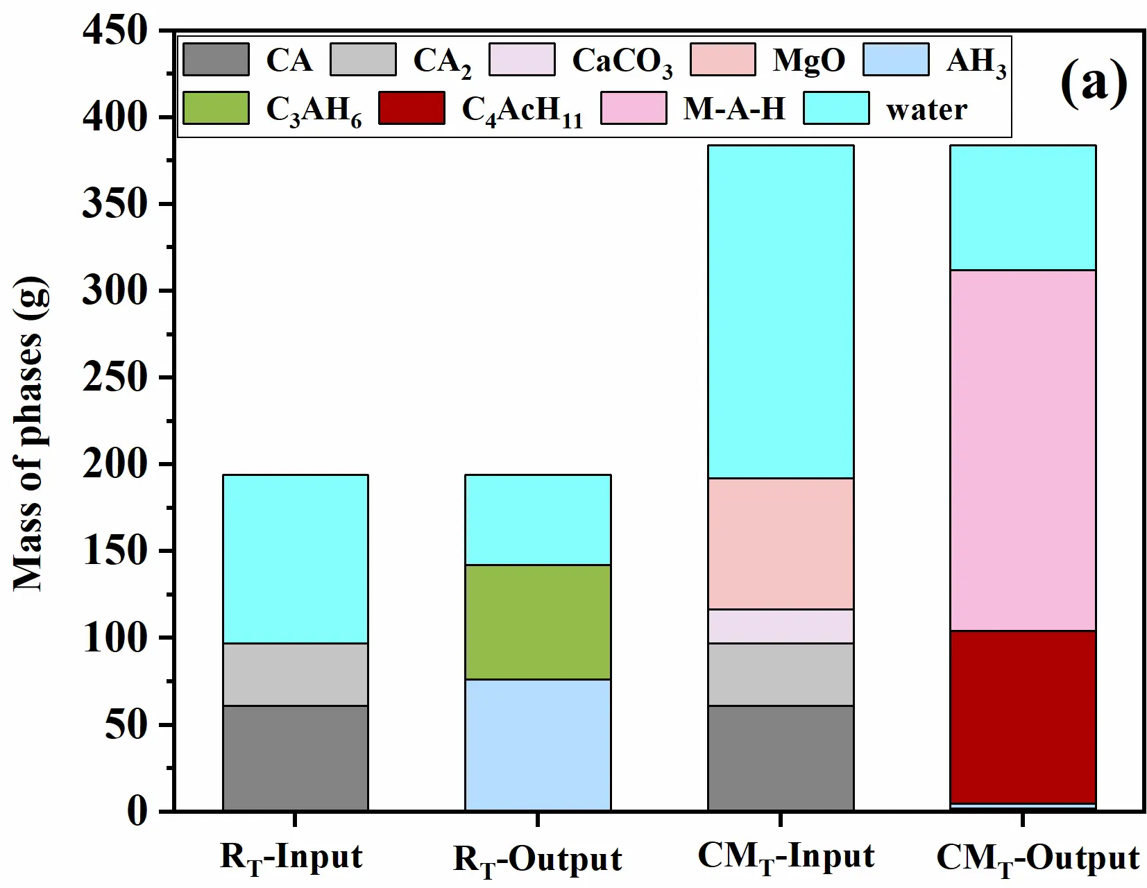
The GEMS software (v3.11.3 for windows) was employed to predict the thermodynamically stable hydrate compositions of pure CAC and CAC with CaCO3 and MgO additions, as shown in Figure 1a. For pure CAC (R and RT samples), the final hydrates stably existed as C3AH6 and AH3. In contrast, upon the introduction of CaCO3 and MgO into CAC, the hydrates transformed into C4AcH11 and M-A-H. This was attributed to the consumption of Al(OH)4− in the system during the formation of C4AcH11 and M-A-H, resulting in a significant reduction in the content of AH3 [30]. Further XRD analysis was conducted to determine the phase compositions of the hydrated samples, with results presented in Figure 1b. After curing at 25 °C for 24 h, the hydrates of the R sample were mainly CAH10 and C2AH8. The continuous consumption of Ca2+ during hydration promotes the formation of AH3. Owing to the relatively low hydration degree at this temperature, diffraction peaks of CA and CA2 were still detectable. For pure CAC subjected to two-step curing (RT sample), the increased hydration temperature not only induced the transformation of metastable hydrates to C3AH6 and AH3 (consistent with the GEMS simulation results) but also significantly enhanced the hydration degree, with no CAC clinker phases (CA and CA2) detected in the sample [31,32,33]. For CMT sample, the addition of CaCO3 facilitated the formation of C4AcH11, and the participation of MgO at 80 °C further promoted the generation of M-A-H, accompanied by the precipitation of a small amount of AH3. Additionally, unreacted CaCO3 and MgO are detected in the system. It should be noted that GEMS simulations predict thermodynamically stable phase assemblages at equilibrium, whereas experimental XRD results reflect actual phase compositions achieved under specific kinetic conditions (curing temperature and time). The presence of metastable CAH10 and C2AH8 in the R specimens is attributed to kinetic limitations at 25 °C. Under the two-step curing regime, the elevated temperature accelerates the transformation to thermodynamically stable C3AH6 and AH3, consistent with GEMS predictions.
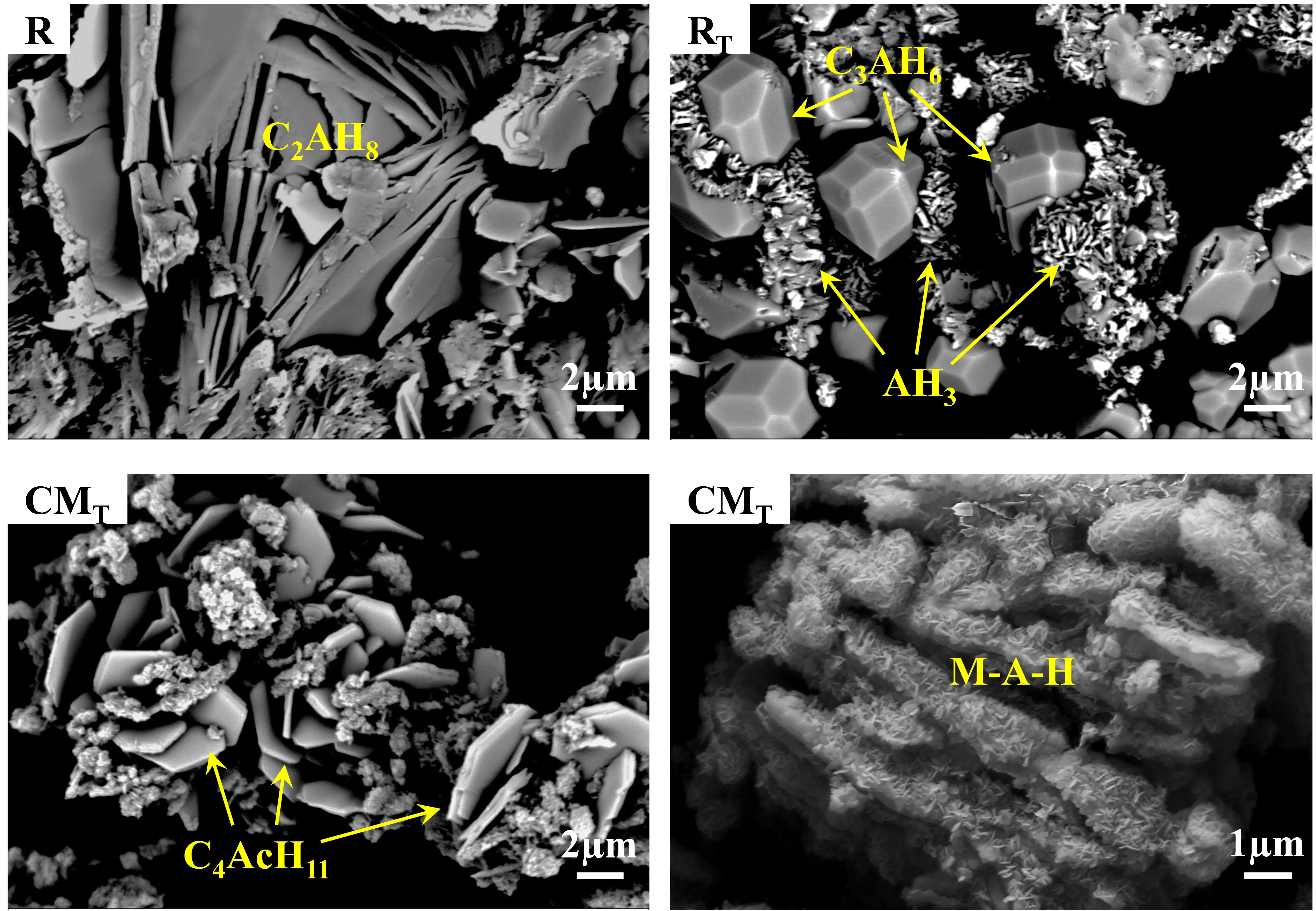
The microstructure of the hydrated samples was observed by SEM (as shown in Figure 2): Lamellar C2AH8 hydrates were clearly observed in the R sample. After two-step curing, the microstructure of RT sample was characterized by granular C3AH6 wrapped in AH3. In the CMT sample with CaCO3 and MgO additions, although the hydrates retain a lamellar morphology, the hydrates had transformed from C2AH8 to C4AcH11, and nano-sized lamellar M-A-H was also observed.
 |
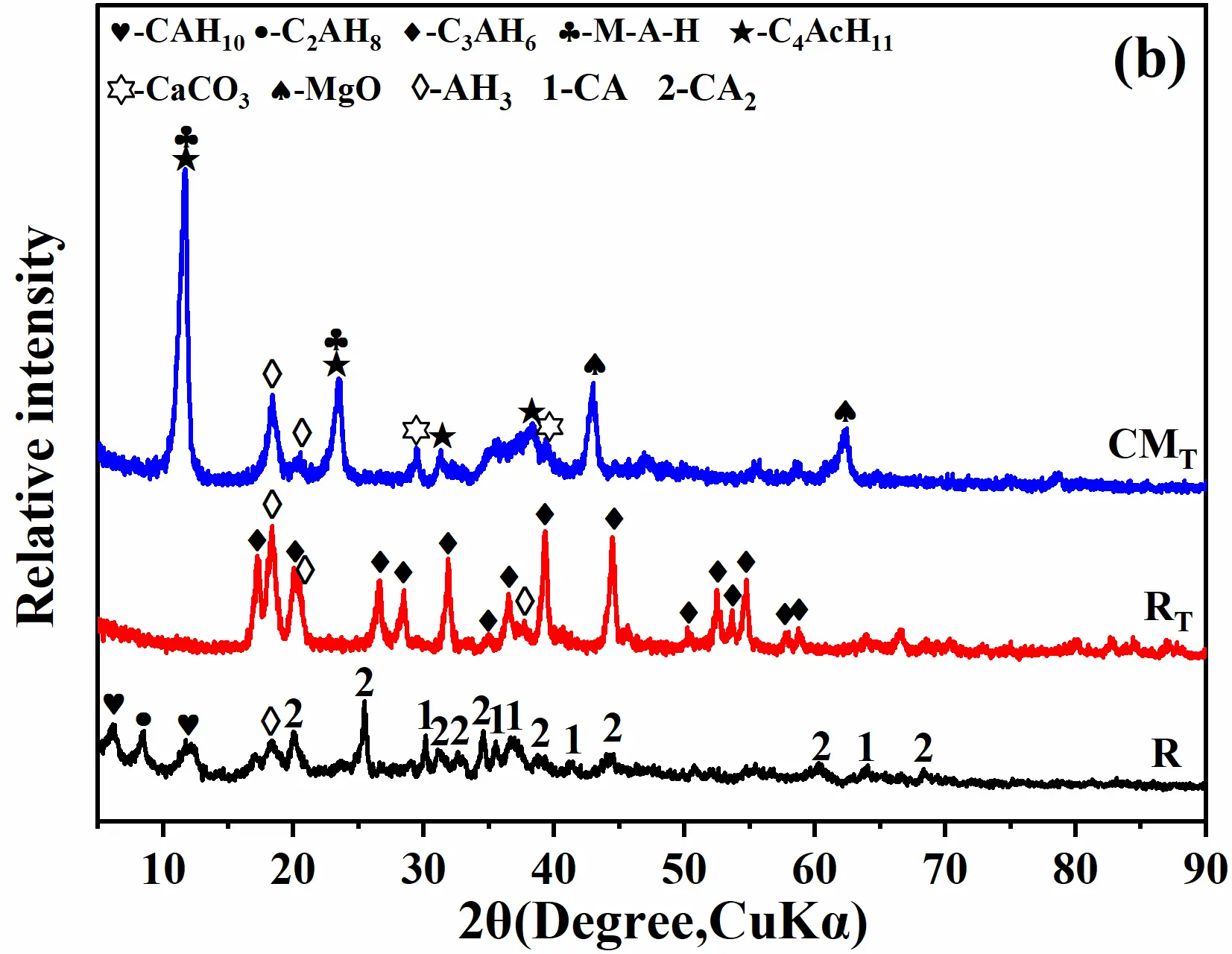 |
Figure 1. Simulated stable hydrates using GEMS (a) and experimental XRD pattern of the hydration samples (b).
3.2. Pore Structure and Fractal Dimension of Castables
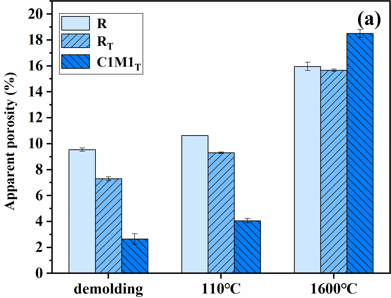
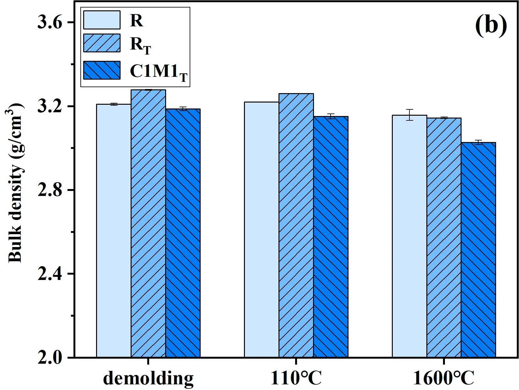
The apparent porosity and bulk density of the castables are shown in Figure 3. After curing at 25 °C, the apparent porosity of the R specimens was 9.5%, while that of the RT specimens was approximately 7% after two-step curing, which was attributed to the enhanced degree of hydration. When micro/nano-lamellar hydrates formed in the matrix, the apparent porosity of the castables decreased significantly. This phenomenon further verified that the interlocking of nano-lamellar M-A-H within the micron-sized hydrates-C4AcH11 contributed to the decreased porosity [22]. Additionally, the apparent porosity of the R specimens was significantly higher than that of other specimens, which may be attributed to its lower degree of hydration and transformation of metastable hydration phases. The decrease in apparent porosity was another reason for the increase in strength. After drying at 110 °C, the apparent porosity of the specimens was consistent with that of the green bodies the C1M1T specimens still maintained a relatively low porosity. After treatment at 1600 °C, although nano-CaCO3 participated in the formation of CAC hydrates, unreacted CaCO3 remained in the system. Consequently, the apparent porosity of the C1M1T specimens exceeded that of the R specimens and RT specimens. The bulk density of the castables is illustrated in Figure 3b. The results demonstrated that the co-incorporation of nano-CaCO3 and MgO into the castables slightly reduced the bulk density of the specimens.
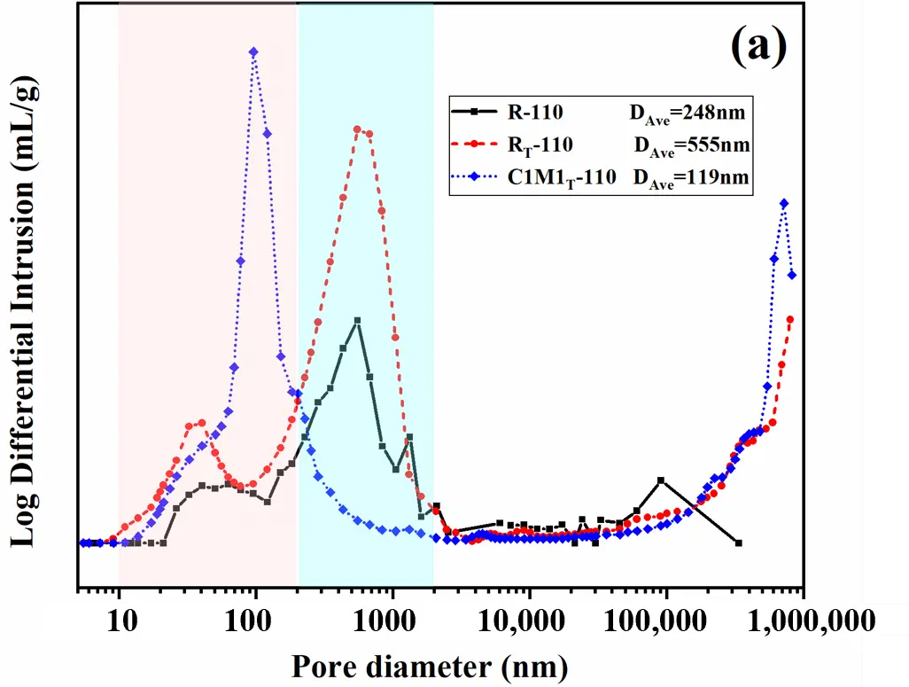
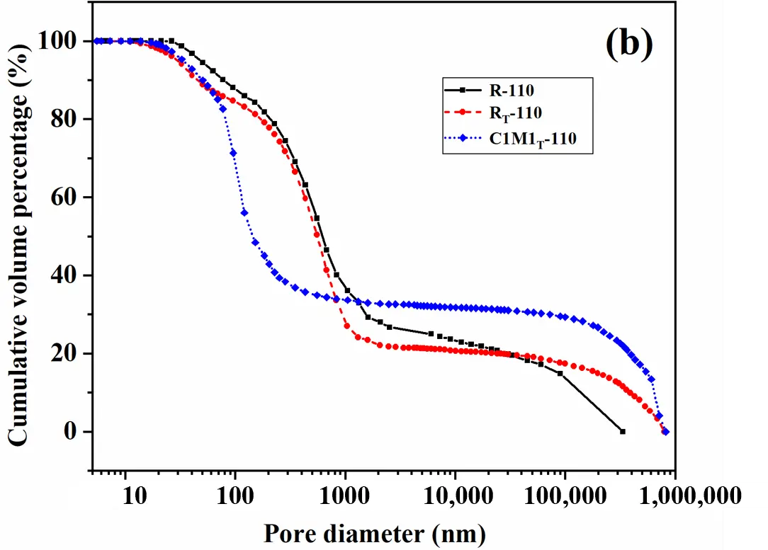
To further investigate the pore characteristics, mercury intrusion porosimetry (MIP) was employed to analyze the pore size distribution of samples treated at 110 °C and 1600 °C, as shown in Figure 4 and Figure 5, respectively. After drying at 110 °C, both the R-110 samples and RT-110 samples exhibited a bimodal pore distribution, with dominant peaks in the 10–200 nm and 200–2000 nm ranges. Additionally, macropores larger than 200,000 nm were detected. In contrast, the R-110 samples showed a reduced distribution in the 200–2000 nm range and a smaller average pore diameter (DAve). In contrast, the C1M1T-110 samples (with co-incorporated nano-CaCO3 and MgO) displayed a unimodal pore distribution, with pores concentrated in the 10–300 nm range and a smallest average pore diameter of 119 nm. Notably, large pores (>200,000 nm) were also observed in this system. Analysis of cumulative mercury intrusion curves (Figure 4b) revealed distinct differences in pore size dominance. Compared to R-110 samples and RT-110 samples. The C1M1T-110 samples exhibited a steeper slope in the <300 nm range. This steeper slope indicates a higher proportion of pores within this size range, as the slope magnitude directly correlates with pore volume concentration at specific sizes.
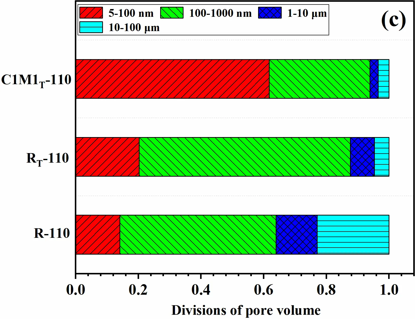
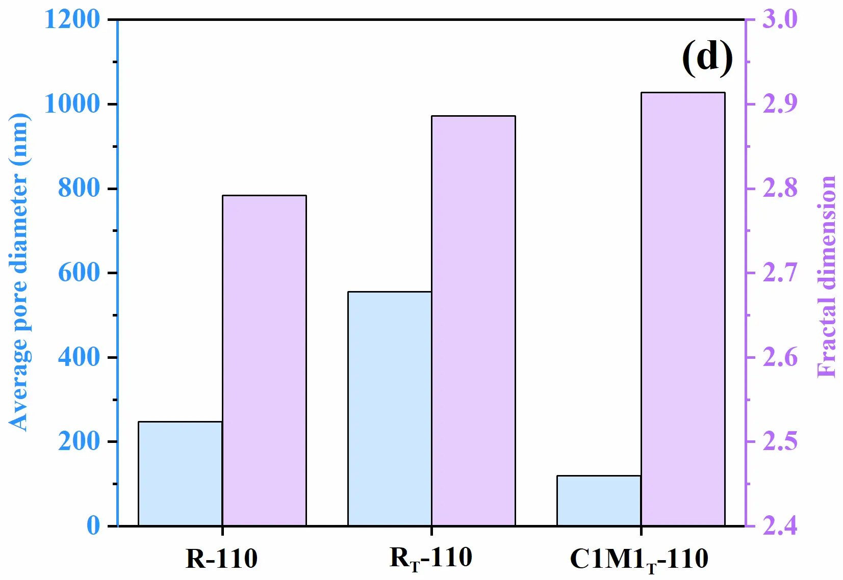
Furthermore, the pore size distribution across different ranges was quantified for the samples, as shown in Figure 4c. Significant differences in pore size distribution were observed among the R samples under different curing regimes. The two-step cured RT-110 sample exhibited a dramatic proportion of pores in the 5–1000 nm range, with a 5–100 nm pore fraction of ~0.17 ± 0.01 and a 100–1000 nm pore fraction of 0.56 ± 0.03. In contrast, the C1M1T-110 samples demonstrated a significant increase in the 5–100 nm pore fraction (0.44 ± 0.03) and a marked decrease in the 100–1000 nm pore fraction (0.23 ± 0.02). The development of micro/nano-structured hydrates facilitated pore refinement, reducing the average pore diameter from 555 nm to 119 nm and transforming 100–1000 nm pores into 5–100 nm pores. The pore surface fractal dimension (Ds), which characterizes the complexity of the pore structure, was calculated using pore data obtained via MIP. The results showed that the pore surface fractal dimensions (Ds) of the R-110 and RT-110 specimens were 2.792 and 2.886, respectively, after drying. In contrast, the filling effect of the micro/nano hydrates not only significantly reduced the average pore size of the specimens but also enhanced the complexity of the pores through pore structure refinement, ultimately increasing the Ds value to 2.914 (C1M1T-110 samples).
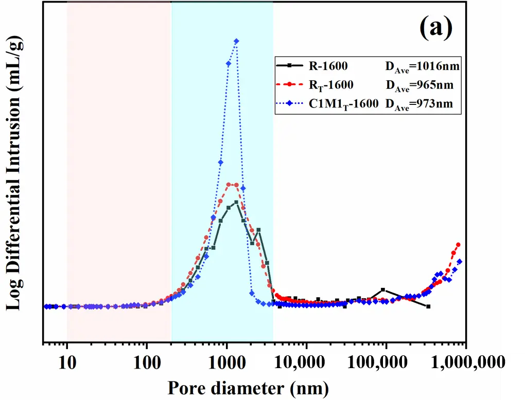
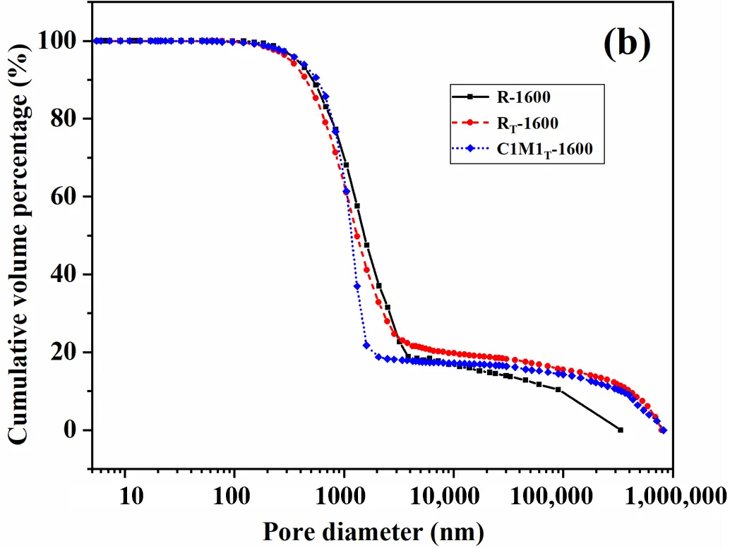
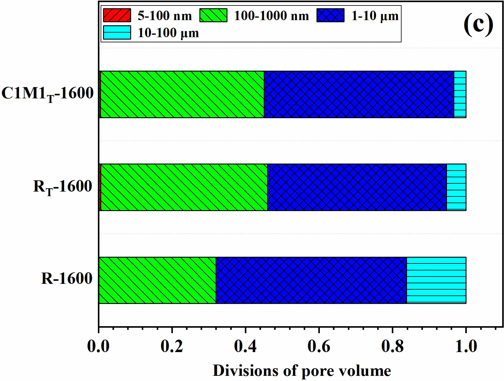
Similarly, the pore size distribution of specimens after 1600 °C heat treatment was analyzed to investigate the inheritance relationship between the early-stage micro/nano-layered hydrates and the pore structure evolution. Unlike samples dried at 110 °C, all 1600 °C-treated samples exhibited unimodal pore size distributions with narrower size ranges. The high-temperature sintering effect eliminated nano-sized pores, driving pore coarsening. Both the R-1600 samples and RT-1600 samples showed a broad pore size distribution (100–3000 nm), among which the R-1600 specimens had a larger average pore diameter (DAve = 1016 nm). In contrast, the C1M1T-1600 samples had a narrower distribution range (100–2000 nm) with a DAve of approximately 973 nm. As shown in Figure 5b, the C1M1T-1600 samples exhibited a steeper slope and higher cumulative mercury intrusion in the <2000 nm range. The quantification of pore fractions (Figure 5c) revealed that 1600 °C-treated samples were mainly concentrated in the 100 nm–10 μm range, with a minimal proportion of 5–100 nm pores due to the sintering effect. Given the influence of different curing regimes on the pore structure of the fired castables, the one-step cured R specimens (R-1600) had a relatively low pore fraction of 0.32 ± 0.02 in the 100–1000 nm range. In contrast, the two-step cured samples had a pore fraction of 0.45 ± 0.02 in this range, representing a 41% increase. This indicates that the enhanced degree of hydration promoted the formation of structural micro-sized pores. All specimens had a similar pore fraction of ~0.51 ± 0.04 in the 1–10 μm range. The difference was that the C1M1T-1600 specimens had a distinct advantage in the 10–100 μm range.
Compared with the dried samples, the pores of the samples after heat treatment at 1600 °C were coarsened, with the average pore diameter of all samples significantly increasing to ~1000 nm and the pore surface fractal dimension (Ds) decreasing to 2.619–2.761. This is because nano-scale and submicron-scale pores gradually disappear during the high-temperature sintering of the matrix, resulting in a reduction in the surface complexity of the pores. A comparison of the Ds values between the R samples and the C1M1T samples showed that the inherited structure of the early micro/nano-lamellar hydrates, together with the structure of in-situ MgAl2O4 microcrystals embedded in the CA6 matrix, synergistically contributes to enhancing the complexity of the pores, which will be discussed in the following section.
 |
 |
 |
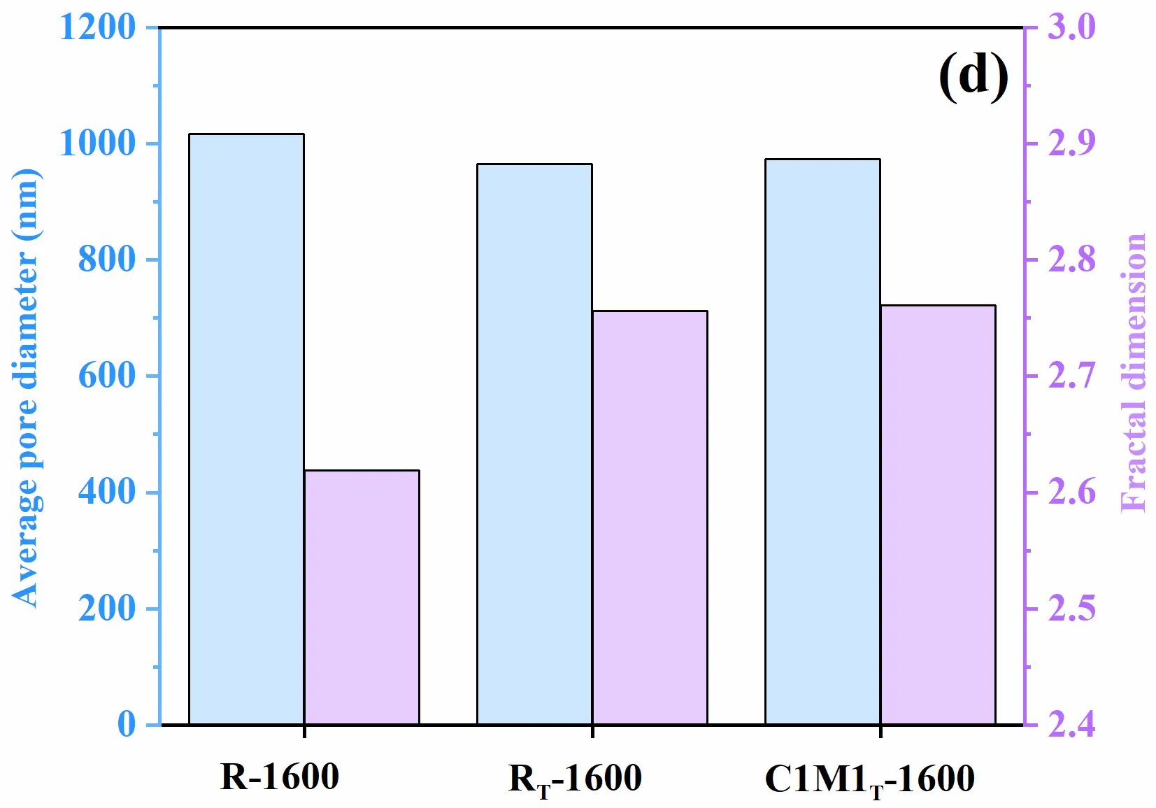 |
Figure 5. MIP pore structure characterization of castable samples treated at 1600 °C (R sample data are from our previous studies [23,24]). (a) Differential pore size distribution curves and D50 values of the samples; (b) Cumulative volume percentage versus pore diameter; (c) Volume fractions of pores in different size ranges (d) Average pore diameter and fractal dimension of the samples.
The above results indicate that the co-incorporation of nano-CaCO3 and MgO influences CAC hydration, thereby affecting the bonding strength and pore structure of the castables. SEM analysis of the 1600 °C-treated samples (Figure 6) reveals strong aggregate-matrix bonding in all specimens, but with distinct pore characteristics. Quantitative assessment at the same scale (50 μm) shows that the R specimen has the coarsest pores (up to ~50 μm), the RT specimen shows intermediate sizes (30–40 μm), and the C1M1T specimen exhibits the finest pore structure (mostly <25 μm). This refinement is attributed to the early-stage filling effect of M-A-H and the subsequent formation of an interlocked CA6-MgAl2O4 structure at high temperatures, consistent with MIP results.
Combined morphological observation and XRD analysis confirm that MgO and its precursor M-A-H react with Al2O3 to form in-situ MgAl2O4 (~1–2 μm) [24], which is embedded between the lamellar CA6 plates in the C1M1T sample, resulting in a mosaic-like microstructure. In contrast, the R and RT samples consist predominantly of interlocking CA6 crystals without such microcrystalline inclusions. This microstructural difference underpins the superior fracture performance of the C1M1T specimen, despite its slightly higher porosity.
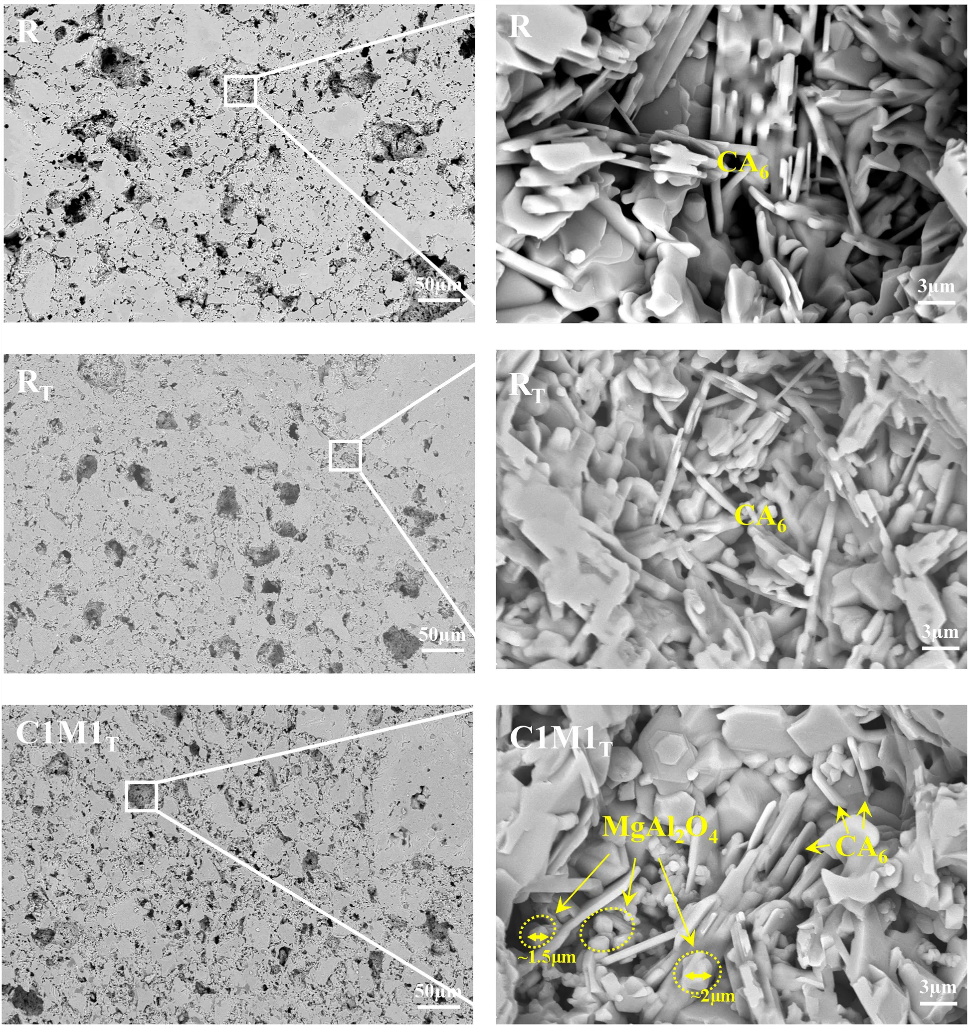
Figure 6. Microstructure of castables after 1600 °C: (left) matrix-aggregate interface; (right) magnified view of boxed region, showing interlocking CA6 plates with in-situ MgAl2O4 microcrystals (~1–2 µm, arrows).
The aforementioned results demonstrated that hydrates with distinct morphologies formed during the curing process exhibited markedly different pore-structuring capabilities. After curing at 25 °C, the initial hydrates were CAH10 and C2AH8, which then transferred to C3AH6 and AH3. During the two-step curing process, a higher degree of hydration and granular hydrates (C3AH6) preferentially facilitated the formation of 50–1000 nm pores, whereas micro/nano-lamellar hydrates (C4AcH11 and M-A-H) predominantly contributed to 5–100 nm pores. These findings further validated that nano-sized M-A-H exhibits superior pore-filling efficiency, serving as the key factor in reducing open porosity and average pore diameter, refining the pore size distribution, and increasing the pore fractal dimension.
After 1600 °C heat treatment, the matrices of both the R and RT samples primarily consisted of plate-like CA6 and Al2O3 fines. In contrast, the co-incorporation of nano-CaCO3 and MgO in the C1M1T samples induced the following phase-evolution pathways: Hydrate-C4AcH11 and unreacted CaCO3 acted as highly reactive CaO sources, reacting with Al2O3 to generate plate-like CA6; The MgO precursor M-A-H and residual MgO reacted with Al2O3 to form in-situ MgAl2O4 spinel microcrystals. Owing to the 1–2 µm grain size of the MgAl2O4 microcrystals, the C1M1T samples developed a higher proportion of 100–1000 nm pores. Concurrently, the interlocking stacking of CA6 plates promoted the formation of 1–10 µm pores, ultimately forming a more complex and refined pore structure. In summary, while the co-incorporation strategy achieved pore refinement in both dried and high-temperature-treated samples, the underlying mechanisms diverged: Refinement originated from nanoscale filling effects of M-A-H of the castables after drying; Pore refinement stems from stacked via in-situ microcrystalline MgAl2O4 formation refinement arose from aggregated MgAl2O4 microcrystals stacking.
3.3. Mechanical Properties of Castables
To investigate the effect of aforementioned structure on the mechanical properties, CMOR and CCS of the castables with and without nano-sized CaCO3 and MgO with different curing regimes were tested. After curing at 25 °C, the CMOR of the R specimens was 6.6 MPa. After the two-step curing, the demolding flexural strength of the RT specimens was 8.5 MPa, while the demolding strength of the C1M1T specimens increased to 12.0 MPa, which was 82% and 41% higher compared to the R and RT specimens, respectively. The difference in early-stage bonding strength was attributed to the degree of CAC and the type of hydrates. The R specimens exhibited low hydration degree, and their hydrates were CAH10 and C2AH8. After two-step curing, the hydration degree of CAC was increased, and the bonding phase of the RT specimens was granular C3AH6 and AH3, whereas the bonding phase of the C1M1T specimens was micro/nano-lamellar C4AcH11 and M-A-H when nano-CaCO3 and MgO were introduced. After drying at 110 °C, the hardening of the hydrates further improved the CMOR of the castables, and the CMOR of the C1M1T specimens was higher than that of the R specimens and RT specimens. Upon firing at 1600 °C, the CMOR of the castables increased to more than 35 MPa owing to the formation of CA6 lamellar interlocking structure in the specimens and the sintering effect of the matrices. However, the CMOR of the C1M1T specimens was slightly lower than that of the R specimens and RT specimens, it can be attributed to two main factors: (1) decomposition of residual CaCO3, generating additional porosity; (2) volume expansion (~5–8%) associated with in-situ MgAl2O4 formation, introducing microstructural stresses. CCS showed a similar trend (Figure 7b).
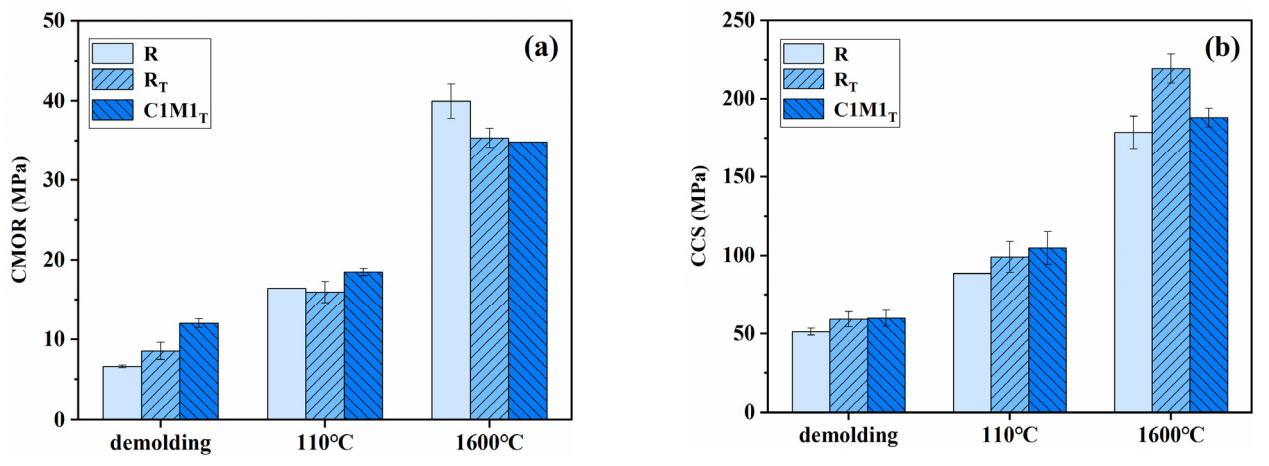
Figure 7. Mechanical properties of the castables after treatment at different temperatures: (a) CMOR; (b) CCS.
3.4. High-Temperature Thermomechanical Stress Damage Mechanisms
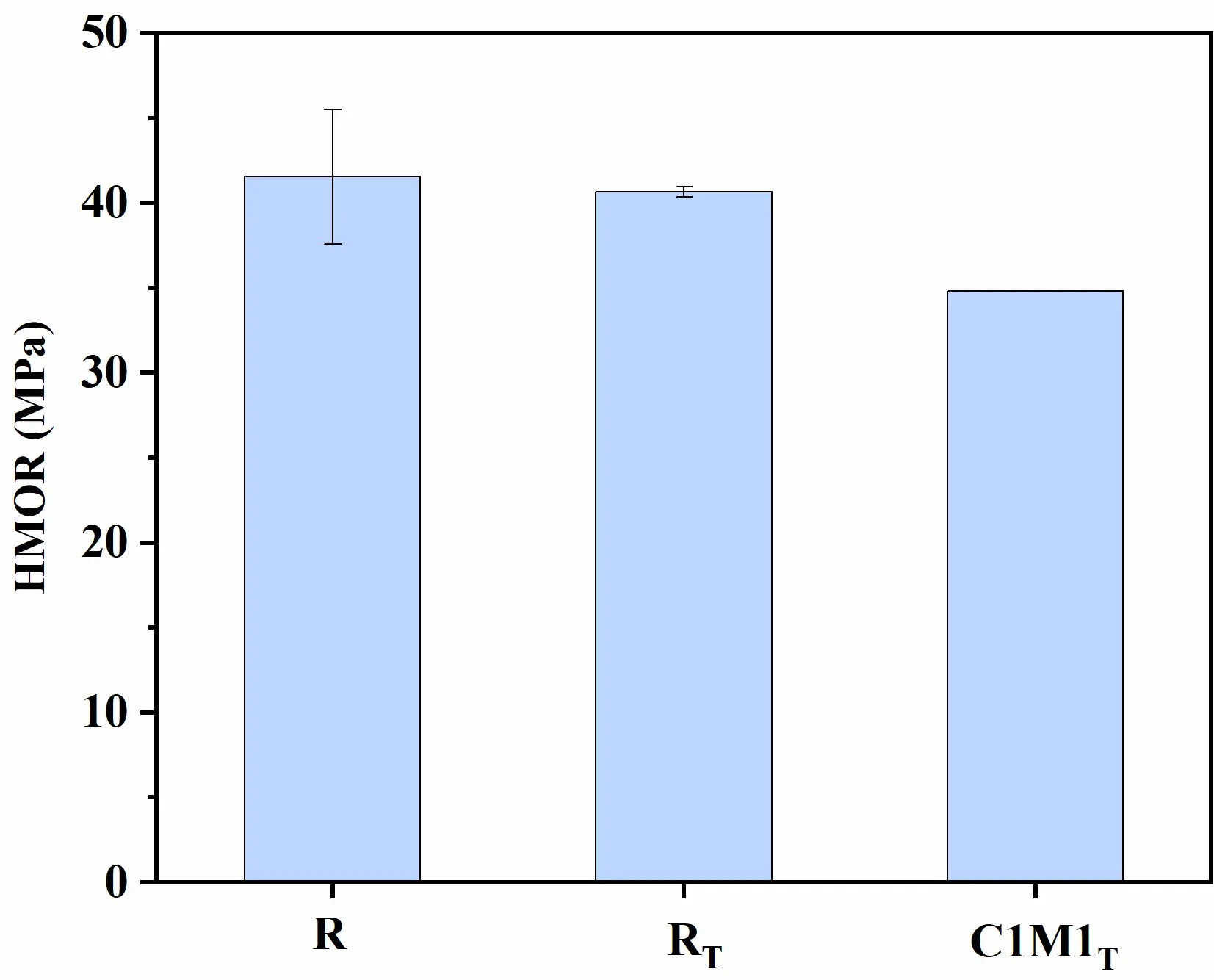
The high-temperature modulus of rupture (HMOR) of the castable specimens was tested at 1400 °C. The results (Figure 8) showed that all specimens exhibited HMOR values exceeding 35 MPa, which was similar to the trend of the CMOR. However, the C1M1T specimens exhibited a slight reduction in strength compared to the specimens without additives, primarily attributed to decomposition-induced pores in their microstructure.
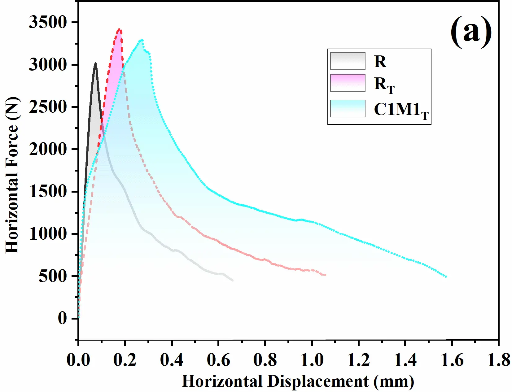
High-temperature wedge splitting tests were conducted to characterize the thermomechanical stress damage resistance mechanism of two-step cured castables. The horizontal load-displacement curves are presented in Figure 9a, and a physical photograph of the specimens after testing is shown in Figure 9b. Upon loading, all specimens rapidly reached their maximum horizontal load (FH,max). Although the R specimens cured at 25 °C exhibited the highest high-temperature modulus of rupture (HMOR), it had the lowest FH,max of approximately 3000 N, which may be related to the presence of more 10–100 μm pores. In contrast, the RT specimen exhibited a slightly higher FH,max than the C1M1T specimen, consistent with their high-temperature modulus of rupture (HMOR) trends. Following peak load, a gradual load reduction phase was observed: the horizontal displacement of the R specimen was only 0.7 mm, that of the two-step cured RT specimen was approximately 1.1 mm, and that of the C1M1T specimen significantly increased to 1.6 mm. These results indicate that the two-step cured specimens exhibit better resistance to crack propagation, and in particular, the C1M1T specimen showing superior thermal stress damage resistance.
 |
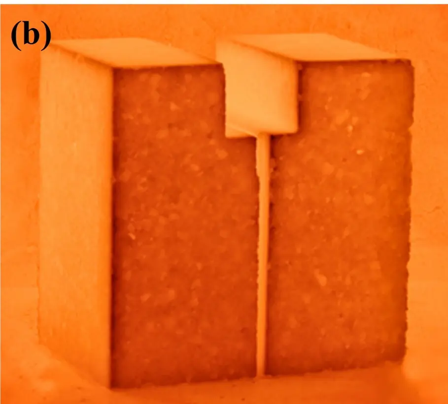 |
Figure 9. Horizontal force-displacement curves (a) and physical photo (b) of alumina-spinel castables evaluated at 1400 °C.
The fracture parameters derived from the horizontal load-displacement curve, including specific fracture energy (Gf′), apparent notched tensile strength (σNT), the ratio Gf′/σNT, characteristic length (lch), and the ((B+C)/A) index, were summarized in Table 2. σNT, which correlates with the maximum horizontal load (FH,max), was slightly lower in the C1M1T specimen than in the additive-free specimen, primarily due to its higher porosity. In terms of specific fracture energy (Gf′), the R specimen cured conventionally at 25 °C exhibited a baseline value of 181.5 J/m2. Two-step curing significantly enhanced Gf′ by 74–163%: the RT specimen reached 315.8 J/m2, while the C1M1T specimen achieved 476.6 J/m2. This increase indicated that the C1M1T specimen dissipated more energy during damage, thereby suppressing the formation of new fracture surfaces. Similarly, Gf′/σNT and lch are key indicators of material toughness, with larger values signifying superior resistance to crack propagation and enhanced toughness [34,35]. The R specimen showed Gf′/σNT and lch values of 38.3 μm and 920.8 mm, respectively, whereas the C1M1T specimen exhibited values approximately three times higher (111.0 μm and 2948.9 mm), highlighting its enhanced toughness. The superior fracture parameters of the C1M1T specimen are attributed to five mechanisms: in-situ MgAl2O4 microcrystals (~1–2 μm) inducing crack deflection and bridging; the highest proportion of fine pores (5–1000 nm, Ds = 2.761) providing multiple energy dissipation pathways; enhanced plastic deformation capacity; multi-scale pore structure enabling synergistic energy dissipation; and structural inheritance from initial hydrates to the high-temperature CA6-MA interlaced phase. These mechanisms collectively explain its superior fracture toughness despite slightly lower strength. It is worth noting that the higher (B+C)/A ratio of the R specimen (7.0) compared to the C1M1T specimen (4.4) does not imply superior crack resistance. Instead, this anomaly arises because the R specimen has the lowest absolute fracture energy (Gf′ = 181.5 J/m2), which minimizes Region A (initiation work) and thus artificially inflates the ratio. Additionally, the characteristic length lch is the key indicator of thermal shock resistance, representing the critical thermal strain dimension a material can sustain before failure—higher values indicate better thermal shock damage tolerance. In contrast, the C1M1T specimen exhibits Gf′, Gf′/σNT, and lch values that are 2.6, 2.9, and 3.2 times those of the R specimen, clearly demonstrating superior energy dissipation and crack resistance. Therefore, (B+C)/A should always be interpreted alongside absolute fracture parameters, rather than as a standalone indicator. In summary, despite slightly lower strength than the R and RT specimens at high temperatures, the C1M1T specimen exhibits significantly higher Gf′, Gf′/σNT, lch, and superior overall fracture performance. These results collectively demonstrate enhanced resistance to crack propagation and thermal stress damage.
The crack propagation paths are typically categorized into three types: through aggregates, along aggregate-matrix interfaces, and through the matrices. To investigate the influence of optimized energy dissipation mechanisms on fracture pathways, the cracks in the samples were observed via scanning electron microscopy (SEM), as shown in Figure 10. For clarity, cracks propagating through aggregates, along interfaces, and through the matrices were marked in red, yellow, and blue, respectively. Statistical analysis of crack paths was summarized in Table 3. Compared to sintered tabular alumina aggregates, the matrices exhibited relatively weaker bonding strength and some softening deformation capacity at high temperatures, leading to a predominance of matrix-dominated fracture paths. The R sample had narrower cracks, while the C1M1T sample demonstrated larger crack opening displacements (COD, crack widths) and greater crack tortuosity than the RT sample. An increase in COD is a key characteristic of enhanced material toughness [36,37], and its core mechanisms in improving toughness can be elaborated in three aspects: energy dissipation, inhibition of crack propagation, and enhancement of plastic deformation capacity. Specifically, a larger COD implies a broader range of plastic deformation at the crack tip, which consumes more external energy; the plastic deformation at the crack tip can reduce stress concentration through a “blunting effect”, thereby delaying crack propagation; moreover, an increase in COD is often related to the complex microstructure inside the material, and the larger opening displacement provides sufficient physical space for crack deflection and branching, extending the crack propagation path and further increasing energy consumption, ultimately enabling the material to exhibit higher toughness.
Based on statistical results, a comparison of the proportion of crack paths between the R and RT samples indicates that the two-step curing system, which increased the degree of early hydration, was beneficial for improving the bonding strength and increasing the proportion of aggregate-penetrating fractures. When nano-CaCO3 and MgO were co-incorporated, the proportion of interface-aligned cracks increased, while the proportions of aggregate-penetrating and matrix-penetrating cracks decreased. The improved toughness originates from two key energy-dissipation mechanisms in its microstructure: (1) In-situ MgAl2O4 microcrystals embedded within plate-like CA6 induce crack deflection and bridging, increasing the tortuosity of crack paths; (2) The superior fracture performance of the C1M1T specimen stems from its refined pore structure, as evidenced by the highest proportion of 5–1000 nm pores and the highest pore surface fractal dimension (Ds = 2.761) after heated. These fine pores act as energy dissipation sites during crack propagation by inducing crack deflection and branching, which increases crack path tortuosity and consumes more fracture energy. In contrast, large pores (>10 μm), which are more prevalent in the R specimen, serve as stress concentrators that facilitate crack initiation and provide preferential pathways for crack propagation.
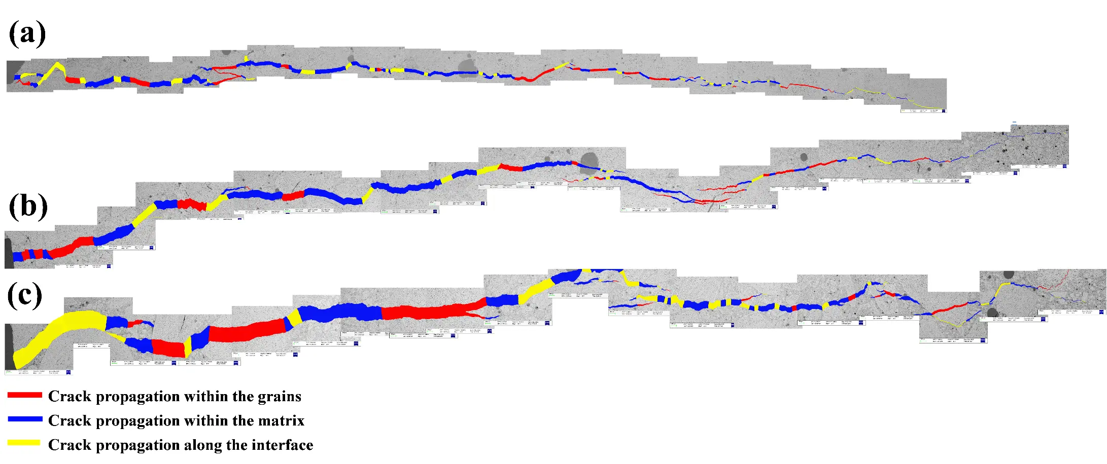
Figure 10. Pictures of crack propagation in alumina-spinel castables under SEM observation: (a) R; (b) RT; (c) C1M1T.
Table 3. Crack propagation path ratio of alumina-spinel castables based on SEM (%).
|
Index |
Aggregates |
Interfaces |
Matrices |
|---|---|---|---|
|
R |
22.4 |
32.9 |
44.7 |
|
RT |
29.9 |
15.0 |
55.2 |
|
C1M1T |
24.4 |
28.0 |
47.6 |
4. Conclusions
Building upon previous research, this work comparatively investigated the effects of curing regimes and additives (nano-CaCO3 and MgO) on the initial hydration phases of CAC. Furthermore, the inheritance of these microstructural features and their influence on high-temperature fracture behavior were systematically analyzed. The following conclusions are drawn:
- (1)
-
The initial hydrates of CAC were influenced by curing regimes and additives. For pure CAC, curing at 25 °C favors the formation of metastable CAH10 and C2AH8, whereas two-step curing induces the formation of granular C3AH6 and AH3. In contrast, CAC containing CaCO3 and MgO promoted the generation of micro/nano-lamellar C4AcH11 and M-A-H.
- (2)
-
The composite hydration phases of C4AcH11 and M-A-H significantly enhanced the early bonding strength of the castables, with the demolding strength increasing by 82% and 41%, respectively, compared to the R and RT samples. Meanwhile, the composite phases effectively refined pores, and after drying at 110 °C, the filling effect of M-A-H introduced more nano-scale pores. With C4AcH11 and M-A-H serving as high-activity CaO and MgO sources, respectively, a CA6-MgAl2O4 interlaced structure was formed after heat treatment at 1600 °C, which increased the proportion of submicron-scale pores and improved the complexity of the pore structure.
- (3)
-
A more refined and complex microporous structure was achieved through the early-stage filling effect of M-A-H and the accumulation of in-situ generated MgAl2O4 microcrystals. This refined CA6-MA structure facilitates energy dissipation and crack deflection while enhancing the capacity for plastic deformation during fracture. As a result, the material exhibits significantly improved fracture toughness, with Gf’ increased by 163% and lch being 3.2 times that of the R specimen, which is beneficial for extending the service life of ladle linings.
Statement of the Use of Generative AI and AI-Assisted Technologies in the Writing Process
Specifically, we have used DeepSeek (Version [DeepSeek-V3]) for English translation and language polishing during the preparation of this manuscript. All AI-generated content has been thoroughly reviewed, verified, and edited by us. We take full responsibility for the accuracy and integrity of the content.
Author Contributions
Conceptualization, Y.L. and S.J.; Methodology, N.L. and W.L.; Software, W.L.; Validation, G.H., W.L.; Formal Analysis, Y.L.; Investigation, N.L.; Resources, S.J.; Data Curation, G.H.; Writing—Original Draft Preparation, G.H.; Writing—Review & Editing, N.L.; Visualization, G.H.; Supervision, Y.L.; Project Administration, N.L.; Funding Acquisition, S.J.
Ethics Statement
Not applicable.
Informed Consent Statement
Not applicable.
Data Availability Statement
All data generated or analyzed during this study are completely presented within the published article. No external datasets were generated or deposited in public repositories, and no additional data links are required.
Funding
This research was funded by [Hubei Provincial Special Fund for Central-Guided Local S&T Development] grant number [2025CSA017]. The APC was also funded by [Hubei Provincial Special Fund for Central-Guided Local S&T Development].
Declaration of Competing Interest
The authors declare that they have no known competing financial interests or personal relationships that could have appeared to influence the work reported in this paper.
References
- Braulio MAL, Rigaud M, Buhr A, Parr C, Pandolfelli VC. Spinel-containing alumina-based refractory castables. Ceram. Int. 2011, 37, 1705–1724. DOI:10.1016/j.ceramint.2011.03.049 [Google Scholar]
- Luz AP, Martinez AGT, Braulio MAL, Pandolfelli VC. Thermodynamic evaluation of spinel containing refractory castables corrosion by secondary metallurgy slag. Ceram. Int. 2011, 37, 1191–1201. DOI:10.1016/j.ceramint.2010.11.043 [Google Scholar]
- Samadi S, Jin S, Gruber D, Harmuth H. Thermomechanical finite element modeling of steel ladle containing alumina spinel refractory lining. Finite Elem. Anal. Des. 2022, 206, 103762. DOI:10.1016/j.finel.2022.103762 [Google Scholar]
- Braulio MAL, Morbioli GG, Milanez DH, Pandolfelli VC. Calcium aluminate cement source evaluation for Al2O3–MgO refractory castables. Ceram. Int. 2011, 37, 215–221. DOI:10.1016/j.ceramint.2010.09.027 [Google Scholar]
- Oliveira IR, Pandolfelli VC. Castable matrix, additives and their role on hydraulic binder hydration. Ceram. Int. 2009, 35, 1453–1460. DOI:10.1016/j.ceramint.2008.07.024 [Google Scholar]
- Idrees M, Ekincioglu O, Sonyal MS. Hydration behavior of calcium aluminate cement mortars with mineral admixtures at different curing temperatures. Constr. Build. Mater. 2021, 285, 122839. DOI:10.1016/j.conbuildmat.2021.122839 [Google Scholar]
- Ahmed AA, Shakouri M, Trejo D, Pavan Vaddey N. Effect of curing temperature and water-to-cement ratio on corrosion of steel in calcium aluminate cement concrete. Constr. Build. Mater. 2022, 350, 128875. DOI:10.1016/j.conbuildmat.2022.128875 [Google Scholar]
- Salomão R, Kawamura MA, Emilio ABV, Sakihama J, Segadães AM. Calcium aluminate cement in castable alumina: From hydrate bonding to the in situ formation of calcium hexaluminate. Ceram. Int. 2021, 47, 15082–15093. DOI:10.1016/j.ceramint.2021.02.066 [Google Scholar]
- Auvray JM, Gault C, Huger M. Evolution of elastic properties and microstructural changes versus temperature in bonding phases of alumina and alumina–magnesia refractory castables. J. Eur. Ceram. Soc. 2007, 27, 3489–3496. DOI:10.1016/j.jeurceramsoc.2007.01.016 [Google Scholar]
- Díaz LA, Torrecillas R. Phase development and high temperature deformation in high alumina refractory castables with dolomite additions. J. Eur. Ceram. Soc. 2007, 27, 67–72. DOI:10.1016/j.jeurceramsoc.2006.02.036 [Google Scholar]
- Liu W, Liao N, Nath M, Li Y, Dai Y, Pan L. Effects of characteristic hydrates on the pore structure and fracture behavior of CAC bonded alumina-spinel castables. Constr. Build. Mater. 2023, 389, 131736. DOI:10.1016/j.conbuildmat.2023.131736 [Google Scholar]
- Antonovič V, Kerienė J, Boris R, Aleknevičius M. The Effect of Temperature on the Formation of the Hydrated Calcium Aluminate Cement Structure. Procedia Eng. 2013, 57, 99–106. DOI:10.1016/j.proeng.2013.04.015 [Google Scholar]
- Liao N, Liu W, Yang J, Nath M, Dai Y, Pan L, et al. In-depth investigation of pore structure and fracture behavior of CAC bonded alumina-spinel castables treated at 110∼1600 °C. Ceram. Int. 2022, 48, 33763–33772. DOI:10.1016/j.ceramint.2022.07.321 [Google Scholar]
- Liu W, Liao N, Nath M, Li Y, Dai Y, Pan L. Assessment of fracture behavior of mono-carbonate (C4AcH11) bonded alumina-spinel castables by wedge splitting test. J. Eur. Ceram. Soc. 2024, 44, 2600–2608. DOI:10.1016/j.jeurceramsoc.2023.11.011 [Google Scholar]
- Renaudin G, Francois M, Evrard O. Order and disorder in the lamellar hydrated tetracalcium monocarboaluminate compound. Cem. Concr. Res. 1999, 29, 63–69. DOI:10.1016/S0008-8846(98)00184-7 [Google Scholar]
- Puerta-Falla G, Balonis M, Le Saout G, Kumar A, Rivera M, Falzone G, et al. The influence of slightly and highly soluble carbonate salts on phase relations in hydrated calcium aluminate cements. J. Mater. Sci. 2016, 51, 6062–6074. DOI:10.1007/s10853-016-9912-9 [Google Scholar]
- Luz AP, Pandolfelli VC. CaCO3 addition effect on the hydration and mechanical strength evolution of calcium aluminate cement for endodontic applications. Ceram. Int. 2012, 38, 1417–1425. DOI:10.1016/j.ceramint.2011.09.021 [Google Scholar]
- Zeng J, Liu M, Zhang Z, Mu Y, Liu C, Ye G. Influence of micro‐sized CaCO3 on hydrates formation of residual calcium aluminate cement in castables during drying. Int. J. Appl. Ceram. Technol. 2024, 21, 1616–1624. DOI:10.1111/ijac.14623 [Google Scholar]
- Goergens J, Manninger T, Goetz-Neunhoeffer F. In-situ XRD study of the temperature-dependent early hydration of calcium aluminate cement in a mix with calcite. Cem. Concr. Res. 2020, 136, 106160. DOI:10.1016/j.cemconres.2020.106160 [Google Scholar]
- Liu M, Zeng J, Jin D, Mu Y, Zhou W, Ye G. Influence of mono-carbonate hydrates on mechanical properties of CAC bonded castables fired at 800 °C. Mater. Chem. Phys. 2023, 301, 127644. DOI:10.1016/j.matchemphys.2023.127644 [Google Scholar]
- Goergens J, Goetz-Neunhoeffer F. Temperature-dependent late hydration of calcium aluminate cement in a mix with calcite—Potential of G-factor quantification combined with GEMS-predicted phase content. Cement 2021, 5, 100011. DOI:10.1016/j.cement.2021.100011 [Google Scholar]
- Liu W, Liao N, Peng H, Li Y, Yan W, Wang X, et al. Design of micron/nano-sized lamellar C(M)-A-H hydrates enhanced alumina-spinel castables through two-step curing. Ceram. Int. 2025, 51, 9852–9863. DOI:10.1016/j.ceramint.2024.12.417 [Google Scholar]
- Liu W, Liao N, Nath M, Ji Z, Dai Y, Pan L, et al. Effects of curing time on the pore structure evolution and fracture behavior of CAC bonded alumina-spinel castables. Ceram. Int. 2022, 48, 25000–25010. DOI:10.1016/j.ceramint.2022.05.153 [Google Scholar]
- Li Y, Liu W, Liao N, Nath M, Tie S, Liu X. Investigation of pore structure and high-temperature fracture behavior of lamellar hydrates bonded alumina-spinel castables. J. Eur. Ceram. Soc. 2024, 44, 5252–5261. DOI:10.1016/j.jeurceramsoc.2024.02.035 [Google Scholar]
- Zhang B, Li S. Determination of the Surface Fractal Dimension for Porous Media by Mercury Porosimetry. Ind. Eng. Chem. Res. 1995, 34, 1383–1386. DOI:10.1021/ie00043a044 [Google Scholar]
- Schmitt Rahner M, Halisch M, Peres Fernandes C, Weller A, Sampaio Santiago dos Santos V. Fractal dimensions of pore spaces in unconventional reservoir rocks using X-ray nano- and micro-computed tomography. J. Nat. Gas Sci. Eng. 2018, 55, 298–311. DOI:10.1016/j.jngse.2018.05.011 [Google Scholar]
- Zhang B, Liu W, Liu X. Scale-dependent nature of the surface fractal dimension for bi- and multi-disperse porous solids by mercury porosimetry. Appl. Surf. Sci. 2006, 253, 1349–1355. DOI:10.1016/j.apsusc.2006.02.009 [Google Scholar]
- Jin S, Gruber D, Harmuth H. Determination of Young’s modulus, fracture energy and tensile strength of refractories by inverse estimation of a wedge splitting procedure. Eng. Fract. Mech. 2014, 116, 228–236. DOI:10.1016/j.engfracmech.2013.11.010 [Google Scholar]
- Ribeiro S, Rodrigues JA. The influence of microstructure on the maximum load and fracture energy of refractory castables. Ceram. Int. 2010, 36, 263–274. DOI:10.1016/j.ceramint.2009.07.033 [Google Scholar]
- Goergens J, Belli R, Schulbert C, Goetz-Neunhoeffer F. Influence of different CA2/CA-ratios on hydration degree, AH3 content and flexural strength investigated for a binder formulation of calcium aluminate cement with calcite. Cem. Concr. Res. 2023, 165, 107090. DOI:10.1016/j.cemconres.2023.107090 [Google Scholar]
- Li J, Chang J, Wang T, Zeng T, Li J, Zhang J. Effects of phosphogypsum on hydration properties and strength of calcium aluminate cement. Constr. Build. Mater. 2022, 347, 128398. DOI:10.1016/j.conbuildmat.2022.128398 [Google Scholar]
- Son HM, Park S, Kim HY, Seo JH, Lee HK. Effect of CaSO4 on hydration and phase conversion of calcium aluminate cement. Constr. Build. Mater. 2019, 224, 40–47. DOI:10.1016/j.conbuildmat.2019.07.004 [Google Scholar]
- Son HM, Park SM, Jang JG, Lee HK. Effect of nano-silica on hydration and conversion of calcium aluminate cement. Constr. Build. Mater. 2018, 169, 819–825. DOI:10.1016/j.conbuildmat.2018.03.011 [Google Scholar]
- Harmuth H. Stability of crack propagation associated with fracture energy determined by wedge splitting specimen. Theor. Appl. Fract. Mech. 1995, 23, 103–108. DOI:10.1016/0167-8442(95)00008-3 [Google Scholar]
- Khlifi I, Pop O, Dupré JC, Doumalin P, Huger M. Investigation of microstructure-property relantionships of magnesia-hercynite refractory composites by a refined digital image correlation technique. J. Eur. Ceram. Soc. 2019, 39, 3893–3902. DOI:10.1016/j.jeurceramsoc.2019.05.010 [Google Scholar]
- Raja RB, Sivakumar MVN, Sai AP. Numerical simulation of wedge splitting test method for evaluating fracture behaviour of self compacting concrete. Comput. Concr. Int. J. 2024, 33, 265–273. DOI:10.12989/cac.2024.33.3.265 [Google Scholar]
- Brochen E, Dannert C, Paul J, Krause O. Investigation of fracture behavior of typical refractory materials up to service temperatures. Int. J. Ceram. Eng. Sci. 2022, 4, 68–76. DOI:10.1002/ces2.10123 [Google Scholar]