
Neurotechnology in Stroke Neurorehabilitation: Towards Precision and Personalized Rehabilitation Treatment
Neurotechnology in Stroke Neurorehabilitation: Towards Precision and Personalized Rehabilitation Treatment
Tianyu Jia
1,2,†
 Jingyao Sun
2,†
Yun Guo
3,†
Chong Li
4,*
Yu Pan
3,*
Jingyao Sun
2,†
Yun Guo
3,†
Chong Li
4,*
Yu Pan
3,*
Received: 05 May 2026 Revised: 10 June 2026 Accepted: 09 July 2026 Published: 20 July 2026

© 2026 The authors. This is an open access article under the Creative Commons Attribution 4.0 International License (https://creativecommons.org/licenses/by/4.0/).
1. Introduction
Globally, stroke is the third most common cause of both death and disability, with an estimated 94 million individuals affected [1]. Given the large global population of stroke survivors and the relative shortage of rehabilitation resources, efficient rehabilitation interventions that help patients regain independence and reintegrate into society are of major clinical importance.
Stroke is a highly heterogeneous disease characterized by neural injury that varies in anatomical location and severity [2,3]. Lesions in different anatomical regions can lead to overlapping neurological symptoms and behavioral deficits, thereby complicating the design of personalized rehabilitation strategies [4]. Accurate prediction of individual post-stroke outcomes plays a pivotal role in patient stratification, which is an essential prerequisite for precision rehabilitation [5]. In this context, neurophysiological and neuroimaging prognostic biomarkers have been developed to characterize recovery potential, and artificial intelligence (AI) models are increasingly being used to help stratify recovery trajectories and inform personalized rehabilitation treatment strategies.
Beyond intelligent prediction and stratification, effective rehabilitation interventions must be tailored to and appropriately delivered to individual patients. Brain-computer interfaces (BCIs) provide a promising solution by closing the loop between motor-related cortical activity and afferent feedback [6]. During BCI-based rehabilitation training, patients can use their motor intentions to control a rehabilitation robot, which then drives the paralysed limb to perform movement. These movements, in turn, generate sensory feedback to the brain, influencing subsequent motor commands. Such sensorimotor feedback can effectively stimulate the cerebral cortex [7] and plays a critical role in promoting neural plasticity and functional reorganization [8]. By establishing a closed loop between neural activity and actual movement, this closed-loop training facilitates the formation of motor sensations [7,9]. It offers a new pathway for active motor rehabilitation, particularly for stroke patients in the flaccid paralysis stage who have little to no voluntary motor ability. Furthermore, this approach can help rebuild patients’ confidence and enhance their willingness to engage in the rehabilitation process, which is an important factor influencing treatment outcomes [10]. However, clinical outcomes after BCI-based rehabilitation vary substantially among stroke patients. This variability is related to individual patient differences and limited alignment between diverse BCI rehabilitation methods and specific patient profiles. Therefore, the development of personalized treatment strategies is essential for effective BCI-based rehabilitation training.
Addressing post-stroke diversity requires tailoring interventions to each patient’s unique clinical profile, thereby optimizing recovery outcomes. Consequently, moving beyond generalized protocols toward personalized rehabilitation is not merely beneficial but essential for advancing post-stroke neurorehabilitation. Therefore, in this review, we focus on the development of neurotechnology for precision and personalized rehabilitation treatment.
As shown in Figure 1, we review the core mechanisms of neurorehabilitation and recent advances in neurotechnology, and discuss how these tools can accelerate neurorehabilitation after stroke. We propose that, despite remaining challenges in the field, integrating AI-driven approaches, multimodal biomarkers, and closed-loop BCI systems will enable more efficient, personalized rehabilitation strategies and move the field toward more tailored, precise, and efficient neurorehabilitation.
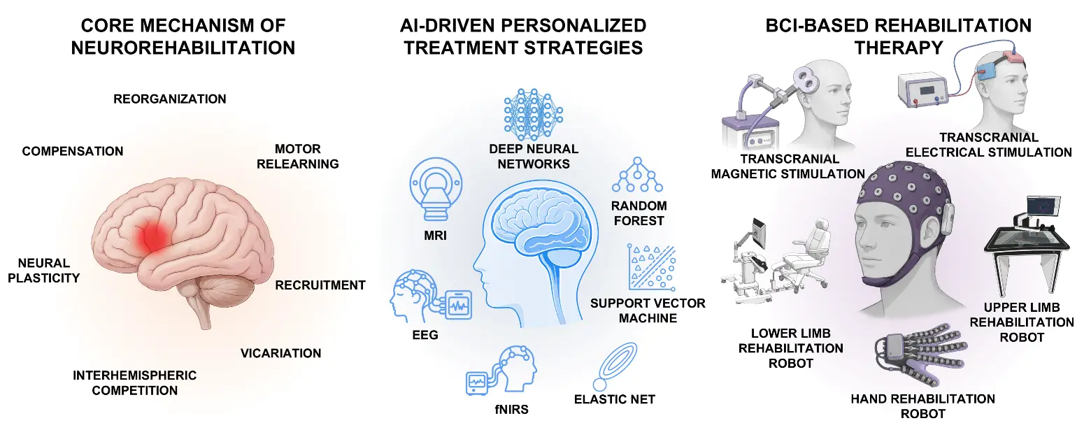
Figure 1. Summary of artificial intelligence (AI) and neurotechnology in stroke neurorehabilitation. This review concisely examines core neurorehabilitation mechanisms and recent advances in multimodal biomarkers, AI-driven personalized treatment strategies, and closed-loop brain-computer interfaces (BCIs) for stroke rehabilitation training.
2. Basic Neurorehabilitation Mechanisms
2.1. Neural Plasticity and Reorganization
Neural plasticity refers to the capacity of the nervous system to remodel its physical architecture, alter its functional properties, and modify its connections in response to internal or external stimuli [11,12,13]. Structural changes involve synapse number and size, axonal arborization, and gray matter density, whereas functional changes encompass alterations in neural activity, motor-map remapping, and intrinsic excitability [12,14,15,16]. Two core mechanistic classes underlie the principles of synaptic plasticity: homeostatic and Hebbian plasticity [17]. Homeostatic plasticity is a form of negative feedback-mediated plasticity that maintains neural network activity within an optimal set point. This mechanism may play a critical role after stroke by initiating pathways that restore synaptic transmission and network function. Hebbian plasticity represents a positive feedback-mediated form of plasticity, in which synapses are strengthened when pre- and postsynaptic neurons are activated coincidentally. This form of plasticity may be critical after stroke for reinforcing and preserving appropriately organized functional connections.
Neural reorganization constitutes the core neurobiological basis of functional recovery after stroke and other central nervous system (CNS) injuries. The balance between inhibition and excitation is disrupted after CNS injury. Several rehabilitation models collectively provide complementary frameworks for understanding post-stroke neuroplasticity. First, the vicariation model assumes that undamaged brain regions can take over functions previously managed by damaged areas. Second, the interhemispheric competition model posits that post-stroke recovery involves rebalancing inhibitory influences between hemispheres, whereby the unaffected hemisphere may excessively inhibit the affected one, hindering recovery. Third, the bimodal balance-recovery model integrates both previous models into a unified explanatory framework by introducing structural reserve, a key parameter that characterizes how well lesion-spared neural pathways and relays support recovery in each patient [18,19]. The level of structural reserve determines whether interhemispheric imbalance or vicariation prevails during recovery [18]. Specifically, high structural reserve favors the interhemispheric competition model, whereas vicariation better explains recovery in patients with limited structural reserve. The bimodal balance-recovery model enables personalized non-invasive brain stimulation strategies tailored to the unique needs of individual patients. Moreover, the brain and spinal cord play essential roles in processing sensory information and in directing and carrying out well-coordinated, skillful actions. Imaging studies have demonstrated altered cortical activation and functional connectivity following spinal cord injury, as well as changes in spinal cord activation and functional connectivity after stroke [20,21,22,23]. Together, these findings provide mechanistic support for the therapeutic use of spinal cord stimulation in both spinal cord and brain disorders. Accordingly, a brain-spinal cord interaction model has been proposed that encompasses stroke-induced changes in the spinal cord and cortical alterations secondary to spinal cord injury.
2.2. Excitability and Connectivity in Neurorehabilitation
CNS injury disrupts the brain’s intricate internal circuitry and external neuronal connections. Following stroke, a dynamic process of neural repair and structural-functional remodeling is spontaneously initiated in residual neural networks. Cortical reorganization emerges as early as 14 days post-stroke, yielding time-varying patterns of motor recovery and structural neuroplasticity [24]. Meanwhile, stroke induces profound physiological alterations in the ipsilesional primary motor cortex (M1), predominantly manifested as reduced corticospinal excitability [25]. In the chronic stroke stage, the severity of motor impairment is closely associated with the extent of structural damage to the corticospinal tract (CST), as well as surrogate markers of blunted cortical excitability and disrupted interhemispheric connectivity [26]. At the network level, motor recovery after stroke exhibits a dynamic trajectory: widespread network recruitment (acute stage), bilateral plastic remodeling (subacute stage), and focused optimization (chronic stage) after ischemic stroke [27]. Structural and functional brain reorganization differ during motor recovery. A multimodal imaging study revealed that, following ischemic stroke, the structure and function of local lesions and their associated distal regions were damaged synchronously; however, during motor recovery, the two modalities exhibited dissociated changes [28,29].
Distinct dimensions of cortical structural reserve support both basic and complex motor control following stroke. Both basic and complex motor performance were found to correlate significantly with structural connectivity involving the bilateral premotor regions and the ipsilesional M1, in addition to interhemispheric connectivity between the left and right M1. Complex motor skills depended on CST integrity, whereas robust M1–M1 connectivity was strongly linked to basic motor control in a CST-independent manner, especially among patients with significant motor recovery [29]. Rehabilitative therapy can effectively promote post-stroke motor recovery. Resting-state functional magnetic resonance imaging (fMRI) evidence has demonstrated that superior improvements in Fugl-Meyer Upper Limb Assessment (FMA-UL) scores among patients receiving mirror therapy are associated with increased fractional amplitude of low-frequency fluctuation in the contralesional M1 and restored interhemispheric functional connectivity between bilateral M1 regions. These changes facilitate motor signal transmission to the affected limb through ipsilateral pathways [30].
The brain and spinal cord interact reciprocally in terms of neural excitability and connectivity. A cross-sectional study showed that, in patients with chronic stroke, activation and connectivity were altered between cortical motor regions and the spinal cord during the affected hand’s execution of a simple force-generation task [20]. Likewise, both short-term skill acquisition and long-term motor memory formation in a novel task are associated with spinal cord plasticity and its bidirectional interactions with the brain [23]. Furthermore, spinal cord injury induced substantial reorganization of the primary somatosensory cortex (S1), characterized by a medial shift of the cortical representation of the little finger toward the region corresponding to the lower body [31].
In summary, rapid advances in neuroimaging and neural decoding have deepened our understanding of neurorehabilitation mechanisms. It is increasingly recognized that stroke represents a highly heterogeneous condition, characterized by neural injury that varies considerably in both anatomical distribution and functional severity. Consequently, a one-size-fits-all approach to post-stroke rehabilitation is no longer tenable. The substantial individual variability in recovery trajectories, lesion profiles, and functional deficits necessitates a paradigm shift toward individualized rehabilitation strategies.
3. Artificial Intelligence-Driven Personalized Treatment Strategies
3.1. Neurophysiological Biomarkers
Neurophysiological biomarkers are indicators obtained using electrophysiological techniques that objectively reflect nervous system function or pathology. These biomarkers are distinguished by high temporal resolution, objective quantifiability, and the capacity for dynamic monitoring. An overview of common neurophysiological biomarkers relevant to stroke rehabilitation is provided in Table 1.
Table 1. Summary of the prognostic biomarkers for stroke recovery.
|
Category |
Technique |
Specific Measure |
|---|---|---|
|
Neurophysiological Biomarker |
TMS |
|
|
Motor-evoked potential latency [34] |
||
|
EEG/MEG |
Event-related desynchronization [35] |
|
|
TMS-EEG |
TMS-evoked potential status [39] |
|
|
EEG response pattern [40] |
||
|
Interhemispheric imbalance [41] |
||
|
EEG-EMG |
Corticomuscular coherence [42] |
|
|
Neuroimaging Biomarker |
DTI |
Fractional anisotropy asymmetry of the corticospinal tract [32,43,44,45] |
|
Corticospinal tract lesion load [46] |
||
|
fMRI |
||
Abbreviations: TMS, transcranial magnetic stimulation; EEG, electroencephalography; MEG, magnetoencephalography; EMG, electromyogram; DTI, diffusion tensor imaging; fMRI, functional magnetic resonance imaging.
Motor-evoked potentials (MEPs) are established indicators for assessing the functional CST integrity [56]. MEPs are typically acquired by delivering transcranial magnetic stimulation (TMS) over the ipsilesional M1 while simultaneously recording electromyogram (EMG) from target muscles of the affected limb. Notably, MEP status in the acute phase constitutes a robust prognostic biomarker for upper-limb motor outcomes [32]. Specifically, patients exhibiting an MEP response (MEP+) in the acute phase often achieve proportional recovery of upper-limb function by the chronic phase, whereas those lacking an MEP response (MEP−) typically demonstrate limited recovery. Moreover, accumulating evidence indicates that MEP status retains prognostic utility in the chronic stage, as even chronic MEP+ patients show a greater potential for meaningful functional gains [33]. However, the predictive value of MEPs for lower-limb recovery remains unclear. Although several studies suggest that MEP+ status is associated with better walking outcomes [57,58], MEPs have not been shown to effectively predict proportional recovery of lower-limb function [59]. Furthermore, among MEP+ patients, those with shorter MEP latencies tend to achieve superior functional outcomes [34].
Electroencephalography (EEG) and magnetoencephalography (MEG) are non-invasive techniques for recording cortical neurophysiological activity and serve as valuable tools for monitoring neuroplasticity and neural reorganization after stroke [60]. Given the high availability of resting-state EEG signals, prognostic research on stroke recovery has largely relied on EEG data acquired during rest [61]. Although alpha- and beta-band event-related desynchronization (ERD) has shown considerable promise for BCI-based rehabilitation training after stroke [62], its prognostic utility remains unclear, largely owing to the substantial inter-individual variability in brain activation patterns [63]. Notably, this prognostic value may differ markedly between the subacute and chronic phases of recovery [35,64]. By contrast, substantial evidence supports the prognostic utility of resting-state EEG power and connectivity [61]. Delta-band power in the ipsilesional M1, as well as delta-band coherence between the ipsilesional and contralesional M1, have been shown to serve as useful biomarkers for stroke recovery [36]. Furthermore, studies indicate that within the first two weeks post-stroke, reduced alpha activity and enhanced delta activity in the ipsilesional hemisphere are typically associated with a poor functional prognosis [37]. Conversely, within three months post-stroke, increased EEG coherence between the ipsilesional M1 and other cortical regions is significantly and positively correlated with improvements in upper-limb motor function [38]. However, current EEG-based biomarker research remains largely confined to group-level statistical correlations and has yet to achieve precise prediction of disease progression at the individual patient level.
A promising approach for exploring motor networks and recovery patterns after stroke is the combined use of EEG and TMS. A significant advantage of TMS-EEG is that it allows investigation of immediate cortical responses induced by TMS and the causal propagation of neural activity [65]. Moreover, unlike conventional MEPs, TMS-EEG does not depend on peripheral pathway integrity or the capacity to generate a behavioral response, rendering it particularly valuable as a biomarker for stroke recovery [40]. Manganotti et al. [39] investigated the prognostic utility of the N100 component of TMS-evoked potentials (TEPs) and found that TEP status in the acute phase can predict motor outcomes at three months post-stroke. Interestingly, some patients with brainstem lesions exhibited normal TEPs in the absence of MEP responses, indicating a cortical-subcortical origin of TEPs and underscoring the complementary roles of MEPs and TEPs. Tscherpel et al. identified two markedly distinct morphologies of TMS-evoked EEG responses after stroke [40]. In patients with favorable outcomes, TMS evoked distinct and sustained EEG waveforms featuring a sequence of deflections, whereas in those with poor outcomes, TMS evoked only slow and simple local responses. Casula et al. employed TMS-EEG to assess interhemispheric imbalance and demonstrated that patients with more favorable recovery exhibited greater TEP symmetry between hemispheres [41]. Overall, TMS-EEG-based biomarkers remain in the exploratory phase, and no consensus features have been established yet.
In addition, corticomuscular coherence (CMC), which quantifies the functional coupling between EEG/MEG and EMG signals, can provide valuable insights into corticospinal communication [66]. Given the central role of CST integrity in prognostic research, CMC represents a particularly promising tool for motor prognosis. Studies investigating the application of CMC in stroke patients have revealed its abnormal modulation following stroke [67,68], as well as longitudinal changes during rehabilitative training [69,70,71]. Notably, a recent study demonstrated that CMC can effectively predict MEP status in subacute stroke patients [42]. However, the sample size was small, and the broader prognostic utility of CMC remains to be fully elucidated.
3.2. Neuroimaging Biomarkers
Neuroimaging biomarkers are quantifiable indicators derived from medical imaging techniques that objectively reflect the structural, functional, or metabolic status of the nervous system. Table 1 summarizes common neuroimaging biomarkers in stroke rehabilitation.
Diffusion tensor imaging (DTI) is a non-invasive technique for quantifying white matter tract damage after stroke [72]. A key metric based on DTI is fractional anisotropy (FA), which reflects both axonal density and myelination. Numerous studies have demonstrated that FA asymmetry at key waypoints along the CST, such as the posterior limb of the internal capsule and pons, serves as a robust predictive factor of motor recovery [32,43,44,45]. In addition, CST lesion load can effectively predict upper-limb motor outcomes [46]. Collectively, these findings show that structural disruption of the ipsilesional CST is strongly correlated with poorer functional recovery. Consequently, DTI-based metrics reflecting CST damage have been established as robust biomarkers for stroke recovery and are ready for use in clinical trials [73]. Notably, other brain regions and neural pathways, including the ipsilesional frontoparietal pathway [47], the M1-ventral premotor cortex pathway [48], the cortico-basal ganglia pathway [49], the cortico-thalamic pathway [50], and the cortico-cerebellar pathway [51], also play a crucial role in motor recovery after stroke. Therefore, achieving more precise motor outcome prediction for individual patients will likely require the integration of white matter integrity measures across multiple regions and pathways. fMRI can measure neural activity by detecting hemodynamic changes induced by neuronal activation. Generally, fMRI-based biomarker techniques can be classified as task-based and resting-state fMRI. Task-based fMRI typically measures task-related cortical activation to identify abnormal activation patterns following stroke. During the acute and subacute phases, mildly impaired patients tend to exhibit ipsilesional activation patterns consistent with those observed in healthy individuals, whereas severely impaired patients demonstrate sustained recruitment of the contralesional hemisphere [35]. The laterality index is commonly used to quantify these activation patterns, with laterality index values closer to those of healthy subjects generally associated with better motor outcomes [52]. However, post-stroke activation patterns are highly dynamic and are substantially influenced by stroke subtype (e.g., cortical versus subcortical) [63]. Consequently, these patterns may primarily reflect the neural mechanisms and efficacy of the recovery process rather than serve as direct predictors of motor prognosis. Resting-state fMRI quantifies functional connectivity across brain regions to characterize functional networks and predict behavioral outcomes [53]. Studies have shown that patients with favorable outcomes exhibit higher functional connectivity than those with poor outcomes [54]. Interhemispheric connectivity is particularly important in post-stroke motor recovery [55]. This association appears to be independent of damage to specific anatomical structures, suggesting that decreased connectivity reflects underlying recovery mechanisms rather than being a direct consequence of stroke-induced injury [74]. Importantly, resting-state functional connectivity can provide additional predictive value beyond that offered by clinical scores and structural CST damage, making it a particularly valuable component for constructing comprehensive prognostic models of post-stroke motor outcomes [54,74].
Given that stroke-related injury and recovery involve widespread structural and functional networks, the integration of multimodal data holds particular promise for predicting motor outcomes. Bowren et al. employed multivariate lesion-behavior and lesion network mapping to predict patients’ motor performance at the 12-month follow-up after stroke, and validated the additive predictive value of structural and functional lesion network mapping beyond that provided by lesion-behavior mapping alone [75]. A recent study emphasized that structural reserve shapes functional network reorganization, suggesting that motor outcomes associated with varying levels of structural reserve are linked to distinct patterns of functional reorganization [76].
3.3. AI-Based Prognostic Prediction
The goal of prognostic biomarkers is to stratify recovery trajectories for individual patients, thereby informing the design of personalized treatment strategies. However, current prognostic biomarkers are largely based on statistical correlations at the group level and remain unable to achieve reliable individual-level prognostication in the early stages of rehabilitation. Modern AI techniques offer new opportunities to bridge the gap between group-level prognostic biomarkers and individualized prognostic models.
Machine learning models have been widely employed to predict motor outcomes after stroke [77]. By combining clinical assessments, MEP status, and FA asymmetry in the posterior limb of the internal capsule within a decision tree framework, the Predict Recovery Potential (PREP) algorithm predicts upper-limb recovery at the individual level [78]. Tozlu et al. evaluated the performance of different methods and found that elastic net (EN) significantly outperformed other approaches in predicting motor outcomes based on demographic and baseline clinical data (R2 = 0.91). Koch et al. employed FA connectomes in conjunction with support vector machine (SVM) models to predict whether patients would achieve proportional recovery, achieving 92% accuracy and 93% precision [79]. Several studies have also adopted deep learning methods for predicting stroke outcomes, leveraging their significant advantage in modeling high-dimensional data. In a study involving 324 stroke patients with large vessel occlusion, a convolutional neural network (CNN) model was designed to extract high-level feature maps from DTI data to predict clinical outcomes [80]. The CNN model demonstrated superior predictive performance compared with conventional models (AUC: 0.81 ± 0.06). Another study employed a 3D-CNN model that used demographic data and structural MRI as input to predict recovery potential, demonstrating superior performance over conventional machine learning methods (accuracy: 92%) [81]. Furthermore, deep learning methods combined with EEG data also hold considerable promise for predicting motor outcomes [82,83,84]. Generally, EEG data are first preprocessed, and then power and functional connectivity in different frequency bands are calculated as input features for CNN models to predict recovery potential. EEG-based prognostic models are promising for clinical practice because of their high availability and low cost. However, given that deep learning methods typically require large sample sizes, further studies are warranted to evaluate their prognostic utility more broadly. The advantages and limitations of different models for prognostic prediction are compared in Table 2.
Table 2. Comparisons of the advantages and limitations of different models for prognostic prediction.
|
Algorithms |
Advantages |
Limitations |
|---|---|---|
|
Elastic Net |
Combines L1 and L2 regularization; performs feature selection and model fitting simultaneously; offers good interpretability. |
Assumes linear relationships; may fail to capture complex nonlinear patterns. |
|
Support Vector Machine |
Effective for high-dimensional, small-sample data; can model nonlinear relationships via kernel functions; strong generalization. |
Requires hyperparameter tuning; limited interpretability with nonlinear kernels. |
|
Artificial Neural Network |
Capable of learning highly complex and nonlinear patterns; flexible for various data types. |
Requires large amounts of data; prone to overfitting; many hyperparameters to tune; poor interpretability. |
|
K-nearest Neighbors |
Simple and intuitive; no training phase. |
Sensitive to feature scaling and noise; suffers from the curse of dimensionality. |
|
Random Forest |
Robust to overfitting; handles high-dimensional data well; provides feature importance; captures nonlinear relationships. |
Large model size; slow prediction speed; can be biased toward majority classes. |
|
Convolutional Neural Network |
Excels at extracting spatial and hierarchical features from neurophysiological and neuroimaging data; high predictive performance. |
Requires large labeled datasets; computationally expensive; complex hyperparameter tuning; poor interpretability; prone to overfitting without sufficient data. |
Model interpretability remains a critical concern in the clinical application of prognostic models. A persistent trade-off exists between model complexity and transparency, making it challenging to optimize both predictive performance and interpretability simultaneously [2]. Currently, most prognostic models are built using machine learning methods that embed interpretability directly into their structure. The predictions of these inherently interpretable models, such as linear regression, can be understood through ante-hoc analysis of their parameters and decision rules [78,85]. However, for models that lack intrinsic interpretability, post-hoc methods are required to explain their predictions in terms of readily interpretable factors [86]. Marialuisa et al. employed four model-interpretability methods, including random forest variable importance (RFI), permutation feature importance (PFI), SHapley Additive exPlanations (SHAP), and Local Interpretable Model-agnostic Explanations (LIME), to interpret the outputs of prognostic models [87]. The results showed that baseline motor impairment was the most influential predictor. A recent study constructed prognostic models using multimodal behavioral data to predict patients’ responses to BCI treatment and applied the SHAP method to interpret the model outputs [88]. The results revealed that baseline motor impairment, muscle spasticity, and balance function were the primary predictors of lower-limb recovery.
In summary, as shown in Figure 2, AI-based prognostic prediction has yielded substantial advances and offers new approaches for stratifying recovery trajectories at the individual level, holding considerable promise for predicting both natural recovery potential and responses to specific treatments. The development of AI-driven prognostic models represents a critical first step toward implementing personalized treatment strategies for stroke patients. However, to maximize rehabilitative efficacy, prognostic stratification must be integrated with personalized intervention protocols. Therefore, it is imperative to design individualized therapies informed by AI-based prognostic models.
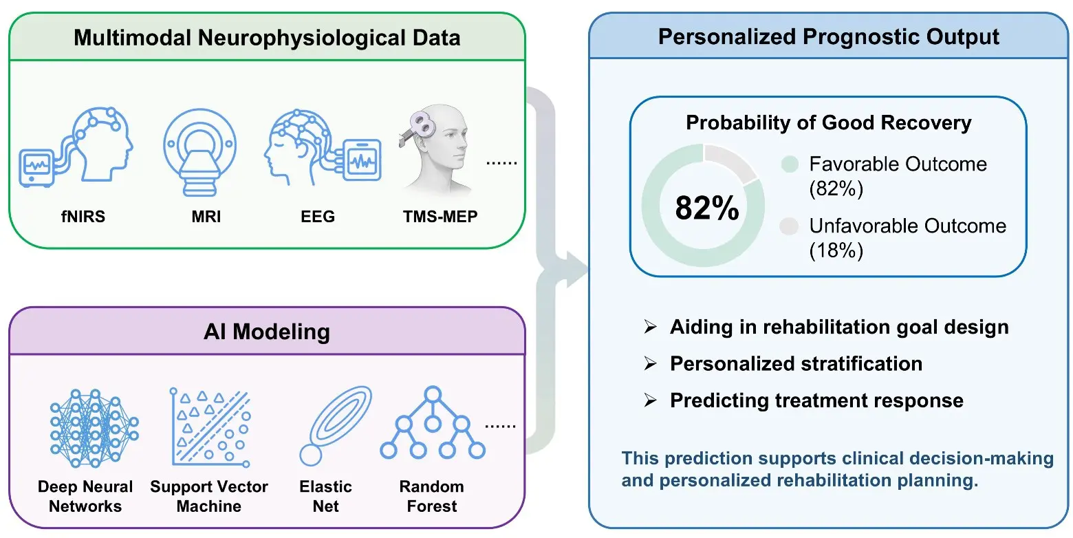
Figure 2. Summary of neurophysiological and neuroimaging prognostic biomarkers developed to characterize recovery potential. The three panels illustrate an integrated framework that combines multimodal neural data (neurophysiological and neuroimaging biomarkers), artificial intelligence (AI) modeling, and personalized prognostic output. These biomarkers help characterize recovery potential, while AI models further stratify individual recovery trajectories and guide the design of tailored rehabilitation strategies.
4. Brain-Computer Interfaces in Neurorehabilitation
4.1. Brain-Computer Interface-Driven Rehabilitation Robots
Given the large global population of stroke survivors and the substantial demand for rehabilitation resources [1], rehabilitation robot-based training can replicate the therapeutic movements of rehabilitation therapists through standardized training tasks, thereby assisting patients in completing their rehabilitation exercises. In this context, rehabilitation therapists can assume the role of high-level supervisors and rehabilitation task designers, overseeing and adjusting the rehabilitation process for multiple patients simultaneously. However, Rodgers et al. [89] conducted a multicentre randomised controlled trial involving 770 stroke patients, comparing the rehabilitation outcomes of robot-assisted training, enhanced upper-limb therapy, and usual care. The results showed that, compared with usual care, neither robot-assisted training nor enhanced upper-limb therapy led to significant improvements in upper-limb function after stroke. These findings do not support the routine clinical implementation of robot-assisted training as delivered in this trial. This study offers new insights for the development of rehabilitation robotics: Rehabilitation robot–based training can now replicate many therapeutic tasks that previously required manual delivery by rehabilitation therapists. However, merely imitating and reproducing these tasks is insufficient to further enhance the efficacy of rehabilitation. The integration of BCIs with rehabilitation robots enables the construction of a closed-loop motor control system by decoding the patient’s movement intentions, using robotic assistance to guide limb movement, and providing sensory feedback from the resulting movement. The establishment of this “central-peripheral-central” closed-loop feedback can promote neural reorganization and the generation of motor sensations [6,90]. The combination of BCIs and rehabilitation robotic systems, therefore, holds promise for substantially improving the efficiency of clinical rehabilitation.
The MIT-Manus, widely regarded as the world’s first upper-limb rehabilitation robot, facilitates coordinated movement of the shoulder and elbow joints by using an end-effector to guide hand motion [91]. Ang et al. [92] examined the efficacy of MIT-Manus, integrated with a motor imagery (MI)-BCI system, in patients with chronic stroke. Motor gains were found to be comparable to those from intensive robotic therapy, even though BCI-MIT-Manus training required fewer arm exercise repetitions by leveraging MI-triggered robotic feedback. In addition to end-effector-type upper-limb rehabilitation robots, exoskeleton-type upper-limb rehabilitation robots have also been developed. For instance, Frisoli et al. [93] developed the L-Exos, a force-feedback-based exoskeleton rehabilitation robot capable of performing point-to-point linear trajectory training tasks in three-dimensional space. To promote the practical use of BCI-exoskeletons by patients, researchers have made efforts to reduce both the time and financial costs associated with their application. Bhagat et al. [94] developed a BCI-based upper-limb exoskeleton (MAHI-Exo II) for stroke rehabilitation, that utilized movement-related cortical potentials (MRCPs) to detect motor intention in chronic stroke patients across days without requiring BCI recalibration, thereby greatly enhancing the convenience of the system for daily use. To enable a user-adaptive rehabilitation architecture, Wu developed an ultra-low-cost BCI system incorporating a lightweight, five-degree-of-freedom forearm exoskeleton equipped with real-time haptic vibration feedback [95]. The integration of BCI with lower-limb rehabilitation robots has also been investigated [96]. In a randomized controlled trial, Zhai et al. [97] provided evidence that BCI-based ankle robot rehabilitation training led to significant lower-limb motor function improvement and spasticity reduction following stroke.
Hand function rehabilitation is critical for performing fine motor control tasks; thus, numerous studies have focused on the development and clinical application of BCI-driven hand exoskeleton rehabilitation systems. To address issues such as poor comfort associated with rigid exoskeletons during human-robot interaction, soft exoskeletons have been proposed as an alternative solution. Guo et al. [98] developed a soft robotic glove (SRG) for paralyzed hand function recovery, in which steady-state visually evoked potentials (SSVEPs) were employed to detect the user’s motor intention and trigger the glove accordingly. After two weeks of training, hand function recovery after SSVEP-BCI-controlled SRG rehabilitation was better than that after robotic glove rehabilitation alone. Cheng et al. [9] used a MI-BCI to drive a SRG during six weeks of rehabilitation training in chronic stroke patients. Their clinical findings indicated that all subjects in the BCI-SRG group experienced a vivid sensation of movement after the intervention. Zhang et al. [99] further developed a hybrid BCI paradigm combining MI and high-frequency SSVEP to enhance BCI control of SRGs for stroke rehabilitation. Evaluation results demonstrated mean accuracies of 95.83 ± 6.83% for 12 healthy subjects and 63.33 ± 10.38% for 9 stroke patients, respectively. Further evidence from a placebo-controlled, multicenter clinical trial demonstrated that integrating BCI control with exoskeleton-assisted physical therapy improves rehabilitation outcomes. Using a randomized controlled functional near-infrared spectroscopy (fNIRS) study, Ji et al. [100] explored the effects and neurorehabilitation mechanisms of BCI-SRG training for stroke rehabilitation and found that the BCI-SRG group showed significant brain cortical activation. Notably, changes in prefrontal activation were positively correlated with functional improvements as measured by the Action Research Arm Test (ARAT), with significant correlations identified specifically in the left and right dorsolateral prefrontal cortex.
It is important to note that the sample sizes of the aforementioned studies are relatively small. To date, no large-scale multicentre trial comparable to the work by Rodgers et al. [89] has been conducted to compare BCI-driven rehabilitation robots, conventional rehabilitation robots, and usual care, to determine whether BCIs offer demonstrable advantages in large-scale clinical applications. Nevertheless, several researchers have attempted to address this question through meta-analyses. Li et al. [90] reviewed twenty-one randomized controlled clinical trials (RCTs) (n = 886 patients) in their meta-analysis and found that BCI combined with rehabilitation robots was effective for patients in the chronic stage. In contrast, Qu et al. [101] found that although BCI-robot systems led to significant and sustained improvements in motor recovery of the hemiparetic upper limb, their meta-analysis showed no significant difference when comparing the BCI-robot group with the robot-alone control group. We identified substantial heterogeneity in the reported clinical studies. This heterogeneity may stem from the absence of a standard protocol for assessing technical and clinical outcomes. In addition, individual clinical variability among patients represents another important source of heterogeneity, as standard BCI-rehabilitation robot training protocol cannot be expected to be effective for all individuals, and a one-size-fits-all BCI training approach is unlikely to be appropriate.
Instead, personalized BCI training strategies tailored to different patients may be necessary. For example, Jia et al. [6] found that individualized differences in post-stroke neural reorganization led to variations in the effectiveness of closed-loop motor control construction during BCI-based rehabilitation robot training. Moreover, the stage of recovery is also an important factor. A meta-analysis showed that BCI-robot intervention improved upper-limb movement function in patients in the chronic stage but not in those in the subacute stage [90]. Tailored BCI-based rehabilitation training could serve as a dependable rehabilitative strategy to promote post-stroke motor recovery.
4.2. Brain-Computer Interface-Triggered Transcranial and Peripheral Neuromodulation
BCIs can directly link patients’ motor intentions to external stimulators acting on the brain (transcranial neuromodulation) or the limbs (peripheral neuromodulation). For peripheral neuromodulation, the rehabilitative mechanism of establishing a “central–peripheral–central” closed loop is similar to that of BCI-based rehabilitation robot training. For transcranial neuromodulation combined with BCI, the addition of transcranial electrical stimulation (tES) or TMS can enhance cortical excitability, motor performance, and motor relearning.
BCI-functional electrical stimulation (FES) and BCI-robot systems share a similar closed-loop mechanism, operating through a “central–peripheral–central” pathway in which cortical activity is decoded to activate peripheral effectors, and the resultant sensory feedback re-enters the central nervous system to complete the loop. By extensively recruiting Golgi tendon organs and muscle spindle feedback circuits, FES not only induces functional movements but also delivers proprioceptive and somatosensory input. Furthermore, evidence from multiple studies suggests that FES can influence cortical excitability [102,103]. Ibáñez et al. [104] validated BCI-actuated FES (BCI–FES) in four patients with chronic stroke, demonstrating the system’s usability and suggesting its potential therapeutic efficacy. Biasiucci et al. [103] further investigated whether BCI–FES targeting hand function outperforms sham-FES in promoting functional recovery and neuroplasticity in 27 patients with chronic stroke. The results confirmed that only the BCI–FES group achieved significant functional recovery, which was sustained for 6–12 months after therapy. In a larger cohort of stroke patients (n = 51 patients), Sebastián-Romagosa et al. [105] further validated the clinical efficacy of BCI–FES, demonstrating that patients achieved a significant 4.68-point improvement assessed by FMA-UL after three months of treatment, which surpasses the threshold for clinically important difference [106]. A meta-analysis of 10 RCTs comprising 290 stroke patients evaluated the effectiveness of BCI–FES training for promoting upper-limb functional recovery. The findings indicate that BCI-FES yields substantial immediate enhancements in upper-limb performance for individuals in both the subacute and chronic stages of stroke, but evidence for long-term effectiveness remains limited [107]. Although the aforementioned studies have demonstrated the clinical efficacy of BCI–FES, certain heterogeneity remains, particularly regarding whether BCI–FES confers long-term benefits. A mechanistic interpretation of BCI–FES is therefore warranted. Biasiucci et al. [103] found that significant EEG differences favoring the BCI–FES group over the sham-FES group, primarily manifested as increased functional connectivity among ipsilesional motor regions, which was associated with functional gains [108]. The BCI-FES-induced simultaneous activation of cerebral motor regions and peripheral effectors may promote Hebbian-like plasticity and reinforced CST projections [103,109,110].
By precisely modulating neural activity and altering cortical excitability, non-invasive brain stimulation (NIBS) techniques, including TMS, transcranial alternating current stimulation (tACS), and transcranial direct current stimulation (tDCS), can promote functional recovery in damaged brain regions. BCI-NIBS primarily adopts two paradigms. The first involves using NIBS to either excite or inhibit specific brain regions prior to BCI-based rehabilitation training, with the goal of enhancing motor learning performance under a favorable cortical excitability state. Jia et al. found that repetitive TMS (rTMS) applied to the sensorimotor cortex before motor imagery could enhance ERD, thereby leading to improved BCI performance in both healthy participants [111] and stroke patients [112]. Hong et al. [113] also reported that long-lasting neuroplasticity was detected in the group that received tDCS prior to MI-BCI. The second paradigm is BCI-triggered NIBS, which establishes a closed loop from central decoding to central stimulation. Accurate temporal coupling between neural state detection and stimulation delivery is critical for potentiating cortical plasticity and facilitating motor recovery [114]. Using state-dependent EEG-triggered TMS (EEG-TMS) in healthy subjects, Zrenner et al. [115] targeted the negative and positive peaks of the sensorimotor µ-rhythm, markers of high versus low corticospinal excitability. Only TMS delivered during the high-excitability state produced long-term potentiation-like changes in corticospinal excitability. Despite the promising clinical outcomes of BCI-NIBS reported above, this approach still faces several technical challenges. The cortical regions from which EEG signals are acquired for BCI decoding are typically not the same as those targeted by stimulation; co-localized recording and stimulation could potentially improve clinical outcomes. The electric and magnetic fields generated during brain stimulation inevitably introduce substantial noise into the simultaneously recorded EEG signals. Therefore, real-time signal denoising algorithms are needed for effective use of BCI-NIBS systems.
In summary, as shown in Figure 3, integrating BCI into rehabilitation robots or neuromodulation can effectively establish a closed-loop system. However, few studies have examined the construction of personalized closed-loop systems based on individual differences, such as patients’ neural reorganization, and this should be a key direction for future research. Moreover, BCI intervention outcomes are strongly influenced by brain-signal stability and closed-loop delay. Therefore, the clinical implementation of BCI requires robust, low-latency signal processing architectures and quick recalibration protocols that optimise the balance between time cost and clinical effectiveness. Furthermore, one important factor currently missing from BCI-based rehabilitation training is the positive affective benefits of interpersonal interaction led by rehabilitation physicians [10,116]. Future development of intelligent rehabilitation technology should explore the affective influences on motor relearning and develop methods for affective human-robot interaction to improve the efficiency of robot-based rehabilitation training.
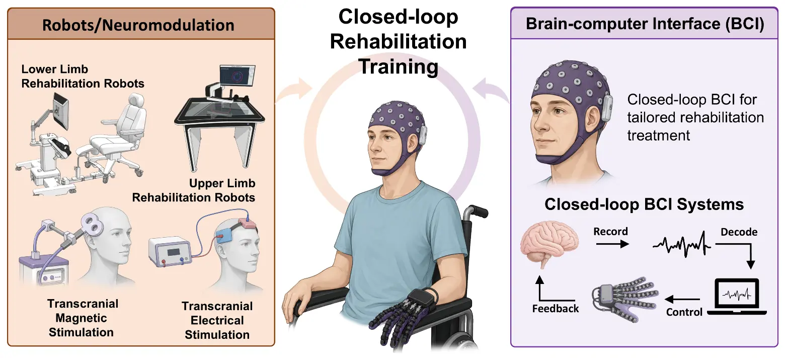
Figure 3. Summary of brain-computer interface (BCI)-driven rehabilitation robots and neuromodulation developed for closed-loop rehabilitation training.
5. Considerations and Limitations
This review highlights recent advances in the development and clinical application of stroke prognostic prediction and BCI-based rehabilitation technologies. It aims to motivate further exploration of how neural reorganization mechanisms can be integrated with AI to develop neurorehabilitation technologies for stroke, thereby enabling personalized rehabilitation treatment. However, careful consideration must be given to the current state of neural signal acquisition and model training, particularly the difficulty of developing a clinically generalizable model from the constrained sample sizes available through routine clinical data collection. Neural activity data, such as EEG, fNIRS, and fMRI, serve as the foundation for implementing these neurorehabilitation technologies. However, the non-uniform and non-standardized acquisition paradigms make large-scale, multicenter data collection difficult. Consequently, limited sample sizes cannot adequately account for the substantial inter-individual variability among stroke patients, rendering the development of a generalized prognostic prediction model or a broadly applicable BCI-training model a distant goal. Therefore, this situation underscores the need to investigate further the mechanisms underlying neural rehabilitation to guide neurotechnology development, rather than relying solely on data-driven approaches. One promising strategy is to stratify patients, for example, by classifying stroke patients into subgroups based on distinct neural reorganization patterns associated with different levels of structural reserve. For each subgroup, BCI closed-loop systems can be constructed around either the lesioned or the contralesional hemisphere, as appropriate. By mitigating certain individual differences, it becomes feasible to develop a relatively generalizable approach within a homogenous patient subgroup, thereby enabling precise rehabilitation training tailored to different populations. Furthermore, employing AI algorithms to adaptively optimize BCI and stimulation parameters, as well as task difficulty, can further optimize the efficacy of the tailored treatment. However, this AI-aided adaptive rehabilitation strategy is still at a relatively early stage of development, and the practical paradigms for such AI-driven optimization have yet to be fully established. Concurrently, the development of neural signal acquisition devices that are easy to use, low-cost, and capable of achieving higher signal-to-noise ratios will promote the widespread adoption of such devices, thereby enabling standardized, multicenter, large-scale clinical trials and providing more robust evidence for validating the clinical efficacy of neurotechnologies for stroke rehabilitation. Moreover, the tailored emotional interaction is also needed during rehabilitation training. Patients’ emotional state during rehabilitation training can strongly influence their training performance. Patients with different personality traits require different training strategies. For example, patients with high mental resilience can be assigned more challenging tasks, whereas those with lower mental resilience should begin with less difficult tasks. Dynamically identifying patients’ emotional states during rehabilitation training and making timely adjustments are also key directions for the development of personalized rehabilitation treatment.
6. Conclusions
This review provides a timely overview of the current status and prospects of the rapidly evolving fields of AI and neurotechnology in stroke neurorehabilitation. Given the large global population of stroke survivors and the substantial demand for rehabilitation resources, there is a clear need for a paradigm shift in stroke rehabilitation: it should be robot-assisted rather than labor-intensive, and intelligently personalized according to individual patient variability rather than one-size-fits-all. Nevertheless, the rapid progress in BCIs, intelligent rehabilitation robots, and personalized treatment techniques provides strong reason to anticipate meaningful advances in stroke neurorehabilitation. Such advancements promise, in turn, to enhance the overall quality of healthcare and rehabilitation services for stroke patients.
Statement of the Use of Generative AI and AI-Assisted Technologies in the Writing Process
During the preparation of this manuscript, the authors used DeepSeek in order to improve the clarity and check the grammar. After using this tool, the authors reviewed and edited the content as needed and take full responsibility for the content of the published article.
Author Contributions
Writing—Original Draft Preparation, T.J., J.S. and Y.G.; Writing—Review & Editing, C.L., and Y.P.; Visualization, T.J. and J.S.; Supervision, C.L. and Y.P.; Project Administration, C.L. and Y.P.; Funding Acquisition, T.J., C.L. and Y.P.
Ethics Statement
Not applicable.
Informed Consent Statement
Not applicable.
Data Availability Statement
No new data were created or analyzed during this study. Data sharing is not applicable to this article.
Funding
This work was supported by the General Program of National Natural Science Foundation of China (grant number 82572934), the National Clinical Key Specialty Construction Project (grant number XKB2023A2001), the High-Level Innovation Talent Program-Dengfeng Initiative (grant number G202511047), and the National Social Science Fund of China (grant number 22&ZD186).
Declaration of Competing Interest
The authors declare that they have no known competing financial interests or personal relationships that could have appeared to influence the work reported in this paper.
References
- Feigin VL, Brainin M, Norrving B, Martins SO, Pandian J, Lindsay P, et al. World Stroke Organization: Global Stroke Fact Sheet 2025. Int. J. Stroke 2025, 20, 132–144. DOI:10.1177/17474930241308142 [Google Scholar]
- Bonkhoff AK, Grefkes C. Precision medicine in stroke: Towards personalized outcome predictions using artificial intelligence. Brain 2022, 145, 457–475. DOI:10.1093/brain/awab439 [Google Scholar]
- Stinear CM, Lang CE, Zeiler S, Byblow WD. Advances and challenges in stroke rehabilitation. Lancet Neurol. 2020, 19, 348–360. DOI:10.1016/S1474-4422(19)30415-6 [Google Scholar]
- Fox MD. Mapping Symptoms to Brain Networks with the Human Connectome. N. Engl. J. Med. 2018, 379, 2237–2245. DOI:10.1056/NEJMra1706158 [Google Scholar]
- Stinear CM. Prediction of motor recovery after stroke: Advances in biomarkers. Lancet Neurol. 2017, 16, 826–836. DOI:10.1016/S1474-4422(17)30283-1 [Google Scholar]
- Jia T, Li C, Mo L, Qian C, Li W, Xu Q, et al. Tailoring brain–machine interface rehabilitation training based on neural reorganization: Towards personalized treatment for stroke patients. Cereb. Cortex 2023, 33, 3043–3052. DOI:10.1093/cercor/bhac259 [Google Scholar]
- Soekadar SR, Birbaumer N, Slutzky MW, Cohen LG. Brain–machine interfaces in neurorehabilitation of stroke. Neurobiol. Dis. 2015, 83, 172–179. DOI:10.1016/j.nbd.2014.11.025 [Google Scholar]
- Chaudhary U, Birbaumer N, Ramos-Murguialday A. Brain–computer interfaces for communication and rehabilitation. Nat. Rev. Neurol. 2016, 12, 513–525. DOI:10.1038/nrneurol.2016.113 [Google Scholar]
- Cheng N, Phua KS, Lai HS, Tam PK, Tang KY, Cheng KK, et al. Brain-Computer Interface-Based Soft Robotic Glove Rehabilitation for Stroke. IEEE Trans. Biomed. Eng. 2020, 67, 3339–3351. DOI:10.1109/TBME.2020.2984003 [Google Scholar]
- Jia T, Pan F, Yang X, Ji L, Farina D, Li C. Artificial Empathy in Therapy and Healthcare: Advancements in Interpersonal Interaction Technologies. Cyborg Bionic Syst. 2025, 6, 473. DOI:10.34133/cbsystems.0473 [Google Scholar]
- Pekna M, Pekny M, Nilsson M. Modulation of Neural Plasticity as a Basis for Stroke Rehabilitation. Stroke 2012, 43, 2819–2828. DOI:10.1161/STROKEAHA.112.654228 [Google Scholar]
- Warraich Z, Kleim JA. Neural Plasticity: The Biological Substrate For Neurorehabilitation. PM R 2010, 2, S208–S219. DOI:10.1016/j.pmrj.2010.10.016 [Google Scholar]
- Wolpaw JR. Harnessing neuroplasticity for clinical applications. Brain 2012, 135, e215. DOI:10.1093/brain/aws017 [Google Scholar]
- Holtmaat A, Svoboda K. Experience-dependent structural synaptic plasticity in the mammalian brain. Nat. Rev. Neurosci. 2009, 10, 647–658. DOI:10.1038/nrn2699 [Google Scholar]
- Hordacre B, Austin D, Brown KE, Graetz L, Pareés I, De Trane S, et al. Evidence for a Window of Enhanced Plasticity in the Human Motor Cortex Following Ischemic Stroke. Neurorehabil. Neural Repair 2021, 35, 307–320. DOI:10.1177/1545968321992330 [Google Scholar]
- Richards LJ, Huang C, Bauer AQ, Lee JM. Long-range axon branching: Contributions to brain network plasticity and repair. Nat. Rev. Neurosci. 2026, 27, 243–259. DOI:10.1038/s41583-025-01008-y [Google Scholar]
- Murphy TH, Corbett D. Plasticity during stroke recovery: From synapse to behaviour. Nat. Rev. Neurosci. 2009, 10, 861–872. DOI:10.1038/nrn2735 [Google Scholar]
- Di Pino G, Pellegrino G, Assenza G, Capone F, Ferreri F, Formica D, et al. Modulation of brain plasticity in stroke: A novel model for neurorehabilitation. Nat. Rev. Neurol. 2014, 10, 597–608. DOI:10.1038/nrneurol.2014.162 [Google Scholar]
- Yan W, Lin Y, Chen YF, Wang Y, Wang J, Zhang M. Enhancing Neuroplasticity for Post-Stroke Motor Recovery: Mechanisms, Models, and Neurotechnology. IEEE Trans. Neural Syst. Rehabil. Eng. 2025, 33, 1156–1168. DOI:10.1109/TNSRE.2025.3551753 [Google Scholar]
- Braaß H, Wolf S, Feldheim J, Chu Y, Tinnermann A, Finsterbusch J, et al. Altered Functional Connectivity Between Cortical Premotor Areas and the Spinal Cord in Chronic Stroke. Stroke 2025, 56, 1159–1168. DOI:10.1161/STROKEAHA.124.048384 [Google Scholar]
- Emmenegger TM, Pfyffer D, Curt A, Schading‐Sassenhausen S, Hupp M, Ashburner J, et al. Longitudinal motor system changes from acute to chronic spinal cord injury. Eur. J. Neurol. 2024, 31, e16196. DOI:10.1111/ene.16196 [Google Scholar]
- Freund P, Weiskopf N, Ashburner J, Wolf K, Sutter R, Altmann DR, et al. MRI investigation of the sensorimotor cortex and the corticospinal tract after acute spinal cord injury: A prospective longitudinal study. Lancet Neurol. 2013, 12, 873–881. DOI:10.1016/S1474-4422(13)70146-7 [Google Scholar]
- Khatibi A, Vahdat S, Lungu O, Finsterbusch J, Büchel C, Cohen-Adad J, et al. Brain-spinal cord interaction in long-term motor sequence learning in human: An fMRI study. NeuroImage 2022, 253, 119111. DOI:10.1016/j.neuroimage.2022.119111 [Google Scholar]
- Liu H, Peng X, Dahmani L, Wang H, Zhang M, Shan Y, et al. Patterns of motor recovery and structural neuroplasticity after basal ganglia infarcts. Neurology 2020, 95, e1174–e1187. DOI:10.1212/WNL.0000000000010149 [Google Scholar]
- McDonnell MN, Stinear CM. TMS measures of motor cortex function after stroke: A meta-analysis. Brain Stimul. 2017, 10, 721–734. DOI:10.1016/j.brs.2017.03.008 [Google Scholar]
- Volz LJ, Sarfeld AS, Diekhoff S, Rehme AK, Pool EM, Eickhoff SB, et al. Motor cortex excitability and connectivity in chronic stroke: A multimodal model of functional reorganization. Brain Struct. Funct. 2015, 220, 1093–1107. DOI:10.1007/s00429-013-0702-8 [Google Scholar]
- Yu P, Dong R, Wang X, Tang Y, Liu Y, Wang C, et al. Neuroimaging of motor recovery after ischemic stroke—Functional reorganization of motor network. NeuroImage Clin. 2024, 43, 103636. DOI:10.1016/j.nicl.2024.103636 [Google Scholar]
- Cui F, Zhao L, Lu M, Liu R, Lv Q, Lin D, et al. Functional and structural brain reorganization in patients with ischemic stroke: A multimodality MRI fusion study. Cereb. Cortex 2023, 33, 10453–10462. DOI:10.1093/cercor/bhad295 [Google Scholar]
- Paul T, Wiemer VM, Hensel L, Cieslak M, Tscherpel C, Grefkes C, et al. Interhemispheric Structural Connectivity Underlies Motor Recovery after Stroke. Ann. Neurol. 2023, 94, 785–797. DOI:10.1002/ana.26737 [Google Scholar]
- Zhang K, Ding L, Wang X, Zhuang J, Tong S, Jia J, et al. Evidence of mirror therapy for recruitment of ipsilateral motor pathways in stroke recovery: A resting fMRI study. Neurotherapeutics 2024, 21, e00320. DOI:10.1016/j.neurot.2024.e00320 [Google Scholar]
- Henderson LA, Gustin SM, Macey PM, Wrigley PJ, Siddall PJ. Functional Reorganization of the Brain in Humans Following Spinal Cord Injury: Evidence for Underlying Changes in Cortical Anatomy. J. Neurosci. 2011, 31, 2630–2637. DOI:10.1523/JNEUROSCI.2717-10.2011 [Google Scholar]
- Byblow WD, Stinear CM, Barber PA, Petoe MA, Ackerley SJ. Proportional recovery after stroke depends on corticomotor integrity. Ann. Neurol. 2015, 78, 848–859. DOI:10.1002/ana.24472 [Google Scholar]
- Stinear CM, Barber PA, Smale PR, Coxon JP, Fleming MK, Byblow WD. Functional potential in chronic stroke patients depends on corticospinal tract integrity. Brain 2006, 130, 170–180. DOI:10.1093/brain/awl333 [Google Scholar]
- Escudero JV, Sancho J, Bautista D, Escudero M, López-Trigo J. Prognostic Value of Motor Evoked Potential Obtained by Transcranial Magnetic Brain Stimulation in Motor Function Recovery in Patients with Acute Ischemic Stroke. Stroke 1998, 29, 1854–1859. DOI:10.1161/01.STR.29.9.1854 [Google Scholar]
- Rehme AK, Fink GR, von Cramon DY, Grefkes C. The Role of the Contralesional Motor Cortex for Motor Recovery in the Early Days after Stroke Assessed with Longitudinal fMRI. Cereb. Cortex 2011, 21, 756–768. DOI:10.1093/cercor/bhq140 [Google Scholar]
- Cassidy JM, Wodeyar A, Wu J, Kaur K, Masuda AK, Srinivasan R, et al. Low-Frequency Oscillations Are a Biomarker of Injury and Recovery After Stroke. Stroke 2020, 51, 1442–1450. DOI:10.1161/STROKEAHA.120.028932 [Google Scholar]
- Finnigan S, van Putten MJAM. EEG in ischaemic stroke: Quantitative EEG can uniquely inform (sub-)acute prognoses and clinical management. Clin. Neurophysiol. 2013, 124, 10–19. DOI:10.1016/j.clinph.2012.07.003 [Google Scholar]
- Nicolo P, Rizk S, Magnin C, Pietro MD, Schnider A, Guggisberg AG. Coherent neural oscillations predict future motor and language improvement after stroke. Brain 2015, 138, 3048–3060. DOI:10.1093/brain/awv200 [Google Scholar]
- Paolo M. TMS-evoked N100 responses as a prognostic factor in acute stroke. Funct. Neurol. 2015, 30, 125–130. DOI:10.11138/FNeur/2015.30.2.125 [Google Scholar]
- Tscherpel C, Dern S, Hensel L, Ziemann U, Fink GR, Grefkes C. Brain responsivity provides an individual readout for motor recovery after stroke. Brain 2020, 143, 1873–1888. DOI:10.1093/brain/awaa127 [Google Scholar]
- Casula EP, Pellicciari MC, Bonnì S, Spanò B, Ponzo V, Salsano I, et al. Evidence for interhemispheric imbalance in stroke patients as revealed by combining transcranial magnetic stimulation and electroencephalography. Hum. Brain Mapp. 2021, 42, 1343–1358. DOI:10.1002/hbm.25297 [Google Scholar]
- Sun J, Zhang Q, Ma D, Jia T, Jia S, Zhai X, et al. Beta-Band Corticomuscular Coherence: A Novel Biomarker of Functional Corticospinal Tract Integrity and Motor Recovery After Stroke. IEEE Trans. Neural Syst. Rehabil. Eng. 2025, 33, 3856–3865. DOI:10.1109/TNSRE.2025.3613417 [Google Scholar]
- Buch ER, Rizk S, Nicolo P, Cohen LG, Schnider A, Guggisberg AG. Predicting motor improvement after stroke with clinical assessment and diffusion tensor imaging. Neurology 2016, 86, 1924–1925. DOI:10.1212/WNL.0000000000002675 [Google Scholar]
- Kumar P, Kathuria P, Nair P, Prasad K. Prediction of Upper Limb Motor Recovery after Subacute Ischemic Stroke Using Diffusion Tensor Imaging: A Systematic Review and Meta-Analysis. J. Stroke 2016, 18, 50–59. DOI:10.5853/jos.2015.01186 [Google Scholar]
- Puig J, Blasco G, Daunis-I-Estadella J, Thomalla G, Castellanos M, Figueras J, et al. Decreased Corticospinal Tract Fractional Anisotropy Predicts Long-term Motor Outcome After Stroke. Stroke 2013, 44, 2016–2018. DOI:10.1161/STROKEAHA.111.000382 [Google Scholar]
- Feng W, Wang J, Chhatbar PY, Doughty C, Landsittel D, Lioutas V, et al. Corticospinal tract lesion load: An imaging biomarker for stroke motor outcomes. Ann. Neurol. 2015, 78, 860–870. DOI:10.1002/ana.24510 [Google Scholar]
- Buch ER, Modir Shanechi A, Fourkas AD, Weber C, Birbaumer N, Cohen LG. Parietofrontal integrity determines neural modulation associated with grasping imagery after stroke. Brain 2012, 135, 596–614. DOI:10.1093/brain/awr331 [Google Scholar]
- Schulz R, Park E, Lee J, Chang WH, Lee A, Kim YH, et al. Interactions Between the Corticospinal Tract and Premotor–Motor Pathways for Residual Motor Output after Stroke. Stroke 2017, 48, 2805–2811. DOI:10.1161/STROKEAHA.117.016834 [Google Scholar]
- Guo L, Kondapavulur S, Lemke SM, Won SJ, Ganguly K. Coordinated increase of reliable cortical and striatal ensemble activations during recovery after stroke. Cell Rep. 2021, 36, 109370. DOI:10.1016/j.celrep.2021.109370 [Google Scholar]
- Sauerbrei BA, Guo JZ, Cohen JD, Mischiati M, Guo W, Kabra M, et al. Cortical pattern generation during dexterous movement is input-driven. Nature 2020, 577, 386–391. DOI:10.1038/s41586-019-1869-9 [Google Scholar]
- Schulz R, Frey BM, Koch P, Zimerman M, Bönstrup M, Feldheim J, et al. Cortico-Cerebellar Structural Connectivity Is Related to Residual Motor Output in Chronic Stroke. Cereb. Cortex 2017, 27, 635–645. DOI:10.1093/cercor/bhv251 [Google Scholar]
- Calautti C, Baron JC. Functional Neuroimaging Studies of Motor Recovery After Stroke in Adults. Stroke 2003, 34, 1553–1566. DOI:10.1161/01.STR.0000071761.36075.A6 [Google Scholar]
- Xu Q, Shao Z, Ma D, Zhai X, Wang Y, Dou W, et al. Predicting rehabilitation outcomes of unilateral stroke after brain-computer interface training based on magnetic resonance imaging data. Medicine 2026, 105, e46280. DOI:10.1097/MD.0000000000046280 [Google Scholar]
- Puig J, Blasco G, Alberich-Bayarri A, Schlaug G, Deco G, Biarnes C, et al. Resting-State Functional Connectivity Magnetic Resonance Imaging and Outcome After Acute Stroke. Stroke 2018, 49, 2353–2360. DOI:10.1161/STROKEAHA.118.021319 [Google Scholar]
- Grefkes C, Fink GR. Connectivity-based approaches in stroke and recovery of function. Lancet Neurol. 2014, 13, 206–216. DOI:10.1016/S1474-4422(13)70264-3 [Google Scholar]
- Vucic S, Stanley Chen KH, Kiernan MC, Hallett M, Benninger DH, Di Lazzaro V, et al. Clinical diagnostic utility of transcranial magnetic stimulation in neurological disorders. Updated report of an IFCN committee. Clin. Neurophysiol. 2023, 150, 131–175. DOI:10.1016/j.clinph.2023.03.010 [Google Scholar]
- Ackerley S, Smith MC, Jordan H, Stinear CM. Biomarkers of Motor Outcomes After Stroke. Phys. Med. Rehabil. Clin. North. Am. 2024, 35, 259–276. DOI:10.1016/j.pmr.2023.06.003 [Google Scholar]
- Smith MC, Scrivener BJ, Stinear CM. Do lower limb motor-evoked potentials predict walking outcomes post-stroke? J. Neurol. Neurosurg. Psychiatry 2024, 95, 348–355. DOI:10.1136/jnnp-2023-332018 [Google Scholar]
- Smith MC, Byblow WD, Barber PA, Stinear CM. Proportional Recovery From Lower Limb Motor Impairment after Stroke. Stroke 2017, 48, 1400–1403. DOI:10.1161/STROKEAHA.116.016478 [Google Scholar]
- Delcamp C, Srinivasan R, Cramer SC. EEG Provides Insights Into Motor Control and Neuroplasticity During Stroke Recovery. Stroke 2024, 55, 2579–2583. DOI:10.1161/STROKEAHA.124.048458 [Google Scholar]
- Vatinno AA, Simpson A, Ramakrishnan V, Bonilha HS, Bonilha L, Seo NJ. The Prognostic Utility of Electroencephalography in Stroke Recovery: A Systematic Review and Meta-Analysis. Neurorehabil. Neural Repair 2022, 36, 255–268. DOI:10.1177/15459683221078294 [Google Scholar]
- Cervera MA, Soekadar SR, Ushiba J, Millán JDR, Liu M, Birbaumer N, et al. Brain‐computer interfaces for post‐stroke motor rehabilitation: A meta‐analysis. Ann. Clin. Transl. Neurol. 2018, 5, 651–663. DOI:10.1002/acn3.544 [Google Scholar]
- Feydy A, Carlier R, Roby-Brami A, Bussel B, Cazalis F, Pierot L, et al. Longitudinal Study of Motor Recovery After Stroke. Stroke 2002, 33, 1610–1617. DOI:10.1161/01.STR.0000017100.68294.52 [Google Scholar]
- Platz T, Kim IH, Engel U, Kieselbach A, Mauritz KH. Brain activation pattern as assessed with multi-modal EEG analysis predict motor recovery among stroke patients with mild arm paresis who receive the Arm Ability Training. Restor. Neurol. Neurosci. 2002, 20, 21–35. DOI:10.3233/RNN-2002-00212 [Google Scholar]
- Keser Z, Buchl SC, Seven NA, Markota M, Clark HM, Jones DT, et al. Electroencephalogram (EEG) With or Without Transcranial Magnetic Stimulation (TMS) as Biomarkers for Post-stroke Recovery: A Narrative Review. Front. Neurol. 2022, 13, 827866. DOI:10.3389/fneur.2022.827866 [Google Scholar]
- Liu J, Sheng Y, Liu H. Corticomuscular Coherence and Its Applications: A Review. Front. Hum. Neurosci. 2019, 13, 100. DOI:10.3389/fnhum.2019.00100 [Google Scholar]
- Mima T, Toma K, Koshy B, Hallett M. Coherence Between Cortical and Muscular Activities After Subcortical Stroke. Stroke 2001, 32, 2597–2601. DOI:10.1161/hs1101.098764 [Google Scholar]
- Xu R, Zhang H, Shi X, Liang J, Wan C, Ming D. Lower-Limb Motor Assessment With Corticomuscular Coherence of Multiple Muscles During Ankle Dorsiflexion After Stroke. IEEE Trans. Neural Syst. Rehabil. Eng. 2023, 31, 160–168. DOI:10.1109/TNSRE.2022.3217571 [Google Scholar]
- Belardinelli P, Laer L, Ortiz E, Braun C, Gharabaghi A. Plasticity of premotor cortico-muscular coherence in severely impaired stroke patients with hand paralysis. NeuroImage Clin. 2017, 14, 726–733. DOI:10.1016/j.nicl.2017.03.005 [Google Scholar]
- Khademi F, Naros G, Nicksirat A, Kraus D, Gharabaghi A. Rewiring Cortico-Muscular Control in the Healthy and Poststroke Human Brain with Proprioceptive β-Band Neurofeedback. J. Neurosci. 2022, 42, 6861–6877. DOI:10.1523/JNEUROSCI.1530-20.2022 [Google Scholar]
- Sun J, Xie R, Yu J, Ji L, Jia T, Pan Y, et al. Dynamic modulation of corticomuscular coherence during ankle dorsiflexion after stroke: Towards hybrid BCI for lower-limb rehabilitation. J. Neural Eng. 2026, 23, 016019. DOI:10.1088/1741-2552/ae3c41 [Google Scholar]
- Puig J, Blasco G, Schlaug G, Stinear CM, Daunis-i-Estadella P, Biarnes C, et al. Diffusion tensor imaging as a prognostic biomarker for motor recovery and rehabilitation after stroke. Neuroradiology 2017, 59, 343–351. DOI:10.1007/s00234-017-1816-0 [Google Scholar]
- Boyd LA, Hayward KS, Ward NS, Stinear CM, Rosso C, Fisher RJ, et al. Biomarkers of Stroke Recovery: Consensus-Based Core Recommendations from the Stroke Recovery and Rehabilitation Roundtable. Neurorehabil. Neural Repair 2017, 31, 864–876. DOI:10.1177/1545968317732680 [Google Scholar]
- Siegel JS, Ramsey LE, Snyder AZ, Metcalf NV, Chacko RV, Weinberger K, et al. Disruptions of network connectivity predict impairment in multiple behavioral domains after stroke. Proc. Natl. Acad. Sci. USA 2016, 113, E4367. DOI:10.1073/pnas.1521083113 [Google Scholar]
- Bowren M, Bruss J, Manzel K, Edwards D, Liu C, Corbetta M, et al. Post-stroke outcomes predicted from multivariate lesion-behaviour and lesion network mapping. Brain 2022, 145, 1338–1353. DOI:10.1093/brain/awac010 [Google Scholar]
- Wiemer VM, Paul T, Hensel L, Cieslak M, Tscherpel C, Grefkes C, et al. Structural reserve-dependent cortical rerouting of motor control after stroke. Brain 2026, 149, 2505–2515. DOI:10.1093/brain/awaf434 [Google Scholar]
- Campagnini S, Arienti C, Patrini M, Liuzzi P, Mannini A, Carrozza MC. Machine learning methods for functional recovery prediction and prognosis in post-stroke rehabilitation: A systematic review. J. NeuroEng. Rehabil. 2022, 19, 54. DOI:10.1186/s12984-022-01032-4 [Google Scholar]
- Stinear CM, Barber PA, Petoe M, Anwar S, Byblow WD. The PREP algorithm predicts potential for upper limb recovery after stroke. Brain 2012, 135, 2527–2535. DOI:10.1093/brain/aws146 [Google Scholar]
- Koch PJ, Park CH, Girard G, Beanato E, Egger P, Evangelista GG, et al. The structural connectome and motor recovery after stroke: Predicting natural recovery. Brain 2021, 144, 2107–2119. DOI:10.1093/brain/awab082 [Google Scholar]
- Nishi H, Oishi N, Ishii A, Ono I, Ogura T, Sunohara T, et al. Deep Learning–Derived High-Level Neuroimaging Features Predict Clinical Outcomes for Large Vessel Occlusion. Stroke 2020, 51, 1484–1492. DOI:10.1161/STROKEAHA.119.028101 [Google Scholar]
- Karakis R, Gurkahraman K, Mitsis GD, Boudrias MH. Deep learning prediction of motor performance in stroke individuals using neuroimaging data. J. Biomed. Inform. 2023, 141, 104357. DOI:10.1016/j.jbi.2023.104357 [Google Scholar]
- Lin PJ, Li W, Zhai X, Li Z, Sun J, Xu Q, et al. Explainable Deep-Learning Prediction for Brain–Computer Interfaces Supported Lower Extremity Motor Gains Based on Multistate Fusion. IEEE Trans. Neural Syst. Rehabil. Eng. 2024, 32, 1546–1555. DOI:10.1109/TNSRE.2024.3384498 [Google Scholar]
- Lin PJ, Jia T, Li C, Li T, Qian C, Li Z, et al. CNN-Based Prognosis of BCI Rehabilitation Using EEG From First Session BCI Training. IEEE Trans. Neural Syst. Rehabil. Eng. 2021, 29, 1936–1943. DOI:10.1109/TNSRE.2021.3112167 [Google Scholar]
- Lin PJ, Zhai X, Li W, Li T, Cheng D, Li C, et al. A Transferable Deep Learning Prognosis Model for Predicting Stroke Patients' Recovery in Different Rehabilitation Trainings. IEEE J. Biomed. Health Inform. 2022, 26, 6003–6011. DOI:10.1109/JBHI.2022.3205436 [Google Scholar]
- Stinear CM, Byblow WD, Ackerley SJ, Smith M, Borges VM, Barber PA. PREP2: A biomarker‐based algorithm for predicting upper limb function after stroke. Ann. Clin. Transl. Neurol. 2017, 4, 811–820. DOI:10.1002/acn3.488 [Google Scholar]
- Angelov PP, Soares EA, Jiang R, Arnold NI, Atkinson PM. Explainable artificial intelligence: An analytical review. WIREs Data Min. Knowl. 2021, 11, e1424. DOI:10.1002/widm.1424 [Google Scholar]
- Gandolfi M, Boscolo Galazzo I, Gasparin Pavan R, Cruciani F, Vale N, Picelli A, et al. eXplainable AI Allows Predicting Upper Limb Rehabilitation Outcomes in Sub-Acute Stroke Patients. IEEE J. Biomed. Health Inform. 2023, 27, 263–273. DOI:10.1109/JBHI.2022.3220179 [Google Scholar]
- Sun J, Lin PJ, Zhai X, Wang W, Jia T, Li Z, et al. Multimodal Behavioral Data Predict Stroke Patient’s Response to BCI Treatment Through Explainable AI. IEEE Trans. Neural Syst. Rehabil. Eng. 2026, 34, 1–9. DOI:10.1109/TNSRE.2025.3625222 [Google Scholar]
- Rodgers H, Bosomworth H, Krebs HI, van Wijck F, Howel D, Wilson N, et al. Robot assisted training for the upper limb after stroke (RATULS): A multicentre randomised controlled trial. Lancet 2019, 394, 51–62. DOI:10.1016/S0140-6736(19)31055-4 [Google Scholar]
- Li D, Li R, Song Y, Qin W, Sun G, Liu Y, et al. Effects of brain-computer interface based training on post-stroke upper-limb rehabilitation: A meta-analysis. J. NeuroEng. Rehabil. 2025, 22, 44. DOI:10.1186/s12984-025-01588-x [Google Scholar]
- Hogan N, Krebs HI, Charnnarong J, Srikrishna P, Sharon A. MIT-MANUS: A workstation for manual therapy and training. I. In Proceedings of the International Workshop on Robot and Human Communication, Tokyo, Japan, 1–3 September 1992; pp. 161–165. DOI:10.1109/ROMAN.1992.253895 [Google Scholar]
- Ang KK, Chua KSG, Phua KS, Wang C, Chin ZY, Kuah CWK, et al. A Randomized Controlled Trial of EEG-Based Motor Imagery Brain-Computer Interface Robotic Rehabilitation for Stroke. Clin. EEG Neurosci. 2015, 46, 310–320. DOI:10.1177/1550059414522229 [Google Scholar]
- Frisoli A, Salsedo F, Bergamasco M, Rossi B, Carboncini MC. A Force‐Feedback Exoskeleton for Upper‐Limb Rehabilitation in Virtual Reality. Appl. Bionics Biomech. 2009, 6, 115–126. DOI:10.1080/11762320902959250 [Google Scholar]
- Bhagat NA, Venkatakrishnan A, Abibullaev B, Artz EJ, Yozbatiran N, Blank AA, et al. Design and Optimization of an EEG-Based Brain Machine Interface (BMI) to an Upper-Limb Exoskeleton for Stroke Survivors. Front. Neurosci. 2016, 10, 122. DOI:10.3389/fnins.2016.00122 [Google Scholar]
- Wu B. A brain–computer-interface driven forearm exoskeleton with adaptive neuroregulation-based feedback for stroke rehabilitation. Alex. Eng. J. 2025, 131, 199–208. DOI:10.1016/j.aej.2025.09.069 [Google Scholar]
- Zongxing L, Jie Z, Ligang Y, Jinshui C, Hongbin L. The Human–Machine Interaction Methods and Strategies for Upper and Lower Extremity Rehabilitation Robots: A Review. IEEE Sens. J. 2024, 24, 13773–13787. DOI:10.1109/JSEN.2024.3374344 [Google Scholar]
- Zhai X, Hao Z, Wang X, Li C, Cao Y, Peng B, et al. Effect of Brain-Computer Interface-Controlled Ankle Robot Training on Post-Stroke Motor Rehabilitation and Resting QEEG Neuroplasticity: A Randomized Controlled Trial. Neurorehabil. Neural Repair 2026, 40, 278–290. DOI:10.1177/15459683251412286 [Google Scholar]
- Guo N, Wang X, Duanmu D, Huang X, Li X, Fan Y, et al. SSVEP-Based Brain Computer Interface Controlled Soft Robotic Glove for Post-Stroke Hand Function Rehabilitation. IEEE Trans. Neural Syst. Rehabil. Eng. 2022, 30, 1737–1744. DOI:10.1109/TNSRE.2022.3185262 [Google Scholar]
- Zhang R, Feng S, Hu N, Low S, Li M, Chen X, et al. Hybrid Brain-Computer Interface Controlled Soft Robotic Glove for Stroke Rehabilitation. IEEE J. Biomed. Health Inform. 2024, 28, 4194–4203. DOI:10.1109/JBHI.2024.3392412 [Google Scholar]
- Ji X, Lu X, Xu Y, Zhang W, Yang H, Yin C, et al. Effects and neural mechanisms of a brain–computer interface-controlled soft robotic glove on upper limb function in patients with subacute stroke: A randomized controlled fNIRS study. J. NeuroEng. Rehabil. 2025, 22, 171. DOI:10.1186/s12984-025-01704-x [Google Scholar]
- Qu H, Zeng F, Tang Y, Shi B, Wang Z, Chen X, et al. The clinical effects of brain–computer interface with robot on upper-limb function for post-stroke rehabilitation: A meta-analysis and systematic review. Disabil. Rehabil. Assist. Technol. 2024, 19, 30–41. DOI:10.1080/17483107.2022.2060354 [Google Scholar]
- Barsi GI, Popovic DB, Tarkka IM, Sinkjær T, Grey MJ. Cortical excitability changes following grasping exercise augmented with electrical stimulation. Exp. Brain Res. 2008, 191, 57–66. DOI:10.1007/s00221-008-1495-5 [Google Scholar]
- Biasiucci A, Leeb R, Iturrate I, Perdikis S, Al-Khodairy A, Corbet T, et al. Brain-actuated functional electrical stimulation elicits lasting arm motor recovery after stroke. Nat. Commun. 2018, 9, 2421. DOI:10.1038/s41467-018-04673-z [Google Scholar]
- Ibáñez J, Monge-Pereira E, Molina-Rueda F, Serrano JI, del Castillo MD, Cuesta-Gómez A, et al. Low Latency Estimation of Motor Intentions to Assist Reaching Movements along Multiple Sessions in Chronic Stroke Patients: A Feasibility Study. Front. Neurosci. 2017, 11, 126. DOI:10.3389/fnins.2017.00126 [Google Scholar]
- Sebastián-Romagosa M, Cho W, Ortner R, Murovec N, Von Oertzen T, Kamada K, et al. Brain Computer Interface Treatment for Motor Rehabilitation of Upper Extremity of Stroke Patients—A Feasibility Study. Front. Neurosci. 2020, 14, 591435. DOI:10.3389/fnins.2020.591435 [Google Scholar]
- Page SJ, Fulk GD, Boyne P. Clinically Important Differences for the Upper-Extremity Fugl-Meyer Scale in People with Minimal to Moderate Impairment Due to Chronic Stroke. Phys. Ther. 2012, 92, 791–798. DOI:10.2522/ptj.20110009 [Google Scholar]
- Ren C, Li X, Gao Q, Pan M, Wang J, Yang F, et al. The effect of brain-computer interface controlled functional electrical stimulation training on rehabilitation of upper limb after stroke: A systematic review and meta-analysis. Front. Hum. Neurosci. 2024, 18, 1438095. DOI:10.3389/fnhum.2024.1438095 [Google Scholar]
- Wu J, Quinlan EB, Dodakian L, McKenzie A, Kathuria N, Zhou RJ, et al. Connectivity measures are robust biomarkers of cortical function and plasticity after stroke. Brain 2015, 138, 2359–2369. DOI:10.1093/brain/awv156 [Google Scholar]
- Markram H. A history of spike-timing-dependent plasticity. Front. Syn. Neurosci. 2011, 3, 4. DOI:10.3389/fnsyn.2011.00004 [Google Scholar]
- Mrachacz-Kersting N, Jiang N, Stevenson AJT, Niazi IK, Kostic V, Pavlovic A, et al. Efficient neuroplasticity induction in chronic stroke patients by an associative brain-computer interface. J. Neurophysiol. 2016, 115, 1410–1421. DOI:10.1152/jn.00918.2015 [Google Scholar]
- Jia T, Mo L, Li C, Liu A, Li Z, Ji L. 5 Hz rTMS improves motor-imagery based BCI classification performance. In Proceedings of the 2021 43rd Annual International Conference of the IEEE Engineering in Medicine & Biology Society (EMBC), Virtual, 1–5 November 2021; pp. 6116–6120. DOI:10.1109/EMBC46164.2021.9630102 [Google Scholar]
- Jia T, Mo L, McGeady C, Sun J, Liu A, Ji L, et al. Cortical Activation Patterns Determine Effectiveness of rTMS-Induced Motor Imagery Decoding Enhancement in Stroke Patients. IEEE Trans. Biomed. Eng. 2025, 72, 1200–1208. DOI:10.1109/TBME.2024.3492977 [Google Scholar]
- Hong X, Lu ZK, Teh I, Nasrallah FA, Teo WP, Ang KK, et al. Brain plasticity following MI-BCI training combined with tDCS in a randomized trial in chronic subcortical stroke subjects: A preliminary study. Sci. Rep. 2017, 7, 9222. DOI:10.1038/s41598-017-08928-5 [Google Scholar]
- Bergmann TO. Brain State-Dependent Brain Stimulation. Front. Psychol. 2018, 9, 2108. DOI:10.3389/fpsyg.2018.02108 [Google Scholar]
- Zrenner C, Desideri D, Belardinelli P, Ziemann U. Real-time EEG-defined excitability states determine efficacy of TMS-induced plasticity in human motor cortex. Brain Stimul. 2018, 11, 374–389. DOI:10.1016/j.brs.2017.11.016 [Google Scholar]
- Jia T, Sun J, McGeady C, Ji L, Li C. Enhancing Brain–Computer Interface Performance by Incorporating Brain-to-Brain Coupling. Cyborg Bionic Syst. 2024, 5, 0116. DOI:10.34133/cbsystems.0116 [Google Scholar]