Fibrinaloid Microclots-Induced Microcirculation Dysfunction: Mechanism and Laser-Based Haemodynamic Validation

Fibrinaloid Microclots-Induced Microcirculation Dysfunction: Mechanism and Laser-Based Haemodynamic Validation
Douglas B. Kell
1,2,*
 Huihui Zhao
3,4
Etheresia Pretorius
1,2,*
Huihui Zhao
3,4
Etheresia Pretorius
1,2,*
Received: 23 January 2026 Revised: 20 March 2026 Accepted: 15 April 2026 Published: 09 May 2026

© 2026 The authors. This is an open access article under the Creative Commons Attribution 4.0 International License (https://creativecommons.org/licenses/by/4.0/).
1. Introduction
1.1. The Microcirculation and Endothelial Dysfunction
The microcirculation represents the terminal elements of the circulation consisting of microvessels, and has been defined as those with diameters less than 20 μm [1] or (more commonly) less than 100 μm [2,3,4,5,6]. As with other blood vessels, the walls of microvessels consist of endothelial cells [7] (we here ignore the glycocalyx [8] and mucins [9]). The microcirculatiion is responsible for perfusing and bringing O2 to tissues throughout the body, and especially at its extremities. By contrast, endothelial dysfunction, manifesting straightforwardly as effects on the microcirculation (e.g., [10,11]), underpins a large variety of diseases and associated symptoms. Thus, Table 1 provides a list of some diseases or syndromes in which the evidence is especially well established. Further details, in terms of the use of laser imaging methods for assessing the microcirculation in these and many other diseases, and whether or not the presence of fibrinaloid microclots has been tested or observed, are given later in Table 2 (laser speckle imaging) and Table 3 (laser Doppler imaging).
Table 1. A summary of some of the diseases or syndromes in which a disruption of the microcirculation is both observed (using any means of observation) and is considered to have aetiological involvement.
|
Disease or Syndrome |
Comments |
Selected References |
|---|---|---|
|
Age-related macular degeneration (AMD) |
Also related to cardiovascular issues. Note that the proteins in drusen, that is the insoluble material often associated with AMD, include amyloid A, amyloid-β, amyloid P, α1-antitrypsin, fibrinogen, etc. [12,13,14], importantly including abundant amyloid structures [15,16,17] that stain with the amyloid stain thioflavin T [18,19] |
|
|
Cancers |
Many vascular changes are involved in all aspects of tumorigenesis, etc. |
|
|
Cardiovascular diseases |
Strong relationship with microcirculation disruption |
|
|
Choroid thickness after haemodialysis |
[48] |
|
|
Chronic fatigue syndrome |
Bears some similarities to Long COVID |
|
|
Chronic venous insufficiency |
[56] |
|
|
COVID and post-COVID |
The key to recovery |
|
|
Diabetes, type 2 |
Recognised as a vascular disease |
|
|
Diabetic complications |
||
|
Fibromyalgia |
Clear likelihood of fibrinaloid microclot deposition |
|
|
General reviews of microcirculation disruption |
||
|
Glaucoma |
Relates to intraocular blood pressure |
|
|
Hypertension |
Capillary rarefaction is seen as a major driver, at least in later stages. Increased blood pressure but lowered flux strongly implies that the latter causes the former. Put another way, there is an increased resistance to flow. This is entirely consistent with the known role of angiogenesis inhibitors in raising blood pressure [116,117]. |
[41,75,116,118,119,120,121,122,123,124,125,126, |
|
Inflammatory bowel disease |
||
|
Metabolic syndrome |
A comorbidity of many cardiovascular diseases |
|
|
Obstructive sleep apnoea |
A common co-morbidity of many of these diseases, which implies the potential for a common aetiology and a common cure |
|
|
Parkinson’s disease |
[158] |
|
|
Pre-eclampsia |
Clear hypertensive disorder, albeit involving cellular senescence [159] and likely an infectious origin [160,161] |
|
|
Psoriasis |
||
|
Raynaud’s phenomenon |
Strongly related to systemic scleroderma |
|
|
Sepsis and septic shock |
One of the most significant examples, with a high level of mortality. Strong evidence that lowered microcirculatory flux relates closely to mortality (and might hence offer protective treatments). |
[175,176,177,178,179,180,181, |
|
Sickle cell disease |
Significant impacts on the microcirculation |
|
|
Stroke (ischaemic) |
Very clear evidence for a relation between microcirculation and multiple factors before and after an ischaemic stroke |
|
|
Subarachnoid haemorrhage |
Erythrocyte sedimentation rate (ESR) was the only measure predictive of a subsequent stroke in a detailed study [226] |
|
|
Systemic sclerosis (scleroderma) |
A major focus in the nailfold capillaroscopy field |
|
|
Traumatic brain injury and other traumas |
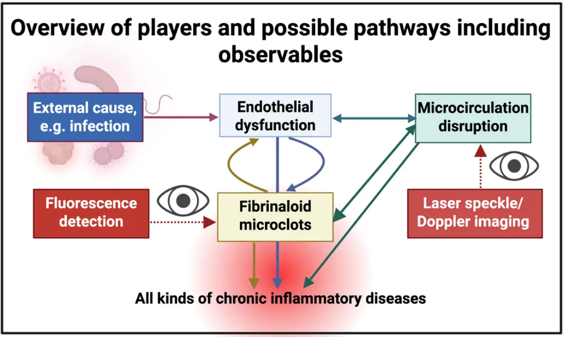
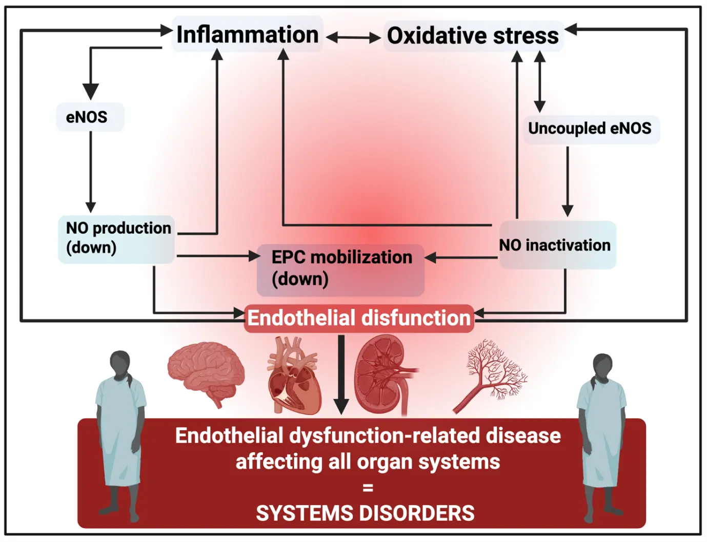
While it is slightly egregious to pick out specific syndromes, we would comment that some, such as ischaemic stroke, are among the main causes of human deaths. All these diseases, especially the chronic diseases [253], display multiple, similar observables, and endothelial dysfunction can both cause and be caused by oxidative stress (from hypoxia and/or reactive oxygen species) (e.g., [254,255,256,257,258,259,260,261,262,263,264,265,266,267,268,269]), mitochondrial dysfunction [270], and inflammation [258,259,271]. Endothelial dysfunction can itself be caused by cellular senescence [272,273,274,275,276,277,278,279,280], and in particular via infection (see Figure 1).
Disseminated intravascular coagulation (DIC) is commonly an accompaniment to sepsis and is characterised by widespread microvascular thrombosis [281,282,283,284,285,286,287,288,289,290,291,292,293,294,295,296,297,298,299] and is associated with a high mortality. A particularly striking recent finding [300] involved the discovery of an unequivocal relationship (odds ratio > 50) between DIC and the presence of fibrinaloid microclots. The directions of causality are not yet known, but this does highlight the potential utility of microcirculation measurements in such patients.
Particular attractions of the microcirculation as an object of study are (i) that it is amenable to non-invasive measurements, in particular via the skin, tongue or retina, and (ii) that it reflects the properties of the far less accessible macrovasculature (see e.g., [126,301,302,303,304,305,306,307,308,309,310,311,312]) and is thus effectively a surrogate for assessing the presence, likelihood, and possibly severity, of a large variety of mainly (cardio)vascular diseases.
1.2. Fibrinaloid Microclots
We discovered long ago that blood can clot into an anomalous amyloid-like form [313,314,315], producing ‘fibrinaloid’ microclots (commonly in the range 2–200 μm in equivalent diameter [316,317,318,319]) that are relatively resistant to degradation. All such diseases in which fibrinaloid microclots formation has been studied are similarly accompanied by the above symptoms. These diseases [320] include acute COVID-19 [321,322,323,324,325,326], Alzheimer’s dementia [313,327,328,329,330], diabetes mellitus type 2 [326,327,331,332,333], Long COVID [317,318,319,334,335,336,337,338,339,340,341,342,343], migraine [344], myalgic encephalopathy/chronic fatigue syndrome (ME/CFS) [345,346,347,348], Parkinson’s disease [327,349,350], rheumatoid diseases [351,352,353], and sepsis/septic shock [300] (see also [294]). It is obvious that such particulate matter as represented by fibrinaloid microclots can block the microcirculation causing local hypoxia, and (focusing on Long COVID) this readily explains phenomena such as blood stasis [354], fatigue [334], post-exertional system exacerbation (previously post-exertional malaise) [355], auto-antibody formation [356], postural orthostatic tachycardia syndrome (POTS) [340], atrial fibrillation [357] and fibromyalgia [358]. Their amyloid nature, as well as their proteome content [335,336,359], straightforwardly explains the relative resistance of fibrinaloid microclots to fibrinolysis [360,361]. We further showed that the macroclots removed by thrombectomy following an ischaemic stroke are also amyloid in character [362,363].
Although other amyloid stains are available, fibrinaloid microclots are typically measured using the classical fluorogenic amyloid stain thioflavin T [364,365,366,367], and the fluorescence is observed using fluorescence microscopy or flow methods. These may be considered to be ‘structural’ methods, while a variety of more ‘functional’ methods are known. We recently suggested [368,369] that one ‘functional’ type of methods of assessing abnormalities in the microcirculation, based on nailfold capillaroscopy (see e.g., [370,371,372,373,374,375,376,377]), might make a useful complement to our ‘structural’ microclot assays.
In addition, other functional methods of measuring the microcirculation are known, including indocyanine green fluorescence [378,379,380,381,382,383], optical coherence tomography angiography (OCTA) [28,179,384,385,386,387,388,389,390,391,392,393,394,395,396], and in particular, as we focus on here, here laser speckle (contrast) imaging (LSI or LSCI) [36,397,398,399,400,401,402,403,404,405,406] and laser Doppler imaging (LDI) [239,402,407,408]. From the physics point of view, the latter two are considered essentially equivalent [409,410]. The chief purpose of this paper is thus to assess LSI and LDI and the findings made with them when they are applied in diseases known to be accompanied by fibrinaloid microclots. Explicitly, methods such as LDI and LSI that can detect the effects of microclots in lowering the rate of blood flow are to be seen as having significant clinical value in terms of admitting treatments that either remove them via fibrinolysis (see e.g., [316,334]) or stop their formationo (e.g., [411,412]). We conclude that, while they are not that cheap, they should prove to be exceptionally useful tools for determining disorders of the microcirculation. A preprint has been posted [413].
1.3. A Note on Systems Biology Explanations of Cause and Effect
We recognise, for non-systems-biologists, that if one is studying a steady state system in which all steps are proceeding at the same rate, it might be seen as odd to argue that some steps are somehow ‘more important’ in determining the speed or course of events than are others. However, this is in fact the case, and it can be quantified precisely. Specifically, the answer lies in what is called sensitivity analysis, in which we study the effects of a normalised change in a parameter (such as the kcat of an enzymatic step) on the normalised value of a variable (in metabolism this is usually a concentration or a flux). Metabolic control analysis [414,415,416,417,418] is exactly such a formalism that applies this to biochemistry, and is based on what is called a local sensitivity analysis [419,420]. Even in very simple systems consisting of just three metabolites (e.g., A → B → C) with the two steps catalysed by enzymes E1 and E2, it is surprisingly tricky to do this well unless one is both informed and careful (see e.g., [421,422,423,424,425]).
This said, in an elementary sense, blood pressure (V), peripheral resistance (R), and the rate of blood flow or flux (I) can be seen as straightforwardly related to each other in a manner entirely analogous to the standard and well-known Ohm’s law relation V = IR of DC electricity. Given this relationship, it is worth pointing out that in such systems, one can establish a setup in which external control is either of the voltage or the current (also in AC systems [426]). Consequently, it is at least reasonable to ask which of the elements contributing to the observable blood flow then normally exerts the greater control. The answer is that it seems clearly to be the case that blood pressure increases that can be observed [41,75,116,118,119,120,121,122,123,124,125,126,127,128,129,130,131,132,133,134,135,136,137,138] seem to be caused mainly by changes in peripheral resistance, i.e., the microcirculation [116,117] rather than anything else controlling the blood pressure more directly. From the perspective of the role of fibrinaloid microclots this is an extremely important recognition.
We next rehearse the role of ‘blood stasis’ in disorders of the microcirculation, before describing LSI.
1.4. The Microcirculation from the Point of View of ‘Blood Stasis’ in Traditional Chinese Medicine
The concept of “blood stasis” in TCM is closely related to microcirculatory disorders in modern medicine. Blood stasis is one of the basic pathological mechanisms in TCM, referring to the pathological state of poor blood circulation and stagnant blood. In recent years, multiple studies have shown a high degree of similarity between the concept of blood stasis and microcirculatory disorders in terms of pathophysiology. Our study on the relationship between blood stasis syndrome and microclotting [354] recognised that abnormal amyloid-like clots, known as fibrinaloid microclots, can form in the blood. These microclots appear in various chronic inflammatory diseases, and they can block microvessels, reduce tissue oxygen transport, and lead to various pathological consequences. Microclots provide a simple mechanism for slowing blood flow by obstructing the transport of red blood cells [334,354].
Blood stasis syndrome is commonly seen in various chronic diseases in TCM clinical practice, and its manifestations are highly consistent with the clinical characteristics of microcirculation disorders. Blood stasis constitution is associated with a number of metabolic abnormalities and microcirculation disorders. The complex interactions between host constitution, gut microbiota, and serum metabolites may indicate potential metabolic vulnerability, even in cases of surface health [427].
The main method of treating blood stasis syndrome in TCM is to promote blood circulation. Many TCM herbal formulas have shown significant effects in improving the microcirculation, not least XueFu ZhuYu (reviewed in [354]). Danshen is another commonly used TCM for promoting blood circulation and removing blood stasis, and studies have shown that it has various pharmacological effects on improving microcirculation [428,429]. Salvia miltiorrhiza extract and its pure compounds have many effects, such as anti atherosclerosis, anti arrhythmia, anti thrombosis, anti hypertension, anti ischemia reperfusion injury, and protection of endothelial function [430]. These effects are closely related to improving the microcirculation [431].
Dang-gui-Si-Ni (DGSN) decoction is another typical formula for promoting blood circulation and removing blood stasis. DGSN can prolong clotting time (PT, TT, and APTT) and reduce fibrinogen (FIB) content. In in vivo experiments, low-dose (500 μg·mL−1) DGSN significantly enhanced cardiac output and blood flow velocity. These findings indicate that DGSN can significantly improve hemodynamics and downregulate coagulation factors, thereby improving the microcirculation [432].
In the treatment of chronic coronary syndrome (CCS), the TCM compound Danshen Dripping Pills has shown significant cardioprotective effects. Compared with Western medicine treatment alone, the combination of TCM and Western medicine improved the effectiveness of electrocardiogram by 8318%, the effectiveness of angina by 20%, and the cessation or reduction of nitroglycerin tablet use by 20%. These effects are likely related to improving coronary microcirculation [433]. Overall, the microcirculation is seen within TCM as contributing strongly to the phenomena of blood stasis. We now turn to LSI.
1.5. Laser Speckle (Contrast) Imaging (LSI/LSCI)
When laser light illuminates an object, the scattered light produces a ‘random’ (actually deterministic, but massively complex) interference effect referred to as a speckle pattern. If the object is moving, the speckles necessarily fluctuate in intensity. Similarly, if the speckle pattern is imaged with an exposure time longer than the shortest speckle fluctuation time, the fluctuations cause a blurring of the speckle, leading to a reduction in the local speckle contrast. This thus encodes the velocities and the distributions thereof as speckle contrast variations; for higher velocity, the speckle contrast is reduced [397,434,435]. Given the size of the speckles, the magnification used, and the typical blood flow rates (~1 mm·s−1 in capillaries [436]), exposure times are typically in the range of 1–10 ms [437]. Typically, the range thereby covered is 0.1–10 mm·s−1. Specific implementations of the general technique are variously referred to as laser speckle imaging (LSI), laser speckle contrast imaging (LSCI), laser speckle contrast analysis (LASCA) and laser speckle flowgraphy (LSFG) (there are slight variations in implementation); we shall normally use the first terminology., and not discriminate them in any real detail A particular attraction is that interrogation can be over a wide area simultaneously (i.e., no scanning or rastering is necessary).
Instruments can be used in ‘spatial mode’ or ‘temporal model’ [397]. Typically, when used in ‘spatial mode’, the speckles are mapped over a small grid of detector pixels (typically 5 × 5) and the contrast is assessed as the standard deviation (SD) of pixel intensities (average pixel intensity = I); SD is low for fast moving speckles (high blood flow) where the image is blurred, and SD is high for slow moving speckles (low blood flow) where the image is not so blurred. The basic formula for LSCI assessment of tissue is thus Flux µ (<I>/SD)2. Note that flux differs from velocity as it also takes into account the concentration of the scattering particles.
In ‘temporal mode’, the intensities of individual pixels during at least 25 successive images are used to calculate average intensities and SDs. Compared to a 5 × 5-pixel set-up, this mode is necessarily at least 25 times slower than spatial mode, but its linear resolution is, of course, 5 times greater.
A typical speckle pattern (taken from [397]) is given in Figure 2, while Figure 3 illustrates the general principle.
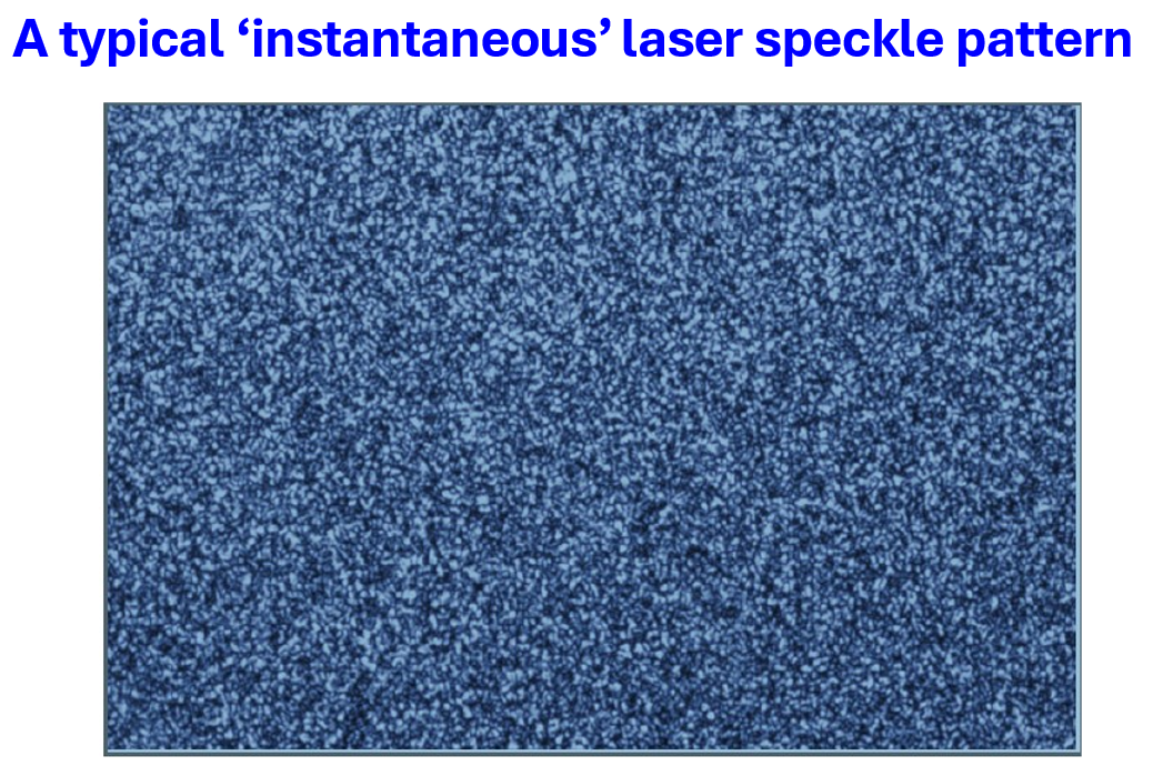
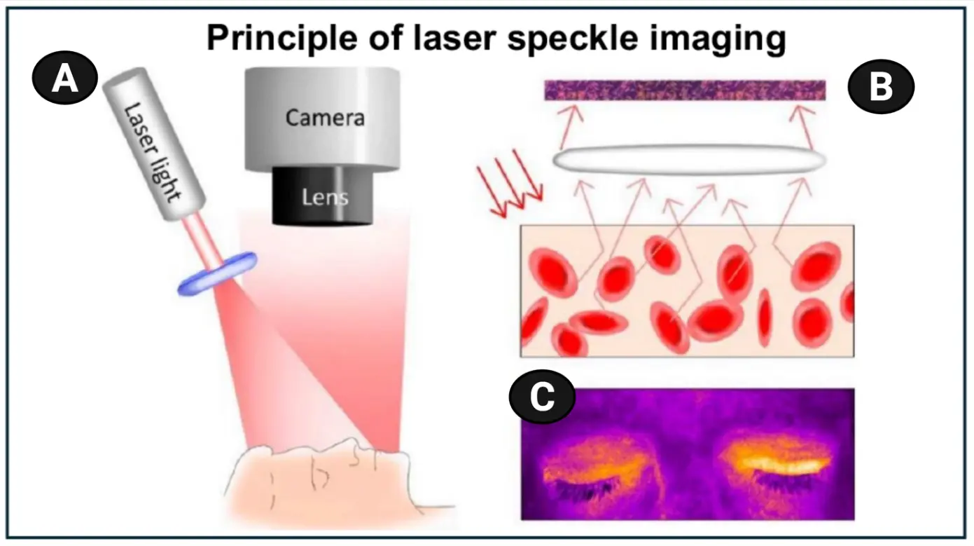
Figure 3. Schematic representation of laser speckle contrast imaging (LSCI). (A) The technique relies on the interference of light backscattered from an interrogation zone, which may include moving particles, creating distinct dark and bright areas (a speckle pattern) that is captured by a camera. The greater the blur or spatial homogeneity, the faster the blood flow. (B) Variations in the speckle pattern, specifically the amount of blur that is observable following a specific imaging window, are predominantly driven by the movement of red blood cells, enabling interpretation as perfusion, whose rate can be estimated. (C) Analysis of speckle-pattern variations yields an image displayed on the monitor, where white and yellow depict areas with high perfusion, contrasting with darker areas indicating lower perfusion areas. Taken from the CC-BY 4.0 publication [401], originally from [438]. For interrogating the subject’s face, only particularly low-power lasers are to be used.
As with our previous review on nailfold capillaroscopy [368,369], we think that the most illuminating strategy for our purposes is to compare diseases assessed using LSCI with those in which fibrinaloid microclots are known to exist experimentally, so as to see how much overlap is already documented. Table 2 sets out such an analysis. Note, of course, that many of these syndromes are diseases of ageing, and that microvascular properties do decline with age [439,440,441,442], so a comparison with age-matched controls is (as usual [443]) required.
Table 2. Some disorders involving the microcirculation in which laser speckle contrast imaging has been found to have diagnostic utility or where fibrinaloid microclots have been demonstrated. Disorders in which fibrinaloid microclots have been demonstrated are rendered in bold face; note that every disorder in which microclots have been demonstrated has microcirculation anomalies when assessed using laser speckle imaging (where this has been applied).
|
Disease or Syndrome |
Comments |
Selected Laser Speckle Imaging References |
Selected Fibrinaloid Microclot References (Where Tested) |
|---|---|---|---|
|
Acute COVID-19 |
Significant evidence of microvascular dysfunction |
||
|
Acute respiratory distress syndrome |
Severity correlates with lowered microcirculation |
||
|
Age-related macular degeneration |
Also related to glaucoma |
||
|
Alzheimer’s dementia (including mild cognitive impairment) |
Significantly lowered cerebral blood flow in Alzheimer’s dementia |
||
|
Antineutrophil Cytoplasmic Antibody-Associated (ANCA) Vasculitis |
Impaired microvascular function and blunted reactivity after occlusion |
[458] |
|
|
Atopic dermatitis |
Review showing marked differences, and treatment |
[459] |
|
|
Behçet’s disease |
Higher baseline flux |
[458] |
|
|
Biliary cirrhosis |
Significant microcirculation lesions |
[460] |
|
|
Burns |
Lesions can occur at places distal to the burn site. Faster though less common than LDI. Useful in burn depth diagnosis. |
[461,462,463,464,465,466,467,468, |
|
|
Cancers |
There is a large literature, indicating issues with the microcirculation. A very small number of reviews at right. |
||
|
Chronic smokers |
Led to Buerger’s disease, successful diagnosed (and cured) |
[477] |
|
|
Cold urticaria |
Attenuated response to cold challenge in patients with cold urticaria |
[478] |
|
|
Connective tissue disorders |
Includes Ehlers-Danlos syndrome |
||
|
Coronary heart disease |
|||
|
Dermatomyositis |
|||
|
Diabetes mellitus, type 1 |
Decreased microcirculation flux. Can be ameliorated by a Chinese herbal formula. |
||
|
Diabetes mellitus, type 2 |
Impaired microcirculation. Correlates with glycosylated haemoglobin A1c levels |
||
|
Diabetic complications |
Review |
[499] |
|
|
Diabetic foot |
|||
|
Diabetic nephropathy |
Decreased blood flow despite no lowering of vessel diameter (consistent with microclots) |
[72] |
|
|
Diabetic neuropathy |
|||
|
Diabetic retinopathy |
Decreased blood flow despite no lowering of vessel diameter (consistent with microclots). Microcirculation decrease precedes retinopathy. |
||
|
Digital ulcers |
|||
|
Endothelial (dys)function |
[342] |
||
|
Erythromelalgia |
[519] |
||
|
Fibromyalgia |
Seemingly, no studies have been done. |
See [358], and for amyloid deposition in skeletal muscle [520] |
|
|
Gaucher disease |
Seemingly, no studies have been done. |
||
|
General reviews |
|||
|
Glaucoma |
Evidence for vasculopathies |
||
|
Heart failure |
|||
|
Hepatitis, viral |
Seemingly, no studies have been done. |
||
|
Hypertension and hypertensives |
As expected, raised blood pressure correlates with lower flow rates (implying that the latter is a cause of the former) |
||
|
Long COVID |
Observable effects on the microcirculation well after the acute phase. Surprisingly few studies. |
||
|
Lupus (systemic lupus erythematosus, SLE) |
Functional and morphological microvascular impairments in patients with SLE |
||
|
Migraine |
Significant microcirculation changes relative to controls |
[344] |
|
|
Myalgic encephalopathy/chronic fatigue syndrome |
Despite the fact that it is clearly an endotheliopathy associated with a deranged microcirculation, and with similarities to Long COVID [52,345,553], we have found no relevant studies |
||
|
Obstructive sleep apnoea |
|||
|
Parkinson’s disease |
Allowed analysis of the function of vasomotor small fibers |
[555] |
|
|
Polycythemia vera |
[556] |
||
|
Polymyositis |
|||
|
Port wine stain |
Convenient non-invasive measurement/diagnostic |
[557] |
|
|
Pre-eclampsia |
Microcirculation impaired |
||
|
Psoriasis |
Perilesional increased perfusion and perfusion inhomogeneity predictive of lesion expansion after two weeks |
||
|
Pulmonary arterial hypertension |
|||
|
Raynaud’s disease or Raynaud’s phenomenon (a transient digital ischaemia, often related to systemic sclerosis) |
Laser speckle analysis is a little-known relative to nailfold capillaroscopy [173]. |
||
|
Rheumatoid arthritis |
|||
|
Sarcopenia |
|||
|
Sepsis and septic shock |
Can discriminate sepsis from septic shock, and lowered blood flow is a high predictor of mortality. The odds ratio of predicting survival based on the presence of fibrinaloid microclots was more than 5 [300]. |
||
|
Sickle cell disease |
Microcirculation significantly impaired |
||
|
Stroke (ischaemic) |
Very useful technique for monitoring and prediction |
||
|
Subarachnoid haemorrhage |
[599] |
||
|
Systemic sclerosis |
Also, the commonest area for nailfold capillaroscopy |
[241,508,509,510,511,512,513,600,601,602, |
|
|
Traumatic brain injury |
Clear effects in decreasing microcirculation |
The widespread occurrence of alterations in the microcirculation, as judged by LSI, is also accompanied by inflammation and oxidative stress, indicating how extensive this is in multiple syndromes (Figure 4), and we would argue that they likely share common causes [97]. In particular, where it was tested, all examples in which fibrinaloid microclots have been measured in plasma also show disorders of the microcirculation, as we would expect. This said, despite extensive detection of microclots in diseases such as myalgic encephalopathy/chronic fatigue syndrome (ME/CFS) and Parkinson’s, laser speckle imaging seems not to have been assessed. This clearly provides some tremendous opportunities.
1.6. Laser Doppler Imaging (LDI)
Detecting properties of moving objects via the Doppler effect is, of course, a method dating back to the 19th century, and a suite of methods referred to under the term laser Doppler imaging (LDI) has also been applied to the non-invasive estimation of blood flow.
Figure 5 illustrates typical arrangements for LDI. In this case, rastering is required, using either a point scan (Figure 5A) or a line scan (Figure 5B).
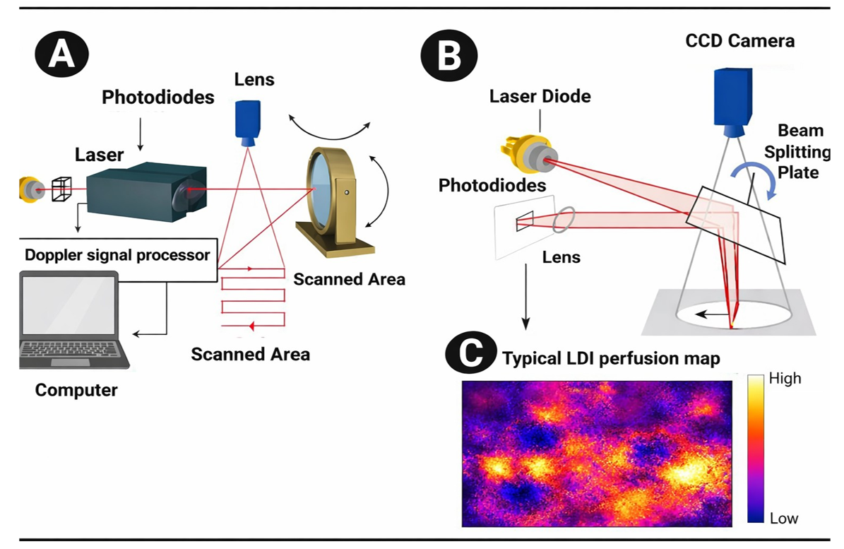
Figure 5. Two styles of laser Doppler imaging in which rastering is achieved via (A) a point scan or (B) a line scan. Figure taken, with permission, and redrawn, from a document provided by Moor Instruments at https://www.moor.co.uk/support/theory/ (accessed on 14 April 2026). Panel (C) shows a representative false-colour perfusion map derived from Doppler signal processing, illustrating the spatial heterogeneity of microvascular blood flow; regions of reduced signal (blue) may reflect locally diminished erythrocyte flux or intermittent capillary obstruction.
In point-scan LDI (Figure 5A), a focused laser beam sequentially interrogates individual pixels across the tissue surface using galvanometric mirrors or mechanical scanning. At each position, the Doppler frequency broadening of backscattered light—arising from moving red blood cells—is recorded to yield a local perfusion signal. A full perfusion map is then reconstructed pixel-by-pixel. This approach provides high spatial resolution and flexible sampling density, but acquisition is relatively slow, making it more susceptible to motion artefacts and limiting temporal resolution. In line-scan LDI (Figure 5B), the laser is shaped into a line (e.g., via cylindrical optics) and projected across the tissue, while a linear detector array (or fast camera) captures Doppler signals simultaneously along that line. The scan proceeds orthogonally to the line direction to build up a 2D image. This parallelisation enables substantially faster acquisition and improved temporal resolution, facilitating dynamic studies of blood flow, albeit sometimes at the expense of spatial resolution and with greater sensitivity to optical heterogeneity along the illuminated line.
In both modalities, the resulting signal is typically expressed in arbitrary perfusion units and reflects an ensemble average over the sampling volume. Importantly, in pathological states characterised by non-uniform or intermittently obstructed microcirculation, the Doppler signal may exhibit non-Gaussian fluctuations and heavy-tailed distributions, reflecting heterogeneous flow velocities and vessel occupancy. The choice of scanning modality therefore influences not only spatial and temporal resolution but also the statistical structure of the measured signal, with implications for quantitative interpretation and for downstream computational analyses, including AI-based detection of abnormal perfusion patterns. These differences are particularly relevant when probing diseases involving microvascular occlusion, where distinguishing true perfusion deficits from sampling or averaging artefacts is critical for linking imaging phenotypes to underlying clot structure and composition.
Our interest again resides in determining the spatial variation of the microcirculation and assessing diseases in which LDI has been used to detect microcirculation dysfunction and where microclots have also been observed. To this end, Table 3 is presented in the style of Table 2, but where the measurement technique is now laser Doppler imaging rather than laser speckle imaging. As with LSI, there is an age dependence in the observables [612] that needs to be taken into account.
Table 3. Some disorders involving the microcirculation in which laser Doppler imaging has been found to have diagnostic utility or where fibrinaloid microclots have been demonstrated. Disorders in which fibrinaloid microclots have been demonstrated are rendered in bold face; note again that every disorder in which microclots have been demonstrated has microcirculation anomalies when assessed using laser Doppler imaging (where this has been applied).
|
Disease or Syndrome |
Comments |
Selected Laser Doppler Imaging References |
Selected Fibrinaloid Microclot References (Where Tested) |
|---|---|---|---|
|
Acute COVID-19 |
Significant evidence of microvascular dysfunction |
||
|
Acute respiratory distress syndrome |
Few studies, but low microcirculation is clearly observable |
||
|
Alzheimer’s dementia (including mild cognitive impairment) |
Significantly lowered cerebral blood flow in Alzheimer’s dementia. Many more studies than with LSI. Care needed with age matching, though [612]. Vascular impairment is clearly related to Aβ deposition. |
||
|
Antineutrophil Cytoplasmic Antibody-Associated (ANCA) Vasculitis |
Impaired microvascular function |
[632] |
|
|
Atopic dermatitis |
Evidence of impaired microvascular function, but surprisingly little recent literature |
||
|
Biliary cirrhosis |
Significant microcirculation lesions |
||
|
Burns |
Utility in burn depth assessment for assisting clinical judgement. Microcirculation problems also occur at distal sites. Seemingly more frequently used here than LSI. |
[464,474,637,638,639,640, |
|
|
Cancer |
Somewhat lesser literature than for LSI (given the importance to tumours of vascularisation), but there are issues with the microcirculation in cancer and its treatment. A very small number of articles on the right. |
||
|
Chronic smokers |
Impaired microcirculation (many more papers than for LSI) |
||
|
Connective tissue disorders |
|||
|
Coronary heart disease |
Surprisingly little directly |
||
|
Dermatomyositis |
Lowered flow rate correlates with disease severity |
||
|
Diabetes mellitus, type 1 |
A large literature implicating microcirculation defects |
||
|
Diabetes mellitus, type 2 |
Impaired microcirculation. Correlates with glycosylated haemoglobin A1c levels |
||
|
Diabetic complications |
Reviews (note that most complications follow from impaired microcirculation) |
||
|
Diabetic foot (ulcers) |
|||
|
Diabetic nephropathy |
Decreased blood flow despite no lowering of vessel diameter (consistent with microclots) |
[682] |
|
|
Diabetic neuropathy |
|||
|
Diabetic retinopathy |
Decreased blood flow despite no lowering of vessel diameter (consistent with microclots). Microcirculation decrease precedes retinopathy. |
||
|
Digital ulcers |
Often coupled to systemic sclerosis |
||
|
Endothelial (dys)function generally |
[342] |
||
|
Fibromyalgia |
Much more frequent use of LDI than of LSI |
[90,93,697,698,699] (and for Complex Regional Pain Syndrome [700]) |
See [358], and for amyloid deposition in skeletal muscle [520] |
|
General reviews |
|||
|
Glaucoma |
|||
|
Heart failure |
Decreased microcirculation is seen as a risk factor (causative) for worse outcomes |
||
|
Hepatitis, viral |
|||
|
Hypertension and hypertensives |
As also seen with LSI, raised blood pressure correlates with lower flow rates (implying that the latter is a cause of the former) |
||
|
Inflammatory bowel disease |
Measured as rectal blood flow |
||
|
Long COVID |
Observable effects on the microcirculation well after the acute phase—surprisingly few studies. |
||
|
Lupus (systemic lupus erythematosus, SLE) |
Functional and morphological microvascular impairments in patients with SLE |
||
|
Migraine |
[730] |
[344] |
|
|
Myalgic encephalopathy/chronic fatigue syndrome |
Despite the fact that it is clearly an endotheliopathy associated with a deranged microcirculation, and with similarities to Long COVID [52,345,553] we have found only one relevant study |
[52] |
|
|
Obstructive sleep apnoea |
Both improved with treatment |
||
|
Parkinson’s disease |
Very few studies |
[734] |
|
|
Peripheral artery disease |
Often related to diabetes |
||
|
Polymyositis |
|||
|
Port wine stain |
Convenient non-invasive measurement/diagnostic |
[557] |
|
|
Pre-eclampsia |
Microcirculation impaired |
||
|
Psoriasis |
|||
|
Raynaud’s disease or Raynaud’s phenomenon (a transient digital ischaemia, often related to systemic sclerosis) |
|||
|
Rheumatoid arthritis |
|||
|
Sarcopenia |
[73] |
||
|
Sepsis and septic shock |
Microcirculation very important in sepsis. As with LSI, it can discriminate sepsis from septic shock, and lowered blood flow is a high predictor of mortality. |
||
|
Sjögren’s syndrome |
[174] |
||
|
Sickle cell disease |
Microcirculation significantly impaired |
||
|
Stroke (ischaemic) |
Very useful technique for monitoring and prediction |
||
|
Subarachnoid haemorrhage |
Note that impaired blood flow (measured by ESR) was the only predictor of a subsequent stroke [226] |
||
|
Systemic sclerosis |
[173,238,239,240,245,247,371,512,513, |
||
|
Traumatic brain injury |
Clear effects in decreasing microcirculation as a result of damage following the trauma |
||
|
Urticaria |
From the perspective of the role of fibrinaloid microclots in affecting the microcirculation, at least two features are of particular note. The first is that blood pressure is raised while flow is lower; this clearly speaks to either or both of capillary rarefaction (decreased density) [123] or to occlusion (or both), and that the raised blood pressure is the effect and not the cause of the change in flow rate. (One might comment that in this sense, blood pressure [792] corresponds to metabolic fluxes in general, as these tend to be regulated by demand and not by supply [793]). Secondly, many studies indicate—not least in diabetes—that changes in the microcirculation leading to hypoxia precede disease, again consistent with an aetiological role. This, of course, raises the significance of these phenomena considerably. In a similar vein, the fact that fibrinaloid microclots accompany so many of these diseases is again consistent with them having an aetiological role rather than being a simple side effect of whatever the core component of the diseases might be considered to be.
While the above table focused on disease, it is worth noting that LDI indicated that there are significant differences in local blood flow at acupuncture points relative to surrounding tissue [794,795,796], and that suitable treatments can affect the microcirculation as measured [797]. Given the significance of blood stasis in a variety of diseases [354], this is definitely noteworthy.
1.7. Comparison of the Two Techniques
Both laser Doppler Imaging and Laser speckle imaging are capable of measuring the microcirculation effectively, are comparably priced, and in skilled hands generally reasonably reproducible [410,798,799,800,801,802,803] depending on the LSI exposure time (though seemingly not when assessed in boys [804]). They are significantly more expensive than nailfold capillaroscopy, but do offer real-time measurements. The general feeling is that LSI is more powerful but that LDI penetrates more deeply if that is important, although this depends on a variety of optical and geometric parameters [805,806,807,808]. In one study of dermal blood flow [809], LSI was considered more sensitive.
2. Discussion
2.1. Comparison of Technological Advantages and Innovative Breakthroughs
Laser speckle imaging (LSI) and laser Doppler imaging (LDI) quantify microvascular blood flow in a non-invasive manner, significantly enhancing the clinical value of microcirculation assessment. LSI captures real-time blood flow velocity and distribution with a high spatial resolution of 10 μm, suitable for dynamic monitoring of superficial organs. LDI is known for its ability to penetrate deeper tissues and locate low perfusion areas in deep regions such as the myocardium. The combined application of the two offers functional complementarity and provides a comprehensive analysis for complex microcirculatory disorders. The introduction of artificial intelligence algorithms has further improved the accuracy of blood flow parameter analysis, promoting the transfer of microcirculation imaging techniques and instrumentation from laboratory research to clinical practice.
2.2. Unity and Specificity of Cross-Disease Mechanisms
Fibrinaloid microthrombi, as the core pathological mediator of microcirculatory disorders, exhibit both mechanistic unity and significant specificity due to differences in precise phenotypes in various diseases. Its unity is reflected in the fact that, whether in acute infection (such as COVID-19), metabolic disorders (such as diabetes), or autoimmune diseases (such as systemic lupus erythematosus), the formation of microthrombosis involves three core links: endothelial cell injury, platelet activation, and a systemic imbalance between coagulation and fibrinolysis. The commonality of these pathological processes suggests that microthrombi may be a common hub for the transformation of various diseases into microcirculatory disorders and vice versa.
2.3. Opportunities and Challenges for Further Clinical Translation
Laser speckle imaging (LSI) and laser Doppler imaging (LDI) bring new opportunities for the diagnosis, treatment and prognosis of microcirculatory disorders: intraoperative blood flow imaging can optimize the effect of cardiovascular surgery, portable equipment can improve the early screening rate of chronic diseases such as diabetes and foot, and objective blood flow parameters may support the evaluation of the efficacy of traditional Chinese medicine. However, the promotion of the technology still faces obstacles: the blood flow calculation standards of different devices are not unified, imaging of deep organs (such as the myocardium) is limited, and high costs constrain grassroots applications.
2.4. Future Research Directions and Technological Innovation
Future research may be expected to focus on a number of major directions: precision imaging technology, developing targeted probes and super-resolution microscopes to achieve subcellular-level visualization of microthrombi; intelligent diagnostic systems, using AI algorithms to automatically analyze blood flow patterns and improve the efficiency of recognition of microthrombi and their effects; and multimodal integration, combining optical, ultrasound and other technologies to simultaneously obtain three-dimensional information such as blood flow and vascular elasticity. As examples of deep learning, Shang et al. [810] used convolutional neural networks to transform speckle dynamics into absolute blood flow rates in mm/s, Yosovich et al. [597] used deep learning to classify flow abnormalities directly from speckle images, Morales-Vargas and colleagues [811] used deep learning for vessel segmentation and depth estimation, Park and Ahn [812] used AI effectively to solve the inverse scattering problem, while Shi and colleagues [813] were able to apply these methods in an intraoperative setting. As to multimodal methods, Wang and colleagues [814] have successfully implemented combined hyperspectral and laser speckle imaging.
Together with biochemical analyses involving multiomics and the data mining thereof, this will greatly promote microcirculation research from “functional observation” to “molecular mechanism analysis”, providing new tools for the diagnosis and treatment of cardiovascular and cerebrovascular diseases.
Acknowledgments
D.B.K. thanks Brian Lock (Moor Instruments) for useful discussions.
Author Contributions
Conceptualization, D.B.K., E.P. and H.Z.; Formal Analysis, D.B.K., E.P. and H.Z.; Resources, D.B.K. and E.P.; Writing—Original Draft Preparation, D.B.K.; Writing—Review & Editing, D.B.K., E.P. and H.Z.; Visualization, D.B.K. and E.P.; Funding Acquisition, D.B.K. and E.P.
Ethics Statement
Not applicable.
Informed Consent Statement
Not applicable.
Data Availability Statement
Not applicable.
Funding
D.B.K. thanks the Balvi Foundation (grant 18) and the Novo Nordisk Foundation for funding (grant NNF20CC0035580). E.P. thanks PolyBio Research Foundation and Kanro Foundation for funding. The content and findings reported and illustrated are the sole deduction, view, and responsibility of the researchers and do not reflect the official position and sentiments of the funders. The funders had no role in study design, data collection and analysis, decision to publish, or preparation of the manuscript.
Declaration of Competing Interests
E.P. is a named inventor on a patent disclosing the use of fluorescence microscopy in Long COVID.
References
-
Guven G, Hilty MP, Ince C. Microcirculation: Physiology, Pathophysiology, and Clinical Application. Blood Purif. 2020, 49, 143–150. DOI:10.1159/000503775 [Google Scholar]
-
Lai C, Teboul JL. Hemodynamic monitoring: Current practice and new perspectives. In The Sepsis Codex; Sa MB, Hidalgo J, Perez-Fernandez J, Eds.; Elsevier: Amsterdam, The Netherlands, 2023; pp. 75–87. [Google Scholar]
-
Munoz CJ, Lucas A, Williams AT, Cabrales P. A Review on Microvascular Hemodynamics: The Control of Blood Flow Distribution and Tissue Oxygenation. Crit. Care Clin. 2020, 36, 293–305. DOI:10.1016/j.ccc.2019.12.011 [Google Scholar]
-
Orellana Jimenez CEA. Sepsis and Microcirculation. In The Sepsis Codex; Sa MB, Hidalgo J, Perez-Fernandez J, Eds.; Elsevier: Amsterdam, The Netherlands, 2023; pp. 29–34. [Google Scholar]
-
Slovinski AP, Hajjar LA, Ince C. Microcirculation in Cardiovascular Diseases. J. Cardiothorac. Vasc. Anesth. 2019, 33, 3458–3468. DOI:10.1053/j.jvca.2019.08.008 [Google Scholar]
-
Yu DY, Cringle SJ, Yu PK, Balaratnasingam C, Mehnert A, Sarunic MV, et al. Retinal capillary perfusion: Spatial and temporal heterogeneity. Prog. Retin. Eye Res. 2019, 70, 23–54. DOI:10.1016/j.preteyeres.2019.01.001 [Google Scholar]
-
Alberts B, Johnson A, Lewis J, Morgan D, Raff M, Roberts K, et al. Molecular Biology of the Cell, 6th ed.; Garland Science: New York, NY, USA, 2016. [Google Scholar]
-
Foote CA, Soares RN, Ramirez-Perez FI, Ghiarone T, Aroor A, Manrique-Acevedo C, et al. Endothelial Glycocalyx. Compr. Physiol. 2022, 12, 3781–3811. DOI:10.1002/cphy.c210029 [Google Scholar]
-
Kesimer M, Ehre C, Burns KA, Davis CW, Sheehan JK, Pickles RJ. Molecular organization of the mucins and glycocalyx underlying mucus transport over mucosal surfaces of the airways. Mucosal Immunol. 2013, 6, 379–392. DOI:10.1038/mi.2012.81 [Google Scholar]
-
Ait-Oufella H, Maury E, Lehoux S, Guidet B, Offenstadt G. The endothelium: Physiological functions and role in microcirculatory failure during severe sepsis. Intensive Care Med. 2010, 36, 1286–1298. DOI:10.1007/s00134-010-1893-6 [Google Scholar]
-
Cusack R, Leone M, Rodriguez AH, Martin-Loeches I. Endothelial Damage and the Microcirculation in Critical Illness. Biomedicines 2022, 10, 3150. DOI:10.3390/biomedicines10123150 [Google Scholar]
-
Crabb JW. The proteomics of drusen. Cold Spring Harb. Perspect. Med. 2014, 4, a017194. DOI:10.1101/cshperspect.a017194 [Google Scholar]
-
Dentchev T, Milam AH, Lee VM, Trojanowski JQ, Dunaief JL. Amyloid-beta is found in drusen from some age-related macular degeneration retinas, but not in drusen from normal retinas. Mol. Vis. 2003, 9, 184–190. Available online: http://www.molvis.org/molvis/v9/a27/v9a27-dentchev.pdf (accessed on 14 April 2026).
-
Wang J, Ohno-Matsui K, Yoshida T, Kojima A, Shimada N, Nakahama K, et al. Altered function of factor I caused by amyloid beta: Implication for pathogenesis of age-related macular degeneration from Drusen. J. Immunol. 2008, 181, 712–720. DOI:10.4049/jimmunol.181.1.712 [Google Scholar]
-
Isas JM, Luibl V, Johnson LV, Kayed R, Wetzel R, Glabe CG, et al. Soluble and mature amyloid fibrils in drusen deposits. Investig. Ophthalmol. Vis. Sci. 2010, 51, 1304–1310. DOI:10.1167/iovs.09-4207 [Google Scholar]
-
Luibl V, Isas JM, Kayed R, Glabe CG, Langen R, Chen J. Drusen deposits associated with aging and age-related macular degeneration contain nonfibrillar amyloid oligomers. J. Clin. Investig. 2006, 116, 378–385. DOI:10.1172/JCI25843 [Google Scholar]
-
Shoda C, Kitagawa Y, Shimada H, Yuzawa M, Tateno A, Okubo Y. Relationship of Area of Soft Drusen in Retina with Cerebral Amyloid-beta Accumulation and Blood Amyloid-beta Level in the Elderly. J. Alzheimers Dis. 2018, 62, 239–245. DOI:10.3233/JAD-170956 [Google Scholar]
-
Anderson DH, Talaga KC, Rivest AJ, Barron E, Hageman GS, Johnson LV. Characterization of beta amyloid assemblies in drusen: The deposits associated with aging and age-related macular degeneration. Exp. Eye Res. 2004, 78, 243–256. DOI:10.1016/j.exer.2003.10.011 [Google Scholar]
-
Mullins RF, Russell SR, Anderson DH, Hageman GS. Drusen associated with aging and age-related macular degeneration contain proteins common to extracellular deposits associated with atherosclerosis, elastosis, amyloidosis, and dense deposit disease. FASEB J. 2000, 14, 835–846. DOI:10.1096/fasebj.14.7.835 [Google Scholar]
-
Friedman E. A hemodynamic model of the pathogenesis of age-related macular degeneration. Am. J. Ophthalmol. 1997, 124, 677–682. DOI:10.1016/s0002-9394(14)70906-7 [Google Scholar]
-
Friedman E. The pathogenesis of age-related macular degeneration. Am. J. Ophthalmol. 2008, 146, 348–349. DOI:10.1016/j.ajo.2008.05.017 [Google Scholar]
-
Kubicka-Trząska A. Macular microcirculation blood flow in patients with age related macular degeneration treated with photodynamic therapy and transpupillary thermotherapy. Klin. Oczna 2007, 109, 138–141. Available online: https://europepmc.org/article/med/17725271 (accessed on 14 April 2026).
-
Lipecz A, Miller L, Kovacs I, Czakó C, Csipo T, Baffi J, et al. Microvascular contributions to age-related macular degeneration (AMD): From mechanisms of choriocapillaris aging to novel interventions. Geroscience 2019, 41, 813–845. DOI:10.1007/s11357-019-00138-3 [Google Scholar]
-
Lylyk I, Bleise C, Lylyk PN, Perez N, Lundquist J, Scrivano E, et al. Ophthalmic artery angioplasty for age-related macular degeneration. J. Neurointerv. Surg. 2022, 14, 968–972. DOI:10.1136/neurintsurg-2021-018222 [Google Scholar]
-
Stefánsson E, Geirsdóttir A, Sigurdsson H. Metabolic physiology in age related macular degeneration. Prog. Retin. Eye Res. 2011, 30, 72–80. DOI:10.1016/j.preteyeres.2010.09.003 [Google Scholar]
-
Alameddine RS, Hamieh L, Shamseddine A. From sprouting angiogenesis to erythrocytes generation by cancer stem cells: Evolving concepts in tumor microcirculation. Biomed. Res. Int. 2014, 2014, 986768. DOI:10.1155/2014/986768 [Google Scholar]
-
Fukumura D, Duda DG, Munn LL, Jain RK. Tumor microvasculature and microenvironment: Novel insights through intravital imaging in pre-clinical models. Microcirculation 2010, 17, 206–225. DOI:10.1111/j.1549-8719.2010.00029.x [Google Scholar]
-
Gao W. Quantitative depth-resolved microcirculation imaging with optical coherence tomography angiography (Part I): Blood flow velocity imaging. Microcirculation 2018, 25, e12375. DOI:10.1111/micc.12375 [Google Scholar]
-
Li HM. Microcirculation of liver cancer, microenvironment of liver regeneration, and the strategy of Chinese medicine. Chin. J. Integr. Med. 2016, 22, 163–167. DOI:10.1007/s11655-016-2460-y [Google Scholar]
-
Mayr NA, Hawighorst H, Yuh WT, Essig M, Magnotta VA, Knopp MV. MR microcirculation assessment in cervical cancer: Correlations with histomorphological tumor markers and clinical outcome. J. Magn. Reson. Imaging 1999, 10, 267–276. DOI:10.1002/(SICI)1522-2586(199909)10:3%3C267::AID-JMRI7%3E3.0.CO;2-Y [Google Scholar]
-
Puleri DF, Balogh P, Randles A. Computational models of cancer cell transport through the microcirculation. Biomech. Model. Mechanobiol. 2021, 20, 1209–1230. DOI:10.1007/s10237-021-01452-6 [Google Scholar]
-
Wei F, Su Y, Quan Y, Li X, Zou Q, Zhang L, et al. Anticoagulants Enhance Molecular and Cellular Immunotherapy of Cancer by Improving Tumor Microcirculation Structure and Function and Redistributing Tumor Infiltrates. Clin. Cancer Res. 2023, 29, 2525–2539. DOI:10.1158/1078-0432.CCR-22-2757 [Google Scholar]
-
Bacelova M, Nikolova J, Alakidi A, Petkova V, Mihaylova V, Dimov I, et al. Microcirculation and cardiovascular risk: Diagnostic value and clinical relevance. Pharmacia 2025, 72, 1–8. DOI:10.3897/pharmacia.72.e154431 [Google Scholar]
-
Ciaramella L, Di Serafino L, Mitrano L, De Rosa ML, Carbone C, Rea FS, et al. Invasive Assessment of Coronary Microcirculation: A State-of-the-Art Review. Diagnostics 2023, 14, 86. DOI:10.3390/diagnostics14010086 [Google Scholar]
-
Kalia N. A historical review of experimental imaging of the beating heart coronary microcirculation in vivo. J. Anat. 2023, 242, 3–16. DOI:10.1111/joa.13611 [Google Scholar]
-
Lazaridis A, Triantafyllou A, Mastrogiannis K, Malliora A, Doumas M, Gkaliagkousi E. Assessing skin microcirculation in patients at cardiovascular risk by using laser speckle contrast imaging. A narrative review. Clin. Physiol. Funct. Imaging 2023, 43, 211–222. DOI:10.1111/cpf.12819 [Google Scholar]
-
Tibiriçá E, Lorenzo A, de Oliveira GMM. Microcirculation and Cardiovascular Diseases. Arq. Bras. Cardiol. 2018, 111, 120–121. DOI:10.5935/abc.20180149 [Google Scholar]
-
Ullrich-Daub H, Daub S, Olschewski M, Münzel T, Gori T. Diseases of the Coronary Microcirculation: Diagnosis and Treatment. Dtsch. Arztebl. Int. 2023, 120, 739–746. DOI:10.3238/arztebl.m2023.0205 [Google Scholar]
-
Widmer RJ, Samuels B, Samady H, Price MJ, Jeremias A, Anderson RD, et al. The functional assessment of patients with non-obstructive coronary artery disease: Expert review from an international microcirculation working group. EuroIntervention 2019, 14, 1694–1702. DOI:10.4244/EIJ-D-18-00982 [Google Scholar]
-
Xu S, Ilyas I, Little PJ, Li H, Kamato D, Zheng X, et al. Endothelial Dysfunction in Atherosclerotic Cardiovascular Diseases and Beyond: From Mechanism to Pharmacotherapies. Pharmacol. Rev. 2021, 73, 924–967. DOI:10.1124/pharmrev.120.000096 [Google Scholar]
-
Pries AR. Microcirculation in hypertension and cardiovascular disease. Eur. Heart J. Suppl. 2014, 16, A28–A29. DOI:10.1093/eurheartj/sut007 [Google Scholar]
-
Pries AR, Kuebler WM, Habazettl H. Coronary Microcirculation in Ischemic Heart Disease. Curr. Pharm. Des. 2018, 24, 2893–2899. DOI:10.2174/1381612824666180625142341 [Google Scholar]
-
Souza ACDAH, Troschel AS, Marquardt JP, Hadžić I, Foldyna B, Moura FA, et al. Skeletal muscle adiposity, coronary microvascular dysfunction, and adverse cardiovascular outcomes. Eur. Heart J. 2025, 46, 1112–1123. DOI:10.1093/eurheartj/ehae827 [Google Scholar]
-
Taqueti VR, Di Carli MF. Coronary Microvascular Disease Pathogenic Mechanisms and Therapeutic Options: JACC State-of-the-Art Review. J. Am. Coll. Cardiol. 2018, 72, 2625–2641. DOI:10.1016/j.jacc.2018.09.042 [Google Scholar]
-
Taqueti VR, Solomon SD, Shah AM, Desai AS, Groarke JD, Osborne MT, et al. Coronary microvascular dysfunction and future risk of heart failure with preserved ejection fraction. Eur. Heart J. 2018, 39, 840–849. DOI:10.1093/eurheartj/ehx721 [Google Scholar]
-
Obert P, Walther G, Dutheil F, Lesourd B, Chapier R, Courteix D, et al. Regional myocardial function abnormalities are associated with macro- and microcirculation dysfunction in the metabolic syndrome: The RESOLVE study. Heart Vessels 2018, 33, 688–694. DOI:10.1007/s00380-017-1108-y [Google Scholar]
-
Uchida Y, Ichimiya S, Ishii H, Kanashiro M, Watanabe J, Yoshikawa D, et al. Impact of metabolic syndrome on various aspects of microcirculation and major adverse cardiac events in patients with ST-segment elevation myocardial infarction. Circ. J. 2012, 76, 1972–1979. DOI:10.1253/circj.cj-11-1299 [Google Scholar]
-
Roskal-Wałek J, Golębiewska J, Mackiewicz J, Wałek P, Bociek A, Biskup M, et al. The Haemodialysis Session Effect on the Choroidal Thickness and Retinal and Choroidal Microcirculation—A Literature Review. J. Clin. Med. 2023, 12, 7729. DOI:10.3390/jcm12247729 [Google Scholar]
-
Chudzik M, Cender A, Mordaka R, Zielinski J, Katarzynska J, Marcinek A, et al. Chronic Fatigue Associated with Post-COVID Syndrome versus Transient Fatigue Caused by High-Intensity Exercise: Are They Comparable in Terms of Vascular Effects? Vasc. Health Risk Manag. 2022, 18, 711–719. DOI:10.2147/VHRM.S371468 [Google Scholar]
-
Haunhorst S, Dudziak D, Scheibenbogen C, Seifert M, Sotzny F, Finke C, et al. Towards an understanding of physical activity-induced post-exertional malaise: Insights into microvascular alterations and immunometabolic interactions in post-COVID condition and myalgic encephalomyelitis/chronic fatigue syndrome. Infection 2024, 53, 1–13. DOI:10.1007/s15010-024-02386-8 [Google Scholar]
-
Khan F, Spence V, Kennedy G, Belch JJ. Prolonged acetylcholine-induced vasodilatation in the peripheral microcirculation of patients with chronic fatigue syndrome. Clin. Physiol. Funct. Imaging 2003, 23, 282–285. DOI:10.1046/j.1475-097x.2003.00511.x [Google Scholar]
-
Ryabkova VA, Gavrilova NY, Fedotkina TV, Churilov LP, Shoenfeld Y. Myalgic Encephalomyelitis/Chronic Fatigue Syndrome and Post-COVID Syndrome: A Common Neuroimmune Ground? Diagnostics 2022, 13, 66. DOI:10.3390/diagnostics13010066 [Google Scholar]
-
Spence VA, Khan F, Kennedy G, Abbot NC, Belch JJ. Acetylcholine mediated vasodilatation in the microcirculation of patients with chronic fatigue syndrome. Prostaglandins Leukot. Essent. Fatty Acids 2004, 70, 403–407. DOI:10.1016/j.plefa.2003.12.016 [Google Scholar]
-
Wirth KJ, Löhn M. Microvascular Capillary and Precapillary Cardiovascular Disturbances Strongly Interact to Severely Affect Tissue Perfusion and Mitochondrial Function in Myalgic Encephalomyelitis/Chronic Fatigue Syndrome Evolving from the Post COVID-19 Syndrome. Medicina 2024, 60, 194. DOI:10.3390/medicina60020194 [Google Scholar]
-
Nunes M, Kell L, Slaghekke A, Wüst RC, Fielding BC, Kell DB, et al. Virus-induced endothelial senescence as a cause and driving factor for ME/CFS and long COVID: Mediated by a dysfunctional immune system. Cell Death Dis. 2026, 17, 16. DOI:10.1038/s41419-025-08162-2 [Google Scholar]
-
Wollina U, Abdel-Naser MB, Mani R. A review of the microcirculation in skin in patients with chronic venous insufficiency: The problem and the evidence available for therapeutic options. Int. J. Low. Extrem. Wounds 2006, 5, 169–180. DOI:10.1177/1534734606291870 [Google Scholar]
-
Abrard S, Coquet T, Riou J, Rineau E, Hersant J, Vincent A, et al. Detection and quantification of microcirculatory dysfunction in severe COVID-19 not requiring mechanical ventilation: A three-arm cohort study. Shock 2024, 62, 673–681. DOI:10.1097/SHK.0000000000002451 [Google Scholar]
-
Belcaro G, Cornelli U, Cesarone MR, Scipione C, Scipione V, Hu S, et al. Preventive effects of Pycnogenol(R) on cardiovascular risk factors (including endothelial function) and microcirculation in subjects recovering from coronavirus disease 2019 (COVID-19). Minerva Med. 2022, 113, 300–308. DOI:10.23736/S0026-4806.21.07650-3 [Google Scholar]
-
Colantuoni A, Martini R, Caprari P, Ballestri M, Capecchi PL, Gnasso A, et al. COVID-19 Sepsis and Microcirculation Dysfunction. Front. Physiol. 2020, 11, 747. DOI:10.3389/fphys.2020.00747 [Google Scholar]
-
Glazkov AA, Ulbashev DS, Borshchev GG, Pulin AA, Glazkova PA, Kulikov DA. Skin microcirculation reactivity to local thermal hyperaemia in patients with COVID-19—A pilot observational study. Clin. Hemorheol. Microcirc. 2023, 83, 19–29. DOI:10.3233/CH-221431 [Google Scholar]
-
Koutsiaris AG. A Blood Supply Pathophysiological Microcirculatory Mechanism for Long COVID. Life 2024, 14, 1076. DOI:10.3390/life14091076 [Google Scholar]
-
Mesquida J, Caballer A, Cortese L, Vila C, Karadeniz U, Pagliazzi M, et al. Peripheral microcirculatory alterations are associated with the severity of acute respiratory distress syndrome in COVID-19 patients admitted to intermediate respiratory and intensive care units. Crit. Care 2021, 25, 381. DOI:10.1186/s13054-021-03803-2 [Google Scholar]
-
Netiazhenko VZ, Mostovyi SI, Safonova OM, Mikhaliev KO. Microcirculatory Alterations in Stable Coronary Artery Disease Patients with Concomitant COVID-19. Wiad. Lek. 2023, 76, 2224–2238. DOI:10.36740/WLek202310115 [Google Scholar]
-
Sabioni L, De Lorenzo A, Castro-Faria-Neto HC, Estato V, Tibirica E. Long-term assessment of systemic microcirculatory function and plasma cytokines after coronavirus disease 2019 (COVID-19). Braz. J. Infect. Dis. 2023, 27, 102719. DOI:10.1016/j.bjid.2022.102719 [Google Scholar]
-
Schlick S, Lucio M, Wallukat G, Bartsch A, Skornia A, Hoffmanns J, et al. Post-COVID-19 Syndrome: Retinal Microcirculation as a Potential Marker for Chronic Fatigue. Int. J. Mol. Sci. 2022, 23, 13683. DOI:10.3390/ijms232213683 [Google Scholar]
-
Szewczykowski C, Mardin C, Lucio M, Wallukat G, Hoffmanns J, Schröder T, et al. Long COVID: Association of Functional Autoantibodies against G-Protein-Coupled Receptors with an Impaired Retinal Microcirculation. Int. J. Mol. Sci. 2022, 23, 7209. DOI:10.3390/ijms23137209 [Google Scholar]
-
Xiang M, Wu X, Jing H, Liu L, Wang C, Wang Y, et al. The impact of platelets on pulmonary microcirculation throughout COVID-19 and its persistent activating factors. Front. Immunol. 2022, 13, 955654. DOI:10.3389/fimmu.2022.955654 [Google Scholar]
-
Zharkikh EV, Loktionova YI, Fedorovich AA, Gorshkov AY, Dunaev AV. Assessment of Blood Microcirculation Changes after COVID-19 Using Wearable Laser Doppler Flowmetry. Diagnostics 2023, 13, 920. DOI:10.3390/diagnostics13050920 [Google Scholar]
-
Lip S, Tran TQB, Hanna R, Nichol S, Guzik TJ, Delles C, et al. Long-term effects of SARS-CoV-2 infection on blood vessels and blood pressure—LOCHINVAR. J. Hypertens. 2025, 43, 1057–1065. DOI:10.1097/HJH.0000000000004013 [Google Scholar]
-
Chang CH, Tsai RK, Wu WC, Kuo SL, Yu HS. Use of dynamic capillaroscopy for studying cutaneous microcirculation in patients with diabetes mellitus. Microvasc. Res. 1997, 53, 121–127. DOI:10.1006/mvre.1996.2003 [Google Scholar]
-
Hansen TW, Ripa RS. Advances in Imaging Techniques for Assessing Myocardial Microcirculation in People with Diabetes: An Overview of Current Techniques, Emerging Techniques, and Clinical Applications. Diabetes Ther. 2025, 16, 785–797. DOI:10.1007/s13300-025-01710-1 [Google Scholar]
-
Iwase T, Ueno Y, Tomita R, Terasaki H. Relationship Between Retinal Microcirculation and Renal Function in Patients with Diabetes and Chronic Kidney Disease by Laser Speckle Flowgraphy. Life 2023, 13, 424. DOI:10.3390/life13020424 [Google Scholar]
-
Jung CH, Cho YY, Choi D, Kim BY, Kim CH, Mok JO. Relationship of Sarcopenia with Microcirculation Measured by Skin Perfusion Pressure in Patients with Type 2 Diabetes. Endocrinol. Metab. 2020, 35, 578–586. DOI:10.3803/EnM.2020.679 [Google Scholar]
-
Koivukangas V, Oikarinen A, Salmela PI, Lahti A. Microcirculatory response of skin to benzoic acid and methyl nicotinate in patients with diabetes. Diabet. Med. 2000, 17, 130–133. DOI:10.1046/j.1464-5491.2000.00248.x [Google Scholar]
-
Nyberg M, Gliemann L, Hellsten Y. Vascular function in health, hypertension, and diabetes: Effect of physical activity on skeletal muscle microcirculation. Scand. J. Med. Sci. Sports 2015, 25 (Suppl. S4), 60–73. DOI:10.1111/sms.12591 [Google Scholar]
-
Sawada S, Tsuchiya S, Kodama S, Kurosawa S, Endo A, Sugawara H, et al. Vascular resistance of carotid and vertebral arteries is associated with retinal microcirculation measured by laser speckle flowgraphy in patients with type 2 diabetes mellitus. Diabetes Res. Clin. Pract. 2020, 165, 108240. DOI:10.1016/j.diabres.2020.108240 [Google Scholar]
-
Strain WD, Paldanius PM. Diabetes, cardiovascular disease and the microcirculation. Cardiovasc. Diabetol. 2018, 17, 57. DOI:10.1186/s12933-018-0703-2 [Google Scholar]
-
Tooke JE. Microcirculation and diabetes. Br. Med. Bull. 1989, 45, 206–223. DOI:10.1093/oxfordjournals.bmb.a072313 [Google Scholar]
-
Zhong M, Song X, Zhang X, Chen J, Wang L, Xia J, et al. Treatment of microcirculation dysfunction in type 2 diabetic mellitus with Shenqi compound prescription: A protocol of systematic review and meta-analysis of randomized clinical trials. Medicine 2020, 99, e22347. DOI:10.1097/MD.0000000000022347 [Google Scholar]
-
Riaz A, Asghar S, Shahid S, Tanvir H, Ejaz MH, Akram M. Prevalence of Metabolic Syndrome and Its Risk Factors Influence on Microvascular Complications in Patients With Type 1 and Type 2 Diabetes Mellitus. Cureus 2024, 16, e55478. DOI:10.7759/cureus.55478 [Google Scholar]
-
Sena CM, Pereira AM, Seiça R. Endothelial dysfunction—A major mediator of diabetic vascular disease. Biochim. Biophys. Acta 2013, 1832, 2216–2231. DOI:10.1016/j.bbadis.2013.08.006 [Google Scholar]
-
Balasubramanian GV, Chockalingam N, Naemi R. The Role of Cutaneous Microcirculatory Responses in Tissue Injury, Inflammation and Repair at the Foot in Diabetes. Front. Bioeng. Biotechnol. 2021, 9, 732753. DOI:10.3389/fbioe.2021.732753 [Google Scholar]
-
Lowry D, Saeed M, Narendran P, Tiwari A. The Difference Between the Healing and the Nonhealing Diabetic Foot Ulcer: A Review of the Role of the Microcirculation. J. Diabetes Sci. Technol. 2017, 11, 914–923. DOI:10.1177/1932296816658054 [Google Scholar]
-
Li Q, Liu X, Jia M, Sun F, Li Y, Zhang H, et al. Assessment of sublingual microcirculation for the screening of diabetic nephropathy. Diabetol. Metab. Syndr. 2022, 14, 90. DOI:10.1186/s13098-022-00864-3 [Google Scholar]
-
Al-Allaf AW, Khan F, Moreland J, Belch JJ, Pullar T. Investigation of cutaneous microvascular activity and flare response in patients with fibromyalgia syndrome. Rheumatology 2001, 40, 1097–1101. DOI:10.1093/rheumatology/40.10.1097 [Google Scholar]
-
Bengtsson A, Bengtsson M. Regional sympathetic blockade in primary fibromyalgia. Pain 1988, 33, 161–167. DOI:10.1016/0304-3959(88)90086-3 [Google Scholar]
-
Casas-Barragán A, Molina F, Tapia-Haro RM, García-Ríos MC, Correa-Rodríguez M, Aguilar-Ferrándiz ME. Association of core body temperature and peripheral blood flow of the hands with pain intensity, pressure pain hypersensitivity, central sensitization, and fibromyalgia symptoms. Ther. Adv. Chronic Dis. 2021, 12, 2040622321997253. DOI:10.1177/2040622321997253 [Google Scholar]
-
Choi DH, Kim HS. Quantitative analysis of nailfold capillary morphology in patients with fibromyalgia. Korean J. Intern. Med. 2015, 30, 531–537. DOI:10.3904/kjim.2015.30.4.531 [Google Scholar]
-
Frödin T, Bengtsson A, Skogh M. Nail fold capillaroscopy findings in patients with primary fibromyalgia. Clin. Rheumatol. 1988, 7, 384–388. DOI:10.1007/BF02239197 [Google Scholar]
-
Jeschonneck M, Grohmann G, Hein G, Sprott H. Abnormal microcirculation and temperature in skin above tender points in patients with fibromyalgia. Rheumatology 2000, 39, 917–921. DOI:10.1093/rheumatology/39.8.917 [Google Scholar]
-
Kasikcioglu E, Dinler M, Berker E. Reduced tolerance of exercise in fibromyalgia may be a consequence of impaired microcirculation initiated by deficient action of nitric oxide. Med. Hypotheses 2006, 66, 950–952. DOI:10.1016/j.mehy.2005.11.028 [Google Scholar]
-
Le Goff P. Is fibromyalgia a muscle disorder? Jt. Bone Spine 2006, 73, 239–242. DOI:10.1016/j.jbspin.2005.03.022 [Google Scholar]
-
Morf S, Amann-Vesti B, Forster A, Franzeck UK, Koppensteiner R, Uebelhart D, et al. Microcirculation abnormalities in patients with fibromyalgia—Measured by capillary microscopy and laser fluxmetry. Arthritis Res. Ther. 2005, 7, R209–R216. DOI:10.1186/ar1459 [Google Scholar]
-
Nigro A. Microvascular and cerebrovascular alterations in Raynaud’s phenomenon and fibromyalgia. Angiogenesis 2025, 28, 22. DOI:10.1007/s10456-025-09974-8 [Google Scholar]
-
Rubio-Zarapuz A, Parraca JA, Tornero-Aguilera JF, Clemente-Suárez VJ. Unveiling the link: Exploring muscle oxygen saturation in fibromyalgia and its implications for symptomatology and therapeutic strategies. Med. Gas. Res. 2025, 15, 58–72. DOI:10.4103/mgr.MEDGASRES-D-24-00013 [Google Scholar]
-
Shang Y, Gurley K, Symons B, Long D, Srikuea R, Crofford LJ, et al. Noninvasive optical characterization of muscle blood flow, oxygenation, and metabolism in women with fibromyalgia. Arthritis Res. Ther. 2012, 14, R236. DOI:10.1186/ar4079 [Google Scholar]
-
Feuer DS, Handberg EM, Mehrad B, Wei J, Bairey Merz CN, Pepine CJ, et al. Microvascular Dysfunction as a Systemic Disease: A Review of the Evidence. Am. J. Med. 2022, 135, 1059–1068. DOI:10.1016/j.amjmed.2022.04.006 [Google Scholar]
-
Gutterman DD, Chabowski DS, Kadlec AO, Durand MJ, Freed JK, Ait-Aissa K, et al. The Human Microcirculation: Regulation of Flow and Beyond. Circ. Res. 2016, 118, 157–172. DOI:10.1161/CIRCRESAHA.115.305364 [Google Scholar]
-
Morris G, Puri BK, Olive L, Carvalho A, Berk M, Walder K, et al. Endothelial dysfunction in neuroprogressive disorders-causes and suggested treatments. BMC Med. 2020, 18, 305. DOI:10.1186/s12916-020-01749-w [Google Scholar]
-
Paříková A. Rheopheresis and Its Use in the Treatment of Diseases with Impaired Microcirculation. A Review. Czech Slovak Ophthalmol. 2022, 79, 3–5. DOI:10.31348/2023/1 [Google Scholar]
-
Rajendran P, Rengarajan T, Thangavel J, Nishigaki Y, Sakthisekaran D, Sethi G, et al. The vascular endothelium and human diseases. Int. J. Biol. Sci. 2013, 9, 1057–1069. DOI:10.7150/ijbs.7502 [Google Scholar]
-
Ray A, Maharana KC, Meenakshi S, Singh S. Endothelial dysfunction and its relation in different disorders: Recent update. Health Sci. Rev. 2023, 7, 100084. DOI:10.1016/j.hsr.2023.100084 [Google Scholar]
-
Hilty MP, Akin S, Boerma C, Donati A, Erdem O, Giaccaglia P, et al. Automated Algorithm Analysis of Sublingual Microcirculation in an International Multicentral Database Identifies Alterations Associated with Disease and Mechanism of Resuscitation. Crit. Care Med. 2020, 48, e864–e875. DOI:10.1097/CCM.0000000000004491 [Google Scholar]
-
Hilty MP, Ince C. Automated quantification of tissue red blood cell perfusion as a new resuscitation target. Curr. Opin. Crit. Care 2020, 26, 273–280. DOI:10.1097/MCC.0000000000000725 [Google Scholar]
-
Bekkers A, Borren N, Ederveen V, Fokkinga E, Andrade De Jesus D, Sanchez Brea L, et al. Microvascular damage assessed by optical coherence tomography angiography for glaucoma diagnosis: A systematic review of the most discriminative regions. Acta Ophthalmol. 2020, 98, 537–558. DOI:10.1111/aos.14392 [Google Scholar]
-
Aizawa N, Kunikata H, Shiga Y, Yokoyama Y, Omodaka K, Nakazawa T. Correlation between structure/function and optic disc microcirculation in myopic glaucoma, measured with laser speckle flowgraphy. BMC Ophthalmol. 2014, 14, 113. DOI:10.1186/1471-2415-14-113 [Google Scholar]
-
Aizawa N, Kunikata H, Yokoyama Y, Nakazawa T. Correlation between optic disc microcirculation in glaucoma measured with laser speckle flowgraphy and fluorescein angiography, and the correlation with mean deviation. Clin. Exp. Ophthalmol. 2014, 42, 293–294. DOI:10.1111/ceo.12130 [Google Scholar]
-
Aizawa N, Kunikata H, Nakazawa T. Diagnostic power of laser speckle flowgraphy-measured optic disc microcirculation for open-angle glaucoma: Analysis of 314 eyes. Clin. Exp. Ophthalmol. 2019, 47, 680–683. DOI:10.1111/ceo.13482 [Google Scholar]
-
Bojikian KD, Nobrega P, Wen JC, Zhang Q, Mudumbai RC, Johnstone MA, et al. Macular Vascular Microcirculation in Eyes with Open-angle Glaucoma Using Different Visual Field Severity Classification Systems. J. Glaucoma 2019, 28, 790–796. DOI:10.1097/IJG.0000000000001308 [Google Scholar]
-
Hohberger B, Lucio M, Schlick S, Wollborn A, Hosari S, Mardin C. OCT-angiography: Regional reduced macula microcirculation in ocular hypertensive and pre-perimetric glaucoma patients. PLoS ONE 2021, 16, e0246469. DOI:10.1371/journal.pone.0246469 [Google Scholar]
-
Hou W, Feng J, Chen J, Li X, Yang G, Sun X. Analysis of the Optic Nerve Head Microcirculation Using Optical Coherence Tomography Angiography and the Upstream Macrocirculation Using Color Doppler Imaging in Low-Tension and High-Tension Glaucoma Patients. Ophthalmic Res. 2023, 66, 579–589. DOI:10.1159/000528521 [Google Scholar]
-
Lin PW, Chiu LW. Evaluation of Optic Nerve Head Microcirculation in Open-Angle Glaucoma Patients with Unilateral Visual Field Defect. Ophthalmic Res. 2024, 67, 257–265. DOI:10.1159/000538407 [Google Scholar]
-
Taylor L, Bojikian KD, Jung H, Chu Z, Zhou X, Zhang Q, et al. Peripapillary and Macular Microcirculation in Glaucoma Patients of African and European Descent Using Optical Coherence Tomography Angiography. J. Glaucoma 2020, 29, 885–889. DOI:10.1097/IJG.0000000000001629 [Google Scholar]
-
Wang T, Ling Q, Shen B, Jia X. The strong correlation between visual function improvement and retinal microcirculation enhancement in glaucoma. Front. Med. 2025, 12, 1537741. DOI:10.3389/fmed.2025.1537741 [Google Scholar]
-
Yokoyama Y, Aizawa N, Chiba N, Omodaka K, Nakamura M, Otomo T, et al. Significant correlations between optic nerve head microcirculation and visual field defects and nerve fiber layer loss in glaucoma patients with myopic glaucomatous disk. Clin. Ophthalmol. 2011, 5, 1721–1727. DOI:10.2147/OPTH.S23204 [Google Scholar]
-
Mourad JJ, des Guetz G, Debbabi H, Levy BI. Blood pressure rise following angiogenesis inhibition by bevacizumab. A crucial role for microcirculation. Ann. Oncol. 2008, 19, 927–934. DOI:10.1093/annonc/mdm550 [Google Scholar]
-
Sane DC, Anton L, Brosnihan KB. Angiogenic growth factors and hypertension. Angiogenesis 2004, 7, 193–201. DOI:10.1007/s10456-004-2699-3 [Google Scholar]
-
Duprez D, De Buyzere M, De Backer T, Vercammen J, Brusselmans F, Clement DL. Impaired microcirculation in mild-to-moderate essential arterial hypertension. J. Hypertens. 1992, 10, 251–254. DOI:10.1097/00004872-199203000-00008 [Google Scholar]
-
Feihl F, Liaudet L, Waeber B, Levy BI. Hypertension: A disease of the microcirculation? Hypertension 2006, 48, 1012–1017. DOI:10.1161/01.HYP.0000249510.20326.72 [Google Scholar]
-
Agabiti-Rosei C, Saxton SN, De Ciuceis C, Lorenza Muiesan M, Rizzoni D, Agabiti Rosei E, et al. Influence of Perivascular Adipose Tissue on Microcirculation: A Link Between Hypertension and Obesity. Hypertension 2024, 81, 24–33. DOI:10.1161/HYPERTENSIONAHA.123.19437 [Google Scholar]
-
De Ciuceis C, Rizzoni D, Palatini P. Microcirculation and Physical Exercise In Hypertension. Hypertension 2023, 80, 730–739. DOI:10.1161/HYPERTENSIONAHA.122.19465 [Google Scholar]
-
Feihl F, Liaudet L, Waeber B. The macrocirculation and microcirculation of hypertension. Curr. Hypertens. Rep. 2009, 11, 182–189. DOI:10.1007/s11906-009-0033-6 [Google Scholar]
-
Flores J, Pena C, Nugent K. Skin microcirculation and hypertension: Is there a connection? J. Bras. Nefrol. 2025, 47, e202440192. DOI:10.1590/2175-8239-JBN-2024-0192en [Google Scholar]
-
Gracia-Sancho J, Marrone G, Fernandez-Iglesias A. Hepatic microcirculation and mechanisms of portal hypertension. Nat. Rev. Gastroenterol. Hepatol. 2019, 16, 221–234. DOI:10.1038/s41575-018-0097-3 [Google Scholar]
-
Hu Y, Hu A, Song S. Photoplethysmography for Assessing Microcirculation in Hypertensive Patients After Taking Antihypertensive Drugs: A Review. J. Multidiscip. Healthc. 2024, 17, 263–274. DOI:10.2147/JMDH.S441440 [Google Scholar]
-
Laurent S, Agabiti-Rosei C, Bruno RM, Rizzoni D. Microcirculation and Macrocirculation in Hypertension: A Dangerous Cross-Link? Hypertension 2022, 79, 479–490. DOI:10.1161/HYPERTENSIONAHA.121.17962 [Google Scholar]
-
Tsioufis C, Dimitriadis K, Katsiki N, Tousoulis D. Microcirculation in Hypertension: An Update on Clinical Significance and Therapy. Curr. Vasc. Pharmacol. 2015, 13, 413–417. DOI:10.2174/1570161113666150206110512 [Google Scholar]
-
Vicaut E. Hypertension and the microcirculation: A brief overview of experimental studies. J. Hypertens. Suppl. 1992, 10, S59–S68. DOI:10.1097/00004872-199207005-00009 [Google Scholar]
-
Vicaut E. Hypertension and the microcirculation. Arch. Mal. Coeur Vaiss. 2003, 96, 893–903, PMID:14571644. [Google Scholar]
-
Cesarone MR, Laurora G, Belcaro GV. Microcirculation in Systemic Hypertension. Angiology 1992, 43, 899–903. DOI:10.1177/000331979204301104 [Google Scholar]
-
Durante A, Mazzapicchi A, Baiardo Redaelli M. Systemic and Cardiac Microvascular Dysfunction in Hypertension. Int. J. Mol. Sci. 2024, 25, 13294. DOI:10.3390/ijms252413294 [Google Scholar]
-
Fedorovich AA, Loktionova YI, Zharkikh EV, Gorshkov AY, Korolev AI, Dadaeva VA, et al. Skin microcirculation in middle-aged men with newly diagnosed arterial hypertension according to remote laser Doppler flowmetry data. Microvasc. Res. 2022, 144, 104419. DOI:10.1016/j.mvr.2022.104419 [Google Scholar]
-
Jan MY, Hsiu H, Hsu TL, Wang YY, Wang WK. The importance of pulsatile microcirculation in relation to hypertension. IEEE Eng. Med. Biol. Mag. 2000, 19, 106–111. DOI:10.1109/51.844389 [Google Scholar]
-
Junqueira CLC, Magalhaes MEC, Brandao AA, Ferreira E, Cyrino F, Maranhao PA, et al. Microcirculation and biomarkers in patients with resistant or mild-to-moderate hypertension: A cross-sectional study. Hypertens. Res. 2018, 41, 515–523. DOI:10.1038/s41440-018-0043-3 [Google Scholar]
-
Korolev AI, Fedorovich AA, Gorshkov AY, Dadaeva VA, Omelyanenko KV, Chashchin MG, et al. Structural and functional state of various parts of skin microcirculation at an early stage of hypertension in working-age men. Microvasc. Res. 2023, 145, 104440. DOI:10.1016/j.mvr.2022.104440 [Google Scholar]
-
Lewandowska K, Marzyńska D, Rzesoś P, Partyka A, Dydowicz F, Lewandowski M, et al. Methods for the assessment of microcirculation in patients with hypertension. Arterial Hypertens. 2023, 27, 1–12. DOI:10.5603/AH.a2023.0004 [Google Scholar]
-
Rizzoni D, Agabiti-Rosei C, Boari GEM, Muiesan ML, De Ciuceis C. Microcirculation in Hypertension: A Therapeutic Target to Prevent Cardiovascular Disease? J. Clin. Med. 2023, 12, 4892. DOI:10.3390/jcm12154892 [Google Scholar]
-
Zweifach BW. The microcirculation in experimental hypertension. State-of-the-art review. Hypertension 1983, 5, I10–I16. DOI:10.1161/01.hyp.5.2_pt_2.i10 [Google Scholar]
-
Bernardino VR, Rodrigues AC, Panarra A. Raynaud’s phenomenon and inflammatory bowel disease: The possible role of microcirculation. Eur. J. Intern. Med. 2019, 62, e16. DOI:10.1016/j.ejim.2019.02.001 [Google Scholar]
-
Caliskan Z, Keles N, Gokturk HS, Ozdil K, Aksu F, Ozturk O, et al. Is activation in inflammatory bowel diseases associated with further impairment of coronary microcirculation? Int. J. Cardiol. 2016, 223, 176–181. DOI:10.1016/j.ijcard.2016.08.141 [Google Scholar]
-
Danese S. Inflammation and the mucosal microcirculation in inflammatory bowel disease: The ebb and flow. Curr. Opin. Gastroenterol. 2007, 23, 384–389. DOI:10.1097/MOG.0b013e32810c8de3 [Google Scholar]
-
Foitzik T, Kruschewski M, Kroesen A, Buhr HJ. Does microcirculation play a role in the pathogenesis of inflammatory bowel diseases? Answers from intravital microscopic studies in animal models. Int. J. Colorectal Dis. 1999, 14, 29–34. DOI:10.1007/s003840050179 [Google Scholar]
-
Khalil PN, Weiler V, Nelson PJ, Khalil MN, Moosmann S, Mutschler WE, et al. Nonmyeloablative stem cell therapy enhances microcirculation and tissue regeneration in murine inflammatory bowel disease. Gastroenterology 2007, 132, 944–954. DOI:10.1053/j.gastro.2006.12.029 [Google Scholar]
-
Laroux FS, Grisham MB. Immunological basis of inflammatory bowel disease: Role of the microcirculation. Microcirculation 2001, 8, 283–301. DOI:10.1038/sj/mn/7800095 [Google Scholar]
-
Frisbee JC, Goodwill AG, Frisbee SJ, Butcher JT, Wu F, Chantler PD. Microvascular perfusion heterogeneity contributes to peripheral vascular disease in metabolic syndrome. J. Physiol. 2016, 594, 2233–2243. DOI:10.1113/jphysiol.2014.285247 [Google Scholar]
-
Jang KW, Hur J, Lee DW, Kim SR. Metabolic Syndrome, Kidney-Related Adiposity, and Kidney Microcirculation: Unraveling the Damage. Biomedicines 2024, 12, 2706. DOI:10.3390/biomedicines12122706 [Google Scholar]
-
Krentz AJ, Clough G, Byrne CD. Vascular disease in the metabolic syndrome: Do we need to target the microcirculation to treat large vessel disease? J. Vasc. Res. 2009, 46, 515–526. DOI:10.1159/000226220 [Google Scholar]
-
Marini E, Mariani PG, Ministrini S, Pippi R, Aiello C, Reginato E, et al. Combined aerobic and resistance training improves microcirculation in metabolic syndrome. J. Sports Med. Phys. Fit. 2019, 59, 1571–1576. DOI:10.23736/S0022-4707.18.09077-1 [Google Scholar]
-
Nikolova D, Kamenov Z. New Markers for the Assessment of Microvascular Complications in Patients with Metabolic Syndrome. Metabolites 2025, 15, 184. DOI:10.3390/metabo15030184 [Google Scholar]
-
Serné EH, de Jongh RT, Eringa EC, Ijzerman RG, de Boer MP, Stehouwer CD. Microvascular dysfunction: Causative role in the association between hypertension, insulin resistance and the metabolic syndrome? Essays Biochem. 2006, 42, 163–176. DOI:10.1042/bse0420163 [Google Scholar]
-
Serné EH, de Jongh RT, Eringa EC, IJzerman RG, Stehouwer CD. Microvascular dysfunction: A potential pathophysiological role in the metabolic syndrome. Hypertension 2007, 50, 204–211. DOI:10.1161/HYPERTENSIONAHA.107.089680 [Google Scholar]
-
Shiba T, Takahashi M, Matsumoto T, Hori Y. Relationship between Metabolic Syndrome and Ocular Microcirculation Shown by Laser Speckle Flowgraphy in a Hospital Setting Devoted to Sleep Apnea Syndrome Diagnostics. J. Diabetes Res. 2017, 2017, 3141678. DOI:10.1155/2017/3141678 [Google Scholar]
-
Wiernsperger N, Nivoit P, De Aguiar LG, Bouskela E. Microcirculation and the metabolic syndrome. Microcirculation 2007, 14, 403–438. DOI:10.1080/10739680701285617 [Google Scholar]
-
Brożyna-Tkaczyk K, Myśliński W, Dybala A, Paprzycki P. Assessment of microcirculation among patients with obstructive sleep apnea after CPAP treatment. Ann. Agric. Environ. Med. 2025, 32, 98–103. DOI:10.26444/aaem/194383 [Google Scholar]
-
Christou EE, Kostikas K, Asproudis C, Zafeiropoulos P, Stefaniotou M, Asproudis I. Retinal microcirculation characteristics in obstructive sleep apnea/hypopnea syndrome evaluated by OCT-angiography: A literature review. Int. Ophthalmol. 2022, 42, 3977–3991. DOI:10.1007/s10792-022-02361-y [Google Scholar]
-
Patt BT, Jarjoura D, Haddad DN, Sen CK, Roy S, Flavahan NA, et al. Endothelial dysfunction in the microcirculation of patients with obstructive sleep apnea. Am. J. Respir. Crit. Care Med. 2010, 182, 1540–1545. DOI:10.1164/rccm.201002-0162OC [Google Scholar]
-
Lin PW, Chiu LW, Lin CW, Chang CT, Lin HC. Alterations on Microcirculation of Optic Nerve Head Before and After OSA Surgery. Nat. Sci. Sleep. 2025, 17, 1249–1258. DOI:10.2147/NSS.S493508 [Google Scholar]
-
Christou EE, Asproudis I, Asproudis C, Giannakis A, Stefaniotou M, Konitsiotis S. Macular microcirculation characteristics in Parkinson’s disease evaluated by OCT-Angiography: A literature review. Semin. Ophthalmol. 2022, 37, 399–407. DOI:10.1080/08820538.2021.1987482 [Google Scholar]
-
Kell DB, Kell L, Kenny LC, Merriel A, Moore JB, Pretorius E. The roles of placental senescence, autophagy and senotherapeutics in the development and prevention of pre-eclampsia: A focus on ergothioneine. J. Reprod. Immunol. 2025, 171, 104621. DOI:10.1016/j.jri.2025.104621 [Google Scholar]
-
Kell DB, Kenny LC. A dormant microbial component in the development of pre-eclampsia. Front. Med. Obs. Gynecol. 2016, 3, 60. DOI:10.3389/fmed.2016.00060 [Google Scholar]
-
Kenny LC, Kell DB. Immunological tolerance, pregnancy and pre-eclampsia: The roles of semen microbes and the father. Front. Med. Obs. Gynecol. 2018, 4, 239. DOI:10.3389/fmed.2017.00239 [Google Scholar]
-
Granger JP, Alexander BT, Llinas MT, Bennett WA, Khalil RA. Pathophysiology of hypertension during preeclampsia linking placental ischemia with endothelial dysfunction. Hypertension 2001, 38, 718–722. DOI:10.1161/01.HYP.38.3.718 [Google Scholar]
-
Lamarca B. Endothelial dysfunction. An important mediator in the pathophysiology of hypertension during pre-eclampsia. Minerva Ginecol. 2012, 64, 309–320. Available online: https://pmc.ncbi.nlm.nih.gov/articles/PMC3796355/ (accessed on 14 April 2026).
-
McElwain CJ, Tuboly E, McCarthy FP, McCarthy CM. Mechanisms of Endothelial Dysfunction in Pre-eclampsia and Gestational Diabetes Mellitus: Windows into Future Cardiometabolic Health? Front. Endocrinol. 2020, 11, 655. DOI:10.3389/fendo.2020.00655 [Google Scholar]
-
Possomato-Vieira JS, Khalil RA. Mechanisms of Endothelial Dysfunction in Hypertensive Pregnancy and Preeclampsia. Adv. Pharmacol. 2016, 77, 361–431. DOI:10.1016/bs.apha.2016.04.008 [Google Scholar]
-
Braverman IM, Sibley J. Role of the microcirculation in the treatment and pathogenesis of psoriasis. J. Investig. Dermatol. 1982, 78, 12–17. DOI:10.1111/1523-1747.ep12497850 [Google Scholar]
-
Ger TY, Lien KH, Chi CC. Association of Psoriasis with Hearing Loss: A Systematic Review and Meta-Analysis. J. Cutan. Med. Surg. 2023, 27, 330–339. DOI:10.1177/12034754231177613 [Google Scholar]
-
Csiki Z, Garai I, Varga J, Szucs G, Galajda Z, Andras C, et al. Microcirculation of the fingers in Raynaud’s syndrome: (99m)Tc-DTPA imaging. Nuklearmedizin 2005, 44, 29–32. DOI:10.1055/s-0038-1623922 [Google Scholar]
-
Gregorczyk-Maga I, Frołow M, Kaczmarczyk P, Maga P. Microcirculation disorders of the oral cavity in patients with primary Raynaud phenomenon. Pol. Arch. Intern. Med. 2019, 129, 36–42. DOI:10.20452/pamw.4389 [Google Scholar]
-
Latuskiewicz-Potemska J, Chmura-Skirlinska A, Gurbiel RJ, Smolewska E. Nailfold capillaroscopy assessment of microcirculation abnormalities and endothelial dysfunction in children with primary or secondary Raynaud syndrome. Clin. Rheumatol. 2016, 35, 1993–2001. DOI:10.1007/s10067-016-3340-8 [Google Scholar]
-
Mosdósi B, Bölcskei K, Helyes Z. Impairment of microcirculation and vascular responsiveness in adolescents with primary Raynaud phenomenon. Pediatr. Rheumatol. Online J. 2018, 16, 20. DOI:10.1186/s12969-018-0237-x [Google Scholar]
-
Radić M, Snow M, Frech TM, Saketkoo LA, Cutolo M, Smith V. Consensus-based evaluation of dermatoscopy versus nailfold videocapillaroscopy in Raynaud’s phenomenon linking USA and Europe: A European League against Rheumatism study group on microcirculation in rheumatic diseases project. Clin. Exp. Rheumatol. 2020, 38 (Suppl. S125), S132–S136. Available online: https://www.clinexprheumatol.org/article.asp?a=15089 (accessed on 14 April 2026).
-
Ingegnoli F, Ughi N, Dinsdale G, Orenti A, Boracchi P, Allanore Y, et al. An international SUrvey on non-iNvaSive tecHniques to assess the mIcrocirculation in patients with RayNaud’s phEnomenon (SUNSHINE survey). Rheumatol. Int. 2017, 37, 1879–1890. DOI:10.1007/s00296-017-3808-0 [Google Scholar]
-
Szabo N, Csiki Z, Szanto A, Danko K, Szodoray P, Zeher M. Functional and morphological evaluation of hand microcirculation with nailfold capillaroscopy and laser Doppler imaging in Raynaud’s and Sjogren’s syndrome and poly/dermatomyositis. Scand. J. Rheumatol. 2008, 37, 23–29. DOI:10.1080/03009740701640209 [Google Scholar]
-
Bourcier S, Joffre J, Dubee V, Preda G, Baudel JL, Bige N, et al. Marked regional endothelial dysfunction in mottled skin area in patients with severe infections. Crit. Care 2017, 21, 155. DOI:10.1186/s13054-017-1742-x [Google Scholar]
-
Lundy DJ, Trzeciak S. Microcirculatory dysfunction in sepsis. Crit. Care Clin. 2009, 25, 721–731. DOI:10.1016/j.ccc.2009.06.002 [Google Scholar]
-
Lundy DJ, Trzeciak S. Microcirculatory dysfunction in sepsis. Crit. Care Nurs. Clin. N. Am. 2011, 23, 67–77. DOI:10.1016/j.ccell.2010.12.004 [Google Scholar]
-
Joffre J, Hellman J, Ince C, Ait-Oufella H. Endothelial Responses in Sepsis. Am. J. Respir. Crit. Care Med. 2020, 202, 361–370. DOI:10.1164/rccm.201910-1911TR [Google Scholar]
-
Alexandre AR, Leitão AT, Póvoa P. Optical coherence tomography angiography as a novel tool to assess microcirculatory dysfunction in septic shock. Intensive Care Med. 2025, 51, 632–634. DOI:10.1007/s00134-025-07816-1 [Google Scholar]
-
Courtie E, Gilani A, Veenith T, Blanch RJ. Optical coherence tomography angiography as a surrogate marker for end-organ resuscitation in sepsis: A review. Front. Med. 2022, 9, 1023062. DOI:10.3389/fmed.2022.1023062 [Google Scholar]
-
Bakker J, Ince C. Monitoring coherence between the macro and microcirculation in septic shock. Curr. Opin. Crit. Care 2020, 26, 267–272. DOI:10.1097/MCC.0000000000000729 [Google Scholar]
-
De Backer D, Creteur J, Preiser JC, Dubois MJ, Vincent JL. Microvascular blood flow is altered in patients with sepsis. Am. J. Respir. Crit. Care Med. 2002, 166, 98–104. DOI:10.1164/rccm.200109-016oc [Google Scholar]
-
De Backer D, Orbegozo Cortes D, Donadello K, Vincent JL. Pathophysiology of microcirculatory dysfunction and the pathogenesis of septic shock. Virulence 2014, 5, 73–79. DOI:10.4161/viru.26482 [Google Scholar]
-
De Backer D, Ricottilli F, Ospina-Tascón GA. Septic shock: A microcirculation disease. Curr. Opin. Anaesthesiol. 2021, 34, 85–91. DOI:10.1097/ACO.0000000000000957 [Google Scholar]
-
González R, Urbano J, López-Herce J. Resuscitating the macro- vs. microcirculation in septic shock. Curr. Opin. Pediatr. 2024, 36, 274–281. DOI:10.1097/MOP.0000000000001345 [Google Scholar]
-
Gruartmoner G, Mesquida J, Ince C. Microcirculatory monitoring in septic patients: Where do we stand? Med. Intensiva 2017, 41, 44–52. DOI:10.1016/j.medin.2016.11.011 [Google Scholar]
-
Hinshaw LB. Sepsis/septic shock: Participation of the microcirculation: An abbreviated review. Crit. Care Med. 1996, 24, 1072–1078. DOI:10.1097/00003246-199606000-00031 [Google Scholar]
-
Lipińska-Gediga M. Sepsis and septic shock—Is a microcirculation a main player? Anaesthesiol. Intensive Ther. 2016, 48, 261–265. DOI:10.5603/AIT.a2016.0037 [Google Scholar]
-
Massey MJ, Hou PC, Filbin M, Wang H, Ngo L, Huang DT, et al. Microcirculatory perfusion disturbances in septic shock: Results from the ProCESS trial. Crit. Care 2018, 22, 308. DOI:10.1186/s13054-018-2240-5 [Google Scholar]
-
Obonyo NG, Fanning JP, Ng AS, Pimenta LP, Shekar K, Platts DG, et al. Effects of volume resuscitation on the microcirculation in animal models of lipopolysaccharide sepsis: A systematic review. Intensive Care Med. Exp. 2016, 4, 38. DOI:10.1186/s40635-016-0112-3 [Google Scholar]
-
Potter EK, Hodgson L, Creagh-Brown B, Forni LG. Manipulating the Microcirculation in Sepsis—The Impact of Vasoactive Medications on Microcirculatory Blood Flow: A Systematic Review. Shock 2019, 52, 5–12. DOI:10.1097/SHK.0000000000001239 [Google Scholar]
-
Saugel B, Trepte CJ, Heckel K, Wagner JY, Reuter DA. Hemodynamic management of septic shock: Is it time for “individualized goal-directed hemodynamic therapy” and for specifically targeting the microcirculation? Shock 2015, 43, 522–529. DOI:10.1097/SHK.0000000000000345 [Google Scholar]
-
Shapiro NI, Angus DC. A review of therapeutic attempts to recruit the microcirculation in patients with sepsis. Minerva Anestesiol. 2014, 80, 225–235. Available online: https://europepmc.org/article/med/24002463 (accessed on 14 April 2026).
-
Siegemund M, Hollinger A, Gebhard EC, Scheuzger JD, Bolliger D. The value of volume substitution in patients with septic and haemorrhagic shock with respect to the microcirculation. Swiss Med. Wkly. 2019, 149, w20007. DOI:10.4414/smw.2019.20007 [Google Scholar]
-
Spronk PE, Zandstra DF, Ince C. Bench-to-bedside review: Sepsis is a disease of the microcirculation. Crit. Care 2004, 8, 462–468. DOI:10.1186/cc2894 [Google Scholar]
-
Tang A, Shi Y, Dong Q, Wang S, Ge Y, Wang C, et al. Prognostic Value of Sublingual Microcirculation in Sepsis: A Systematic Review and Meta-analysis. J. Intensive Care Med. 2024, 39, 1221–1230. DOI:10.1177/08850666241253800 [Google Scholar]
-
Wang H, Ding H, Wang ZY, Zhang K. Research progress on microcirculatory disorders in septic shock: A narrative review. Medicine 2024, 103, e37273. DOI:10.1097/MD.0000000000037273 [Google Scholar]
-
Elbers PWG, Ince C. The Microcirculation Is a Vulnerable Organ in Sepsis. In Mechanisms of Sepsis-Induced Organ Dysfunction and Recovery. Update in Intensive Care and Emergency Medicine; Abraham E, Singer M, Eds.; Springer: Berlin/Heidelberg, Germany, 2007; pp. 249–262. [Google Scholar]
-
Gomez H, Ince C, De Backer D, Pickkers P, Payen D, Hotchkiss J, et al. A unified theory of sepsis-induced acute kidney injury: Inflammation, microcirculatory dysfunction, bioenergetics, and the tubular cell adaptation to injury. Shock 2014, 41, 3–11. DOI:10.1097/SHK.0000000000000052 [Google Scholar]
-
Ince C, Mik EG. Microcirculatory and mitochondrial hypoxia in sepsis, shock, and resuscitation. J. Appl. Physiol. 2016, 120, 226–235. DOI:10.1152/japplphysiol.00298.2015 [Google Scholar]
-
Legrand M, Klijn E, Payen D, Ince C. The response of the host microcirculation to bacterial sepsis: Does the pathogen matter? J. Mol. Med. 2010, 88, 127–133. DOI:10.1007/s00109-009-0585-6 [Google Scholar]
-
Lima A, van Rooij T, Ergin B, Sorelli M, Ince Y, Specht PAC, et al. Dynamic Contrast-Enhanced Ultrasound Identifies Microcirculatory Alterations in Sepsis-Induced Acute Kidney Injury. Crit. Care Med. 2018, 46, 1284–1292. DOI:10.1097/CCM.0000000000003209 [Google Scholar]
-
Sakr Y, Dubois MJ, De Backer D, Creteur J, Vincent JL. Persistent microcirculatory alterations are associated with organ failure and death in patients with septic shock. Crit. Care Med. 2004, 32, 1825–1831. DOI:10.1097/01.ccm.0000138558.16257.3f [Google Scholar]
-
Top APC, Ince C, de Meij N, van Dijk M, Tibboel D. Persistent low microcirculatory vessel density in nonsurvivors of sepsis in pediatric intensive care. Crit. Care Med. 2011, 39, 8–13. DOI:10.1097/CCM.0b013e3181fb7994 [Google Scholar]
-
Birkhoff W, de Vries J, Dent G, Verma A, Kerkhoffs JL, van Meurs AHF, et al. Retinal microcirculation imaging in sickle cell disease patients. Microvasc. Res. 2018, 116, 1–5. DOI:10.1016/j.mvr.2017.09.001 [Google Scholar]
-
Catella J, Turpin E, Connes P, Nader E, Carin R, Martin M, et al. Impaired microvascular function in patients with sickle cell anemia and leg ulcers improved with healing. Br. J. Haematol. 2024, 205, 2459–2469. DOI:10.1111/bjh.19785 [Google Scholar]
-
Clarke K, Mannath A, Anastasi M, Nasr M, Pan S, Balaskas K, et al. Optical coherence tomography angiography as a tool for diagnosis and monitoring of sickle cell related eye disease: A systematic review and meta-analysis. Eye 2025, 39, 2112–2123. DOI:10.1038/s41433-025-03814-1 [Google Scholar]
-
Grego L, Pignatto S, Alfier F, Arigliani M, Rizzetto F, Rassu N, et al. Optical coherence tomography (OCT) and OCT angiography allow early identification of sickle cell maculopathy in children and correlate it with systemic risk factors. Graefe’s Arch. Clin. Exp. Ophthalmol. 2020, 258, 2551–2561. DOI:10.1007/s00417-020-04764-y [Google Scholar]
-
Mgboji GE, Cain D, Scott AW. Conjunctival optical coherence tomography angiography imaging in sickle cell maculopathy. Am. J. Ophthalmol. Case Rep. 2022, 26, 101428. DOI:10.1016/j.ajoc.2022.101428 [Google Scholar]
-
Möckesch B, Charlot K, Jumet S, Romana M, Divialle-Doumdo L, Hardy-Dessources MD, et al. Micro- and macrovascular function in children with sickle cell anaemia and sickle cell haemoglobin C disease. Blood Cells Mol. Dis. 2017, 64, 23–29. DOI:10.1016/j.bcmd.2017.02.001 [Google Scholar]
-
Raffa EH, Raffa L, Almadani S, Murad W, Alshanti H. Optical Coherence Tomography Angiography of Macular Microangiopathy in Children with Sickle Cell Disease. J. Pediatr. Hematol. Oncol. 2024, 46, 349–355. DOI:10.1097/MPH.0000000000002934 [Google Scholar]
-
Sapozhnikov M, Rehman M, Johnson C, Daich J, Salciccioli L, Gillette P, et al. Characterization of microvascular disease in patients with sickle cell disease using nailfold capillaroscopy. Microvasc. Res. 2019, 125, 103877. DOI:10.1016/j.mvr.2019.04.007 [Google Scholar]
-
Arsava EM, Arat A, Topcuoglu MA, Peker A, Yemisci M, Dalkara T. Angiographic Microcirculatory Obstructions Distal to Occlusion Signify Poor Outcome after Endovascular Treatment for Acute Ischemic Stroke. Transl. Stroke Res. 2018, 9, 44–50. DOI:10.1007/s12975-017-0562-2 [Google Scholar]
-
Charidimou A, Kakar P, Fox Z, Werring DJ. Cerebral microbleeds and recurrent stroke risk: Systematic review and meta-analysis of prospective ischemic stroke and transient ischemic attack cohorts. Stroke 2013, 44, 995–1001. DOI:10.1161/STROKEAHA.111.000038 [Google Scholar]
-
Dalkara T, Arsava EM. Can restoring incomplete microcirculatory reperfusion improve stroke outcome after thrombolysis? J. Cereb. Blood Flow. Metab. 2012, 32, 2091–2099. DOI:10.1038/jcbfm.2012.139 [Google Scholar]
-
Deng G, Chu YH, Xiao J, Shang K, Zhou LQ, Qin C, et al. Risk factors, pathophysiologic mechanisms, and potential treatment strategies of futile recanalization after endovascular therapy in acute ischemic stroke. Aging Dis. 2023, 14, 2096–2112. DOI:10.14336/AD.2023.0321-1 [Google Scholar]
-
Ginsberg MD. Visualizing the cortical microcirculation in patients with stroke. Crit. Care Med. 2011, 39, 1228–1230. DOI:10.1097/CCM.0b013e318211fa5d [Google Scholar]
-
Hu J, Nan D, Lu Y, Niu Z, Ren Y, Qu X, et al. Microcirculation No-Reflow Phenomenon after Acute Ischemic Stroke. Eur. Neurol. 2023, 86, 85–94. DOI:10.1159/000528250 [Google Scholar]
-
Ishikawa M, Cooper D, Russell J, Salter JW, Zhang JH, Nanda A, et al. Molecular determinants of the prothrombogenic and inflammatory phenotype assumed by the postischemic cerebral microcirculation. Stroke 2003, 34, 1777–1782. DOI:10.1161/01.STR.0000074921.17767.F2 [Google Scholar]
-
Jin H, Chen Y, Ding C, Lin Y, Chen Y, Jiang D, et al. Microcirculatory Disorders and Protective Role of Xuebijing in Severe Heat Stroke. Sci. Rep. 2018, 8, 4553. DOI:10.1038/s41598-018-22812-w [Google Scholar]
-
Liu J, Ding N, Yu Y, Liu L, Yuan X, Lv H, et al. Whole-brain microcirculation detection after ischemic stroke based on swept-source optical coherence tomography. J. Biophotonics 2019, 12, e201900122. DOI:10.1002/jbio.201900122 [Google Scholar]
-
Swanepoel AC, Pretorius E. Scanning electron microscopy analysis of erythrocytes in thromboembolic ischemic stroke. Int. J. Lab. Hematol. 2012, 34, 185–191. DOI:10.1111/j.1751-553X.2011.01379.x [Google Scholar]
-
Xu Q, Liu J, Guo X, Tang Y, Zhou G, Liu Y, et al. Xuebijing injection reduces organ injuries and improves survival by attenuating inflammatory responses and endothelial injury in heatstroke mice. BMC Complement. Altern. Med. 2015, 15, 4. DOI:10.1186/s12906-015-0519-5 [Google Scholar]
-
Xu K, Deng B, Jia T, Ren M, Chen H, Zhang J, et al. A review of the Bovis Calculus’s intervention mechanism and clinical application in ischemic stroke. Front. Pharmacol. 2024, 15, 1510779. DOI:10.3389/fphar.2024.1510779 [Google Scholar]
-
Zhang X, Pei J, Xue L, Zhao Z, Xu R, Zhang C, et al. An-Gong-Niu-Huang-Wan (AGNHW) regulates cerebral blood flow by improving hypoperfusion, cerebrovascular reactivity and microcirculation disturbances after stroke. Chin. Med. 2024, 19, 73. DOI:10.1186/s13020-024-00945-7 [Google Scholar]
-
McMahon CJ, Hopkins S, Vail A, King AT, Smith D, Illingworth KJ, et al. Inflammation as a predictor for delayed cerebral ischemia after aneurysmal subarachnoid haemorrhage. J. Neurointerv. Surg. 2013, 5, 512–517. DOI:10.1136/neurintsurg-2012-010386 [Google Scholar]
-
Clarke JV, Suggs JM, Diwan D, Lee JV, Lipsey K, Vellimana AK, et al. Microvascular platelet aggregation and thrombosis after subarachnoid hemorrhage: A review and synthesis. J. Cereb. Blood Flow. Metab. 2020, 40, 1565–1575. DOI:10.1177/0271678X20921974 [Google Scholar]
-
Dóczi TP. Impact of cerebral microcirculatory changes on cerebral blood flow during cerebral vasospasm after aneurysmal subarachnoid hemorrhage. Stroke 2001, 32, 817. DOI:10.1161/01.str.32.3.817 [Google Scholar]
-
Li S, Wu L, Li N, Zhao X. Early Microcirculatory Dysfunction on Perfusion CT Is Related to Prognosis After Aneurysmal Subarachnoid Hemorrhage. Transl. Stroke Res. 2025, 16, 1523–1535. DOI:10.1007/s12975-024-01323-z [Google Scholar]
-
Naraoka M, Shimamura N, Ohkuma H. Cilostazol Alleviates Delayed Cerebral Ischemia After Subarachnoid Hemorrhage by Attenuating Microcirculatory Dysfunction. Transl. Stroke Res. 2024, 16, 1285–1292. DOI:10.1007/s12975-024-01308-y [Google Scholar]
-
Ohkuma H, Manabe H, Tanaka M, Suzuki S. Impact of cerebral microcirculatory changes on cerebral blood flow during cerebral vasospasm after aneurysmal subarachnoid hemorrhage. Stroke 2000, 31, 1621–1627. DOI:10.1161/01.str.31.7.1621 [Google Scholar]
-
Østergaard L, Aamand R, Karabegovic S, Tietze A, Blicher JU, Mikkelsen IK, et al. The role of the microcirculation in delayed cerebral ischemia and chronic degenerative changes after subarachnoid hemorrhage. J. Cereb. Blood Flow. Metab. 2013, 33, 1825–1837. DOI:10.1038/jcbfm.2013.173 [Google Scholar]
-
Sabri M, Ai J, Lakovic K, Macdonald RL. Mechanisms of microthrombosis and microcirculatory constriction after experimental subarachnoid hemorrhage. In Cerebral Vasospasm: Neurovascular Events After Subarachnoid Hemorrhage; Springer: Vienna, Austria, 2013; Volume 115, pp. 185–192. DOI:10.1007/978-3-7091-1192-5_35 [Google Scholar]
-
Song JN, Chen H, Zhang M, Zhao YL, Ma XD. Dynamic change in cerebral microcirculation and focal cerebral metabolism in experimental subarachnoid hemorrhage in rabbits. Metab. Brain Dis. 2013, 28, 33–43. DOI:10.1007/s11011-012-9369-8 [Google Scholar]
-
Terpolilli NA, Brem C, Buhler D, Plesnila N. Are We Barking Up the Wrong Vessels? Cerebral Microcirculation After Subarachnoid Hemorrhage. Stroke 2015, 46, 3014–3019. DOI:10.1161/STROKEAHA.115.006353 [Google Scholar]
-
Tso MK, Macdonald RL. Subarachnoid hemorrhage: A review of experimental studies on the microcirculation and the neurovascular unit. Transl. Stroke Res. 2014, 5, 174–189. DOI:10.1007/s12975-014-0323-4 [Google Scholar]
-
Zhou J, Guo P, Guo Z, Sun X, Chen Y, Feng H. Fluid metabolic pathways after subarachnoid hemorrhage. J. Neurochem. 2022, 160, 13–33. DOI:10.1111/jnc.15458 [Google Scholar]
-
Blaise S, Boulon C, Mangin M, Senet P, Lazareth I, Imbert B, et al. Digital ulcers in systemic sclerosis are strongly associated with digital arterial disease: A finger-by-finger analysis of finger brachial pressure index measurements. Rheumatology 2025, 64, 1975–1980. DOI:10.1093/rheumatology/keae478 [Google Scholar]
-
Correa MJ, Andrade LE, Kayser C. Comparison of laser Doppler imaging, fingertip lacticemy test, and nailfold capillaroscopy for assessment of digital microcirculation in systemic sclerosis. Arthritis Res. Ther. 2010, 12, R157. DOI:10.1186/ar3112 [Google Scholar]
-
Cutolo M, Smith V. Detection of microvascular changes in systemic sclerosis and other rheumatic diseases. Nat. Rev. Rheumatol. 2021, 17, 665–677. DOI:10.1038/s41584-021-00685-0 [Google Scholar]
-
Della Rossa A, Cazzato M, d’Ascanio A, Tavoni A, Bencivelli W, Pepe P, et al. Alteration of microcirculation is a hallmark of very early systemic sclerosis patients: A laser speckle contrast analysis. Clin. Exp. Rheumatol. 2013, 31, 109–114. Available online: https://www.clinexprheumatol.org/article.asp?a=6441 (accessed on 14 April 2026).
-
Mandujano A, Golubov M. Animal Models of Systemic Sclerosis: Using Nailfold Capillaroscopy as a Potential Tool to Evaluate Microcirculation and Microangiopathy: A Narrative Review. Life 2022, 12, 703. DOI:10.3390/life12050703 [Google Scholar]
-
Mostmans Y, Cutolo M, Giddelo C, Decuman S, Melsens K, Declercq H, et al. The role of endothelial cells in the vasculopathy of systemic sclerosis: A systematic review. Autoimmun. Rev. 2017, 16, 774–786. DOI:10.1016/j.autrev.2017.05.024 [Google Scholar]
-
Paolino S, Gotelli E, Goegan F, Casabella A, Ferrari G, Patane M, et al. Body composition and bone status in relation to microvascular damage in systemic sclerosis patients. J. Endocrinol. Investig. 2021, 44, 255–264. DOI:10.1007/s40618-020-01234-4 [Google Scholar]
-
Ruaro B, Sulli A, Pizzorni C, Paolino S, Smith V, Cutolo M. Correlations between skin blood perfusion values and nailfold capillaroscopy scores in systemic sclerosis patients. Microvasc. Res. 2016, 105, 119–124. DOI:10.1016/j.mvr.2016.02.007 [Google Scholar]
-
Vanhaecke A, Debusschere C, Cutolo M, Smith V, EULAR Study Group on Microcirculation in Rheumatic Diseases. Predictive value of laser speckle contrast analysis in systemic sclerosis. A systematic review and pilot study. Eur. J. Clin. Investig. 2022, 52, e13672. DOI:10.1111/eci.13672 [Google Scholar]
-
Yu S, Hu SC, Yu HS, Chin YY, Cheng YC, Lee CH. Early sign of microangiopathy in systemic sclerosis: The significance of cold stress test in dynamic laser Doppler flowmetry. Clin. Hemorheol. Microcirc. 2019, 71, 373–378. DOI:10.3233/CH-180419 [Google Scholar]
-
Bragin DE, Bragina OA, Trofimov AO, Huang PL, Atochin DN. Involvement of Endothelial Nitric Oxide Synthase in Cerebral Microcirculation and Oxygenation in Traumatic Brain Injury. In Oxygen Transport to Tissue XLIII; Springer: Cham, Switzerland, 2022; Volume 1395, pp. 3–7. DOI:10.1007/978-3-031-14190-4_1 [Google Scholar]
-
Rafols JA, Kreipke CW, Petrov T. Alterations in cerebral cortex microvessels and the microcirculation in a rat model of traumatic brain injury: A correlative EM and laser Doppler flowmetry study. Neurol. Res. 2007, 29, 339–347. DOI:10.1179/016164107X204648 [Google Scholar]
-
Trofimov A, Dubrovin A, Martynov D, Agarkova D, Trofimova K, Zorkova A, et al. Microcirculatory Biomarkers of Secondary Cerebral Ischemia in Traumatic Brain Injury. In Intracranial Pressure and Neuromonitoring XVII; Springer: Cham, Switzerland, 2021; Volume 131, pp. 3–5. DOI:10.1007/978-3-030-59436-7_1 [Google Scholar]
-
Villalba N, Sackheim AM, Nunez IA, Hill-Eubanks DC, Nelson MT, Wellman GC, et al. Traumatic Brain Injury Causes Endothelial Dysfunction in the Systemic Microcirculation through Arginase-1-Dependent Uncoupling of Endothelial Nitric Oxide Synthase. J. Neurotrauma 2017, 34, 192–203. DOI:10.1089/neu.2015.4340 [Google Scholar]
-
Domizi R, Damiani E, Scorcella C, Carsetti A, Castagnani R, Vannicola S, et al. Association between sublingual microcirculation, tissue perfusion and organ failure in major trauma: A subgroup analysis of a prospective observational study. PLoS ONE 2019, 14, e0213085. DOI:10.1371/journal.pone.0213085 [Google Scholar]
-
Kell DB, Pretorius E. No effects without causes. The Iron Dysregulation and Dormant Microbes hypothesis for chronic, inflammatory diseases. Biol. Rev. 2018, 93, 1518–1557. DOI:10.1111/brv.12407 [Google Scholar]
-
Abrashev H, Abrasheva D, Nikolov N, Ananiev J, Georgieva E. A Systematic Review of Endothelial Dysfunction in Chronic Venous Disease-Inflammation, Oxidative Stress, and Shear Stress. Int. J. Mol. Sci. 2025, 26, 3660. DOI:10.3390/ijms26083660 [Google Scholar]
-
Angjelova A, Jovanova E, Polizzi A, Lagana L, Santonocito S, Ragusa R, et al. Impact of Periodontitis on Endothelial Risk Dysfunction and Oxidative Stress Improvement in Patients with Cardiovascular Disease. J. Clin. Med. 2024, 13, 3781. DOI:10.3390/jcm13133781 [Google Scholar]
-
Dou B, Zhu Y, Sun M, Wang L, Tang Y, Tian S, et al. Mechanisms of Flavonoids and Their Derivatives in Endothelial Dysfunction Induced by Oxidative Stress in Diabetes. Molecules 2024, 29, 3265. DOI:10.3390/molecules29143265 [Google Scholar]
-
Faro DC, Di Pino FL, Monte IP. Inflammation, Oxidative Stress, and Endothelial Dysfunction in the Pathogenesis of Vascular Damage: Unraveling Novel Cardiovascular Risk Factors in Fabry Disease. Int. J. Mol. Sci. 2024, 25, 8273. DOI:10.3390/ijms25158273 [Google Scholar]
-
Fodor A, Tiperciuc B, Login C, Orasan OH, Lazar AL, Buchman C, et al. Endothelial Dysfunction, Inflammation, and Oxidative Stress in COVID-19-Mechanisms and Therapeutic Targets. Oxid. Med. Cell Longev. 2021, 2021, 8671713. DOI:10.1155/2021/8671713 [Google Scholar]
-
Higashi Y. Roles of Oxidative Stress and Inflammation in Vascular Endothelial Dysfunction-Related Disease. Antioxidants 2022, 11, 1958. DOI:10.3390/antiox11101958 [Google Scholar]
-
Incalza MA, D’Oria R, Natalicchio A, Perrini S, Laviola L, Giorgino F. Oxidative stress and reactive oxygen species in endothelial dysfunction associated with cardiovascular and metabolic diseases. Vasc. Pharmacol. 2018, 100, 1–19. DOI:10.1016/j.vph.2017.05.005 [Google Scholar]
-
Janaszak-Jasiecka A, Siekierzycka A, Płoska A, Dobrucki IT, Kalinowski L. Endothelial Dysfunction Driven by Hypoxia-The Influence of Oxygen Deficiency on NO Bioavailability. Biomolecules 2021, 11, 982. DOI:10.3390/biom11070982 [Google Scholar]
-
Joffre J, Hellman J. Oxidative Stress and Endothelial Dysfunction in Sepsis and Acute Inflammation. Antioxid. Redox Signal. 2021, 35, 1291–1307. DOI:10.1089/ars.2021.0027 [Google Scholar]
-
Matsuoka H. Endothelial dysfunction associated with oxidative stress in human. Diabetes Res. Clin. Pract. 2001, 54 (Suppl. S2), S65–S72. DOI:10.1016/s0168-8227(01)00337-0 [Google Scholar]
-
Nguyen TTU, Yeom JH, Kim W. Beneficial Effects of Vitamin E Supplementation on Endothelial Dysfunction, Inflammation, and Oxidative Stress Biomarkers in Patients Receiving Hemodialysis: A Systematic Review and Meta-Analysis of Randomized Controlled Trials. Int. J. Mol. Sci. 2021, 22, 11923. DOI:10.3390/ijms222111923 [Google Scholar]
-
Schulz E, Gori T, Munzel T. Oxidative stress and endothelial dysfunction in hypertension. Hypertens. Res. 2011, 34, 665–673. DOI:10.1038/hr.2011.39 [Google Scholar]
-
Sena CM, Leandro A, Azul L, Seica R, Perry G. Vascular Oxidative Stress: Impact and Therapeutic Approaches. Front. Physiol. 2018, 9, 1668. DOI:10.3389/fphys.2018.01668 [Google Scholar]
-
Shaito A, Aramouni K, Assaf R, Parenti A, Orekhov A, Yazbi AE, et al. Oxidative Stress-Induced Endothelial Dysfunction in Cardiovascular Diseases. Front. Biosci. 2022, 27, 105. DOI:10.31083/j.fbl2703105 [Google Scholar]
-
Shi X, Li P, Liu H, Prokosch V. Oxidative Stress, Vascular Endothelium, and the Pathology of Neurodegeneration in Retina. Antioxidants 2022, 11, 543. DOI:10.3390/antiox11030543 [Google Scholar]
-
Silva BR, Pernomian L, Bendhack LM. Contribution of oxidative stress to endothelial dysfunction in hypertension. Front. Physiol. 2012, 3, 441. DOI:10.3389/fphys.2012.00441 [Google Scholar]
-
Prajapat SK, Maharana KC, Singh S. Mitochondrial dysfunction in the pathogenesis of endothelial dysfunction. Mol. Cell Biochem. 2024, 479, 1999–2016. DOI:10.1007/s11010-023-04835-8 [Google Scholar]
-
Pober JS, Sessa WC. Evolving functions of endothelial cells in inflammation. Nat. Rev. Immunol. 2007, 7, 803–815. DOI:10.1038/nri2171 [Google Scholar]
-
Bloom SI, Islam MT, Lesniewski LA, Donato AJ. Mechanisms and consequences of endothelial cell senescence. Nat. Rev. Cardiol. 2023, 20, 38–51. DOI:10.1038/s41569-022-00739-0 [Google Scholar]
-
González I, Maldonado-Agurto R. The role of cellular senescence in endothelial dysfunction and vascular remodelling in arteriovenous fistula maturation. J. Physiol. 2025. DOI:10.1113/JP287387. [Google Scholar]
-
Graves SI, Baker DJ. Implicating endothelial cell senescence to dysfunction in the ageing and diseased brain. Basic. Clin. Pharmacol. Toxicol. 2020, 127, 102–110. DOI:10.1111/bcpt.13403 [Google Scholar]
-
Honda S, Ikeda K, Urata R, Yamazaki E, Emoto N, Matoba S. Cellular senescence promotes endothelial activation through epigenetic alteration, and consequently accelerates atherosclerosis. Sci. Rep. 2021, 11, 14608. DOI:10.1038/s41598-021-94097-5 [Google Scholar]
-
Jia G, Aroor AR, Jia C, Sowers JR. Endothelial cell senescence in aging-related vascular dysfunction. Biochim. Biophys. Acta Mol. Basis Dis. 2019, 1865, 1802–1809. DOI:10.1016/j.bbadis.2018.08.008 [Google Scholar]
-
Kasal DA, Sena V, Huguenin GVB, De Lorenzo A, Tibirica E. Microvascular endothelial dysfunction in vascular senescence and disease. Front. Cardiovasc. Med. 2025, 12, 1505516. DOI:10.3389/fcvm.2025.1505516 [Google Scholar]
-
Kim SY, Cheon J. Senescence-associated microvascular endothelial dysfunction: A focus on the blood-brain and blood-retinal barriers. Ageing Res. Rev. 2024, 100, 102446. DOI:10.1016/j.arr.2024.102446 [Google Scholar]
-
Nunes M, Kell L, Slaghekke A, Wüst R, Fielding B, Kell DB, et al. Virus-induced endothelial senescence as a cause and driving factor for ME/CFS and Long COVID: Mediated by a dysfunctional immune system. Preprints 2025. DOI:10.20944/preprints202505.1875.v1 [Google Scholar]
-
Csik B, Nyúl-Tóth Á, Gulej R, Patai R, Kiss T, Delfavero J, et al. Senescent Endothelial Cells in Cerebral Microcirculation Are Key Drivers of Age-Related Blood-Brain Barrier Disruption, Microvascular Rarefaction, and Neurovascular Coupling Impairment in Mice. Aging Cell 2025, 24, e70048. DOI:10.1111/acel.70048 [Google Scholar]
-
Duangchan T, Kotepui M, Sukati S, Rattanapan Y, Wangdi K. A Systematic Review and Meta-Analysis of the Proportion Estimates of Disseminated Intravascular Coagulation (DIC) in Malaria. Trop. Med. Infect. Dis. 2023, 8, 289. DOI:10.3390/tropicalmed8060289 [Google Scholar]
-
Gando S, Levi M, Toh CH. Disseminated intravascular coagulation. Nat. Rev. Dis. Primers 2016, 2, 16037. DOI:10.1038/nrdp.2016.37 [Google Scholar]
-
Gando S, Levi M, Toh CH. Trauma-induced innate immune activation and disseminated intravascular coagulation. J. Thromb. Haemost. 2024, 22, 337–351. DOI:10.1016/j.jtha.2023.09.028 [Google Scholar]
-
Gong F, Zheng X, Zhao S, Liu H, Chen E, Xie R, et al. Disseminated intravascular coagulation: Cause, molecular mechanism, diagnosis, and therapy. MedComm 2025, 6, e70058. DOI:10.1002/mco2.70058 [Google Scholar]
-
Iba T, Levi M, Thachil J, Levy JH. Disseminated Intravascular Coagulation: The Past, Present, and Future Considerations. Semin. Thromb. Hemost. 2022, 48, 978–987. DOI:10.1055/s-0042-1756300 [Google Scholar]
-
Kiya GT, Abebe G, Mekonnen Z, Tadasa E, Milkias G, Asefa ET. A comparison of disseminated intravascular coagulation scoring systems and their performance to predict mortality in sepsis patients: A systematic review and meta-analysis. PLoS ONE 2025, 20, e0315797. DOI:10.1371/journal.pone.0315797 [Google Scholar]
-
Levi M, van der Poll T. A short contemporary history of disseminated intravascular coagulation. Semin. Thromb. Hemost. 2014, 40, 874–880. DOI:10.1055/s-0034-1395155 [Google Scholar]
-
Levi M, Thachil J. Coronavirus Disease 2019 Coagulopathy: Disseminated Intravascular Coagulation and Thrombotic Microangiopathy-Either, Neither, or Both. Semin. Thromb. Hemost. 2020, 46, 781–784. DOI:10.1055/s-0040-1712156 [Google Scholar]
-
Li W, Sheng S, Zhu F. Efficacy and safety of antithrombin or recombinant human thrombomodulin in the treatment of disseminated intravascular coagulation: A systematic review and meta-analysis. Thromb. Res. 2025, 249, 109302. DOI:10.1016/j.thromres.2025.109302 [Google Scholar]
-
Okamoto K, Tamura T, Sawatsubashi Y. Sepsis and disseminated intravascular coagulation. J. Intensive Care 2016, 4, 23. DOI:10.1186/s40560-016-0149-0 [Google Scholar]
-
Toh CH, Dennis M. Disseminated intravascular coagulation: Old disease, new hope. BMJ 2003, 327, 974–977. DOI:10.1136/bmj.327.7421.974 [Google Scholar]
-
Umemura Y, Scarlatescu E, Nwagha TU, Levy JH, Othman M, Moore H, et al. Mortality, diagnosis, and etiology of disseminated intravascular coagulation-a systematic review and meta-analysis: Communication from the ISTH SSC subcommittee on disseminated intravascular coagulation. J. Thromb. Haemost. 2025, 23, 2663–2679. DOI:10.1016/j.jtha.2025.04.033 [Google Scholar]
-
Unar A, Bertolino L, Patauner F, Gallo R, Durante-Mangoni E. Decoding Sepsis-Induced Disseminated Intravascular Coagulation: A Comprehensive Review of Existing and Emerging Therapies. J. Clin. Med. 2023, 12, 6128. DOI:10.3390/jcm12196128 [Google Scholar]
-
Kell DB, Pretorius E. To what extent are the terminal stages of sepsis, septic shock, SIRS, and multiple organ dysfunction syndrome actually driven by a toxic prion/amyloid form of fibrin? Semin. Thromb. Hemost. 2018, 44, 224–238. DOI:10.1055/s-0037-1604108 [Google Scholar]
-
Niu CY, Zhao ZG, Zhang YP, Hou YL, Li JJ, Jiang H, et al. Exogenous normal lymph alleviates microcirculation disturbances and abnormal hemorheological properties in rats with disseminated intravascular coagulation. Braz. J. Med. Biol. Res. 2013, 46, 138–147. DOI:10.1590/1414-431x20122378 [Google Scholar]
-
Popescu NI, Lupu C, Lupu F. Disseminated intravascular coagulation and its immune mechanisms. Blood 2022, 139, 1973–1986. DOI:10.1182/blood.2020007208 [Google Scholar]
-
ten Cate H, Schoenmakers SHHF, Franco R, Timmerman JJ, Groot AP, Spek CA, et al. Microvascular coagulopathy and disseminated intravascular coagulation. Crit. Care Med. 2001, 29, S95–S97. DOI:10.1097/00003246-200107001-00030 [Google Scholar]
-
Vignolo-Scalone WH, Vignolo-Puglia WH, Kitchens CS. Microvascular alterations in thrombin-induced experimental disseminated intravascular coagulation in the dog. Angiology 1984, 35, 261–268. DOI:10.1177/000331978403500501 [Google Scholar]
-
Trevino-Peinado C, Zubieta JL, Fernandez MM. Subcortical Microbleeds in Disseminated Intravascular Coagulation Mimicking Amyloid Angiopathy. J. Neuroimaging 2015, 25, 660–661. DOI:10.1111/jon.12208 [Google Scholar]
-
Schofield J, Abrams ST, Jenkins R, Lane S, Wang G, Toh CH. Microclots, as defined by amyloid-fibrinogen aggregates, predict risks of disseminated intravascular coagulation and mortality. Blood Adv. 2024, 8, 2499–2508. DOI:10.1182/bloodadvances.2023012473 [Google Scholar]
-
Adingupu DD, Thorn CE, Casanova F, Elyas S, Gooding K, Gilchrist M, et al. Blood Oxygen Saturation After Ischemia is Altered with Abnormal Microvascular Reperfusion. Microcirculation 2015, 22, 294–305. DOI:10.1111/micc.12198 [Google Scholar]
-
Casanova F, Adingupu DD, Adams F, Gooding KM, Looker HC, Aizawa K, et al. The impact of cardiovascular co-morbidities and duration of diabetes on the association between microvascular function and glycaemic control. Cardiovasc. Diabetol. 2017, 16, 114. DOI:10.1186/s12933-017-0594-7 [Google Scholar]
-
Elyas S, Adingupu D, Aizawa K, Casanova F, Gooding K, Fulford J, et al. Cerebral small vessel disease, systemic vascular characteristics and potential therapeutic targets. Aging 2021, 13, 22030–22039. DOI:10.18632/aging.203557 [Google Scholar]
-
Levy BI, Schiffrin EL, Mourad JJ, Agostini D, Vicaut E, Safar ME, et al. Impaired tissue perfusion: A pathology common to hypertension, obesity, and diabetes mellitus. Circulation 2008, 118, 968–976. DOI:10.1161/CIRCULATIONAHA.107.763730 [Google Scholar]
-
Loader J, Khouri C, Taylor F, Stewart S, Lorenzen C, Cracowski JL, et al. The continuums of impairment in vascular reactivity across the spectrum of cardiometabolic health: A systematic review and network meta-analysis. Obes. Rev. 2019, 20, 906–920. DOI:10.1111/obr.12831 [Google Scholar]
-
Roustit M, Cracowski JL. Assessment of endothelial and neurovascular function in human skin microcirculation. Trends Pharmacol. Sci. 2013, 34, 373–384. DOI:10.1016/j.tips.2013.05.007 [Google Scholar]
-
Strain WD, Chaturvedi N, Hughes A, Nihoyannopoulos P, Bulpitt CJ, Rajkumar C, et al. Associations between cardiac target organ damage and microvascular dysfunction: The role of blood pressure. J. Hypertens. 2010, 28, 952–958. DOI:10.1097/HJH.0b013e328336ad6c [Google Scholar]
-
Thorn CE, Gates PE, Casanova F, Ramalli A, Tortoli P, Palombo C, et al. Interaction of macro- and microvascular function underlies brachial artery flow-mediated dilation in humans. Am. J. Physiol. Heart Circ. Physiol. 2024, 327, H268–H274. DOI:10.1152/ajpheart.00158.2024 [Google Scholar]
-
Williams J, Gilchrist M, Strain D, Fraser D, Shore A. The systemic microcirculation in dialysis populations. Microcirculation 2020, 27, e12613. DOI:10.1111/micc.12613 [Google Scholar]
-
Strain WD, Adingupu DD, Shore AC. Microcirculation on a large scale: Techniques, tactics and relevance of studying the microcirculation in larger population samples. Microcirculation 2012, 19, 37–46. DOI:10.1111/j.1549-8719.2011.00140.x [Google Scholar]
-
Maniewski R, Liebert A. Manifestation of internal organs malfunction by laser Doppler study on microcirculation. Front. Med. Biol. Eng. 2000, 10, 233–238. DOI:10.1163/15685570052062701 [Google Scholar]
-
Deegan AJ, Wang RK. Microvascular imaging of the skin. Phys. Med. Biol. 2019, 64, 07TR01. DOI:10.1088/1361-6560/ab03f1 [Google Scholar]
-
Pretorius E, Bester J, Kell DB. A bacterial component to Alzheimer-type dementia seen via a systems biology approach that links iron dysregulation and inflammagen shedding to disease J. Alzheimers’s Dis. 2016, 53, 1237–1256. DOI:10.3233/JAD-160318 [Google Scholar]
-
Pretorius E, Mbotwe S, Bester J, Robinson CJ, Kell DB. Acute induction of anomalous and amyloidogenic blood clotting by molecular amplification of highly substoichiometric levels of bacterial lipopolysaccharide. J. R. Soc. Interface 2016, 123, 20160539. DOI:10.1098/rsif.2016.0539 [Google Scholar]
-
Kell DB, Pretorius E. Proteins behaving badly. Substoichiometric molecular control and amplification of the initiation and nature of amyloid fibril formation: Lessons from and for blood clotting. Prog. Biophys. Mol. Biol. 2017, 123, 16–41. DOI:10.1016/j.pbiomolbio.2016.08.006 [Google Scholar]
-
Grixti JM, Theron CW, Salcedo-Sora JE, Pretorius E, Kell DB. Automated microscopic measurement of fibrinaloid microclots and their degradation by nattokinase, the main natto protease. J. Exp. Clin. Appl. Chin. Med. 2024, 5, 30–55. DOI:10.62767/jecacm504.6557 [Google Scholar]
-
Pretorius E, Nunes M, Pretorius J, Kell DB. Flow clotometry: Measuring amyloid microclots in ME/CFS, long COVID, and healthy samples with imaging flow cytometry. Res. Sq. 2024. DOI:10.21203/rs.3.rs-4507472/v1 [Google Scholar]
-
Pretorius E, Thierry A, Sanchez C, Ha T, Pastor B, Mirandola A, et al. Circulating microclots are structurally associated with Neutrophil Extracellular Traps and their amounts are strongly elevated in long COVID patients. Res. Sq. 2024. DOI:10.21203/rs.3.rs-4666650/v1 [Google Scholar]
-
Turner S, Laubscher GJ, Khan MA, Kell DB, Pretorius E. Accelerating discovery: A novel flow cytometric method for detecting fibrin(ogen) amyloid microclots using long COVID as a model. Heliyon 2023, 9, e19605. DOI:10.1016/j.heliyon.2023.e19605 [Google Scholar]
-
Pretorius E, Kell DB. A perspective on how fibrinaloid microclots and platelet pathology may be applied in clinical investigations. Semin. Thromb. Hemost. 2024, 50, 537–551. DOI:10.1055/s-0043-1774796 [Google Scholar]
-
Bunch CM, Moore EE, Moore HB, Neal MD, Thomas AV, Zackariya N, et al. Immuno-thrombotic Complications of COVID-19: Implications for Timing of Surgery and Anticoagulation. Front. Surg. 2022, 9, 889999. DOI:10.3389/fsurg.2022.889999 [Google Scholar]
-
Grobbelaar LM, Venter C, Vlok M, Ngoepe M, Laubscher GJ, Lourens PJ, et al. SARS-CoV-2 spike protein S1 induces fibrin(ogen) resistant to fibrinolysis: Implications for microclot formation in COVID-19. Biosci. Rep. 2021, 41, BSR20210611. DOI:10.1042/BSR20210611 [Google Scholar]
-
Grobbelaar LM, Kruger A, Venter C, Burger EM, Laubscher GJ, Maponga TG, et al. Relative hypercoagulopathy of the SARS-CoV-2 Beta and Delta variants when compared to the less severe Omicron variants is related to TEG parameters, the extent of fibrin amyloid microclots, and the severity of clinical illness. Semin. Thromb. Haemost. 2022, 48, 858–868. DOI:10.1055/s-0042-1756306 [Google Scholar]
-
Grobler C, Maphumulo SC, Grobbelaar LM, Bredenkamp J, Laubscher J, Lourens PJ, et al. COVID-19: The Rollercoaster of Fibrin(ogen), D-dimer, von Willebrand Factor, P-selectin and Their Interactions with Endothelial Cells, Platelets and Erythrocytes. Int. J. Mol. Sci. 2020, 21, 5168. DOI:10.3390/ijms21145168 [Google Scholar]
-
Laubscher GJ, Lourens PJ, Venter C, Kell DB, Pretorius E. TEG®, Microclot and Platelet Mapping for Guiding Early Management of Severe COVID-19 Coagulopathy. J. Clin. Med. 2021, 10, 5381. DOI:10.3390/jcm10225381 [Google Scholar]
-
Pretorius E, Venter C, Laubscher GJ, Lourens PJ, Steenkamp J, Kell DB. Prevalence of readily detected amyloid blood clots in ‘unclotted’ Type 2 Diabetes Mellitus and COVID-19 plasma: A preliminary report. Cardiovasc. Diabetol. 2020, 19, 193. DOI:10.1186/s12933-020-01165-7 [Google Scholar]
-
de Waal GM, Engelbrecht L, Davis T, de Villiers WJS, Kell DB, Pretorius E. Correlative Light-Electron Microscopy detects lipopolysaccharide and its association with fibrin fibres in Parkinson’s Disease, Alzheimer’s Disease and Type 2 Diabetes Mellitus. Sci. Rep. 2018, 8, 16798. DOI:10.1038/s41598-018-35009-y [Google Scholar]
-
Grobler C, van Tongeren M, Gettemans J, Kell D, Pretorius E. Alzheimer-type dementia: A systems view provides a unifying explanation of its development. J. Alzheimer’s Dis. 2023, 91, 43–70. DOI:10.3233/JAD-220720 [Google Scholar]
-
Pretorius E, Bester J, Page MJ, Kell DB. The potential of LPS-binding protein to reverse amyloid formation in plasma fibrin of individuals with Alzheimer-type dementia. Front. Aging Neurosci. 2018, 10, 257. DOI:10.3389/fnagi.2018.00257 [Google Scholar]
-
Pretorius L, Kell DB, Pretorius E. Iron Dysregulation and Dormant Microbes as Causative Agents for Impaired Blood Rheology and Pathological Clotting in Alzheimer’s Type Dementia. Front. Neurosci. 2018, 12, 851. DOI:10.3389/fnins.2018.00851 [Google Scholar]
-
Pretorius E, Oberholzer HM, van der Spuy WJ, Swanepoel AC, Soma P. Qualitative scanning electron microscopy analysis of fibrin networks and platelet abnormalities in diabetes. Blood Coagul. Fibrinolysis 2011, 22, 463–467. DOI:10.1097/MBC.0b013e3283468a0d [Google Scholar]
-
Pretorius E, Bester J, Vermeulen N, Alummoottil S, Soma P, Buys AV, et al. Poorly controlled type 2 diabetes is accompanied by significant morphological and ultrastructural changes in both erythrocytes and in thrombin-generated fibrin: Implications for diagnostics. Cardiovasc. Diabetol. 2015, 134, 30. DOI:10.1186/s12933-015-0192-5 [Google Scholar]
-
Pretorius E, Page MJ, Engelbrecht L, Ellis GC, Kell DB. Substantial fibrin amyloidogenesis in type 2 diabetes assessed using amyloid-selective fluorescent stains. Cardiovasc. Diabetol. 2017, 16, 141. DOI:10.1186/s12933-017-0624-5 [Google Scholar]
-
Kell DB, Laubscher GJ, Pretorius E. A central role for amyloid fibrin microclots in long COVID/PASC: Origins and therapeutic implications. Biochem. J. 2022, 479, 537–559. DOI:10.1042/BCJ20220016 [Google Scholar]
-
Kruger A, Vlok M, Turner S, Venter C, Laubscher GJ, Kell DB, et al. Proteomics of fibrin amyloid microclots in Long COVID/Post-Acute Sequelae of COVID-19 (PASC) shows many entrapped pro-inflammatory molecules that may also contribute to a failed fibrinolytic system. Cardiovasc. Diabetol. 2022, 21, 190. DOI:10.1186/s12933-022-01623-4 [Google Scholar]
-
Pretorius E, Vlok M, Venter C, Bezuidenhout JA, Laubscher GJ, Steenkamp J, et al. Persistent clotting protein pathology in Long COVID/Post-Acute Sequelae of COVID-19 (PASC) is accompanied by increased levels of antiplasmin. Cardiovasc. Diabetol. 2021, 20, 172. DOI:10.1186/s12933-021-01359-7 [Google Scholar]
-
Pretorius E, Venter C, Laubscher GJ, Kotze MJ, Oladejo S, Watson LR, et al. Prevalence of symptoms, comorbidities, fibrin amyloid microclots and platelet pathology in individuals with Long COVID/Post-Acute Sequelae of COVID-19 (PASC) Cardiovasc. Diabetol. 2022, 21, 148. DOI:10.1186/s12933-022-01579-5 [Google Scholar]
-
Turner S, Khan MA, Putrino D, Woodcock A, Kell DB, Pretorius E. Long COVID: Pathophysiological factors and abnormal coagulation. Trends Endocrinol. Metab. 2023, 34, 321–344. DOI:10.1016/j.tem.2023.03.002 [Google Scholar]
-
Dalton CF, de Oliveira MIR, Stafford P, Peake N, Kane B, Higham A, et al. Increased fibrinaloid microclot counts in platelet-poor plasma are associated with Long COVID. medRxiv 2024, 2024-04. DOI:10.1101/2024.04.04.24305318 [Google Scholar]
-
Kell DB, Khan MA, Kane B, Lip GYH, Pretorius E. Possible role of fibrinaloid microclots in Postural Orthostatic Tachycardia Syndrome (POTS): Focus on Long COVID. J. Pers. Med. 2024, 14, 170. DOI:10.3390/jpm14020170 [Google Scholar]
-
Kell DB, Khan MA, Pretorius E. Fibrinaloid microclots in Long COVID: Assessing the actual evidence properly. Res. Pract. Thromb. Haemost. 2024, 8, 102566. DOI:10.1016/j.rpth.2024.102566 [Google Scholar]
-
Kruger A, Joffe D, Lloyd-Jones G, Khan MA, Šalamon Š, Laubscher GJ, et al. Vascular pathogenesis in acute and long covid: Current insights and therapeutic outlook Semin. Thromb. Hemost. 2025, 51, 256–271. DOI:10.1055/s-0044-1790603 [Google Scholar]
-
Dalton CJ, Lemmon CA. Fibronectin: Molecular Structure, Fibrillar Structure and Mechanochemical Signaling. Cells 2021, 10, 2443. DOI:10.3390/cells10092443 [Google Scholar]
-
de Villiers S, Bester J, Kell DB, Pretorius E. Erythrocyte health and the possible role of amyloidogenic blood clotting in the evolving haemodynamics of female migraine-with-aura pathophysiology: Results from a pilot study. Front. Neurol. 2019, 10, 1262. DOI:10.3389/fneur.2019.01262 [Google Scholar]
-
Arron HE, Marsh BD, Kell DB, Khan MA, Jaeger BR, Pretorius E. Myalgic Encephalomyelitis/Chronic Fatigue Syndrome: The biology of a neglected disease. Front. Immunol. 2024, 15, 1386607. DOI:10.3389/fimmu.2024.1386607 [Google Scholar]
-
Nunes JM, Kruger A, Proal A, Kell DB, Pretorius E. The occurrence of hyperactivated platelets and fibrinaloid microclots in Myalgic Encephalomyelitis/Chronic Fatigue Syndrome (ME/CFS). Pharmaceuticals 2022, 15, 931. DOI:10.3390/ph15080931 [Google Scholar]
-
Nunes JM, Kell DB, Pretorius E. Cardiovascular and haematological pathology in Myalgic Encephalomyelitis/Chronic Fatigue Syndrome (ME/CFS): A role for Viruses. Blood Rev. 2023, 60, 101075. DOI:10.1016/j.blre.2023.101075 [Google Scholar]
-
Nunes JM, Kell DB, Pretorius E. Herpesvirus Infection of Endothelial Cells as a Systemic Pathological Axis in Myalgic Encephalomyelitis/Chronic Fatigue Syndrome. Viruses 2024, 16, 572. DOI:10.3390/v16040572 [Google Scholar]
-
Pretorius E, Page MJ, Mbotwe S, Kell DB. Lipopolysaccharide-binding protein (LBP) can reverse the amyloid state of fibrin seen or induced in Parkinson’s disease. PLoS ONE 2018, 13, e0192121. DOI:10.1371/journal.pone.0192121 [Google Scholar]
-
van Vuuren MJ, Nell TA, Carr JA, Kell DB, Pretorius E. Iron dysregulation and inflammagens related to oral and gut health are central to the development of Parkinson’s disease. Biomolecules 2021, 11, 30. DOI:10.3390/biom11010030 [Google Scholar]
-
Bezuidenhout J, Venter C, Roberts T, Tarr G, Kell D, Pretorius E. The Atypical Fibrin Fibre Network in Rheumatoid Arthritis and its Relation to Autoimmunity, Inflammation and Thrombosis. bioRxiv 2020, 2020-05. DOI:10.1101/2020.05.28.121301 [Google Scholar]
-
Pretorius E, Oberholzer HM, van der Spuy WJ, Swanepoel AC, Soma P. Scanning electron microscopy of fibrin networks in rheumatoid arthritis: A qualitative analysis. Rheumatol. Int. 2012, 32, 1611–1615. DOI:10.1007/s00296-011-1805-2 [Google Scholar]
-
Pretorius E, Akeredolu O-O, Soma P, Kell DB. Major involvement of bacterial components in rheumatoid arthritis and its accompanying oxidative stress, systemic inflammation and hypercoagulability. Exp. Biol. Med. 2017, 242, 355–373. DOI:10.1177/1535370216681549 [Google Scholar]
-
Kell DB, Pretorius E, Zhao H. A direct relationship between ‘blood stasis’ and fibrinaloid microclots in chronic, inflammatory and vascular diseases, and some traditional natural products approaches to treatment. Pharmaceuticals 2025, 18, 712. DOI:10.3390/ph18050712 [Google Scholar]
-
Kell DB, Pretorius E. The potential role of ischaemia-reperfusion injury in chronic, relapsing diseases such as rheumatoid arthritis, long COVID and ME/CFS: Evidence, mechanisms, and therapeutic implications. Biochem. J. 2022, 479, 1653–1708. DOI:10.1042/BCJ20220154 [Google Scholar]
-
Kell DB, Pretorius E. Are fibrinaloid microclots a cause of autoimmunity in Long Covid and other post-infection diseases? Biochem. J. 2023, 480, 1217–1240. DOI:10.1042/BCJ20230241 [Google Scholar]
-
Kell DB, Lip GYH, Pretorius E. Fibrinaloid Microclots and Atrial Fibrillation. Biomedicines 2024, 12, 891. DOI:10.3390/biomedicines12040891 [Google Scholar]
-
Kell DB, Pretorius E. Some potential roles of fibrin amyloid (‘fibrinaloid’) microclots in fibromyalgia syndrome. Int. J. Adv. Med. Clin. Therapeut 2026, 3, 1–38. Available online: https://ijamct.com/ijamct/article/view/24 (accessed on 14 April 2026).
-
Nunes JM, Vlok M, Proal A, Kell DB, Pretorius E. Data-independent LC-MS/MS analysis of ME/CFS plasma reveals a dysregulated coagulation system, endothelial dysfunction, downregulation of complement machinery. Cardiovasc. Diabetol. 2024, 23, 254. DOI:10.1186/s12933-024-02315-x [Google Scholar]
-
Kell DB, Pretorius E. Proteomic evidence for amyloidogenic cross-seeding in fibrinaloid microclots. Int. J. Mol. Sci. 2024, 25, 10809. DOI:10.3390/ijms251910809 [Google Scholar]
-
Kell DB, Pretorius E. The proteome content of blood clots observed under different conditions: Successful role in predicting clot amyloid(ogenicity). Molecules 2025, 30, 668. DOI:10.3390/molecules30030668 [Google Scholar]
-
Grixti JM, Chandran A, Pretorius JH, Walker M, Sekhar A, Pretorius E, et al. The clots removed from ischaemic stroke patients by mechanical thrombectomy are amyloid in nature. medRxiv 2024, 2024-11. DOI:10.1101/2024.11.01.24316555 [Google Scholar]
-
Grixti JM, Chandran A, Pretorius JH, Walker M, Sekhar A, Pretorius E, et al. Amyloid presence in acute ischemic stroke thrombi: Observational evidence for fibrinolytic resistance. Stroke 2025, 56, e165–e167. DOI:10.1161/STROKEAHA.124.050033 [Google Scholar]
-
Biancalana M, Koide S. Molecular mechanism of Thioflavin-T binding to amyloid fibrils. Biochim. Biophys. Acta 2010, 1804, 1405–1412. DOI:10.1016/j.bbapap.2010.04.001 [Google Scholar]
-
Biancardi A, Biver T, Mennucci B. Fluorescent dyes in the context of DNA-binding: The case of Thioflavin T. Int. J. Quant. Chem. 2017, 117, e25349. DOI:10.1002/qua.25349 [Google Scholar]
-
Gade Malmos K, Blancas-Mejia LM, Weber B, Buchner J, Ramirez-Alvarado M, Naiki H, et al. ThT 101: A primer on the use of thioflavin T to investigate amyloid formation. Amyloid 2017, 24, 1–16. DOI:10.1080/13506129.2017.1304905 [Google Scholar]
-
Xue C, Lin TY, Chang D, Guo Z. Thioflavin T as an amyloid dye: Fibril quantification, optimal concentration and effect on aggregation. R. Soc. Open Sci. 2017, 4, 160696. DOI:10.1098/rsos.160696 [Google Scholar]
-
Kell DB, Pretorius E. On the utility of nailfold capillaroscopy in detecting the effects of fibrinaloid microclots in diseases involving blood stasis. Preprints. 2025, 2025052356. DOI:10.20944/preprints202505.2356.v1 10.20944/preprints202505.2356.v1 [Google Scholar]
-
Kell DB, Pretorius E. On the utility of nailfold capillaroscopy in detecting the effects of fibrinaloid microclots in diseases involving blood stasis. Immune Discov. 2025, 1, 10011. DOI:10.70322/immune.2025.10011 [Google Scholar]
-
Ciaffi J, Ajasllari N, Mancarella L, Brusi V, Meliconi R, Ursini F. Nailfold capillaroscopy in common non-rheumatic conditions: A systematic review and applications for clinical practice. Microvasc. Res. 2020, 131, 104036. DOI:10.1016/j.mvr.2020.104036 [Google Scholar]
-
Cutolo M, Smith V. State of the art on nailfold capillaroscopy: A reliable diagnostic tool and putative biomarker in rheumatology? Rheumatology 2013, 52, 1933–1940. DOI:10.1093/rheumatology/ket153 [Google Scholar]
-
Herrick AL, Berks M, Taylor CJ. Quantitative nailfold capillaroscopy-update and possible next steps. Rheumatology 2021, 60, 2054–2065. DOI:10.1093/rheumatology/keab006 [Google Scholar]
-
Kintrup S, Listkiewicz L, Arnemann PH, Wagner NM. Nailfold videocapillaroscopy—A novel method for the assessment of hemodynamic incoherence on the ICU. Crit. Care 2024, 28, 400. DOI:10.1186/s13054-024-05194-6 [Google Scholar]
-
Komai M, Takeno D, Fujii C, Nakano J, Ohsaki Y, Shirakawa H. Nailfold capillaroscopy: A comprehensive review on its usefulness in both clinical diagnosis and improving unhealthy dietary lifestyles. Nutrients 2024, 16, 1914. DOI:10.3390/nu16121914 [Google Scholar]
-
Lim MWS, Setjiadi D, Dobbin SJH, Lang NN, Delles C, Connelly PJ. Nailfold video-capillaroscopy in the study of cardiovascular disease: A systematic review. Blood Press. Monit. 2023, 28, 24–32. DOI:10.1097/MBP.0000000000000624 [Google Scholar]
-
Mansueto N, Rotondo C, Corrado A, Cantatore FP. Nailfold capillaroscopy: A comprehensive review on common findings and clinical usefulness in non-rheumatic disease. J. Med. Investig. 2021, 68, 6–14. DOI:10.2152/jmi.68.6 [Google Scholar]
-
Silva I, Teixeira A, Oliveira J, Almeida I, Almeida R, Aguas A, et al. Endothelial dysfunction and nailfold videocapillaroscopy pattern as predictors of digital ulcers in systemic sclerosis: A cohort study and review of the literature. Clin. Rev. Allergy Immunol. 2015, 49, 240–252. DOI:10.1007/s12016-015-8500-0 [Google Scholar]
-
Balamurugan S, Agrawal A, Kato Y, Sano H. Intra operative indocyanine green video-angiography in cerebrovascular surgery: An overview with review of literature. Asian J. Neurosurg. 2011, 6, 88–93. DOI:10.4103/1793-5482.92168 [Google Scholar]
-
Breuking EA, de Fraiture EJ, Krijgh DD, van Wessem K, de Bruin IGJM, Hietbrink F, et al. Current applications of indocyanine green fluorescence angiography in trauma patients and its potential impact: A systematic review. BMJ Open 2025, 15, e099755. DOI:10.1136/bmjopen-2025-099755 [Google Scholar]
-
Iwamoto M, Ueda K, Kawamura J. A Narrative Review of the Usefulness of Indocyanine Green Fluorescence Angiography for Perfusion Assessment in Colorectal Surgery. Cancers 2022, 14, 5623. DOI:10.3390/cancers14225623 [Google Scholar]
-
Le-Nguyen A, O’Neill Trudeau M, Dodin P, Keezer MR, Faure C, Piché N. The Use of Indocyanine Green Fluorescence Angiography in Pediatric Surgery: A Systematic Review and Narrative Analysis. Front. Pediatr. 2021, 9, 736242. DOI:10.3389/fped.2021.736242 [Google Scholar]
-
Li K, Zhang Z, Nicoli F, D’Ambrosia C, Xi W, Lazzeri D, et al. Application of Indocyanine Green in Flap Surgery: A Systematic Review. J. Reconstr. Microsurg. 2018, 34, 77–86. DOI:10.1055/s-0037-1606536 [Google Scholar]
-
Vega-Moreno DA, Janković D, Azouz H, Nakipuria M, Kato Y. Dual Microscope Indocyanine Green Video Angiography and Endoscopic Review to Treat Intracranial Aneurysm: A Review of the Literature Regarding a Case. Asian J. Neurosurg. 2023, 18, 701–707. DOI:10.1055/s-0043-1775584 [Google Scholar]
-
Aumann S, Donner S, Fischer J, Muller F. Optical Coherence Tomography (OCT): Principle and Technical Realization. In High Resolution Imaging in Microscopy and Ophthalmology: New Frontiers in Biomedical Optics; Bille JF, Ed.; Springer: Cham, Switzerland, 2019; pp. 59–85. [Google Scholar]
-
Greig EC, Duker JS, Waheed NK. A practical guide to optical coherence tomography angiography interpretation. Int. J. Retina Vitreous 2020, 6, 55. DOI:10.1186/s40942-020-00262-9 [Google Scholar]
-
Akil H, Karst S, Heisler M, Etminan M, Navajas E, Maberley D. Application of optical coherence tomography angiography in diabetic retinopathy: A comprehensive review. Can. J. Ophthalmol. 2019, 54, 519–528. DOI:10.1016/j.jcjo.2019.02.010 [Google Scholar]
-
de Carlo TE, Romano A, Waheed NK, Duker JS. A review of optical coherence tomography angiography (OCTA). Int. J. Retina Vitreous 2015, 1, 5. DOI:10.1186/s40942-015-0005-8 [Google Scholar]
-
Ge JY, Teo ZL, Loo JL. Recent advances in the use of optical coherence tomography in neuro-ophthalmology: A review. Clin. Exp. Ophthalmol. 2024, 52, 220–233. DOI:10.1111/ceo.14341 [Google Scholar]
-
Koutsiaris AG, Batis V, Liakopoulou G, Tachmitzi SV, Detorakis ET, Tsironi EE. Optical Coherence Tomography Angiography (OCTA) of the eye: A review on basic principles, advantages, disadvantages and device specifications. Clin. Hemorheol. Microcirc. 2023, 83, 247–271. DOI:10.3233/CH-221634 [Google Scholar]
-
Langlo CS, Amin A, Park SS. Optical coherence tomography retinal imaging: Narrative review of technological advancements and clinical applications. Ann. Transl. Med. 2025, 13, 17. DOI:10.21037/atm-24-211 [Google Scholar]
-
Lohrasbi F, Karimi E, Gouravani M, Fooladi Sarabi S, Mafi A, Beikmarzehei A, et al. Analysis of Retinal and Choroidal Microvasculature in Systemic Sclerosis Using Optical Coherence Tomography Angiography: A Systematic Review and Meta-Analysis. Ophthalmic Res. 2025, 68, 23–40. DOI:10.1159/000542413 [Google Scholar]
-
Pradeep K, Jeyakumar V, Bhende M, Shakeel A, Mahadevan S. Artificial intelligence and hemodynamic studies in optical coherence tomography angiography for diabetic retinopathy evaluation: A review. Proc. Inst. Mech. Eng. Part H 2024, 238, 3–21. DOI:10.1177/09544119231213443 [Google Scholar]
-
Wan KH, Leung CKS. Optical coherence tomography angiography in glaucoma: A mini-review. F1000Research 2017, 6, 1686. DOI:10.12688/f1000research.11691.1 [Google Scholar]
-
Yao X, Alam MN, Le D, Toslak D. Quantitative optical coherence tomography angiography: A review. Exp. Biol. Med. 2020, 245, 301–312. DOI:10.1177/1535370219899893 [Google Scholar]
-
Ring HC, Themstrup L, Banzhaf CA, Jemec GB, Mogensen M. Dynamic Optical Coherence Tomography Capillaroscopy: A New Imaging Tool in Autoimmune Connective Tissue Disease. JAMA Dermatol. 2016, 152, 1142–1146. DOI:10.1001/jamadermatol.2016.2027 [Google Scholar]
-
Themstrup L, Welzel J, Ciardo S, Kaestle R, Ulrich M, Holmes J, et al. Validation of Dynamic optical coherence tomography for non-invasive, in vivo microcirculation imaging of the skin. Microvasc. Res. 2016, 107, 97–105. DOI:10.1016/j.mvr.2016.05.004 [Google Scholar]
-
Briers D, Duncan DD, Hirst E, Kirkpatrick SJ, Larsson M, Steenbergen W, et al. Laser speckle contrast imaging: Theoretical and practical limitations. J. Biomed. Opt. 2013, 18, 066018. DOI:10.1117/1.JBO.18.6.066018 [Google Scholar]
-
Couturier A, Bouvet R, Cracowski JL, Roustit M. Reproducibility of high-resolution laser speckle contrast imaging to assess cutaneous microcirculation for wound healing monitoring in mice. Microvasc. Res. 2022, 141, 104319. DOI:10.1016/j.mvr.2022.104319 [Google Scholar]
-
Dunn AK. Laser speckle contrast imaging of cerebral blood flow. Ann. Biomed. Eng. 2012, 40, 367–377. DOI:10.1007/s10439-011-0469-0 [Google Scholar]
-
Hellmann M, Kalinowski L, Cracowski JL. Laser speckle contrast imaging to assess microcirculation. Cardiol. J. 2022, 29, 1028–1030. DOI:10.5603/CJ.a2022.0097 [Google Scholar]
-
Hren R, Brezar SK, Marhl U, Sersa G. Laser speckle contrast imaging of perfusion in oncological clinical applications: A literature review. Radiol. Oncol. 2024, 58, 326–334. DOI:10.2478/raon-2024-0042 [Google Scholar]
-
Humeau-Heurtier A, Guerreschi E, Abraham P, Mahé G. Relevance of laser Doppler and laser speckle techniques for assessing vascular function: State of the art and future trends. IEEE Trans. Biomed. Eng. 2013, 60, 659–666. DOI:10.1109/TBME.2013.2243449 [Google Scholar]
-
Kazmi SM, Richards LM, Schrandt CJ, Davis MA, Dunn AK. Expanding applications, accuracy, and interpretation of laser speckle contrast imaging of cerebral blood flow. J. Cereb. Blood Flow. Metab. 2015, 35, 1076–1084. DOI:10.1038/jcbfm.2015.84 [Google Scholar]
-
Linkous C, Pagan AD, Shope C, Andrews L, Snyder A, Ye T, et al. Applications of Laser Speckle Contrast Imaging Technology in Dermatology. JID Innov. 2023, 3, 100187. DOI:10.1016/j.xjidi.2023.100187 [Google Scholar]
-
Senarathna J, Rege A, Li N, Thakor NV. Laser Speckle Contrast Imaging: Theory, instrumentation and applications. IEEE Rev. Biomed. Eng. 2013, 6, 99–110. DOI:10.1109/RBME.2013.2243140 [Google Scholar]
-
Vaz PG, Humeau-Heurtier A, Figueiras E, Correia C, Cardoso J. Laser speckle imaging to monitor microvascular blood flow: A review. IEEE Rev. Biomed. Eng. 2016, 9, 106–120. DOI:10.1109/RBME.2016.2532598 [Google Scholar]
-
Girkantaite Z, Laucyte-Cibulskiene A, Ryliskyte L, Juceviciene A, Badariene J. Laser Doppler flowmetry evaluation of skin microvascular endothelial function in patients with metabolic syndrome. Microvasc. Res. 2022, 142, 104373. DOI:10.1016/j.mvr.2022.104373 [Google Scholar]
-
Magnain C, Castel A, Boucneau T, Simonutti M, Ferezou I, Rancillac A, et al. Holographic laser Doppler imaging of microvascular blood flow. J. Opt. Soc. Am. A Opt. Image Sci. Vis. 2014, 31, 2723–2735. DOI:10.1364/JOSAA.31.002723 [Google Scholar]
-
Briers JD. Laser Doppler and time-varying speckle: A reconciliation. J. Opt. Soc. Am. A 1996, 13, 345–350. DOI:10.1364/JOSAA.13.000345 [Google Scholar]
-
Tew GA, Klonizakis M, Crank H, Briers JD, Hodges GJ. Comparison of laser speckle contrast imaging with laser Doppler for assessing microvascular function. Microvasc. Res. 2011, 82, 326–332. DOI:10.1016/j.mvr.2011.07.007 [Google Scholar]
-
Pretorius E, Venter C, Laubsher GJ, Kotze MJ, Moremi K, Oladejo S, et al. Combined triple treatment of fibrin amyloid microclots and platelet pathology in individuals with Long COVID/Post-Acute Sequelae of COVID-19 (PASC) can resolve their persistent symptoms. Res. Sq. 2021. DOI:10.21203/rs.3.rs-1205453/v1 [Google Scholar]
-
Wright C, Kell DB, Pretorius E, Putrino D. Treatment of Long Covid with enoxaparin. Cardiopulm. Phys. Ther. J. 2025, 36, 70–73. DOI:10.1097/CPT.0000000000000276 [Google Scholar]
-
Kell DB, Zhao H, Pretorius E. Assessment of the impacts of fibrinaloid microclots on the microcirculation and endothelial function, using laser speckle and laser Doppler imaging. Preprints 2025, 2025062239. DOI:10.20944/preprints202506.2239.v1 [Google Scholar]
-
Fell DA. Understanding the Control of Metabolism; Portland Press: London, UK, 1996. [Google Scholar]
-
Heinrich R, Rapoport TA. A linear steady-state treatment of enzymatic chains. General properties, control and effector strength. Eur. J. Biochem. 1974, 42, 89–95. DOI:10.1111/j.1432-1033.1974.tb03318.x [Google Scholar]
-
Heinrich R, Schuster S. The Regulation of Cellular Systems; Chapman & Hall: New York, NY, USA, 1996. [Google Scholar]
-
Kacser H, Burns JA. The control of flux. In Rate Control of Biological Processes. Symposium No. 27 of the Society for Experimental Biology; Davies DD, Ed.; Cambridge University Press: Cambridge, UK, 1973; pp. 65–104. [Google Scholar]
-
Kell DB, Westerhoff HV. Metabolic control theory: Its role in microbiology and biotechnology. FEMS Microbiol. Rev. 1986, 39, 305–320. DOI:10.1016/0378-1097(86)90020-0 [Google Scholar]
-
Minas G, Rand DA. Parameter sensitivity analysis for biochemical reaction networks. Math. Biosci. Eng. 2019, 16, 3965–3987. DOI:10.3934/mbe.2019196 [Google Scholar]
-
Rand DA. Mapping global sensitivity of cellular network dynamics: Sensitivity heat maps and a global summation law. J. R. Soc. Interface 2008, 5 (Suppl. S1), S59–S69. DOI:10.1098/rsif.2008.0084.focus [Google Scholar]
-
Cornish-Bowden A. Failure of channelling to maintain low concentrations of metabolic intermediates. Eur. J. Biochem. 1991, 195, 103–108. DOI:10.1111/j.1432-1033.1991.tb15681.x [Google Scholar]
-
Cornish-Bowden A, Cardenas ML. Channelling can affect concentrations of metabolic intermediates at constant net flux: Artefact or reality? Eur. J. Biochem. 1993, 213, 87–92. DOI:10.1111/j.1432-1033.1993.tb17737.x [Google Scholar]
-
Mendes P, Kell DB, Westerhoff HV. Channelling can decrease pool size. Eur. J. Biochem. 1992, 204, 257–266. DOI:10.1111/j.1432-1033.1992.tb16632.x [Google Scholar]
-
Mendes P, Kell DB. On the role of enzyme kinetic parameters in determining the effectiveness with which channelling can decrease the size of a metabolite pool. Acta Biotheor. 1993, 41, 63–73. DOI:10.1007/bf00712775 [Google Scholar]
-
Mendes P, Kell DB, Welch GR. Metabolic channeling in organized enzyme systems: Experiments and models. In Enzymology In Vivo; Brindle KM, Ed.; JAI Press: London, UK, 1995; pp. 1–19. [Google Scholar]
-
Pethig R, Kell DB. The passive electrical properties of biological systems: Their significance in physiology, biophysics and biotechnology. Phys. Med. Biol. 1987, 32, 933–970. DOI:10.1088/0031-9155/32/8/001 [Google Scholar]
-
Sun C, Liu Y, Huang W, Chen Y, Deng Y, Yuan J, et al. Uric acid, high density lipoprotein cholesterol levels and their ratio are related to microbial enterotypes and serum metabolites in females with a blood stasis constitution. Lipids Health Dis. 2024, 23, 90. DOI:10.1186/s12944-024-02066-4 [Google Scholar]
-
Qu C, Pu ZJ, Zhou GS, Wang J, Zhu ZH, Yue SJ, et al. Comparative analysis of main bio-active components in the herb pair Danshen-Honghua and its single herbs by ultra-high performance liquid chromatography coupled to triple quadrupole tandem mass spectrometry. J. Sep. Sci. 2017, 40, 3392–3401. DOI:10.1002/jssc.201700384 [Google Scholar]
-
Zhang Q, Liang Z, Wang X, Zhang S, Yang Z. Exploring the potential mechanisms of Danshen against COVID-19 via network pharmacology analysis and molecular docking. Sci. Rep. 2024, 14, 12780. DOI:10.1038/s41598-024-62363-x [Google Scholar]
-
Wei B, Sun C, Wan H, Shou Q, Han B, Sheng M, et al. Bioactive components and molecular mechanisms of Salvia miltiorrhiza Bunge in promoting blood circulation to remove blood stasis. J. Ethnopharmacol. 2023, 317, 116697. DOI:10.1016/j.jep.2023.116697 [Google Scholar]
-
Lan T, Yu D, Zhao Q, Qu C, Wu Q. Ethnomedicine, phytochemistry, pharmacology, pharmacokinetics, and clinical application of Salvia miltiorrhiza Bunge (Lamiaceae): A comprehensive review. J. Ethnopharmacol. 2025, 350, 120032. DOI:10.1016/j.jep.2025.120032 [Google Scholar]
-
Li Y, Ren TT, Liu SS, Zhang L, Yi H, Li C, et al. Fingerprint analysis of dang-gui-Si-Ni decoction and its anticoagulant activity in vivo-in vitro. J. Ethnopharmacol. 2024, 325, 117890. DOI:10.1016/j.jep.2024.117890 [Google Scholar]
-
Ma XH, Chen Y, Huang XY, Jiang JR, Liu X, An DQ, et al. Characteristics and efficacy of traditional Chinese medicine in the therapeutic strategy of chronic coronary syndrome: A systematic review and meta-analysis. Phytomedicine 2024, 129, 155579. DOI:10.1016/j.phymed.2024.155579 [Google Scholar]
-
Draijer M, Hondebrink E, van Leeuwen T, Steenbergen W. Review of laser speckle contrast techniques for visualizing tissue perfusion. Lasers Med. Sci. 2009, 24, 639–651. DOI:10.1007/s10103-008-0626-3 [Google Scholar]
-
Qureshi MM, Allam N, Im J, Kwon HS, Chung E, Vitkin IA. Advances in laser speckle imaging: From qualitative to quantitative hemodynamic assessment. J. Biophotonics 2024, 17, e202300126. DOI:10.1002/jbio.202300126 [Google Scholar]
-
Hudetz AG. Blood flow in the cerebral capillary network: A review emphasizing observations with intravital microscopy. Microcirculation 1997, 4, 233–252. DOI:10.3109/10739689709146787 [Google Scholar]
-
Yuan S, Devor A, Boas DA, Dunn AK. Determination of optimal exposure time for imaging of blood flow changes with laser speckle contrast imaging. Appl. Opt. 2005, 44, 1823–1830. DOI:10.1364/ao.44.001823 [Google Scholar]
-
Berggren JV, Stridh M, Malmsjo M. Perfusion Monitoring During Oculoplastic Reconstructive Surgery: A Comprehensive Review. Ophthalmic Plast. Reconstr. Surg. 2022, 38, 522–534. DOI:10.1097/IOP.0000000000002114 [Google Scholar]
-
Aizawa N, Kunikata H, Nitta F, Shiga Y, Omodaka K, Tsuda S, et al. Age- and Sex-Dependency of Laser Speckle Flowgraphy Measurements of Optic Nerve Vessel Microcirculation. PLoS ONE 2016, 11, e0148812. DOI:10.1371/journal.pone.0148812 [Google Scholar]
-
Csipo T, Lipecz A, Mukli P, Péterfi A, Szarvas Z, Ungvari A, et al. Advancing prediction of age-related vascular cognitive impairment based on peripheral and retinal vascular health in a pilot study: A novel comprehensive assessment developed for a prospective workplace-based cohort (The Semmelweis Study). Geroscience 2025, 47, 1329–1344. DOI:10.1007/s11357-024-01447-y [Google Scholar]
-
Khalil A, Humeau-Heurtier A, Gascoin L, Abraham P, Mahe G. Aging effect on microcirculation: A multiscale entropy approach on laser speckle contrast images. Med. Phys. 2016, 43, 4008. DOI:10.1118/1.4953189 [Google Scholar]
-
Luft N, Wozniak PA, Aschinger GC, Fondi K, Bata AM, Werkmeister RM, et al. Ocular Blood Flow Measurements in Healthy White Subjects Using Laser Speckle Flowgraphy. PLoS ONE 2016, 11, e0168190. DOI:10.1371/journal.pone.0168190 [Google Scholar]
-
Broadhurst DI, Kell DB. Statistical strategies for avoiding false discoveries in metabolomics and related experiments. Metabolomics 2006, 2, 171–196. DOI:10.1007/s11306-006-0037-z [Google Scholar]
-
Gille-Johnson P, Kessler I, Tehrani S. Impaired Microvascular Function in Patients with Critical COVID-19. Crit. Care Explor. 2022, 4, e0643. DOI:10.1097/CCE.0000000000000643 [Google Scholar]
-
Tehrani S, Gille-Johnson P. Microvascular Dysfunction in Patients with Critical COVID-19, a Pilot Study. Shock 2021, 56, 964–968. DOI:10.1097/SHK.0000000000001803 [Google Scholar]
-
Orbegozo Cortés D, Rahmania L, Irazabal M, Santacruz C, Fontana V, De Backer D, et al. Microvascular reactivity is altered early in patients with acute respiratory distress syndrome. Respir. Res. 2016, 17, 59. DOI:10.1186/s12931-016-0375-y [Google Scholar]
-
Linton EF, Ahmad NU, Filister R, Wang JK, Sohn EH, Kardon RH. Laser Speckle Flowgraphy Reveals Widespread Reductions in Ocular Blood Flow in nonexudative Age-Related Macular Degeneration. Am. J. Ophthalmol. 2025, 273, 92–106. DOI:10.1016/j.ajo.2025.01.012 [Google Scholar]
-
Takizawa H, Yasuda M, Hoshi K, Okabe T, Kunikata H, Nakazawa T. Changes in ocular blood flow in patients with neovascular age-related macular degeneration after intravitreal injection of ranibizumab biosimilar and brolucizumab. Int. Ophthalmol. 2024, 44, 181. DOI:10.1007/s10792-024-03107-8 [Google Scholar]
-
Clemons GA, Silva ACE, Acosta CH, Udo MSB, Tesic V, Rodgers KM, et al. Protein arginine methyltransferase 4 modulates nitric oxide synthase uncoupling and cerebral blood flow in Alzheimer’s disease. J. Cell Physiol. 2024, 239, e30858. DOI:10.1002/jcp.30858 [Google Scholar]
-
Crouzet C, Phan T, Wilson RH, Shin TJ, Choi B. Intrinsic, widefield optical imaging of hemodynamics in rodent models of Alzheimer’s disease and neurological injury. Neurophotonics 2023, 10, 020601. DOI:10.1117/1.NPh.10.2.020601 [Google Scholar]
-
Jiang Y, Lin Y, Tan Y, Shen X, Liao M, Wang H, et al. Electroacupuncture ameliorates cerebrovascular impairment in Alzheimer’s disease mice via melatonin signaling. CNS Neurosci. Ther. 2023, 29, 917–931. DOI:10.1111/cns.14027 [Google Scholar]
-
Korte N, Barkaway A, Wells J, Freitas F, Sethi H, Andrews SP, et al. Inhibiting Ca2+ channels in Alzheimer’s disease model mice relaxes pericytes, improves cerebral blood flow and reduces immune cell stalling and hypoxia. Nat. Neurosci. 2024, 27, 2086–2100. DOI:10.1038/s41593-024-01753-w [Google Scholar]
-
Tarantini S, Fulop GA, Kiss T, Farkas E, Zolei-Szenasi D, Galvan V, et al. Demonstration of impaired neurovascular coupling responses in TG2576 mouse model of Alzheimer’s disease using functional laser speckle contrast imaging. Geroscience 2017, 39, 465–473. DOI:10.1007/s11357-017-9980-z [Google Scholar]
-
Udo MSB, Zaccarelli-Magalhães J, Clemons GA, Citadin CT, Langman J, Smith DJ, et al. Blockade of A2AR improved brain perfusion and cognitive function in a mouse model of Alzheimer’s disease. Geroscience 2025, 47, 4153–4167. DOI:10.1007/s11357-025-01526-8 [Google Scholar]
-
Ye C, Liang Y, Chen Y, Xiong Y, She Y, Zhong X, et al. Berberine Improves Cognitive Impairment by Simultaneously Impacting Cerebral Blood Flow and beta-Amyloid Accumulation in an APP/tau/PS1 Mouse Model of Alzheimer’s Disease. Cells 2021, 10, 1161. DOI:10.3390/cells10051161 [Google Scholar]
-
Yu T, Wei Z, Wang J, Song C, Huang W, Zhang P, et al. Ginkgo biloba Extract GBE50 ameliorates cerebrovascular dysfunction and cognitive impairment in a mouse model of Alzheimer’s disease. Phytomedicine 2025, 141, 156646. DOI:10.1016/j.phymed.2025.156646 [Google Scholar]
-
Zhao P, Cheng P, Wang J, Zhu G, Wang X. Shenqi Yizhi prescription prevents AbetaO-induced memory impairment in mice by regulating the contractility of brain pericytes. Phytomedicine 2024, 129, 155639. DOI:10.1016/j.phymed.2024.155639 [Google Scholar]
-
Dolgyras P, Lazaridis A, Anyfanti P, Gavriilaki E, Koletsos N, Triantafyllou A, et al. Microcirculation dynamics in systemic vasculitis: Evidence of impaired microvascular response regardless of cardiovascular risk factors. Rheumatology 2023, 62, 2510–2516. DOI:10.1093/rheumatology/keac652 [Google Scholar]
-
Tkaczyk E. Innovations and Developments in Dermatologic Non-invasive Optical Imaging and Potential Clinical Applications. Acta Derm. Venereol. 2017, 218, 5–13. DOI:10.2340/00015555-2717 [Google Scholar]
-
Yan J, Kang Y, Xu S, Ong LL, Zhuo S, Bunte RM, et al. In vivo label-free quantification of liver microcirculation using dual-modality microscopy. J. Biomed. Opt. 2014, 19, 116006. DOI:10.1117/1.JBO.19.11.116006 [Google Scholar]
-
Burmeister DM, Ponticorvo A, Yang B, Becerra SC, Choi B, Durkin AJ, et al. Utility of spatial frequency domain imaging (SFDI) and laser speckle imaging (LSI) to non-invasively diagnose burn depth in a porcine model. Burns 2015, 41, 1242–1252. DOI:10.1016/j.burns.2015.03.001 [Google Scholar]
-
Crouzet C, Nguyen JQ, Ponticorvo A, Bernal NP, Durkin AJ, Choi B. Acute discrimination between superficial-partial and deep-partial thickness burns in a preclinical model with laser speckle imaging. Burns 2015, 41, 1058–1063. DOI:10.1016/j.burns.2014.11.018 [Google Scholar]
-
De Mey K, De Decker I, Gush R, Hoeksema H, Verbelen J, De Coninck P, et al. Validity of laser speckle contrast imaging for predicting wound healing potential in burns: A critical examination. Burns 2025, 51, 107449. DOI:10.1016/j.burns.2025.107449 [Google Scholar]
-
Dijkstra A, Guven G, van Baar ME, Trommel N, Hofland HWC, Kuijper TM, et al. Laser speckle contrast imaging, an alternative to laser doppler imaging in clinical practice of burn wound care derivation of a color code. Burns 2023, 49, 1907–1915. DOI:10.1016/j.burns.2023.04.009 [Google Scholar]
-
Mirdell R, Farnebo S, Sjoberg F, Tesselaar E. Interobserver reliability of laser speckle contrast imaging in the assessment of burns. Burns 2019, 45, 1325–1335. DOI:10.1016/j.burns.2019.01.011 [Google Scholar]
-
Mirdell R, Farnebo S, Sjoberg F, Tesselaar E. Using blood flow pulsatility to improve the accuracy of laser speckle contrast imaging in the assessment of burns. Burns 2020, 46, 1398–1406. DOI:10.1016/j.burns.2020.03.008 [Google Scholar]
-
Ponticorvo A, Burmeister DM, Yang B, Choi B, Christy RJ, Durkin AJ. Quantitative assessment of graded burn wounds in a porcine model using spatial frequency domain imaging (SFDI) and laser speckle imaging (LSI). Biomed. Opt. Express 2014, 5, 3467–3481. DOI:10.1364/BOE.5.003467 [Google Scholar]
-
Ponticorvo A, Burmeister DM, Rowland R, Baldado M, Kennedy GT, Saager R, et al. Quantitative long-term measurements of burns in a rat model using Spatial Frequency Domain Imaging (SFDI) and Laser Speckle Imaging (LSI). Lasers Surg. Med. 2017, 49, 293–304. DOI:10.1002/lsm.22647 [Google Scholar]
-
Ponticorvo A, Rowland R, Baldado M, Burmeister DM, Christy RJ, Bernal NP, et al. Evaluating clinical observation versus Spatial Frequency Domain Imaging (SFDI), Laser Speckle Imaging (LSI) and thermal imaging for the assessment of burn depth. Burns 2019, 45, 450–460. DOI:10.1016/j.burns.2018.09.026 [Google Scholar]
-
Qin J, Reif R, Zhi Z, Dziennis S, Wang R. Hemodynamic and morphological vasculature response to a burn monitored using a combined dual-wavelength laser speckle and optical microangiography imaging system. Biomed. Opt. Express 2012, 3, 455–466. DOI:10.1364/BOE.3.000455 [Google Scholar]
-
Ragol S, Remer I, Shoham Y, Hazan S, Willenz U, Sinelnikov I, et al. Static laser speckle contrast analysis for noninvasive burn diagnosis using a camera-phone imager. J. Biomed. Opt. 2015, 20, 86009. DOI:10.1117/1.JBO.20.8.086009 [Google Scholar]
-
Ragol S, Remer I, Shoham Y, Hazan S, Willenz U, Sinelnikov I, et al. In vivo burn diagnosis by camera-phone diffuse reflectance laser speckle detection. Biomed. Opt. Express 2016, 7, 225–237. DOI:10.1364/BOE.7.000225 [Google Scholar]
-
Sadhwani A, Schomacker KT, Tearney GJ, Nishioka NS. Determination of Teflon thickness with laser speckle. I. Potential for burn depth diagnosis. Appl. Opt. 1996, 35, 5727–5735. DOI:10.1364/AO.35.005727 [Google Scholar]
-
Stewart CJ, Frank R, Forrester KR, Tulip J, Lindsay R, Bray RC. A comparison of two laser-based methods for determination of burn scar perfusion: Laser Doppler versus laser speckle imaging. Burns 2005, 31, 744–752. DOI:10.1016/j.burns.2005.04.004 [Google Scholar]
-
Zheng KJ, Middelkoop E, Stoop M, van Zuijlen PPM, Pijpe A. Validity of laser speckle contrast imaging for the prediction of burn wound healing potential. Burns 2022, 48, 319–327. DOI:10.1016/j.burns.2021.04.028 [Google Scholar]
-
Hajjarian Z, Nadkarni SK. Technological perspectives on laser speckle micro-rheology for cancer mechanobiology research. J. Biomed. Opt. 2021, 26, 090601. DOI:10.1117/1.JBO.26.9.090601 [Google Scholar]
-
Omarjee L, Larralde A, Jaquinandi V, Stivalet O, Mahe G. Performance of noninvasive laser Doppler flowmetry and laser speckle contrast imaging methods in diagnosis of Buerger disease: A case report. Medicine 2018, 97, e12979. DOI:10.1097/MD.0000000000012979 [Google Scholar]
-
Meyer J, Gorbach AM, Liu WM, Medic N, Young M, Nelson C, et al. Mast cell dependent vascular changes associated with an acute response to cold immersion in primary contact urticaria. PLoS ONE 2013, 8, e56773. DOI:10.1371/journal.pone.0056773 [Google Scholar]
-
Molnár B, Molnár E, Fazekas R, Gánti B, Mikecs B, Vág J. Assessment of Palatal Mucosal Wound Healing Following Connective-Tissue Harvesting by Laser Speckle Contrast Imaging: An Observational Case Series Study. Int. J. Periodontics Restor. Dent. 2019, 39, e64–e70. DOI:10.11607/prd.3878 [Google Scholar]
-
Ruaro B, Bruni C, Wade B, Baratella E, Confalonieri P, Antonaglia C, et al. Laser speckle contrast analysis: Functional evaluation of microvascular damage in connective tissue diseases. is there evidence of correlations with organ involvement, such as pulmonary damage? Front. Physiol. 2021, 12, 710298. DOI:10.3389/fphys.2021.710298 [Google Scholar]
-
Sulli A, Hysa E, Cere A, Lalli F, Pinelli A, Sammori S, et al. Microvascular status and skin thickness in adults with hypermobile Ehlers-Danlos syndrome: A pilot investigation. Clin. Exp. Rheumatol. 2024, 42, 682–688. DOI:10.55563/clinexprheumatol/nrs4mp [Google Scholar]
-
Yilmaz OK, Haeberle S, Zhang M, Fritzler MJ, Enk AH, Hadaschik EN. Scurfy Mice Develop Features of Connective Tissue Disease Overlap Syndrome and Mixed Connective Tissue Disease in the Absence of Regulatory T Cells. Front. Immunol. 2019, 10, 881. DOI:10.3389/fimmu.2019.00881 [Google Scholar]
-
Ziółkowska K, Słabón A, Glik J, Maj M, Olszak M, Mikuś-Zagórska K, et al. The Successful Treatment of a Patient with Ehlers-Danlos Syndrome (EDS) After an Extensive Burn Injury: A Case Report. Medicina 2025, 61, 554. DOI:10.3390/medicina61040554 [Google Scholar]
-
Dal Canto E, van Deursen L, Hoek AG, Elders PJM, den Ruijter HM, van der Velden J, et al. Microvascular endothelial dysfunction in skin is associated with higher risk of heart failure with preserved ejection fraction in women with type 2 diabetes: The Hoorn Diabetes Care System Cohort. Cardiovasc. Diabetol. 2023, 22, 234. DOI:10.1186/s12933-023-01935-z [Google Scholar]
-
El-Awaisi J, Kavanagh DPJ, Kalia N. Monitoring coronary blood flow by laser speckle contrast imaging after myocardial ischaemia reperfusion injury in adult and aged mice. Front. Cardiovasc. Med. 2024, 11, 1358472. DOI:10.3389/fcvm.2024.1358472 [Google Scholar]
-
Liu HL, Yuan Y, Han L, Bi Y, Yu WY, Yu Y. Wide dynamic range measurement of blood flow in vivo using laser speckle contrast imaging. J. Biomed. Opt. 2024, 29, 016009. DOI:10.1117/1.JBO.29.1.016009 [Google Scholar]
-
Pang W, Yuan C, Zhong T, Huang X, Pan Y, Qu J, et al. Diagnostic and therapeutic optical imaging in cardiovascular diseases. iScience 2024, 27, 111216. DOI:10.1016/j.isci.2024.111216 [Google Scholar]
-
Woudstra J, Mourmans SGJ, Vink CEM, Marques KMJ, de Jong EAM, Haddad RYR, et al. Relationship between peripheral and intracoronary blood flow in patients with angina and nonobstructive coronary arteries. Am. J. Physiol. Heart Circ. Physiol. 2024, 327, H1086–H1097. DOI:10.1152/ajpheart.00335.2024 [Google Scholar]
-
Betteridge Z, Gunawardena H, North J, Slinn J, McHugh N. Identification of a novel autoantibody directed against small ubiquitin-like modifier activating enzyme in dermatomyositis. Arthritis Rheum. 2007, 56, 3132–3137. DOI:10.1002/art.22862 [Google Scholar]
-
Abendroth D, Schmand J, Landgraf R, Illner WD, Land W. Diabetic microangiopathy in type 1 (insulin-dependent) diabetic patients after successful pancreatic and kidney or solitary kidney transplantation. Diabetologia 1991, 34 (Suppl. S1), S131–S134. DOI:10.1007/BF00587639 [Google Scholar]
-
Matheus ASDM, Clemente EL, de Lourdes Guimaraes Rodrigues M, Valença DCT, Gomes MB. Assessment of microvascular endothelial function in type 1 diabetes using laser speckle contrast imaging. J. Diabetes Complicat. 2017, 31, 753–757. DOI:10.1016/j.jdiacomp.2016.12.007 [Google Scholar]
-
Matheus ASM, da Matta MDFB, Clemente ELS, Rodrigues MLG, Valença DCT, Gomes MB. Sensibility and specificity of laser speckle contrast imaging according to Endo-PAT index in type 1 diabetes. Microvasc. Res. 2018, 117, 10–15. DOI:10.1016/j.mvr.2017.11.002 [Google Scholar]
-
Yu H, Yang C, Wang G, Lv J, Li X, Qi W, et al. Qizhi Kebitong Formula Ameliorates Sciatic Nerve Injury in Streptozocin-induced Diabetic Mice through PERK/ATF4/CHOP Endoplasmic Reticulum Stress Signaling Pathway. Curr. Pharm. Des. 2025, 31, 2370–2384. DOI:10.2174/0113816128362557250314054528 [Google Scholar]
-
Lamprou S, Koletsos N, Zografou I, Lazaridis A, Mintziori G, Trakatelli CM, et al. Skin Microvascular Dysfunction in Type 2 Diabetes Mellitus Using Laser Speckle Contrast Analysis and Association with Carotid Intima-Media Thickness. J. Clin. Med. 2024, 13, 4957. DOI:10.3390/jcm13164957 [Google Scholar]
-
Shiba C, Shiba T, Takahashi M, Matsumoto T, Hori Y. Relationship between glycosylated hemoglobin A1c and ocular circulation by laser speckle flowgraphy in patients with/without diabetes mellitus. Graefes Arch. Clin. Exp. Ophthalmol. 2016, 254, 1801–1809. DOI:10.1007/s00417-016-3437-4 [Google Scholar]
-
Su ZZS, Ang BCH, Balne PK, Tun SBB, Htoon HM, Schmetterer L, et al. Effect of anti-VEGF on retinal blood flow in diabetic mice using laser speckle flowgraphy. Acta Ophthalmol. 2024, 102, e926–e934. DOI:10.1111/aos.16672 [Google Scholar]
-
Tahrani AA, Ali A, Raymond NT, Begum S, Dubb K, Mughal S, et al. Obstructive sleep apnea and diabetic neuropathy: A novel association in patients with type 2 diabetes. Am. J. Respir. Crit. Care Med. 2012, 186, 434–441. DOI:10.1164/rccm.201112-2135OC [Google Scholar]
-
Unal-Cevik I, Orhan D, Acar-Ozen NP, Mamak-Ekinci EB. Small Fiber Functionality in Patients with Diabetic Neuropathic Pain. Pain. Med. 2021, 22, 2068–2078. DOI:10.1093/pm/pnab150 [Google Scholar]
-
Zharkikh E, Dremin V, Zherebtsov E, Dunaev A, Meglinski I. Biophotonics methods for functional monitoring of complications of diabetes mellitus. J. Biophotonics 2020, 13, e202000203. DOI:10.1002/jbio.202000203 [Google Scholar]
-
Mennes OA, van Netten JJ, van Baal JG, Steenbergen W. Assessment of microcirculation in the diabetic foot with laser speckle contrast imaging. Physiol. Meas. 2019, 40, 065002. DOI:10.1088/1361-6579/ab2058 [Google Scholar]
-
Mennes OA, Selles M, van Netten JJ, van Baal JG, Steenbergen W, Slart R. Semi-automatic tracking of laser speckle contrast images of microcirculation in diabetic foot ulcers. Diagnostics 2020, 10, 1054. DOI:10.3390/diagnostics10121054 [Google Scholar]
-
Mennes OA, van Netten JJ, van Baal JG, Slart R, Steenbergen W. The Association between Foot and Ulcer Microcirculation Measured with Laser Speckle Contrast Imaging and Healing of Diabetic Foot Ulcers. J. Clin. Med. 2021, 10, 3844. DOI:10.3390/jcm10173844 [Google Scholar]
-
Sato T, Mito K, Ishii H. Relationship between impaired parasympathetic vasodilation and hyposalivation in parotid glands associated with type 2 diabetes mellitus. Am. J. Physiol. Regul. Integr. Comp. Physiol. 2020, 318, R940–R949. DOI:10.1152/ajpregu.00016.2019 [Google Scholar]
-
Yen S, Wang Y, Liao LD. Exploring the translational impact of type 1 diabetes on cerebral neurovascular function through ECoG-LSCI. APL Bioeng. 2024, 8, 036108. DOI:10.1063/5.0193267 [Google Scholar]
-
Hanaguri J, Yokota H, Watanabe M, Yamagami S, Kushiyama A, Kuo L, et al. Retinal blood flow dysregulation precedes neural retinal dysfunction in type 2 diabetic mice. Sci. Rep. 2021, 11, 18401. DOI:10.1038/s41598-021-97651-3 [Google Scholar]
-
Nitta F, Kunikata H, Aizawa N, Omodaka K, Shiga Y, Yasuda M, et al. The effect of intravitreal bevacizumab on ocular blood flow in diabetic retinopathy and branch retinal vein occlusion as measured by laser speckle flowgraphy. Clin. Ophthalmol. 2014, 8, 1119–1127. DOI:10.2147/OPTH.S62022 [Google Scholar]
-
Patel DD, Lipinski DM. Validating a low-cost laser speckle contrast imaging system as a quantitative tool for assessing retinal vascular function. Sci. Rep. 2020, 10, 7177. DOI:10.1038/s41598-020-64204-z [Google Scholar]
-
Barsotti S, d’Ascanio A, Valentina V, Chiara S, Silvia B, Laura A, et al. Is there a role for laser speckle contrast analysis (LASCA) in predicting the outcome of digital ulcers in patients with systemic sclerosis? Clin. Rheumatol. 2020, 39, 69–75. DOI:10.1007/s10067-019-04662-7 [Google Scholar]
-
Di Battista M, Della Rossa A, Da Rio M, De Mattia G, Morganti R, Mosca M. Preliminary Clinical and Laser Speckle Contrast Analysis Data on Selexipag Efficacy for the Treatment of Digital Vasculopathy in Systemic Sclerosis. J. Rheumatol. 2023, 50, 1029–1031. DOI:10.3899/jrheum.221113 [Google Scholar]
-
Di Battista M, Della Rossa A, Mosca M. Long-Term Data on Efficacy and Safety of Selexipag for Digital Systemic Sclerosis Vasculopathy. J. Rheumatol. 2024, 51, 899–903. DOI:10.3899/jrheum.2024-0103 [Google Scholar]
-
Gigante A, Villa A, Rosato E. Laser speckle contrast analysis predicts major vascular complications and mortality of patients with systemic sclerosis. Rheumatology 2021, 60, 1850–1857. DOI:10.1093/rheumatology/keaa514 [Google Scholar]
-
Marjanovic E, Moore TL, Manning JB, Dinsdale G, Wilkinson S, Dickinson MR, et al. Systemic sclerosis-related digital calcinosis; a pilot study of cutaneous oxygenation and perfusion. Rheumatology 2020, 59, 3573–3575. DOI:10.1093/rheumatology/keaa280 [Google Scholar]
-
Pauling JD, Hackett N, Guida A, Merkel PA. Performance of laser-derived imaging for assessing digital perfusion in clinical trials of systemic sclerosis-related digital vasculopathy: A systematic literature review. Semin. Arthritis Rheum. 2020, 50, 1114–1130. DOI:10.1016/j.semarthrit.2020.06.018 [Google Scholar]
-
Ruaro B, Paolino S, Pizzorni C, Cutolo M, Sulli A. Assessment of treatment effects on digital ulcer and blood perfusion by laser speckle contrast analysis in a patient affected by systemic sclerosis. Reumatismo 2017, 69, 134–136. DOI:10.4081/reumatismo.2017.986 [Google Scholar]
-
Barcelos A, Tibirica E, Lamas C. Evaluation of microvascular endothelial function and capillary density in patients with infective endocarditis using laser speckle contrast imaging and video-capillaroscopy. Microvasc. Res. 2018, 118, 61–68. DOI:10.1016/j.mvr.2018.02.007 [Google Scholar]
-
Puissant C, Abraham P, Durand S, Humeau-Heurtier A, Faure S, Leftheriotis G, et al. Reproducibility of non-invasive assessment of skin endothelial function using laser Doppler flowmetry and laser speckle contrast imaging. PLoS ONE 2013, 8, e61320. DOI:10.1371/journal.pone.0061320 [Google Scholar]
-
Souza EG, De Lorenzo A, Huguenin G, Oliveira GM, Tibirica E. Impairment of systemic microvascular endothelial and smooth muscle function in individuals with early-onset coronary artery disease: Studies with laser speckle contrast imaging. Coron. Artery Dis. 2014, 25, 23–28. DOI:10.1097/MCA.0000000000000055 [Google Scholar]
-
Verri V, Brandao A, Tibirica E. The evaluation of penile microvascular endothelial function using laser speckle contrast imaging in healthy volunteers. Microvasc. Res. 2015, 99, 96–101. DOI:10.1016/j.mvr.2015.03.006 [Google Scholar]
-
Hellmann M, Imbert B, Cracowski JL. Microvascular imaging of primary erythromelalgia. Pol. Arch. Intern. Med. 2019, 129, 632–633. DOI:10.20452/pamw.14890 [Google Scholar]
-
Appelman B, Charlton BT, Goulding RP, Kerkhoff TJ, Breedveld EA, Noort W, et al. Muscle abnormalities worsen after post-exertional malaise in long COVID. Nat. Commun. 2024, 15, 17. DOI:10.1038/s41467-023-44432-3 [Google Scholar]
-
Basak K, Manjunatha M, Dutta PK. Review of laser speckle-based analysis in medical imaging. Med. Biol. Eng. Comput. 2012, 50, 547–558. DOI:10.1007/s11517-012-0902-z [Google Scholar]
-
Cutolo M, Vanhaecke A, Ruaro B, Deschepper E, Ickinger C, Melsens K, et al. Is laser speckle contrast analysis (LASCA) the new kid on the block in systemic sclerosis? A systematic literature review and pilot study to evaluate reliability of LASCA to measure peripheral blood perfusion in scleroderma patients. Autoimmun. Rev. 2018, 17, 775–780. DOI:10.1016/j.autrev.2018.01.023 [Google Scholar]
-
Nadort A, Kalkman K, van Leeuwen TG, Faber DJ. Quantitative blood flow velocity imaging using laser speckle flowmetry. Sci. Rep. 2016, 6, 25258. DOI:10.1038/srep25258 [Google Scholar]
-
Vennemann P, Lindken R, Westerweel J. In vivo whole-field blood velocity measurement techniques. Exp. Fluids 2007, 42, 495–511. DOI:10.1007/s00348-007-0276-4 [Google Scholar]
-
Fan N, Wang P, Tang L, Liu X. Ocular Blood Flow and Normal Tension Glaucoma. Biomed. Res. Int. 2015, 2015, 308505. DOI:10.1155/2015/308505 [Google Scholar]
-
Vinnett A, Kandukuri J, Le C, Cho KA, Sinha A, Asanad S, et al. Dynamic Alterations in Blood Flow in Glaucoma Measured with Laser Speckle Contrast Imaging. Ophthalmol. Glaucoma 2022, 5, 250–261. DOI:10.1016/j.ogla.2021.10.005 [Google Scholar]
-
Gu C, Li A, Yu L. Diagnostic performance of laser speckle flowgraphy in glaucoma: A systematic review and meta-analysis. Int. Ophthalmol. 2021, 41, 3877–3888. DOI:10.1007/s10792-021-01954-3 [Google Scholar]
-
Kiyota N, Kunikata H, Shiga Y, Omodaka K, Nakazawa T. Ocular microcirculation measurement with laser speckle flowgraphy and optical coherence tomography angiography in glaucoma. Acta Ophthalmol. 2018, 96, e485–e492. DOI:10.1111/aos.13639 [Google Scholar]
-
Fulop GA, Ahire C, Csipo T, Tarantini S, Kiss T, Balasubramanian P, et al. Cerebral venous congestion promotes blood-brain barrier disruption and neuroinflammation, impairing cognitive function in mice. Geroscience 2019, 41, 575–589. DOI:10.1007/s11357-019-00110-1 [Google Scholar]
-
Gkaliagkousi E, Lazaridis A, Anyfanti P, Stavropoulos K, Imprialos K, Triantafyllou A, et al. Assessment of skin microcirculation in primary aldosteronism: Impaired microvascular responses compared to essential hypertensives and normotensives. J. Hum. Hypertens. 2022, 36, 1066–1071. DOI:10.1038/s41371-021-00639-9 [Google Scholar]
-
Lorenzo A, Santos EMD, Bello Moreira AS, Huguenin GVB, Tibirica E. Dietary supplementation with whey protein improves systemic microvascular function in heart failure patients: A pilot study. Braz. J. Med. Biol. Res. 2021, 54, e10577. DOI:10.1590/1414-431X202010577 [Google Scholar]
-
Marino P, de Oliveira Lopes G, Pereira Borges J, Carolina Terra Cola M, Arkader Kopiler D, Tibirica E. Evaluation of systemic microvascular reactivity in adults with congenital heart disease. Congenit. Heart Dis. 2018, 13, 978–987. DOI:10.1111/chd.12660 [Google Scholar]
-
Mourmans SGJ, Achten A, Hermans R, Scheepers MJE, D’Alessandro E, Swennen G, et al. The effect of empagliflozin on peripheral microvascular dysfunction in patients with heart failure with preserved ejection fraction. Cardiovasc. Diabetol. 2025, 24, 182. DOI:10.1186/s12933-025-02679-8 [Google Scholar]
-
Calcinaghi N, Wyss MT, Jolivet R, Singh A, Keller AL, Winnik S, et al. Multimodal imaging in rats reveals impaired neurovascular coupling in sustained hypertension. Stroke 2013, 44, 1957–1964. DOI:10.1161/STROKEAHA.111.000185 [Google Scholar]
-
Crahim V, Verri V, De Lorenzo A, Tibirica E. Reduced systemic microvascular function in patients with resistant hypertension and microalbuminuria: An observational study. J. Hum. Hypertens. 2024, 38, 806–813. DOI:10.1038/s41371-024-00958-7 [Google Scholar]
-
Lazaridis A, Triantafyllou A, Dipla K, Dolgyras P, Koletsos N, Anyfanti P, et al. Skin microvascular function, as assessed with laser speckle contrast imaging, is impaired in untreated essential and masked hypertension. Hypertens. Res. 2022, 45, 445–454. DOI:10.1038/s41440-021-00816-w [Google Scholar]
-
Liu L, He W, Liu S, Li Y, Wang P, Yan F, et al. The flavonoid of Dracocephalum heterophyllum Benth. ameliorates cerebral small vessel disease by inhibiting the autophagy via Angs-Tie2 signaling pathway. Front. Pharmacol. 2025, 16, 1500307. DOI:10.3389/fphar.2025.1500307 [Google Scholar]
-
Mottard N, Berkowitz DE, Santhanam L. Assessing Renal Microvascular Reactivity by Laser Speckle-Contrast Imaging in Angiotensin-II-Treated Mice. Int. J. Nephrol. Renovasc Dis. 2020, 13, 45–51. DOI:10.2147/IJNRD.S240147 [Google Scholar]
-
Pappelis K, Choritz L, Jansonius NM. Microcirculatory model predicts blood flow and autoregulation range in the human retina: In vivo investigation with laser speckle flowgraphy. Am. J. Physiol. Heart Circ. Physiol. 2020, 319, H1253–H1273. DOI:10.1152/ajpheart.00404.2020 [Google Scholar]
-
Tamplin MR, Broadhurst KA, Vitale AH, Hashimoto R, Kardon RH, Grumbach IM. Longitudinal Testing of Retinal Blood Flow in a Mouse Model of Hypertension by Laser Speckle Flowgraphy. Transl. Vis. Sci. Technol. 2021, 10, 16. DOI:10.1167/tvst.10.2.16 [Google Scholar]
-
Zou G, Yu R, Zhao D, Duan Z, Guo S, Wang T, et al. Celastrol ameliorates energy metabolism dysfunction of hypertensive rats by dilating vessels to improve hemodynamics. J. Nat. Med. 2024, 78, 191–207. DOI:10.1007/s11418-023-01759-x [Google Scholar]
-
Pai V, Bileck A, Hommer N, Janku P, Lindner T, Kauer V, et al. Impaired retinal oxygen metabolism and perfusion are accompanied by plasma protein and lipid alterations in recovered COVID-19 patients. Sci. Rep. 2024, 14, 8395. DOI:10.1038/s41598-024-56834-4 [Google Scholar]
-
Frodlund M, Wetterö J, Dahle C, Dahlström Ö, Skogh T, Rönnelid J, et al. Longitudinal anti-nuclear antibody (ANA) seroconversion in systemic lupus erythematosus: A prospective study of Swedish cases with recent-onset disease. Clin. Exp. Immunol. 2020, 199, 245–254. DOI:10.1111/cei.13402 [Google Scholar]
-
Koletsos N, Gkaliagkousi E, Lazaridis A, Triantafyllou A, Anyfanti P, Dolgyras P, et al. Skin microvascular dysfunction in systemic lupus erythematosus patients with and without cardiovascular risk factors. Rheumatology 2021, 60, 2834–2841. DOI:10.1093/rheumatology/keaa722 [Google Scholar]
-
Koletsos N, Lazaridis A, Triantafyllou A, Anyfanti P, Lamprou S, Stoimeni A, et al. Accumulation of Microvascular Target Organ Damage in Systemic Lupus Erythematosus Patients Is Associated with Increased Cardiovascular Risk. J. Clin. Med. 2024, 13, 2140. DOI:10.3390/jcm13072140 [Google Scholar]
-
Ruaro B, Sulli A, Casabella A, Pizzorni C, Paolino S, Smith V, et al. Peripheral blood perfusion in patients with systemic lupus erythematosus and in primary Raynaud’s phenomenon. Eur. J. Rheumatol. 2021, 8, 7–11. DOI:10.5152/eurjrheum.2020.20027 [Google Scholar]
-
Dönmez-Demir B, Yemisci M, Kılıç K, Gürsoy-Özdemir Y, Söylemezoğlu F, Moskowitz M, et al. Microembolism of single cortical arterioles can induce spreading depression and ischemic injury; a potential trigger for migraine and related MRI lesions. Brain Res. 2018, 1679, 84–90. DOI:10.1016/j.brainres.2017.11.023 [Google Scholar]
-
Eikermann-Haerter K, Lee JH, Yuzawa I, Liu CH, Zhou Z, Shin HK, et al. Migraine mutations increase stroke vulnerability by facilitating ischemic depolarizations. Circulation 2012, 125, 335–345. DOI:10.1161/CIRCULATIONAHA.111.045096 [Google Scholar]
-
Eikermann-Haerter K, Arbel-Ornath M, Yalcin N, Yu ES, Kuchibhotla KV, Yuzawa I, et al. Abnormal synaptic Ca2+ homeostasis and morphology in cortical neurons of familial hemiplegic migraine type 1 mutant mice. Ann. Neurol. 2015, 78, 193–210. DOI:10.1002/ana.24449 [Google Scholar]
-
Staehr C, Rajanathan R, Postnov DD, Hangaard L, Bouzinova EV, Lykke-Hartmann K, et al. Abnormal neurovascular coupling as a cause of excess cerebral vasodilation in familial migraine. Cardiovasc. Res. 2020, 116, 2009–2020. DOI:10.1093/cvr/cvz306 [Google Scholar]
-
Unal-Cevik I, Yilmaz E. Quantification of Trigeminovascular Hypersensitivity Using Laser Speckle Contrast Analysis in a Patient with Chronic Migraine. Pain. Pract. 2020, 20, 204–210. DOI:10.1111/papr.12834 [Google Scholar]
-
Xing Z, Peng F, Chen Y, Wan F, Peng C, Li D. Metabolomic profiling integrated with molecular exploring delineates the action of Ligusticum chuanxiong hort. on migraine. Phytomedicine 2024, 134, 155977. DOI:10.1016/j.phymed.2024.155977 [Google Scholar]
-
Seifart U. Post-COVID-More than chronic fatigue? Herz 2023, 48, 229–233. DOI:10.1007/s00059-023-05170-w [Google Scholar]
-
Lin PW, Chiu LW, Chang CT, Lin HC. Impaired blood flow of optic nerve head in patients with severe obstructive sleep apnea/hypopnea syndrome. J. Sleep Res. 2024, 34, e14422. DOI:10.1111/jsr.14422 [Google Scholar]
-
Hijazi MM, Buchmann SJ, Sedghi A, Illigens BM, Reichmann H, Schackert G, et al. Assessment of cutaneous axon-reflex responses to evaluate functional integrity of autonomic small nerve fibers. Neurol. Sci. 2020, 41, 1685–1696. DOI:10.1007/s10072-020-04293-w [Google Scholar]
-
Noda T, Noda K, Hirooka K, Kase S, Ishida S. Subretinal fluid accumulation in a patient with polycythemia vera after receiving a prostaglandin I2 analogue treatment. Am. J. Ophthalmol. Case Rep. 2022, 27, 101568. DOI:10.1016/j.ajoc.2022.101568 [Google Scholar]
-
Buch J, Karagaiah P, Raviprakash P, Patil A, Kroumpouzos G, Kassir M, et al. Noninvasive diagnostic techniques of port wine stain. J. Cosmet. Dermatol. 2021, 20, 2006–2014. DOI:10.1111/jocd.14087 [Google Scholar]
-
Barr LC, Pudwell J, Smith GN. Postpartum microvascular functional alterations following severe preeclampsia. Am. J. Physiol. Heart Circ. Physiol. 2021, 320, H1393–H1402. DOI:10.1152/ajpheart.00767.2020 [Google Scholar]
-
Cumsille P, Lara E, Verdugo-Hernandez P, Acurio J, Escudero C. A robust quantitative approach for laser speckle contrast imaging perfusion analysis revealed anomalies in the brain blood flow in offspring mice of preeclampsia. Microvasc. Res. 2022, 144, 104418. DOI:10.1016/j.mvr.2022.104418 [Google Scholar]
-
Sato T, Sugawara J, Aizawa N, Iwama N, Takahashi F, Nakamura-Kurakata M, et al. Longitudinal changes of ocular blood flow using laser speckle flowgraphy during normal pregnancy. PLoS ONE 2017, 12, e0173127. DOI:10.1371/journal.pone.0173127 [Google Scholar]
-
Chizari A, Schaap MJ, Knop T, Boink YE, Seyger MMB, Steenbergen W. Handheld versus mounted laser speckle contrast perfusion imaging demonstrated in psoriasis lesions. Sci. Rep. 2021, 11, 16646. DOI:10.1038/s41598-021-96218-6 [Google Scholar]
-
Chizari A, Schaap MJ, Knop T, Seyger MMB, Steenbergen W. Mitigation of Motion Artifacts in Handheld Laser Speckle Contrast Imaging Illustrated on Psoriasis Lesions. IEEE Trans. Biomed. Eng. 2025, 72, 70–78. DOI:10.1109/TBME.2024.3438375 [Google Scholar]
-
Margouta A, Anyfanti P, Lazaridis A, Nikolaidou B, Mastrogiannis K, Malliora A, et al. Blunted Microvascular Reactivity in Psoriasis Patients in the Absence of Cardiovascular Disease, as Assessed by Laser Speckle Contrast Imaging. Life 2022, 12, 1796. DOI:10.3390/life12111796 [Google Scholar]
-
Schaap MJ, Chizari A, Knop T, Groenewoud HMM, van Erp PEJ, de Jong EMGJ, et al. Perfusion measured by laser speckle contrast imaging as a predictor for expansion of psoriasis lesions. Skin. Res. Technol. 2022, 28, 104–110. DOI:10.1111/srt.13098 [Google Scholar]
-
Trombetta AC, Pizzorni C, Ruaro B, Paolino S, Sulli A, Smith V, et al. Effects of Longterm Treatment with Bosentan and Iloprost on Nailfold Absolute Capillary Number, Fingertip Blood Perfusion, and Clinical Status in Systemic Sclerosis. J. Rheumatol. 2016, 43, 2033–2041. DOI:10.3899/jrheum.160592 [Google Scholar]
-
Aleksiev T, Ivanova Z, Dobrev H, Atanasov N. Application of a novel finger temperature device in the assessment of subjects with Raynaud’s phenomenon. Skin Res. Technol. 2021, 27, 1110–1115. DOI:10.1111/srt.13070 [Google Scholar]
-
Herrick AL, Oogarah PK, Freemont AJ, Marcuson R, Haeney M, Jayson MI. Vasculitis in patients with systemic sclerosis and severe digital ischaemia requiring amputation. Ann. Rheum. Dis. 1994, 53, 323–326. DOI:10.1136/ard.53.5.323 [Google Scholar]
-
Huntgeburth M, Kiessling J, Weimann G, Wilberg V, Saleh S, Hunzelmann N, et al. Riociguat for the Treatment of Raynaud’s Phenomenon: A Single-Dose, Double-Blind, Randomized, Placebo-Controlled Cross-Over Pilot Study (DIGIT). Clin. Drug Investig. 2018, 38, 1061–1069. DOI:10.1007/s40261-018-0698-1 [Google Scholar]
-
van Roon AM, Kuijpers M, van de Zande SC, Abdulle AE, van Roon AM, Bos R, et al. Treatment of resistant Raynaud’s phenomenon with single-port thoracoscopic sympathicotomy: A novel minimally invasive endoscopic technique. Rheumatology 2020, 59, 1021–1025. DOI:10.1093/rheumatology/kez386 [Google Scholar]
-
Rosato E, Borghese F, Pisarri S, Salsano F. Laser Doppler perfusion imaging is useful in the study of Raynaud’s phenomenon and improves the capillaroscopic diagnosis. J. Rheumatol. 2009, 36, 2257–2263. DOI:10.3899/jrheum.090187 [Google Scholar]
-
Rosato E, Molinaro I, Rossi C, Pisarri S, Salsano F. The combination of laser Doppler perfusion imaging and photoplethysmography is useful in the characterization of scleroderma and primary Raynaud’s phenomenon. Scand. J. Rheumatol. 2011, 40, 292–298. DOI:10.3109/03009742.2010.530293 [Google Scholar]
-
Anyfanti P, Gavriilaki E, Nikolaidou B, Triantafyllou A, Dolgyras P, Zarifis H, et al. Impaired skin microcirculation dynamics in patients with rheumatoid arthritis: Association with arterial stiffness and coronary microvascular perfusion. J. Hypertens. 2022, 40 (Suppl. S1), e51–e52. DOI:10.1097/01.hjh.0000835696.61506.de [Google Scholar]
-
Anyfanti P, Gavriilaki E, Dolgyras P, Nikolaidou B, Dimitriadou A, Lazaridis A, et al. Skin microcirculation dynamics are impaired in patients with rheumatoid arthritis and no cardiovascular comorbidities. Clin. Exp. Rheumatol. 2023, 41, 1507–1515. DOI:10.55563/clinexprheumatol/nf05o4 [Google Scholar]
-
Dunn JF, Forrester KR, Martin L, Tulip J, Bray RC. A transmissive laser speckle imaging technique for measuring deep tissue blood flow: An example application in finger joints. Lasers Surg. Med. 2011, 43, 21–28. DOI:10.1002/lsm.21018 [Google Scholar]
-
Vanden Bulcke M, Vanhaecke A, Deschepper E, Cutolo M, Jacques P, Smith V. Laser speckle contrast analysis in rheumatoid arthritis: A pilot study. Clin. Exp. Rheumatol. 2022, 40, 129–134. DOI:10.55563/clinexprheumatol/00egbl [Google Scholar]
-
Paolino S, Goegan F, Cimmino MA, Casabella A, Pizzorni C, Patane M, et al. Advanced microvascular damage associated with occurence of sarcopenia in systemic sclerosis patients: Results from a retrospective cohort study. Clin. Exp. Rheumatol. 2020, 38 (Suppl. S125), 65–72. Available online: https://www.clinexprheumatol.org/article.asp?a=15153 (accessed on 14 April 2026).
-
Qi H, Zhang X, Zhang Z, Gao Y, Tian D, Zhao G, et al. The extract of chrysanthemum flos mitigates post-stroke sarcopenia by inhibiting PANoptosis and restoring muscle homeostasis. Phytomedicine 2025, 142, 156784. DOI:10.1016/j.phymed.2025.156784 [Google Scholar]
-
Qi H, Gao Y, Zhang Z, Zhang X, Tian D, Jiang Y, et al. HouShiHeiSan attenuates sarcopenia in middle cerebral artery occlusion (MCAO) rats. J. Ethnopharmacol. 2025, 337, 118917. DOI:10.1016/j.jep.2024.118917 [Google Scholar]
-
De Backer D, Donadello K, Sakr Y, Ospina-Tascon G, Salgado D, Scolletta S, et al. Microcirculatory alterations in patients with severe sepsis: Impact of time of assessment and relationship with outcome. Crit. Care Med. 2013, 41, 791–799. DOI:10.1097/CCM.0b013e3182742e8b [Google Scholar]
-
Hsieh MC, Hu JJ, Lin YR, Li SY, Hsieh PY, Shing Ching CT, et al. Improving the early diagnosis and clinical outcomes of shock patients via laser speckle contrast imaging assessment of peripheral hemodynamics. iScience 2024, 27, 111307. DOI:10.1016/j.isci.2024.111307 [Google Scholar]
-
Ruan Z, Li R, Dong W, Cui Z, Yang H, Ren R. Laser speckle contrast imaging to monitor microcirculation: An effective method to predict outcome in patients with sepsis and septic shock. Front. Bioeng. Biotechnol. 2022, 10, 1067739. DOI:10.3389/fbioe.2022.1067739 [Google Scholar]
-
Sand CA, Starr A, Wilder CD, Rudyk O, Spina D, Thiemermann C, et al. Quantification of microcirculatory blood flow: A sensitive and clinically relevant prognostic marker in murine models of sepsis. J. Appl. Physiol. 2015, 118, 344–354. DOI:10.1152/japplphysiol.00793.2014 [Google Scholar]
-
Panda A, Revadi G, Sharma JP, Pakhare A, Singhai A, Joshi R, et al. On Admission, Microcirculation Abnormality is an Independent Predictor of Sepsis and Sepsis-related Mortality: A Hospital-based Study. Indian J. Crit. Care Med. 2022, 26, 294–301. DOI:10.5005/jp-journals-10071-24110 [Google Scholar]
-
Bian EJ, Chen CW, Cheng CM, Kuan CY, Sun YY. Impaired post-stroke collateral circulation in sickle cell anemia mice. Front. Neurol. 2023, 14, 1215876. DOI:10.3389/fneur.2023.1215876 [Google Scholar]
-
Minniti CP, Gorbach AM, Xu D, Hon YY, Delaney KM, Seidel M, et al. Topical sodium nitrite for chronic leg ulcers in patients with sickle cell anaemia: A phase 1 dose-finding safety and tolerability trial. Lancet Haematol. 2014, 1, e95–e103. DOI:10.1016/s2352-3026(14)00019-2 [Google Scholar]
-
Minniti CP, Delaney KM, Gorbach AM, Xu D, Lee CC, Malik N, et al. Vasculopathy, inflammation, and blood flow in leg ulcers of patients with sickle cell anemia. Am. J. Hematol. 2014, 89, 1–6. DOI:10.1002/ajh.23571 [Google Scholar]
-
Armitage GA, Todd KG, Shuaib A, Winship IR. Laser speckle contrast imaging of collateral blood flow during acute ischemic stroke. J. Cereb. Blood Flow. Metab. 2010, 30, 1432–1436. DOI:10.1038/jcbfm.2010.73 [Google Scholar]
-
Chen Y, Wang L, Zhou Y, Wang Y, Qin W, Wang M, et al. Exendin-4 improves cerebral ischemia by relaxing microvessels, rapidly increasing cerebral blood flow after reperfusion. Basic Res. Cardiol. 2025, 120, 423–441. DOI:10.1007/s00395-025-01096-y [Google Scholar]
-
Hecht N, Muller MM, Sandow N, Pinczolits A, Vajkoczy P, Woitzik J. Infarct prediction by intraoperative laser speckle imaging in patients with malignant hemispheric stroke. J. Cereb. Blood Flow. Metab. 2016, 36, 1022–1032. DOI:10.1177/0271678X15612487 [Google Scholar]
-
Hu J, Li Y, Quan X, Han Y, Chen J, Yuan M, et al. Shengui Sansheng San alleviates the worsening of blood-brain barrier integrity resulted from delayed tPA administration through VIP/VIPR1 pathway. Chin. Med. 2025, 20, 38. DOI:10.1186/s13020-025-01079-0 [Google Scholar]
-
Huang YX, Mahler S, Abedi A, Tyszka JM, Lo YT, Lyden PD, et al. Correlating stroke risk with non-invasive cerebrovascular perfusion dynamics using a portable speckle contrast optical spectroscopy laser device. Biomed. Opt. Express 2024, 15, 6083–6097. DOI:10.1364/BOE.534796 [Google Scholar]
-
Li Y, Zhu S, Yuan L, Lu H, Li H, Tong S. Predicting the ischemic infarct volume at the first minute after occlusion in rodent stroke model by laser speckle imaging of cerebral blood flow. J. Biomed. Opt. 2013, 18, 76024. DOI:10.1117/1.JBO.18.7.076024 [Google Scholar]
-
Liu Q, Chen S, Soetikno B, Liu W, Tong S, Zhang HF. Monitoring Acute Stroke in Mouse Model Using Laser Speckle Imaging-Guided Visible-Light Optical Coherence Tomography. IEEE Trans. Biomed. Eng. 2018, 65, 2136–2142. DOI:10.1109/TBME.2017.2706976 [Google Scholar]
-
Lu H, Li Y, Yuan L, Li H, Lu X, Tong S. Induction and imaging of photothrombotic stroke in conscious and freely moving rats. J. Biomed. Opt. 2014, 19, 96013. DOI:10.1117/1.JBO.19.9.096013 [Google Scholar]
-
Roberts L, Coutts G, Dickie BR, Smith CJ, South K, Allan SM. Comparison of the Novel Thrombolytic Constitutively Active ADAMTS13 With Clinical Thrombolytics in a Murine Stroke Model. Stroke 2025, 56, 1589–1595. DOI:10.1161/STROKEAHA.125.050848 [Google Scholar]
-
Taha BA, Kadhim AC, Addie AJ, Al-Jubouri Q, Azzahrani AS, Haider AJ, et al. Optical Spectroscopy of Cerebral Blood Flow for Tissue Interrogation in Ischemic Stroke Diagnosis. ACS Chem. Neurosci. 2025, 16, 895–907. DOI:10.1021/acschemneuro.4c00809 [Google Scholar]
-
Yosovich A, Agdarov S, Beiderman Y, Beiderman Y, Zalevsky Z. Speckle pattern analysis with deep learning for low-cost stroke detection: A phantom-based feasibility study. J. Biomed. Opt. 2025, 30, 056003. DOI:10.1117/1.JBO.30.5.056003 [Google Scholar]
-
Zhu L, Huang R, Feng JR, Zhang M, Huang XJ, Chen Z, et al. Shexiang Tongxin Dropping Pills attenuate ischemic microvascular dysfunction via suppressing P66Shc-mediated mitochondrial respiration deficits. J. Ethnopharmacol. 2025, 346, 119664. DOI:10.1016/j.jep.2025.119664 [Google Scholar]
-
Neulen A, Pantel T, Kosterhon M, Kramer A, Kunath S, Petermeyer M, et al. Neutrophils mediate early cerebral cortical hypoperfusion in a murine model of subarachnoid haemorrhage. Sci. Rep. 2019, 9, 8460. DOI:10.1038/s41598-019-44906-9 [Google Scholar]
-
Cutolo CA, Cere A, Toma P, Cannavacciuolo T, Toma C, Balito S, et al. Peripheral and ocular microvascular alterations in systemic sclerosis: Observations from capillaroscopic assessments, perfusion peripheral analysis, and optical coherence tomography angiography. Rheumatol. Int. 2024, 44, 107–118. DOI:10.1007/s00296-023-05495-z [Google Scholar]
-
Gonzalez JM, Valenzuela A. Vascular, Soft Tissue, and Musculoskeletal Imaging in Systemic Sclerosis. Rheum. Dis. Clin. North. Am. 2024, 50, 661–681. DOI:10.1016/j.rdc.2024.07.006 [Google Scholar]
-
Lambrecht V, Cutolo M, De Keyser F, Decuman S, Ruaro B, Sulli A, et al. Reliability of the quantitative assessment of peripheral blood perfusion by laser speckle contrast analysis in a systemic sclerosis cohort. Ann. Rheum. Dis. 2016, 75, 1263–1264. DOI:10.1136/annrheumdis-2015-208857 [Google Scholar]
-
Ruaro B, Sulli A, Alessandri E, Pizzorni C, Ferrari G, Cutolo M. Laser speckle contrast analysis: A new method to evaluate peripheral blood perfusion in systemic sclerosis patients. Ann. Rheum. Dis. 2014, 73, 1181–1185. DOI:10.1136/annrheumdis-2013-203514 [Google Scholar]
-
Sulli A, Ruaro B, Cutolo M. Evaluation of blood perfusion by laser speckle contrast analysis in different areas of hands and face in patients with systemic sclerosis. Ann. Rheum. Dis. 2014, 73, 2059–2061. DOI:10.1136/annrheumdis-2014-205528 [Google Scholar]
-
Wilkinson JD, Leggett SA, Marjanovic EJ, Moore TL, Allen J, Anderson ME, et al. A Multicenter Study of the Validity and Reliability of Responses to Hand Cold Challenge as Measured by Laser Speckle Contrast Imaging and Thermography: Outcome Measures for Systemic Sclerosis-Related Raynaud’s Phenomenon. Arthritis Rheumatol. 2018, 70, 903–911. DOI:10.1002/art.40457 [Google Scholar]
-
Willems S, Smith V, Wallaert S, Gotelli E, Du Four T, Wyckstandt K, et al. Description of Peripheral Blood Perfusion by Laser Speckle Contrast Analysis (LASCA) in ‘Early’ versus ‘Clinically Overt’ Systemic Sclerosis in Routine Clinics. Diagnostics 2023, 13, 1566. DOI:10.3390/diagnostics13091566 [Google Scholar]
-
Rosato E, Rossi C, Molinaro I, Giovannetti A, Pisarri S, Salsano F. Laser Doppler perfusion imaging in systemic sclerosis impaired response to cold stimulation involves digits and hand dorsum. Rheumatology 2011, 50, 1654–1658. DOI:10.1093/rheumatology/ker188 [Google Scholar]
-
Langri DS, Sunar U. Non-Invasive Continuous Optical Monitoring of Cerebral Blood Flow after Traumatic Brain Injury in Mice Using Fiber Camera-Based Speckle Contrast Optical Spectroscopy. Brain Sci. 2023, 13, 1365. DOI:10.3390/brainsci13101365 [Google Scholar]
-
Liu H, He J, Zhang Z, Liu L, Huo G, Sun X, et al. Evolution of cerebral perfusion in the peri-contusional cortex in mice revealed by in vivo laser speckle imaging after traumatic brain injury. Brain Res. 2018, 1700, 118–125. DOI:10.1016/j.brainres.2018.07.006 [Google Scholar]
-
Kofman I, Abookasis D. Dual-wavelength laser speckle imaging for monitoring brain metabolic and hemodynamic response to closed head traumatic brain injury in mice. J. Biomed. Opt. 2015, 20, 106009. DOI:10.1117/1.JBO.20.10.106009 [Google Scholar]
-
Wang G, Zhang YP, Gao Z, Shields LBE, Li F, Chu T, et al. Pathophysiological and behavioral deficits in developing mice following rotational acceleration-deceleration traumatic brain injury. Dis. Model. Mech. 2018, 11, dmm030387. DOI:10.1242/dmm.030387 [Google Scholar]
-
Bari F, Tóth-Szüki V, Domoki F, Kálmán J. Flow motion pattern differences in the forehead and forearm skin: Age-dependent alterations are not specific for Alzheimer’s disease. Microvasc. Res. 2005, 70, 121–128. DOI:10.1016/j.mvr.2005.09.001 [Google Scholar]
-
Lobanov AA, Irina AG, Andronov SV, Gleb NB, Andrey IP, Anatoliy DF, et al. Can aquatic exercises contribute to the improvement of the gait stereotype function in patients with Long COVID outcomes? Eur. J. Transl. Myol. 2022, 32, 10698. DOI:10.4081/ejtm.2022.10698 [Google Scholar]
-
Raia L, Urbina T, Gabarre P, Bonny V, Hariri G, Ehrminger S, et al. Impaired skin microvascular endothelial reactivity in critically ill COVID-19 patients. Ann. Intensive Care 2022, 12, 51. DOI:10.1186/s13613-022-01027-3 [Google Scholar]
-
Sabioni LR, Tibirica E, Lamas CC, Amorim GD, De Lorenzo A. Systemic microvascular dysfunction in COVID-19. Am. J. Cardiovasc. Dis. 2020, 10, 386–391. Available online: https://pmc.ncbi.nlm.nih.gov/articles/PMC7675177/ (accessed on 14 April 2026).
-
Sabioni L, De Lorenzo A, Lamas C, Muccillo F, Castro-Faria-Neto HC, Estato V, et al. Systemic microvascular endothelial dysfunction and disease severity in COVID-19 patients: Evaluation by laser Doppler perfusion monitoring and cytokine/chemokine analysis. Microvasc. Res. 2021, 134, 104119. DOI:10.1016/j.mvr.2020.104119 [Google Scholar]
-
Mazela J, Merritt A, Terry MH, Gregory TJ, Blood AB. Comparison of poractant alfa and lyophilized lucinactant in a preterm lamb model of acute respiratory distress. Pediatr. Res. 2012, 72, 32–37. DOI:10.1038/pr.2012.46 [Google Scholar]
-
Troiani S, Cardona A, Milioni M, Monacelli D, Verrotti A, Gehring M, et al. Evidence of impaired microvascular dilatation in preterms with acute respiratory distress syndrome. Int. J. Cardiol. 2017, 241, 83–86. DOI:10.1016/j.ijcard.2017.04.018 [Google Scholar]
-
Berisha F, Feke GT, Trempe CL, McMeel JW, Schepens CL. Retinal abnormalities in early Alzheimer’s disease. Investig. Ophthalmol. Vis. Sci. 2007, 48, 2285–2289. DOI:10.1167/iovs.06-1029 [Google Scholar]
-
Connelly PJ, Adams F, Tayar ZI, Khan F. Peripheral vascular responses to acetylcholine as a predictive tool for response to cholinesterase inhibitors in Alzheimer’s disease. BMC Neurol. 2019, 19, 88. DOI:10.1186/s12883-019-1316-4 [Google Scholar]
-
Dai SJ, Zhang JY, Bao YT, Zhou XJ, Lin LN, Fu YB, et al. Intracerebroventricular injection of Abeta1-42 combined with two-vessel occlusion accelerate Alzheimer’s disease development in rats. Pathol. Res. Pract. 2018, 214, 1583–1595. DOI:10.1016/j.prp.2018.07.020 [Google Scholar]
-
Feke GT, Hyman BT, Stern RA, Pasquale LR. Retinal blood flow in mild cognitive impairment and Alzheimer’s disease. Alzheimer’s Dement. 2015, 1, 144–151. DOI:10.1016/j.dadm.2015.01.004 [Google Scholar]
-
Frost S, Kanagasingam Y, Sohrabi H, Vignarajan J, Bourgeat P, Salvado O, et al. Retinal vascular biomarkers for early detection and monitoring of Alzheimer’s disease. Transl. Psychiatry 2013, 3, e233. DOI:10.1038/tp.2012.150 [Google Scholar]
-
Khalil Z, Poliviou H, Maynard CJ, Beyreuther K, Masters CL, Li QX. Mechanisms of peripheral microvascular dysfunction in transgenic mice overexpressing the Alzheimer’s disease amyloid Abeta protein. J. Alzheimer’s Dis. 2002, 4, 467–478. DOI:10.3233/jad-2002-4603 [Google Scholar]
-
Li Z, Zhao G, Qian S, Yang Z, Chen X, Chen J, et al. Cerebrovascular protection of beta-asarone in Alzheimer’s disease rats: A behavioral, cerebral blood flow, biochemical and genic study. J. Ethnopharmacol. 2012, 144, 305–312. DOI:10.1016/j.jep.2012.09.013 [Google Scholar]
-
Lin AJ, Liu G, Castello NA, Yeh JJ, Rahimian R, Lee G, et al. Optical imaging in an Alzheimer’s mouse model reveals amyloid-beta-dependent vascular impairment. Neurophotonics 2014, 1, 011005. DOI:10.1117/1.NPh.1.1.011005 [Google Scholar]
-
Liu D, Ahmet I, Griess B, Tweedie D, Greig NH, Mattson MP. Age-related impairment of cerebral blood flow response to K(ATP) channel opener in Alzheimer’s disease mice with presenilin-1 mutation. J. Cereb. Blood Flow. Metab. 2021, 41, 1579–1591. DOI:10.1177/0271678X20964233 [Google Scholar]
-
Liu D, Hsueh SC, Tweedie D, Price N, Glotfelty E, Lecca D, et al. Chronic inflammation with microglia senescence at basal forebrain: Impact on cholinergic deficit in Alzheimer’s brain haemodynamics. Brain Commun. 2024, 6, fcae204. DOI:10.1093/braincomms/fcae204 [Google Scholar]
-
Sarkaki A, Farbood Y, Badavi M, Ghadiri A, Ghasemi Dehcheshmeh M, Mansouri E, et al. The protective effect of betulinic acid on microvascular responsivity and protein expression in alzheimer disease induced by cerebral micro-injection of beta-amyloid and streptozotocin. Microcirculation 2018, 25, e12503. DOI:10.1111/micc.12503 [Google Scholar]
-
Ye X, Shao S, Wang Y, Su W. Ginsenoside Rg2 alleviates neurovascular damage in 3xTg-AD mice with Alzheimer’s disease through the MAPK-ERK pathway. J. Chem. Neuroanat. 2023, 133, 102346. DOI:10.1016/j.jchemneu.2023.102346 [Google Scholar]
-
Yu S, Hsu CY, Chuang HY, Yang CC, Lai CL, Yu HS. Abnormalities in Cutaneous Microcirculation in Patients with Alzheimer’s Disease, Mild Cognitive Impairment, and Chronic Insomnia Disorder. J. Clin. Med. 2021, 10, 5718. DOI:10.3390/jcm10245718 [Google Scholar]
-
Filer AD, Gardner-Medwin JM, Thambyrajah J, Raza K, Carruthers DM, Stevens RJ, et al. Diffuse endothelial dysfunction is common to ANCA associated systemic vasculitis and polyarteritis nodosa. Ann. Rheum. Dis. 2003, 62, 162–167. DOI:10.1136/ard.62.2.162 [Google Scholar]
-
Fluhr JW, Zuberbier T, Darlenski R. Noninvasive measures in atopic dermatitis. Curr. Opin. Allergy Clin. Immunol. 2018, 18, 417–424. DOI:10.1097/ACI.0000000000000476 [Google Scholar]
-
Stücker M, Heese A, Hoffmann K, El Gammal C, Altmeyer P. Quantification of vascular dysregulation in atopic dermatitis using laser Doppler perfusion imaging. Skin. Res. Technol. 1998, 4, 9–13. DOI:10.1111/j.1600-0846.1998.tb00078.x [Google Scholar]
-
Ferraz JG, McKnight W, Sharkey KA, Wallace JL. Impaired vasodilatory responses in the gastric microcirculation of anesthetized rats with secondary biliary cirrhosis. Gastroenterology 1995, 108, 1183–1191. DOI:10.1016/0016-5085(95)90218-x [Google Scholar]
-
Stevens S, Allen J, Murray A, Jones D, Newton J. Microvascular optical assessment confirms the presence of peripheral autonomic dysfunction in primary biliary cirrhosis. Liver Int. 2009, 29, 1467–1472. DOI:10.1111/j.1478-3231.2009.02079.x [Google Scholar]
-
Asif M, Chin AGM, Lagziel T, Klifto KM, Modica AD, Duraes E, et al. The Added Benefit of Combining Laser Doppler Imaging with Clinical Evaluation in Determining the Need for Excision of Indeterminate-Depth Burn Wounds. Cureus 2020, 12, e8774. DOI:10.7759/cureus.8774 [Google Scholar]
-
Claes KEY, De Decker I, Vyncke T, Verbelen J, Dhooghe N, Monstrey S, et al. Enzymatic Debridement with Nexobrid®Reduces Surgery in Laser Doppler Imaging-Confirmed Deep Burns. Ann. Burns Fire Disasters 2023, 36, 347–354. Available online: https://pmc.ncbi.nlm.nih.gov/articles/PMC11041878/ (accessed on 14 April 2026).
-
Jan SN, Khan FA, Bashir MM, Nasir M, Ansari HH, Shami HB, et al. Comparison of Laser Doppler Imaging (LDI) and clinical assessment in differentiating between superficial and deep partial thickness burn wounds. Burns 2018, 44, 405–413. DOI:10.1016/j.burns.2017.08.020 [Google Scholar]
-
Jaskille AD, Ramella-Roman JC, Shupp JW, Jordan MH, Jeng JC. Critical review of burn depth assessment techniques: Part II. Review of laser doppler technology. J. Burn. Care Res. 2010, 31, 151–157. DOI:10.1097/BCR.0b013e3181c7ed60 [Google Scholar]
-
Khatib M, Jabir S, Fitzgerald O’Connor E, Philp B. A systematic review of the evolution of laser Doppler techniques in burn depth assessment. Plast. Surg. Int. 2014, 2014, 621792. DOI:10.1155/2014/621792 [Google Scholar]
-
Owoso T, Kankam HKN, Abdulsalam A, Lewis D. The Use of Laser Doppler Imaging in Nitric Acid Burns: A Case Report and Literature Review. J. Burn. Care Res. 2023, 44, 1440–1444. DOI:10.1093/jbcr/irad044 [Google Scholar]
-
Rozo A, Miskovic V, Rose T, Keersebilck E, Iorio C, Varon C. U-Net based Mapping from Digital Images to Laser Doppler Imaging for Burn Assessment. Annu. Int. Conf. IEEE Eng. Med. Biol. Soc. 2022, 2022, 459–462. DOI:10.1109/EMBC48229.2022.9871759 [Google Scholar]
-
Ryabkov MG, Peretyagin PV, Shestakova SA, Ptushko SS, Koshmanev MS, Bederina YL, et al. Diagnosis of Skin Burn-Induced Colon Circulatory Disorders Using Optical Coherence Tomography Angiography and Laser Doppler Flowmetry (Experimental Study). Sovrem. Tekhnologii Med. 2024, 16, 47–55. DOI:10.17691/stm2024.16.2.05 [Google Scholar]
-
Shahid S, Duarte MC, Zhang J, Markeson D, Barnes D. Laser doppler imaging—The role of poor burn perfusion in predicting healing time and guiding operative management. Burns 2023, 49, 129–136. DOI:10.1016/j.burns.2022.02.009 [Google Scholar]
-
Sharma VP, O’Boyle CP, Jeffery SL. Man or machine? The clinimetric properties of laser Doppler imaging in burn depth assessment. J. Burn. Care Res. 2011, 32, 143–149. DOI:10.1097/BCR.0b013e3182033346 [Google Scholar]
-
Shin JY, Yi HS. Diagnostic accuracy of laser Doppler imaging in burn depth assessment: Systematic review and meta-analysis. Burns 2016, 42, 1369–1376. DOI:10.1016/j.burns.2016.03.012 [Google Scholar]
-
Stewart TL, Ball B, Schembri PJ, Hori K, Ding J, Shankowsky HA, et al. The use of laser Doppler imaging as a predictor of burn depth and hypertrophic scar postburn injury. J. Burn. Care Res. 2012, 33, 764–771. DOI:10.1097/BCR.0b013e318257db36 [Google Scholar]
-
Wang R, Zhao J, Zhang Z, Cao C, Zhang Y, Mao Y. Diagnostic Accuracy of Laser Doppler Imaging for the Assessment of Burn Depth: A Meta-analysis and Systematic Review. J. Burn. Care Res. 2020, 41, 619–625. DOI:10.1093/jbcr/irz203 [Google Scholar]
-
Enejder AM, af Klinteberg C, Wang I, Andersson-Engels S, Bendsoe N, Svanberg S, et al. Blood perfusion studies on basal cell carcinomas in conjunction with photodynamic therapy and cryotherapy employing laser-Doppler perfusion imaging. Acta Derm. Venereol. 2000, 80, 19–23. DOI:10.1080/000155500750012441 [Google Scholar]
-
Kondziołka J, Wilczyński S, Michalecki L. Potential Use of Novel Image and Signal Processing Methods to Develop a Quantitative Assessment of the Severity of Acute Radiation Dermatitis in Breast Cancer Radiotherapy. Clin. Cosmet. Investig. Dermatol. 2022, 15, 725–733. DOI:10.2147/CCID.S354320 [Google Scholar]
-
Wårdell K, Richter J, Zsigmond P. Cerebral Microcirculation: Progress and Outlook of Laser Doppler Flowmetry in Neurosurgery and Neurointensive Care. Microcirculation 2024, 31, e12884. DOI:10.1111/micc.12884 [Google Scholar]
-
Dalla Vecchia L, Palombo C, Ciardetti M, Porta A, Milani O, Kozàkovà M, et al. Contrasting effects of acute and chronic cigarette smoking on skin microcirculation in young healthy subjects. J. Hypertens. 2004, 22, 129–135. DOI:10.1097/00004872-200401000-00022 [Google Scholar]
-
Low BH, Lin YD, Huang BW, Chia T, Bau JG, Huang HY. Impaired Microvascular Response to Muscle Stretching in Chronic Smokers with Type 2 Diabetes. Front. Bioeng. Biotechnol. 2020, 8, 602. DOI:10.3389/fbioe.2020.00602 [Google Scholar]
-
Omae T, Nagaoka T, Yoshida A. Effects of Habitual Cigarette Smoking on Retinal Circulation in Patients with Type 2 Diabetes. Investig. Ophthalmol. Vis. Sci. 2016, 57, 1345–1351. DOI:10.1167/iovs.15-18813 [Google Scholar]
-
Rose K, Flanagan JG, Patel SR, Cheng R, Hudson C. Retinal blood flow and vascular reactivity in chronic smokers. Investig. Ophthalmol. Vis. Sci. 2014, 55, 4266–4276. DOI:10.1167/iovs.14-14022 [Google Scholar]
-
Tehan PE, Mills J, Leask S, Oldmeadow C, Peterson B, Sebastian M, et al. Toe-brachial index and toe systolic blood pressure for the diagnosis of peripheral arterial disease. Cochrane Database Syst. Rev. 2024, 10, CD013783. DOI:10.1002/14651858.CD013783.pub2 [Google Scholar]
-
Uehara K, Sone R, Yamazaki F. Cigarette smoking following a prolonged mental task exaggerates vasoconstriction in glabrous skin in habitual smokers. J. UOEH 2010, 32, 303–316. DOI:10.7888/juoeh.32.303 [Google Scholar]
-
Booi DI, Debats IB, Boeckx WD, van der Hulst RR. A study of perfusion of the distal free-TRAM flap using laser Doppler flowmetry. J. Plast. Reconstr. Aesthetic Surg. 2008, 61, 282–288. DOI:10.1016/j.bjps.2007.07.014 [Google Scholar]
-
Eustachio RR, Ferreira R, Brondino CMN, Damante CA, De Rezende MLR, Sant’ana ACP, et al. Clinical parameters, histological analysis, and laser Doppler flowmetry of different subepithelial connective tissue grafts. J. Indian Soc. Periodontol. 2018, 22, 348–352. DOI:10.4103/jisp.jisp_245_18 [Google Scholar]
-
Forrester K, Doschak M, Bray R. In vivo comparison of scanning technique and wavelength in laser Doppler perfusion imaging: Measurement in knee ligaments of adult rabbits. Med. Biol. Eng. Comput. 1997, 35, 581–586. DOI:10.1007/BF02510964 [Google Scholar]
-
Hellekes D, Hettich R. Countinuous Laser-Doppler free-flap monitoring via the telephone line. Preliminary experiences. Int. J. Clin. Monit. Comput. 1995, 12, 241–244. DOI:10.1007/BF01207205 [Google Scholar]
-
Kouadio AA, Jordana F, Koffi NJ, Le Bars P, Soueidan A. The use of laser Doppler flowmetry to evaluate oral soft tissue blood flow in humans: A review. Arch. Oral. Biol. 2018, 86, 58–71. DOI:10.1016/j.archoralbio.2017.11.009 [Google Scholar]
-
Saemann L, Grosskopf A, Hoorn F, Veres G, Guo Y, Korkmaz-Icöz S, et al. Relationship of Laser-Doppler-Flow and coronary perfusion and a concise update on the importance of coronary microcirculation in donor heart machine perfusion. Clin. Hemorheol. Microcirc. 2021, 79, 121–128. DOI:10.3233/CH-219116 [Google Scholar]
-
Saemann L, Kohl M, Veres G, Korkmaz-Icöz S, Grosskopf A, Karck M, et al. Prediction Model for Contractile Function of Circulatory Death Donor Hearts Based on Microvascular Flow Shifts During Ex Situ Hypothermic Cardioplegic Machine Perfusion. J. Am. Heart Assoc. 2022, 11, e027146. DOI:10.1161/JAHA.122.027146 [Google Scholar]
-
Łoś A, Walczak I, Bieńkowski M, Kutryb-Zając B, Hellmann M. New insight into the aortic microcirculation in coronary disease: Intraoperative laser Doppler flow measurement and vasa vasorum imaging. Kardiol. Pol. 2024, 82, 1008–1009. DOI:10.33963/v.phj.101739 [Google Scholar]
-
Agarwal SC, Allen J, Murray A, Purcell IF. Laser Doppler assessment of dermal circulatory changes in people with coronary artery disease. Microvasc. Res. 2012, 84, 55–59. DOI:10.1016/j.mvr.2012.02.002 [Google Scholar]
-
Dawn A, Thevarajah S, Cayce KA, Carroll CL, Duque ML, Chan YH, et al. Cutaneous blood flow in dermatomyositis and its association with disease severity. Skin Res. Technol. 2007, 13, 285–292. DOI:10.1111/j.1600-0846.2007.00226.x [Google Scholar]
-
Gunawardena H, Harris ND, Carmichael C, McHugh NJ. Microvascular responses following digital thermal hyperaemia and iontophoresis measured by laser Doppler imaging in idiopathic inflammatory myopathy. Rheumatology 2007, 46, 1483–1486. DOI:10.1093/rheumatology/kem175 [Google Scholar]
-
Au M, Rattigan S. Barriers to the management of Diabetes Mellitus—Is there a future role for Laser Doppler Flowmetry? Australas. Med. J. 2012, 5, 627–632. DOI:10.4066/AMJ.2012.1526 [Google Scholar]
-
Fuchs D, Dupon PP, Schaap LA, Draijer R. The association between diabetes and dermal microvascular dysfunction non-invasively assessed by laser Doppler with local thermal hyperemia: A systematic review with meta-analysis. Cardiovasc. Diabetol. 2017, 16, 11. DOI:10.1186/s12933-016-0487-1 [Google Scholar]
-
Jan YK, Kelhofer N, Tu T, Mansuri O, Onyemere K, Dave S, et al. Diagnosis, Pathophysiology and Management of Microvascular Dysfunction in Diabetes Mellitus. Diagnostics 2024, 14, 2830. DOI:10.3390/diagnostics14242830 [Google Scholar]
-
Clough GF, Kuliga KZ, Chipperfield AJ. Flow motion dynamics of microvascular blood flow and oxygenation: Evidence of adaptive changes in obesity and type 2 diabetes mellitus/insulin resistance. Microcirculation 2017, 24, e12331. DOI:10.1111/micc.12331 [Google Scholar]
-
Hu HF, Hsiu H, Sung CJ, Lee CH. Combining laser-Doppler flowmetry measurements with spectral analysis to study different microcirculatory effects in human prediabetic and diabetic subjects. Lasers Med. Sci. 2017, 32, 327–334. DOI:10.1007/s10103-016-2117-2 [Google Scholar]
-
López-Galán E, Montoya-Pedrón A, Barrio-Deler R, Sánchez-Hechavarría ME, Muñoz-Bustos ME, Muñoz-Bustos GA. Reactive Hyperemia and Cardiovascular Autonomic Neuropathy in Type 2 Diabetic Patients: A Systematic Review of Randomized and Nonrandomized Clinical Trials. Medicina 2023, 59, 770. DOI:10.3390/medicina59040770 [Google Scholar]
-
Zhao X, Schalkwijk C, Kroon A, Schram MT, Stehouwer C, Houben A. Different Measures of Hyperglycemia Are Negatively Associated With Skin Microvascular Flowmotion: The Maastricht Study. Microcirculation 2024, 31, e12882. DOI:10.1111/micc.12882 [Google Scholar]
-
Zharkikh E, Loktionova Y, Dunaev A. Microcirculatory Dysfunction in Patients with Diabetes Mellitus Detected by a Distributed System of Wearable Laser Doppler Flowmetry Analysers. J. Biophotonics 2024, 17, e202400297. DOI:10.1002/jbio.202400297 [Google Scholar]
-
Cleofort V, Attal R, Sayegh J, Yannoutsos A, Lazareth I, Emmerich J, et al. Evaluation of the ankle brachial index and toe brachial index for peripheral arterial disease diagnosis in patients over 70 years with lower limb ulcers. J. Med. Vasc. 2023, 48, 11–17. DOI:10.1016/j.jdmv.2023.01.002 [Google Scholar]
-
Cobb J, Claremont D. An in-shoe laser Doppler sensor for assessing plantar blood flow in the diabetic foot. Med. Eng. Phys. 2001, 23, 417–425. DOI:10.1016/s1350-4533(01)00060-1 [Google Scholar]
-
Godavarty A, Leiva K, Amadi N, Klonoff DC, Armstrong DG. Diabetic Foot Ulcer Imaging: An Overview and Future Directions. J. Diabetes Sci. Technol. 2023, 17, 1662–1675. DOI:10.1177/19322968231187660 [Google Scholar]
-
Held M, Bender D, Krauss S, Wenger A, Daigeler A, Rothenberger J. Quantitative Analysis of Heel Skin Microcirculation Using Laser Doppler Flowmetry and Tissue Spectrophotometry. Adv. Skin Wound Care 2019, 32, 88–92. DOI:10.1097/01.ASW.0000549610.99102.01 [Google Scholar]
-
Nemcsik J, Cseprekál O, Egresits J, Kielstein J, Kümpers P, Lukasz A, et al. The role of laser Doppler flowmetry tests, serum angiopoietin-2, asymmetric and symmetric dimethylarginine to predict outcome in chronic kidney disease. J. Hypertens. 2017, 35, 1109–1118. DOI:10.1097/HJH.0000000000001256 [Google Scholar]
-
Green AQ, Krishnan S, Finucane FM, Rayman G. Altered C-fiber function as an indicator of early peripheral neuropathy in individuals with impaired glucose tolerance. Diabetes Care 2010, 33, 174–176. DOI:10.2337/dc09-0101 [Google Scholar]
-
Park HS, Yun HM, Jung IM, Lee T. Role of Laser Doppler for the Evaluation of Pedal Microcirculatory Function in Diabetic Neuropathy Patients. Microcirculation 2016, 23, 44–52. DOI:10.1111/micc.12254 [Google Scholar]
-
Aso Y, Inukai T, Takemura Y. Evaluation of microangiopathy of the skin in patients with non-insulin-dependent diabetes mellitus by laser Doppler flowmetry; microvasodilatory responses to beraprost sodium. Diabetes Res. Clin. Pract. 1997, 36, 19–26. DOI:10.1016/s0168-8227(97)01385-5 [Google Scholar]
-
Pemp B, Schmetterer L. Ocular blood flow in diabetes and age-related macular degeneration. Can. J. Ophthalmol. 2008, 43, 295–301. DOI:10.3129/i08-049 [Google Scholar]
-
Blaise S, Boulon C, Mangin M, Senet P, Lazareth I, Imbert B, et al. Finger Systolic Blood Pressure Index Measurement: A Useful Tool for the Evaluation of Arterial Disease in Patients with Systemic Sclerosis. Arthritis Care Res. 2022, 74, 828–832. DOI:10.1002/acr.24527 [Google Scholar]
-
Herrick AL. Raynaud’s phenomenon and digital ulcers: Advances in evaluation and management. Curr. Opin. Rheumatol. 2021, 33, 453–462. DOI:10.1097/BOR.0000000000000826 [Google Scholar]
-
Herrick AL, Hughes M, Murray A. Recent advances in non-invasive imaging of systemic sclerosis-related digital ulcers. J. Scleroderma Relat. Disord. 2025, 10, 189–199. DOI:10.1177/23971983251339703 [Google Scholar]
-
Busila I, Onofriescu M, Gramaticu A, Hogas S, Covic A, Florea L. Endothelial Dysfunction Assessed by Laser Doppler Post-Occlusive Hyperemia in Chronic Kidney Disease Patients. Rev. Med. Chir. Soc. Med. Nat. Iaşi 2015, 119, 1001–1009. Available online: https://www.revmedchir.ro/index.php/revmedchir/article/view/332 (accessed on 14 April 2026).
-
Gomes V, Gomes MB, Tibirica E, Lessa MA. Post-operative endothelial dysfunction assessment using laser Doppler perfusion measurement in cardiac surgery patients. Acta Anaesthesiol. Scand. 2014, 58, 468–477. DOI:10.1111/aas.12286 [Google Scholar]
-
Hogas SM, Voroneanu L, Serban DN, Segall L, Hogas MM, Serban IL, et al. Methods and potential biomarkers for the evaluation of endothelial dysfunction in chronic kidney disease: A critical approach. J. Am. Soc. Hypertens. 2010, 4, 116–127. DOI:10.1016/j.jash.2010.03.008 [Google Scholar]
-
Kruger A, Stewart J, Sahityani R, O’Riordan E, Thompson C, Adler S, et al. Laser Doppler flowmetry detection of endothelial dysfunction in end-stage renal disease patients: Correlation with cardiovascular risk. Kidney Int. 2006, 70, 157–164. DOI:10.1038/sj.ki.5001511 [Google Scholar]
-
Škrha J, Prázný M, Haas T, Kvasnička J, Kalvodová B. Comparison of laser-Doppler flowmetry with biochemical indicators of endothelial dysfunction related to early microangiopathy in Type 1 diabetic patients. J. Diabetes Complicat. 2001, 15, 234–240. DOI:10.1016/s1056-8727(01)00152-0 [Google Scholar]
-
Stiefel P, Moreno-Luna R, Vallejo-Vaz AJ, Beltran LM, Costa A, Gomez L, et al. Which parameter is better to define endothelial dysfunction in a test of postocclusive hyperemia measured by laser-Doppler flowmetry? Coron. Artery Dis. 2012, 23, 57–61. DOI:10.1097/MCA.0b013e32834e4f34 [Google Scholar]
-
Tao J, Jin YF, Yang Z, Wang LC, Gao XR, Lui L, et al. Reduced arterial elasticity is associated with endothelial dysfunction in persons of advancing age: Comparative study of noninvasive pulse wave analysis and laser Doppler blood flow measurement. Am. J. Hypertens. 2004, 17, 654–659. DOI:10.1016/j.amjhyper.2004.03.678 [Google Scholar]
-
Esen E, Çetin A. Microvascular functions in patients with fibromyalgia syndrome: Effects of physical exercise. Turk. J. Phys. Med. Rehabil. 2017, 63, 215–223. DOI:10.5606/tftrd.2017.351 [Google Scholar]
-
Schley M, Legler A, Skopp G, Schmelz M, Konrad C, Rukwied R. Delta-9-THC based monotherapy in fibromyalgia patients on experimentally induced pain, axon reflex flare, and pain relief. Curr. Med. Res. Opin. 2006, 22, 1269–1276. DOI:10.1185/030079906x112651 [Google Scholar]
-
Souza-Silva E, Ascenso R, Tonussi CR, da Silva-Santos JE. Detection of blood flow perfusion and post—Occlusive reactive hyperemia in the skeletal muscle of rats. Life Sci. 2021, 278, 119571. DOI:10.1016/j.lfs.2021.119571 [Google Scholar]
-
Gorodkin R, Herrick AL, Murray AK. Microvascular Response in Patients with Complex Regional Pain Syndrome as Measured by Laser Doppler Imaging. Microcirculation 2016, 23, 379–383. DOI:10.1111/micc.12286 [Google Scholar]
-
Lenasi H. Assessment of Human Skin Microcirculation and Its Endothelial Function Using Laser Doppler Flowmetry. In Medical Imaging; Erondu OF, Ed.; InTech: Rijeka, Croatia, 2011; pp. 271–296. [Google Scholar]
-
Schabauer AM, Rooke TW. Cutaneous laser Doppler flowmetry: Applications and findings. Mayo Clin. Proc. 1994, 69, 564–574. DOI:10.1016/s0025-6196(12)62249-6 [Google Scholar]
-
McIllhatton A, Lanting S, Chuter V. The Effect of Overweight/Obesity on Cutaneous Microvascular Reactivity as Measured by Laser-Doppler Fluxmetry: A Systematic Review. Biomedicines 2024, 12, 2488. DOI:10.3390/biomedicines12112488 [Google Scholar]
-
Murray AK, Herrick AL, King TA. Laser Doppler imaging: A developing technique for application in the rheumatic diseases. Rheumatology 2004, 43, 1210–1218. DOI:10.1093/rheumatology/keh275 [Google Scholar]
-
Ben Simon GJ, Moroz I, Goldenfeld M, Melamed S. Scanning laser Doppler flowmetry of nonperfused regions of the optic nerve head in patients with glaucoma. Ophthalmic Surg. Lasers Imaging 2003, 34, 245–250. DOI:10.3928/1542-8877-20030501-19 [Google Scholar]
-
Holló G, van den Berg TJTP, Greve EL. Scanning laser Doppler flowmetry in glaucoma. Int. Ophthalmol. 1996, 20, 63–70. DOI:10.1007/BF00212948 [Google Scholar]
-
Hosking SL, Embleton SJ, Cunliffe IA. Application of a local search strategy improves the detection of blood flow deficits in the neuroretinal rim of glaucoma patients using scanning laser Doppler flowmetry. Br. J. Ophthalmol. 2001, 85, 1298–1302. DOI:10.1136/bjo.85.11.1298 [Google Scholar]
-
Dubiel M, Królczyk J, Gasowski J, Grodzicki T. Skin microcirculation and echocardiographic and biochemical indices of left ventricular dysfunction in non-diabetic patients with heart failure. Cardiol. J. 2011, 18, 270–276. Available online: https://journals.viamedica.pl/cardiology_journal/article/view/21243 (accessed on 14 April 2026).
-
Edvinsson ML, Uddman E, Andersson SE. Deteriorated function of cutaneous microcirculation in chronic congestive heart failure. J. Geriatr. Cardiol. 2011, 8, 82–87. DOI:10.3724/SP.J.1263.2011.00082 [Google Scholar]
-
Edvinsson ML, Uddman E, Edvinsson L, Andersson SE. Brain natriuretic peptide is a potent vasodilator in aged human microcirculation and shows a blunted response in heart failure patients. J. Geriatr. Cardiol. 2014, 11, 50–56. Available online:https://pmc.ncbi.nlm.nih.gov/articles/PMC3981984/ (accessed on 14 April 2026).
-
Green DJ, Maiorana AJ, Siong JH, Burke V, Erickson M, Minson CT, et al. Impaired skin blood flow response to environmental heating in chronic heart failure. Eur. Heart J. 2006, 27, 338–343. DOI:10.1093/eurheartj/ehi655 [Google Scholar]
-
Hoit BD, Walsh RA, Shao Y, Gabel M, Millard R. Comparative assessment of regional left atrial perfusion by laser Doppler and radionuclide microsphere techniques. Cardiovasc. Res. 1993, 27, 508–514. DOI:10.1093/cvr/27.3.508 [Google Scholar]
-
Maréchaux S, Samson R, van Belle E, Breyne J, de Monte J, Dedrie C, et al. Vascular and Microvascular Endothelial Function in Heart Failure With Preserved Ejection Fraction. J. Card. Fail. 2016, 22, 3–11. DOI:10.1016/j.cardfail.2015.09.003 [Google Scholar]
-
Marshall RA, Luchkanych AMS, Morton JS, Boyes NG, Zhai A, Marciniuk DD, et al. Cerebral haemodynamics during arrhythmia in health, ischaemic heart disease and heart failure with reduced ejection fraction, and in a preclinical swine model. J. Physiol. 2022, 600, 2311–2325. DOI:10.1113/JP283112 [Google Scholar]
-
Paine NJ, Hinderliter AL, Blumenthal JA, Adams KF, Jr., Sueta CA, Chang PP, et al. Reactive hyperemia is associated with adverse clinical outcomes in heart failure. Am. Heart J. 2016, 178, 108–114. DOI:10.1016/j.ahj.2016.05.008 [Google Scholar]
-
Sansone R, Stanske B, Keymel S, Schuler D, Horn P, Saeed D, et al. Macrovascular and microvascular function after implantation of left ventricular assist devices in end-stage heart failure: Role of microparticles. J. Heart Lung Transplant. 2015, 34, 921–932. DOI:10.1016/j.healun.2015.03.004 [Google Scholar]
-
Shantsila E, Wrigley B, Shantsila A, Tapp LD, Blann AD, Gill PS, et al. Ethnic differences in macrovascular and microvascular function in systolic heart failure. Circ. Heart Fail. 2011, 4, 754–762. DOI:10.1161/CIRCHEARTFAILURE.111.962365 [Google Scholar]
-
Tikhomirova I, Petrochenko E, Muravyov A, Malysheva Y, Petrochenko A, Yakusevich V, et al. Microcirculation and blood rheology abnormalities in chronic heart failure. Clin. Hemorheol. Microcirc. 2017, 65, 383–391. DOI:10.3233/CH-16206 [Google Scholar]
-
Muñoz-Hernández R, Ampuero J, Millán R, Gil-Gómez A, Rojas Á, Macher HC, et al. Hepatitis C Virus Clearance by Direct-Acting Antivirals Agents Improves Endothelial Dysfunction and Subclinical Atherosclerosis: HEPCAR Study. Clin. Transl. Gastroenterol. 2020, 11, e00203. DOI:10.14309/ctg.0000000000000203 [Google Scholar]
-
Nagaoka T, Sato E, Takahashi A, Yokohama S, Yoshida A. Retinal circulatory changes associated with interferon-induced retinopathy in patients with hepatitis C. Investig. Ophthalmol. Vis. Sci. 2007, 48, 368–375. DOI:10.1167/iovs.06-0182 [Google Scholar]
-
Cankar K, Potocnik N, Strucl M. Alteration of skin laser-Doppler flux response to local cooling in gestational hypertension. Clin. Auton. Res. 2010, 20, 183–190. DOI:10.1007/s10286-009-0050-1 [Google Scholar]
-
Cesarone MR, Belcaro G, Rulo A, Griffin M, Ricci A, Ippolito E, et al. Microcirculatory effects of total triterpenic fraction of Centella asiatica in chronic venous hypertension: Measurement by laser Doppler, TcPO2-CO2, and leg volumetry. Angiology 2001, 52 (Suppl. S2), S45–S48. DOI:10.1177/000331970105202S09 [Google Scholar]
-
Farkas K, Kolossvary E, Járai Z, Nemcsik J, Farsang C. Non-invasive assessment of microvascular endothelial function by laser Doppler flowmetry in patients with essential hypertension. Atherosclerosis 2004, 173, 97–102. DOI:10.1016/j.atherosclerosis.2003.11.015 [Google Scholar]
-
Yuan X, Wu Q, Shang F, Li B, Liu M, Wang B, et al. A comparison of the cutaneous microvascular properties of the Spontaneously Hypertensive and the Wistar-Kyoto rats by Spectral analysis of Laser Doppler. Clin. Exp. Hypertens. 2019, 41, 342–352. DOI:10.1080/10641963.2018.1481424 [Google Scholar]
-
Emmanuel AV, Kamm MA. Laser Doppler measurement of rectal mucosal blood flow. Gut 1999, 45, 64–69. DOI:10.1136/gut.45.1.64 [Google Scholar]
-
Guslandi M, Sorghi S, Polli D, Tittobello A. Measurement of rectal blood flow by laser Doppler flowmetry in inflammatory bowel disease. Hepatogastroenterology 1998, 45, 445–446. Available online: https://europepmc.org/article/med/9638422 (accessed on 14 April 2026).
-
Anania C, Norman M, Heimburger M, Gustafsson T, Jogestrand T, Hafström I, et al. Microcirculation as determined by iontophoresis in SLE-patients and controls. Lupus 2012, 21, 815–820. DOI:10.1177/0961203312439117 [Google Scholar]
-
Svensson C, Eriksson P, Bjarnegård N, Jonasson H, Strömberg T, Sjöwall C, et al. Impaired Microcirculation and Vascular Hemodynamics in Relation to Macrocirculation in Patients With Systemic Lupus Erythematosus. Front. Med. 2021, 8, 722758. DOI:10.3389/fmed.2021.722758 [Google Scholar]
-
Yusof MYM, Britton J, Edward S, Hensor EMA, Goodfield MJ, Laws PM, et al. Validity and sensitivity to change of laser Doppler imaging as a novel objective outcome measure for cutaneous lupus erythematosus. Lupus 2019, 28, 1320–1328. DOI:10.1177/0961203319873977 [Google Scholar]
-
Ibrahimi K, Vermeersch S, Frederiks P, Geldhof V, Draulans C, Buntinx L, et al. The influence of migraine and female hormones on capsaicin-induced dermal blood flow. Cephalalgia 2017, 37, 1164–1172. DOI:10.1177/0333102416668659 [Google Scholar]
-
Trzepizur W, Gagnadoux F, Abraham P, Rousseau P, Meslier N, Saumet JL, et al. Microvascular endothelial function in obstructive sleep apnea: Impact of continuous positive airway pressure and mandibular advancement. Sleep Med. 2009, 10, 746–752. DOI:10.1016/j.sleep.2008.06.013 [Google Scholar]
-
Yim-Yeh S, Rahangdale S, Nguyen AT, Jordan AS, Novack V, Veves A, et al. Obstructive sleep apnea and aging effects on macrovascular and microcirculatory function. Sleep 2010, 33, 1177–1183. DOI:10.1093/sleep/33.9.1177 [Google Scholar]
-
Yim-Yeh S, Rahangdale S, Nguyen AT, Stevenson KE, Novack V, Veves A, et al. Vascular dysfunction in obstructive sleep apnea and type 2 diabetes mellitus. Obesity 2011, 19, 17–22. DOI:10.1038/oby.2010.116 [Google Scholar]
-
Siepmann T, Frenz E, Penzlin AI, Goelz S, Zago W, Friehs I, et al. Pilomotor function is impaired in patients with Parkinson’s disease: A study of the adrenergic axon-reflex response and autonomic functions. Park. Relat. Disord. 2016, 31, 129–134. DOI:10.1016/j.parkreldis.2016.08.001 [Google Scholar]
-
Chen Q, Rosenson RS. Systematic Review of Methods Used for the Microvascular Assessment of Peripheral Arterial Disease. Cardiovasc. Drugs Ther. 2018, 32, 301–310. DOI:10.1007/s10557-018-6797-7 [Google Scholar]
-
Oh DG, Hansen L, Taylor WR. Is Laser Doppler Perfusion Imaging Truly a “Gold Standard” for Preclinical Peripheral Artery Disease Research? JACC Basic Transl. Sci. 2025, 10, 104–106. DOI:10.1016/j.jacbts.2024.11.005 [Google Scholar]
-
Palzkill VR, Tan J, Moparthy D, Tice AL, Ferreira LF, Ryan TE. A 6-Minute Limb Function Assessment for Therapeutic Testing in Experimental Peripheral Artery Disease Models. JACC Basic Transl. Sci. 2025, 10, 88–103. DOI:10.1016/j.jacbts.2024.08.011 [Google Scholar]
-
Sanip Z, Pahimi N, Bokti NA, Yusof Z, Mohamed MS, Isa WYHW, et al. Impaired peripheral microvascular reactivity in patients with nonobstructive coronary artery disease. Microcirculation 2023, 30, e12807. DOI:10.1111/micc.12807 [Google Scholar]
-
Valentini J, Sigl M, Dunckel C, Krisam J, Amendt K, Greten HJ. Can acupuncture increase microcirculation in peripheral artery disease and diabetic foot syndrome?—A pilot study. Front. Med. 2024, 11, 1371056. DOI:10.3389/fmed.2024.1371056 [Google Scholar]
-
de Mul FFM, Blaauw J, Aarnoudse JG, Smit AJ, Rakhorst G. Diffusion model for iontophoresis measured by laser-Doppler perfusion flowmetry, applied to normal and preeclamptic pregnancies. J. Biomed. Opt. 2007, 12, 014032. DOI:10.1117/1.2671053 [Google Scholar]
-
de Mul FF, Blaauw J, Smit RJ, Rakhorst G, Aarnoudse JG. Time development models for perfusion provocations studied with laser-Doppler flowmetry, applied to iontophoresis and PORH. Microcirculation 2009, 16, 559–571. DOI:10.1080/10739680902956107 [Google Scholar]
-
Porto LB, Brandao AHF, Leite HV, Cabral ACV. Longitudinal evaluation of uterine perfusion, endothelial function and central blood flow in early onset pre-eclampsia. Pregnancy Hypertens. 2017, 10, 161–164. DOI:10.1016/j.preghy.2017.08.005 [Google Scholar]
-
Pyevich M, Alexander LM, Stanhewicz AE. Women with a history of preeclampsia have preserved sensory nerve-mediated dilatation in the cutaneous microvasculature. Exp. Physiol. 2022, 107, 175–182. DOI:10.1113/EP090177 [Google Scholar]
-
Ramsay JE, Stewart F, Greer IA, Sattar N. Microvascular dysfunction: A link between pre-eclampsia and maternal coronary heart disease. BJOG 2003, 110, 1029–1031. DOI:10.1111/j.1471-0528.2003.02069.x [Google Scholar]
-
Alba BK, Greaney JL, Ferguson SB, Alexander LM. Endothelial function is impaired in the cutaneous microcirculation of adults with psoriasis through reductions in nitric oxide-dependent vasodilation. Am. J. Physiol. Heart Circ. Physiol. 2018, 314, H343–H349. DOI:10.1152/ajpheart.00446.2017 [Google Scholar]
-
Coates LC, Anderson RR, Fitzgerald O, Gottlieb AB, Kelly SG, Lubrano E, et al. Clues to the pathogenesis of psoriasis and psoriatic arthritis from imaging: A literature review. J. Rheumatol. 2008, 35, 1438–1442. Available online: https://www.jrheum.org/content/35/7/1438.short (accessed on 14 April 2026).
-
Grajdeanu IA, Statescu L, Vata D, Popescu IA, Porumb-Andrese E, Patrascu AI, et al. Imaging techniques in the diagnosis and monitoring of psoriasis. Exp. Ther. Med. 2019, 18, 4974–4980. DOI:10.3892/etm.2019.7957 [Google Scholar]
-
Hendriks AGM, Steenbergen W, Hondebrink E, van Hespen JCG, van de Kerkhof PCM, Seyger MMB. Whole field laser Doppler imaging of the microcirculation in psoriasis and clinically unaffected skin. J. Dermatolog. Treat. 2014, 25, 18–21. DOI:10.3109/09546634.2013.777382 [Google Scholar]
-
Hendriks AGM, van de Kerkhof PCM, de Jonge CS, Lucas M, Steenbergen W, Seyger MMB. Clearing of psoriasis documented by laser Doppler perfusion imaging contrasts remaining elevation of dermal expression levels of CD31. Skin Res. Technol. 2015, 21, 340–345. DOI:10.1111/srt.12198 [Google Scholar]
-
Lacarrubba F, Pellacani G, Gurgone S, Verzi AE, Micali G. Advances in non-invasive techniques as aids to the diagnosis and monitoring of therapeutic response in plaque psoriasis: A review. Int. J. Dermatol. 2015, 54, 626–634. DOI:10.1111/ijd.12870 [Google Scholar]
-
Engelhart M, Kristensen JK. Raynaud’s phenomenon: Blood supply to fingers during indirect cooling, evaluated by laser Doppler flowmetry. Clin. Physiol. 1986, 6, 481–488. DOI:10.1111/j.1475-097x.1986.tb00781.x [Google Scholar]
-
Markousis-Mavrogenis G, Bournia VK, Sfikakis PP, Mavrogeni SI. Raynaud phenomenon and microvasculopathy in systemic sclerosis: Multi-modality imaging for diagnosis and evaluation. Curr. Opin. Rheumatol. 2023, 35, 324–333. DOI:10.1097/BOR.0000000000000965 [Google Scholar]
-
Wollersheim H, Reyenga J, Thien T. Postocclusive reactive hyperemia of fingertips, monitored by laser Doppler velocimetry in the diagnosis of Raynaud’s phenomenon. Microvasc. Res. 1989, 38, 286–295. DOI:10.1016/0026-2862(89)90006-x [Google Scholar]
-
Kanetaka T, Komiyama T, Onozuka A, Miyata T, Shigematsu H. Laser Doppler skin perfusion pressure in the assessment of Raynaud’s phenomenon. Eur. J. Vasc. Endovasc. Surg. 2004, 27, 414–416. DOI:10.1016/j.ejvs.2003.12.017 [Google Scholar]
-
Maga P, Henry BM, Kmiotek EK, Gregorczyk-Maga I, Kaczmarczyk P, Tomaszewski KA, et al. Postocclusive Hyperemia Measured with Laser Doppler Flowmetry and Transcutaneous Oxygen Tension in the Diagnosis of Primary Raynaud’s Phenomenon: A Prospective, Controlled Study. Biomed. Res. Int. 2016, 2016, 9645705. DOI:10.1155/2016/9645705 [Google Scholar]
-
Picart C, Carpentier PH, Brasseur S, Galliard H, Piau JM. Systemic sclerosis: Blood rheometry and laser Doppler imaging of digital cutaneous microcirculation during local cold exposure. Clin. Hemorheol. Microcirc. 1998, 18, 47–58. Available online: https://journals.sagepub.com/doi/abs/10.3233/CHM-1998-065 (accessed on 14 April 2026).
-
Dimitroulas T, Hodson J, Sandoo A, Smith J, Kitas GD. Endothelial injury in rheumatoid arthritis: A crosstalk between dimethylarginines and systemic inflammation. Arthritis Res. Ther. 2017, 19, 32. DOI:10.1186/s13075-017-1232-1 [Google Scholar]
-
Fenton SAM, Sandoo A, Metsios GS, Duda JL, Kitas GD, Veldhuijzen van Zanten JJCS. Sitting time is negatively related to microvascular endothelium-dependent function in rheumatoid arthritis. Microvasc. Res. 2018, 117, 57–60. DOI:10.1016/j.mvr.2018.01.005 [Google Scholar]
-
Khan F, Galarraga B, Belch JJ. The role of endothelial function and its assessment in rheumatoid arthritis. Nat. Rev. Rheumatol. 2010, 6, 253–261. DOI:10.1038/nrrheum.2010.44 [Google Scholar]
-
Klimek E, Sulicka J, Gryglewska B, Skalska A, Kwaśny-Krochin B, Korkosz M, et al. Alterations in skin microvascular function in patients with rheumatoid arthritis and ankylosing spondylitis. Clin. Hemorheol. Microcirc. 2017, 65, 77–91. DOI:10.3233/CH-15112 [Google Scholar]
-
Meyer MF, Schmidt O, Hellmich B, Schatz H, Klein HH, Braun J. Microvascular dysfunction in rheumatoid arthritis assessed by laser Doppler anemometry: Relationship to soluble adhesion molecules and extraarticular manifestations. Rheumatol. Int. 2007, 28, 145–152. DOI:10.1007/s00296-007-0397-3 [Google Scholar]
-
Sandoo A, Carroll D, Metsios GS, Kitas GD, Veldhuijzen van Zanten JJ. The association between microvascular and macrovascular endothelial function in patients with rheumatoid arthritis: A cross-sectional study. Arthritis Res. Ther. 2011, 13, R99. DOI:10.1186/ar3374 [Google Scholar]
-
Sandoo A, Hodson J, Douglas KM, Smith JP, Kitas GD. The association between functional and morphological assessments of endothelial function in patients with rheumatoid arthritis: A cross-sectional study. Arthritis Res. Ther. 2013, 15, R107. DOI:10.1186/ar4287 [Google Scholar]
-
Ferrell WR, Balint PV, Egan CG, Lockhart JC, Sturrock RD. Metacarpophalangeal joints in rheumatoid arthritis: Laser Doppler imaging—Initial experience. Radiology 2001, 220, 257–262. DOI:10.1148/radiology.220.1.r01jl26257 [Google Scholar]
-
Ait-Oufella H, Bourcier S, Alves M, Galbois A, Baudel JL, Margetis D, et al. Alteration of skin perfusion in mottling area during septic shock. Ann. Intensive Care 2013, 3, 31. DOI:10.1186/2110-5820-3-31 [Google Scholar]
-
Contreras R, Hernández G, Valenzuela ED, González C, Ulloa R, Soto D, et al. Exploring the relationship between capillary refill time, skin blood flow and microcirculatory reactivity during early resuscitation of patients with septic shock: A pilot study. J. Clin. Monit. Comput. 2023, 37, 839–845. DOI:10.1007/s10877-022-00946-7 [Google Scholar]
-
Post EH, Su F, Hosokawa K, Taccone FS, Herpain A, Creteur J, et al. Changes in kidney perfusion and renal cortex metabolism in septic shock: An experimental study. J. Surg. Res. 2017, 207, 145–154. DOI:10.1016/j.jss.2016.08.083 [Google Scholar]
-
Schulz J, Vollmer C, Truse R, Bauer I, Beck C, Picker O, et al. Effect of Pravastatin Pretreatment and Hypercapnia on Intestinal Microvascular Oxygenation and Blood Flow During Sepsis. Shock 2020, 53, 88–94. DOI:10.1097/SHK.0000000000001323 [Google Scholar]
-
Stark RJ. Endothelial-Dependent Responses Correlate with Pediatric SOFA Scores During Severe Sepsis and Septic Shock. J. Cardiovasc. Transl. Res. 2022, 15, 903–905. DOI:10.1007/s12265-021-10202-z [Google Scholar]
-
Sturm T, Leiblein J, Schneider-Lindner V, Kirschning T, Thiel M. Association of Microcirculation, Macrocirculation, and Severity of Illness in Septic Shock: A Prospective Observational Study to Identify Microcirculatory Targets Potentially Suitable for Guidance of Hemodynamic Therapy. J. Intensive Care Med. 2018, 33, 256–266. DOI:10.1177/0885066616671689 [Google Scholar]
-
Tang AL, Shen MJ, Zhang GQ. Intestinal microcirculation dysfunction in sepsis: Pathophysiology, clinical monitoring, and therapeutic interventions. World J. Emerg. Med. 2022, 13, 343–348. DOI:10.5847/wjem.j.1920-8642.2022.031 [Google Scholar]
-
Tannert A, Ramoji A, Neugebauer U, Popp J. Photonic monitoring of treatment during infection and sepsis: Development of new detection strategies and potential clinical applications. Anal. Bioanal. Chem. 2018, 410, 773–790. DOI:10.1007/s00216-017-0713-z [Google Scholar]
-
Trzeciak S, Rivers EP. Clinical manifestations of disordered microcirculatory perfusion in severe sepsis. Crit. Care 2005, 9 (Suppl. S4), S20–S26. DOI:10.1186/cc3744 [Google Scholar]
-
Connes P, Möckesch B, Tudor Ngo Sock E, Hardy-Dessources MD, Reminy K, Skinner S, et al. Oxidative stress, inflammation, blood rheology, and microcirculation in adults with sickle cell disease: Effects of hydroxyurea treatment and impact of sickle cell syndrome. Eur. J. Haematol. 2021, 106, 800–807. DOI:10.1111/ejh.13607 [Google Scholar]
-
Mohan JS, Vigilance JE, Marshall JM, Hambleton IR, Reid HL, Serjeant GR. Abnormal venous function in patients with homozygous sickle cell (SS) disease and chronic leg ulcers. Clin. Sci. 2000, 98, 667–672. DOI:10.1042/cs0980667 [Google Scholar]
-
Mohan JS, Lip GYH, Blann AD, Bareford D, Marshall JM. Endothelium-dependent and endothelium-independent vasodilatation of the cutaneous circulation in sickle cell disease. Eur. J. Clin. Investig. 2011, 41, 546–551. DOI:10.1111/j.1365-2362.2010.02444.x [Google Scholar]
-
Cai Q, Xu G, Liu J, Wang L, Deng G, Liu J, et al. A modification of intraluminal middle cerebral artery occlusion/reperfusion model for ischemic stroke with laser Doppler flowmetry guidance in mice. Neuropsychiatr. Dis. Treat. 2016, 12, 2851–2858. DOI:10.2147/NDT.S118531 [Google Scholar]
-
Cuccione E, Versace A, Cho TH, Carone D, Berner LP, Ong E, et al. Multi-site laser Doppler flowmetry for assessing collateral flow in experimental ischemic stroke: Validation of outcome prediction with acute MRI. J. Cereb. Blood Flow. Metab. 2017, 37, 2159–2170. DOI:10.1177/0271678X16661567 [Google Scholar]
-
Morais A, Locascio JJ, Sansing LH, Lamb J, Nagarkatti K, Imai T, et al. Embracing Heterogeneity in The Multicenter Stroke Preclinical Assessment Network (SPAN) Trial. Stroke 2023, 54, 620–631. DOI:10.1161/STROKEAHA.122.040638 [Google Scholar]
-
Dreier JP, Major S, Manning A, Woitzik J, Drenckhahn C, Steinbrink J, et al. Cortical spreading ischaemia is a novel process involved in ischaemic damage in patients with aneurysmal subarachnoid haemorrhage. Brain 2009, 132, 1866–1881. DOI:10.1093/brain/awp102 [Google Scholar]
-
Mauritzon S, Ginstman F, Hillman J, Wardell K. Analysis of laser Doppler flowmetry long-term recordings for investigation of cerebral microcirculation during neurointensive care. Front. Neurosci. 2022, 16, 1030805. DOI:10.3389/fnins.2022.1030805 [Google Scholar]
-
Rejmstad P, Akesson G, Aneman O, Wardell K. A laser Doppler system for monitoring cerebral microcirculation: Implementation and evaluation during neurosurgery. Med. Biol. Eng. Comput. 2016, 54, 123–131. DOI:10.1007/s11517-015-1332-5 [Google Scholar]
-
Schubert GA, Poli S, Mendelowitsch A, Schilling L, Thome C. Hypothermia reduces early hypoperfusion and metabolic alterations during the acute phase of massive subarachnoid hemorrhage: A laser-Doppler-flowmetry and microdialysis study in rats. J. Neurotrauma 2008, 25, 539–548. DOI:10.1089/neu.2007.0500 [Google Scholar]
-
Tapper S, Mauritzon S, Martins MP, Ginstman F, Tisell A, Zsigmond P, et al. Simultaneous MRI and laser Doppler Flowmetry: Assessing cerebral Macro- and microcirculation in neurointensive care. Neuroimage Clin. 2025, 47, 103821. DOI:10.1016/j.nicl.2025.103821 [Google Scholar]
-
Herrick AL, Dinsdale G, Murray A. New perspectives in the imaging of Raynaud’s phenomenon. Eur. J. Rheumatol. 2020, 7, S212–S221. DOI:10.5152/eurjrheum.2020.19124 [Google Scholar]
-
Pellicano C, Vaiarello V, Colalillo A, Gigante A, Iannazzo F, Rosato E. Role of kinurenic acid in the systemic sclerosis renal involvement. Clin. Exp. Med. 2023, 23, 1713–1719. DOI:10.1007/s10238-022-00962-6 [Google Scholar]
-
Rotondo C, Nivuori M, Chiala A, Praino E, Matucci Cerinic M, Cutolo M, et al. Evidence for increase in finger blood flow, evaluated by laser Doppler flowmetry, following iloprost infusion in patients with systemic sclerosis: A week-long observational longitudinal study. Scand. J. Rheumatol. 2018, 47, 311–318. DOI:10.1080/03009742.2017.1397187 [Google Scholar]
-
Lin BS, Wang CC, Chang MH, Chio CC. Evaluation of traumatic brain injury by optical technique. BMC Neurol. 2015, 15, 202. DOI:10.1186/s12883-015-0465-3 [Google Scholar]
-
Mendez DR, Cherian L, Robertson CS. Laser Doppler flow and brain tissue PO2 after cortical impact injury complicated by secondary ischemia in rats treated with arginine. J. Trauma 2004, 57, 244–250. DOI:10.1097/01.ta.0000103983.22362.67 [Google Scholar]
-
Marrakchi S, Maibach HI. Functional map and age-related differences in the human face: Nonimmunologic contact urticaria induced by hexyl nicotinate. Contact Dermat. 2006, 55, 15–19. DOI:10.1111/j.1600-0536.2006.00815.x [Google Scholar]
-
Skrebova N, Takiwaki H, Miyaoka Y, Arase S. Localized heat urticaria: A clinical study using laser Doppler flowmetry. J. Dermatol. Sci. 2001, 26, 112–118. DOI:10.1016/s0923-1811(00)00162-6 [Google Scholar]
-
Secomb TW. Theoretical models for regulation of blood flow. Microcirculation 2008, 15, 765–775. DOI:10.1080/10739680802350112 [Google Scholar]
-
Hofmeyr JHS, Cornish-Bowden A. Regulating the cellular economy of supply and demand. FEBS Lett. 2000, 476, 47–51. DOI:10.1016/S0014-5793(00)01668-9 [Google Scholar]
-
Hsiu H, Huang SM, Chao PT, Hsu WC, Hsu CL, Jan MY, et al. Study on the microcirculatory blood velocity of acupoint monitored by laser Doppler signal. Annu. Int. Conf. IEEE Eng. Med. Biol. Soc. 2007, 2007, 959–962. DOI:10.1109/IEMBS.2007.4352451 [Google Scholar]
-
Hsiu H, Huang SM, Chao PT, Jan MY, Hsu TL, Wang WK, et al. Microcirculatory characteristics of acupuncture points obtained by laser Doppler flowmetry. Physiol. Meas. 2007, 28, N77–N86. DOI:10.1088/0967-3334/28/10/N01 [Google Scholar]
-
Hsiu H, Hsu WC, Hsu CL, Huang SM, Lin YY. Microcirculatory changes by laser Doppler after infrared heating over acupuncture points—Relevance to moxibustion. Photomed. Laser Surg. 2009, 27, 855–861. DOI:10.1089/pho.2008.2390 [Google Scholar]
-
Nielsen A, Knoblauch NT, Dobos GJ, Michalsen A, Kaptchuk TJ. The effect of Gua Sha treatment on the microcirculation of surface tissue: A pilot study in healthy subjects. Explore 2007, 3, 456–466. DOI:10.1016/j.explore.2007.06.001 [Google Scholar]
-
Guigui A, Loader J, Bellier A, Roustit M. Commentary: Multiple laser doppler flowmetry probes increase the reproducibility of skin blood flow measurements. Front. Physiol. 2022, 13, 1025905. DOI:10.3389/fphys.2022.1025905 [Google Scholar]
-
Guven G, Dijkstra A, Kuijper TM, Trommel N, van Baar ME, Topeli A, et al. Comparison of laser speckle contrast imaging with laser Doppler perfusion imaging for tissue perfusion measurement. Microcirculation 2023, 30, e12795. DOI:10.1111/micc.12795 [Google Scholar]
-
Mahé G, Durand S, Humeau-Heurtier A, Leftheriotis G, Abraham P. Impact of experimental conditions on noncontact laser recordings in microvascular studies. Microcirculation 2012, 19, 669–675. DOI:10.1111/j.1549-8719.2012.00205.x [Google Scholar]
-
Millet C, Roustit M, Blaise S, Cracowski JL. Comparison between laser speckle contrast imaging and laser Doppler imaging to assess skin blood flow in humans. Microvasc. Res. 2011, 82, 147–151. DOI:10.1016/j.mvr.2011.06.006 [Google Scholar]
-
Roustit M, Millet C, Blaise S, Dufournet B, Cracowski JL. Excellent reproducibility of laser speckle contrast imaging to assess skin microvascular reactivity. Microvasc. Res. 2010, 80, 505–511. DOI:10.1016/j.mvr.2010.05.012 [Google Scholar]
-
Sun S, Hayes-Gill BR, He D, Zhu Y, Huynh NT, Morgan SP. Comparison of laser Doppler and laser speckle contrast imaging using a concurrent processing system. Optic Lasers Eng. 2016, 83, 1–9. DOI:10.1016/j.optlaseng.2016.02.021 [Google Scholar]
-
Hodges GJ, Klentrou P, Cheung SS, Falk B. Comparison of laser speckle contrast imaging and laser-Doppler fluxmetry in boys and men. Microvasc. Res. 2020, 128, 103927. DOI:10.1016/j.mvr.2019.103927 [Google Scholar]
-
Fredriksson I, Larsson M, Stromberg T. Measurement depth and volume in laser Doppler flowmetry. Microvasc. Res. 2009, 78, 4–13. DOI:10.1016/j.mvr.2009.02.008 [Google Scholar]
-
Jakobsson A, Nilsson GE. Prediction of sampling depth and photon pathlength in laser Doppler flowmetry. Med. Biol. Eng. Comput. 1993, 31, 301–307. DOI:10.1007/BF02458050 [Google Scholar]
-
O’Doherty J, McNamara P, Clancy NT, Enfield JG, Leahy MJ. Comparison of instruments for investigation of microcirculatory blood flow and red blood cell concentration. J. Biomed. Opt. 2009, 14, 034025. DOI:10.1117/1.3149863 [Google Scholar]
-
Thompson OB, Hirst ER, Andrews MK. Is there a difference between laser speckle and laser Doppler in depth sensitivity? In Dynamics and Fluctuations in Biomedical Photonics VIII; SPIE: Bellingham, WA, USA, 2011; Volume 7898, pp. 64–69. DOI:10.1117/12.874265 [Google Scholar]
-
Bamps D, Macours L, Buntinx L, de Hoon J. Laser speckle contrast imaging, the future DBF imaging technique for TRP target engagement biomarker assays. Microvasc. Res. 2020, 129, 103965. DOI:10.1016/j.mvr.2019.103965 [Google Scholar]
-
Shang KJ, Yuan Y, Liu HL, Wang RB, Gao WN, Bi Y, et al. Estimation of absolute wide-range blood flow by deep learning-based laser speckle contrast imaging. Optics Lasers Eng. 2025, 193, 109056. DOI:10.1016/j.optlaseng.2025.109056 [Google Scholar]
-
Morales-Vargas E, Peregrina-Barreto H, Fuentes-Aguilar RQ, Padilla-Martinez JP, Garcia-Suastegui WA, Ramirez-San-Juan JC. Improving Blood Vessel Segmentation and Depth Estimation in Laser Speckle Images Using Deep Learning. Information 2024, 15, 185. DOI:10.3390/info15040185 [Google Scholar]
-
Park H-S, Ahn Y-C. Method for measuring blood flow and depth of blood vessel based on laser speckle contrast imaging using 3D convolutional neural network: A preliminary study. Optics Laser Technol. 2024, 179, 111367. DOI:10.1016/j.optlastec.2024.111367 [Google Scholar]
-
Shi Y, Zhao D, Yu J, Ni W, Li P, Gu Y, et al. Label-free intraoperative imaging of hemodynamics using deep learning. Biomed. Optics Expr. 2026, 17, 1427–1441. DOI:10.1364/BOE.582171 [Google Scholar]
-
Wang J, Giannoni L, Yenicelik AG, Giama E, Lange F, Smith KJ, et al. Multimodal Imaging System Combining Hyperspectral and Laser Speckle Imaging for In Vivo Hemodynamic and Metabolic Monitoring. arXiv 2026. arXiv:2601.15947. Available online: https://ui.adsabs.harvard.edu/abs/2026arXiv260115947W (accessed on 1 January 2026).