
Navigating the Size Paradox: The Journey of a Procollagen Molecule from Liquid State to Fibrillar Matrix

Navigating the Size Paradox: The Journey of a Procollagen Molecule from Liquid State to Fibrillar Matrix

Received: 14 August 2026 Revised: 04 September 2026 Accepted: 19 October 2026 Published: 29 October 2026

© 2026 The authors. This is an open access article under the Creative Commons Attribution 4.0 International License (https://creativecommons.org/licenses/by/4.0/).
1. Introduction
Collagen is the foundational architectural element of metazoans and is the primary structural scaffold protein that defines the form, mechanical integrity, and spatial organization of multicellular organisms [1,2,3]. Far from being a static background filler, this ubiquitous extracellular matrix (ECM) component constitutes roughly one-third of the total protein mass in mammals, underpinning everything from the tensile resilience of tendons and skin to the compressive strength of bone and cartilage [4,5,6,7,8].
The collagen family’s extraordinary functional diversity, comprising 28 distinct types in humans, stems from its signature structural motif: the right-handed triple helix [9]. This rigid, rod-like conformation is formed by the tight intertwining of three polyproline II-like alpha-chains [10], which is heavily reliant on a characteristic Glycine-X-Y amino acid repeat sequence [11,12]. Within the extracellular space, these individual triple-helical building blocks spontaneously self-assemble into highly organized supramolecular networks and fibrils [4,5,6,7,8].
Under standard cellular conditions, anterograde transport from the ER to the Golgi apparatus relies on coat protein complex II (COPII) transport vesicles [13,14]. They are conventional carriers that are assembled into rigid, spherical structures with a tightly constrained diameter, typically between 60 and 90 nm [15]. They are highly efficient at transferring small, compact, globular proteins, but they create a severe physical bottleneck when managing asymmetric macro-cargo, such as collagen.
A fully assembled procollagen triple-helical rod is an extraordinarily bulky, rigid molecule that reaches lengths of 300 to 400 nm, five times the standard vesicle’s maximum diameter. If a conventional spherical COPII vesicle were to prematurely bud and pinch off around a procollagen bundle using its default assembly kinetics, it would either cleave the rigid cargo or fail to achieve membrane fission. To bypass this restriction and safely export these large proteins, the cell must use alternative vesicular trafficking pathways. Rather than generating the default small spheres, the secretory machinery at the ER exit site (ERES) dynamically remodels into a specialized, flexible vesicle capable of delaying normal vesicle closure and, at the same time, orchestrating membrane expansion to accommodate the procollagen molecule.
To comprehensively understand how the cell orchestrates the biosynthesis and trafficking of one of its largest structural cargoes, this review traces procollagen through its entire lifecycle in a strictly chronological narrative. By tracing this journey sequentially, we map the complex intersection of structural biochemistry, membrane cell biology, and extracellular pathology.
The manuscript follows the molecule through four distinct operational phases. (1) Phase I (Birth and core folding in the ER lumen). Here, we examine the initiation of individual alpha-chains, their unique post-translational modifications, and the chaperone-mediated quality-control mechanisms that ensure proper assembly of the triple helix. (2) Phase II (ERES and navigating the size paradox): we dissect the mechanical core of the secretory paradox. This section focuses on how the master regulator TANGO1, specialized COPII components, size-regulation delay factors, and retrograde membrane recruitment collectively remodel the ERES to build a mega-carrier. (3) Phase III (Transit through the secretory pathway): We outline the transport of the mega-carrier through the ER-Golgi intermediate compartment and its subsequent processing and sorting throughout the Golgi cisternal stack. (4) Phase IV (Exocytosis and extracellular maturation): Finally, we follow the molecule past exocytosis into the ECM, and discuss the proteolytic cleavage of its propeptides, spontaneous fibrillogenesis, and covalent cross-linking.
By following this molecular trajectory from the ribosome to the matrix, we provide a unified conceptual framework that bridges basic intrinsic intracellular trafficking and the systemic pathology of fibrotic remodeling and genetic connective tissue disorders.
2. Phase I—Birth and Core Folding in the ER Lumen
2.1. Biosynthesis and Triple-Helix Assembly
The biosynthetic lifecycle of type I procollagen begins with the co-translational translocation of nascent $$\alpha$$-chains through the Sec61 translocon into the oxidizing lumen of the rough ER [16,17,18,19,20]. Upon entry, linear $$\alpha$$-chains undergo mandatory post-translational modifications that govern both structural stability and downstream assembly. Key among these, prolyl 4-hydroxylase converts targeted proline residues into hydroxyproline [21,22], a reaction that requires ascorbic acid as an essential cofactor [12,23] to provide the thermal stability of the triple helix at physiological temperatures. Concurrently, lysyl hydroxylases modify specific lysine residues to form hydroxylysine [24], creating essential baseline anchors for enzymatic glycosylation (via galactosyl and glucosyl transferases) and subsequent extracellular cross-linking [24,25,26]. Simultaneously, protein disulfide isomerase catalyzes intra- and inter-chain disulfide bond formation within the bulky, globular C-terminal propeptide domains [23,27], aligning the three independent chains in precise structural registration to initiate folding [27] (Figure 1).
Once C-terminal registration is established, the three modified $$\alpha$$-chains spontaneously wrap around one another, zipping in a strict C-to-N terminal direction to generate a right-handed triple-helical rod approximately 300 nm in length [28,29,30,31,32]. The interlocked geometry of this fold forces every third amino acid into the sterically crowded central core of the helix, imposing an absolute requirement for a Glycine-X-Y repeating sequence where X and Y are predominantly occupied by proline and hydroxyproline [31,32]. Resident molecular chaperones, particularly HSP47 [33], bind selectively to the fully folded native triple helix to stabilize its rigid structure and shield it from premature intracellular aggregation. Together, these coordinated luminal modifications and folding events transform flexible nascent chains into a ~300–400 nm macromolecule, setting the stage for the spatial bottleneck encountered at the ER exit site.
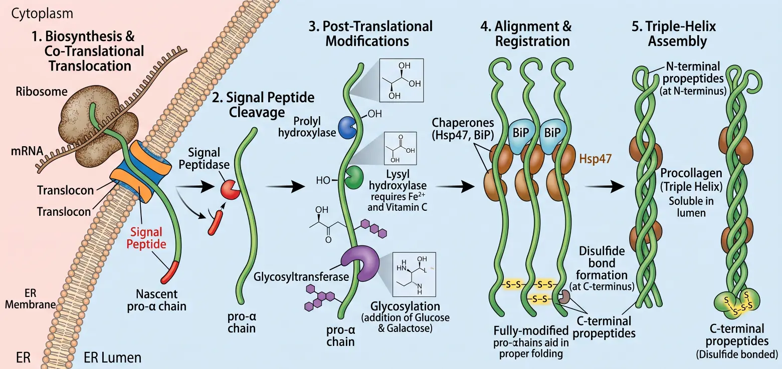
Figure 1. Biosynthesis, Co-Translational Translocation, Post-Translational Modification, and Assembly of Procollagen in the ER. (1) Biosynthesis and Co-Translational Translocation: Ribosomes actively translate pro-$$\alpha$$ collagen chain mRNA on the cytosolic surface of the rER membrane. Guided by an N-terminal signal peptide (red), the nascent polypeptide threads directly through the translocon channel into the ER lumen. (2) Signal Peptide Cleavage: Inside the ER lumen, signal peptidase (notched red circle) proteolytically cleaves the hydrophobic signal peptide, releasing the soluble, linear pro-$$\alpha$$ chain (green). (3) Post-Translational Modifications: The unstructured pro-$$\alpha$$ chain undergoes extensive, coordinated enzymatic processing. Prolyl hydroxylase (notched blue circle) hydroxylates designated proline residues. Lysyl hydroxylase (notched green circle; dependent on $${\text{Fe}}^{2+}$$ and vitamin C/ascorbate as essential cofactors) hydroxylates lysine residues. Glycosyltransferase (large purple element) then adds carbohydrates (glucose, 3 purple hexagons, and galactose, 5 purple hexagons) to selected hydroxylysine residues. (4) Alignment & Registration: ER resident molecular chaperones BiP (light blue) and Hsp47 (brown) interact with the pro-$$\alpha$$ chains to prevent non-productive interactions and guide proper chain folding. Alignment begins at the globular C-terminal propeptides (bottom), which pair through interchain disulfide bond formation (−S−S−), ensuring all three chains register correctly with their free N-terminal propeptides oriented upward. (5) Triple-Helix Assembly: Following C-terminal nucleation, the three registered pro-$$\alpha$$ chains fold in a C-to-N direction into the characteristic triple-helical conformation. Monomeric Hsp47 chaperones (brown) bind along the triple helix to stabilize the helical structure and prevent premature intracellular aggregation. The resulting soluble procollagen molecule, capped by N-terminal propeptides at one end and disulfide-bonded C-terminal propeptides at the other, is fully assembled and ready for ER export.
2.2. Liquid Droplets vs. Rigid Rods
The newly published article from Professor Malhotra’s laboratory [34] represents a paradigm shift in structural cell biology. It directly challenges a multi-decade-old dogma by showing that procollagen is not maintained as an unyielding, isolated rigid rod throughout its entire stay in the ER lumen; instead, it behaves as a flexible component of a liquid biomolecular condensate. The old dogma suggested that the resulting triple-helical structure converts the once-flexible polypeptide strands into a rigid, rod-like procollagen macromolecule [35,36,37]. However, Bhattacharyya et al. [34] proved, using high-resolution live-cell imaging, that procollagen I undergoes liquid-liquid phase separation. It condenses into dynamic spherical droplets within the ER lumen. These droplets behave like fluid condensates: they merge, split, and exchange material with their surroundings (Figure 2). This fluid state solves an evolutionary puzzle: if procollagen molecules function as highly interactive building blocks, they could spontaneously stack and form fibers inside the ER. As Malhotra proposes, premature intracellular fiber formation would kill the cell [38]. By keeping procollagen in a pliably packed vesicle, the fluid procollagen condensate prevents toxic aggregation, shields the cell from devastating proteotoxicity, and accommodates massive bulk cargo volume. The study also revealed that the procollagen condensates form a highly organized, multi-component folding environment. They are heavily enriched with HSP47, calreticulin, PDIA1, and PDIA6 [34]. Crucially, they lack the general unfolded protein sensor BiP [34], meaning that the cell organizes these dense pools specifically to enable efficient procollagen transport, while deliberately keeping them invisible to the ER stress/degradation sensors under normal conditions.
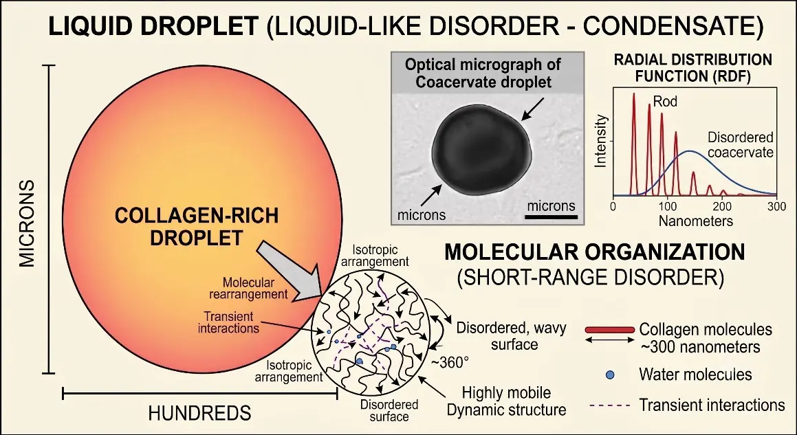
Figure 2. Structural and dynamic characteristics of a collagen liquid-like coacervate droplet. Liquid-liquid phase separation drives collagen into a dense, spherical, macromolecule-rich coacervate droplet suspended within a dilute aqueous phase. The optical micrograph (inset) demonstrates the characteristic high-contrast, isotropic appearance of a fluid condensate on a substrate. Molecular Organization: The highly mobile structure highlights the condensate’s internal liquid-like disorder. Collagen triple helices adopt an isotropic, dynamic arrangement characterized by rapid molecular rearrangement and transient non-covalent interactions in an aqueous environment. Radial Distribution Function (RDF): Comparative structural order between crystalline-like rigid rods and disordered coacervates. While rigid collagen rods display sharp, discrete diffraction peaks corresponding to defined long-range periodicity (e.g., 67 nm D-periodicity of procollagen), the liquid coacervate exhibits a broad, featureless distribution curve characteristic of short-range disorder and fluid-like spatial arrangement.
Within the ER lumen and throughout transit, we proposed that the massive, globular COOH-propeptide is positioned directly adjacent to the hydrophilic carboxy-terminal region of the telopeptide [39] and functions to maintain this fluid state. The globular COOH-propeptide would amplify the local electrostatic repulsion and hydrophilic shielding, ensuring that even when the collagen molecules come close, their interacting regions remain structurally isolated. Thus, the hydrophobic amino-terminal portions of the telopeptides would be kept buried or sterically blocked from aligning with neighboring rods by the sheer bulk of the terminal propeptide caps. We believe that this sheer physical bulk creates a structural barrier that keeps the central triple-helical domains spaced apart, mechanically preventing them from coming close enough to align or stack side by side. Also, we believe the massive concentration of HSP47 coating the collagen molecule [40] could act as a fluid molecular mesh. Therefore, the folded helices could be densely packed, and yet remain highly mobile and disorganized, gliding past one another like a viscous fluid. Taken together, we believe this combination of physical shielding (steric hindrance) and electrical pushing (electrostatic repulsion) keeps the individual folded triple helices highly soluble, preventing them from aggregating into stiff structural fibers.
2.3. The Luminal Gatekeeper and Quality Control
When the phase-separated procollagen condensates pass luminal quality control, they must be accurately positioned for export at specialized macro-domains in the exit site. The discovery of the fluid state of procollagen fundamentally rewrites the classical model of cargo selection. TANGO1-Long, the master transmembrane regulator protein localized at the ERES, does not function as a traditional stoichiometric cargo receptor that picks up individual procollagen. Instead, it acts as a spatial mooring point that chemically captures and anchors the massive, multi-protein fluid procollagen condensate directly over the ERES boundary.
Because structural defects in the triple helix can compromise tissue integrity, the ER lumen enforces a quality-control checkpoint before allowing any cargo to proceed into the export machinery. The central coordinator of this procollagen gatekeeping step is HSP47 [33]. Unlike general chaperones that bind unfolded proteins, HSP47 recognizes and binds exclusively to the fully folded, native trimeric structure of the procollagen triple helix [33,41,42]. This chaperone interaction serves a dual purpose. It structurally stabilizes the procollagen within the crowded lumen and also shields it from premature aggregation [42,43]. TANGO1 cannot efficiently recognize bare procollagen [44]; rather, because of its HSP47-binding site, it relies entirely on the bound HSP47/procollagen complex to identify acceptable, properly matured cargo [45]. Misfolded or unstable helices that fail to bind HSP47 are actively retained in the ER and eventually shunted toward the ER-associated degradation pathway for destruction [46,47]. Once the collagen is extruded from the ER, HSP47 remains behind [33].
3. Phase II—ERES and Navigating the Size Paradox
3.1. Cargo Selection and the Transmembrane Bridge
When a properly folded procollagen macromolecule passes the luminal quality-control checkpoints, it is directed to specialized macro-domains known as the ERES. At this junction, the cell must translate an internal structural signal into an active, outward budding event. This critical communication is mediated by TANGO1-Long, a master transmembrane regulatory protein specifically localized to the ERES membrane [48,49]. TANGO1-Long functions as a physical bridge across the ER lipid bilayer, and on its luminal side, TANGO1 projects an SH3-like domain into the ER interior. On its cytoplasmic side, TANGO1 contains active structural domains that directly interface with the cellular transport machinery. By simultaneously anchoring the interior cargo (HSP47/procollagen) and engaging exterior proteins, this transmembrane bridge directly couples cargo selection inside the ER lumen to the dynamic recruitment and expansion of the transport carrier on the outside. Interestingly, when TANGO1 is depleted, the fluid procollagen droplets still form perfectly in the lumen but drift aimlessly, failing to dock at the exit sites [34], suggesting that this transmembrane bridge couples the fluid-state physics of the ER lumen to the dynamic recruitment and expansion of the transport carrier on the outside. Rather than shipping a rigid array of 400-nm spears, this structural arrangement enables fluid extrusion, allowing the pliable procollagen pool to flow smoothly across the ERES boundary, much like fluid pushed through a tight orifice, thereby perfectly filling the expanding mega-carrier.
3.2. Erecting the Export Perimeter: The TANGO1 Ring as a Fluid-Extrusion Funnel
The change in thinking, introducing fluid-state procollagen droplets within the ER, fundamentally reconfigures our understanding of the cytosolic machinery at the ERES. In the classical model, the assembly of the TANGO1 ring was thought to act purely as a restrictive perimeter fence, preventing standard spherical coat proteins from closing around an unyielding 400 nm rod. Given the discovery of fluid procollagen condensate phase separation, this macromolecular assembly takes on a far more dynamic, physical role: it functions as a structural funnel that constrains and channels the procollagen condensate as it flows out of the ER (Figure 3).
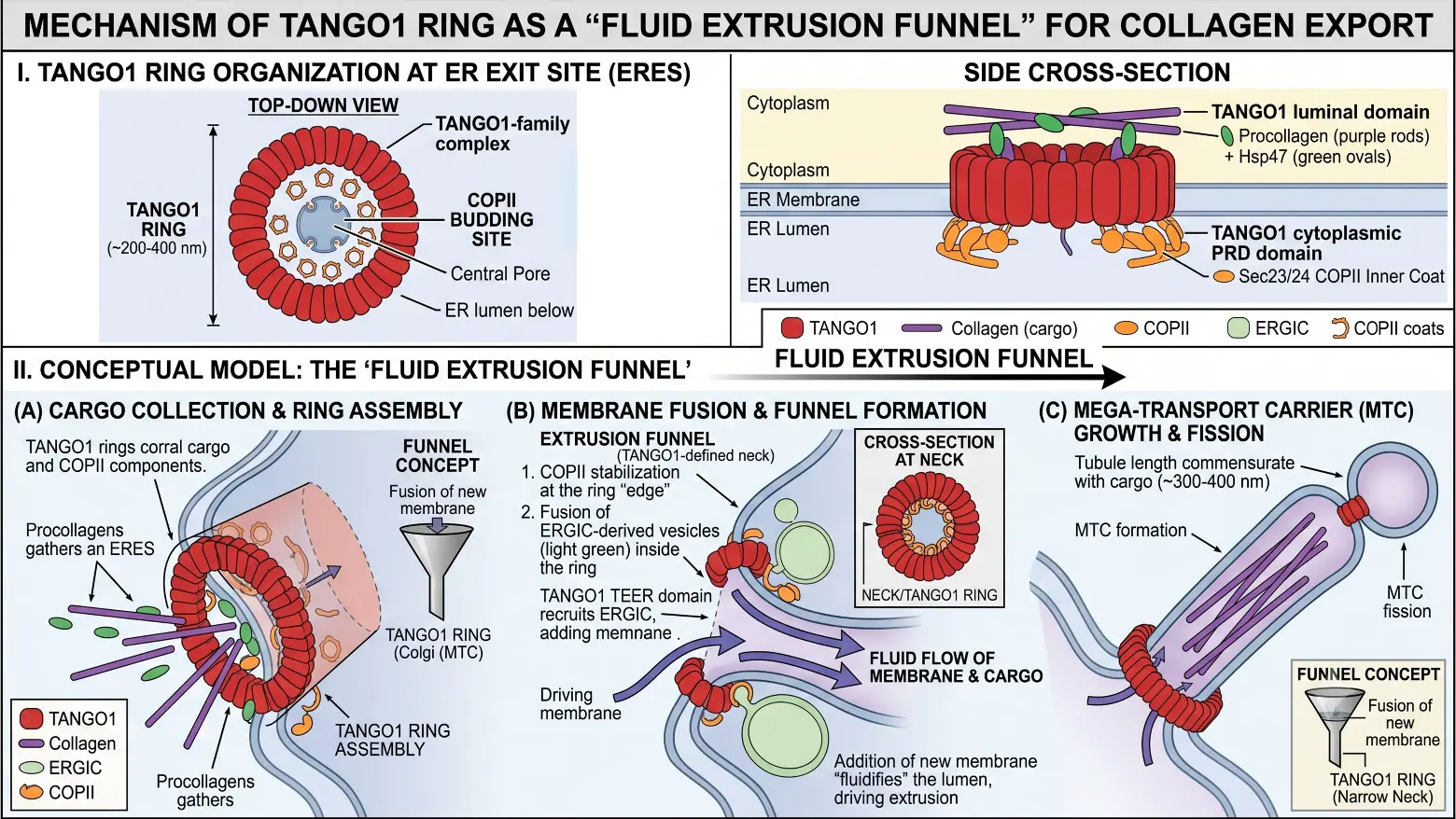
Figure 3. Structural organization and proposed “fluid extrusion funnel” mechanism of the TANGO1 ring during large-cargo ER export. (I). TANGO1 Ring Organization at the Endoplasmic Reticulum Exit Site (ERES). Top-Down View: TANGO1 complexes assemble into a large ring structure (~200–400 nm in diameter) at the ERES membrane. The ring creates a central pore, corralling inner COPII coat components (Sec23/Sec24) and establishing a defined zone for mega-carrier formation. Side Cross-Section: TANGO1 acts as a transmembrane bridge. Its luminal domains bind procollagen cargo via HSP47 chaperones, while its cytoplasmic proline-rich domains (PRDs) interact directly with COPII coat components at the base of the ER membrane. (II). Conceptual Model: The “Fluid Extrusion Funnel”. (A) Cargo Collection & Ring Assembly: TANGO1 rings corral bulky procollagen rods alongside essential COPII inner coat machinery at the ERES to initiate assembly of the outgoing carrier. (B) Membrane Fusion & Funnel Formation: The TANGO1 ring defines a constricted neck region. TANGO1’s tether for ERGIC at the ER (TEER) domain recruits ERGIC-derived membrane vesicles into the ring’s interior. Fusion of these membranes provides the necessary surface area and fluid flow to drive the continuous extrusion of bulky cargo through the neck. (C) Mega-Transport Carrier (MTC) Growth & Fission: As incoming membrane fluidifies and expands the growing tubule, the carrier elongates proportionally to fit large procollagen bundles (~300–400 nm) before undergoing fission to yield a mature Mega-Transport Carrier.
This molecular funnel is erected through a highly coordinated, multi-protein oligomerization program on the cytosolic leaflet of the ERES. The insertion of TANGO1-Long across the bilayer causes it to self-assemble into a vast, highly ordered ring structure measuring approximately 200 nm in diameter [50]. This architecture is structurally supported and stabilized by its alternative splice isoform, TANGO1-Short, and its coreceptor cutaneous T-cell lymphoma-associated antigen 5 (cTAGE5), which physically locks the structural subunits into place [51,52,53]. Rather than grouping randomly, the proline-rich domains decorating the cytoplasmic portion of the TANGO1 ring project outward to directly bind Sec23, a core component of the inner COPII coat [37,50,51]. By physically anchoring the Sec23/Sec24 complex directly over the rim of the 200 nm aperture, TANGO1 corrals the inner COPII coat components along the perimeter [50]. This halts their default thermodynamic drive to spontaneously polymerize into small spheres [54], effectively keeping the throat of the budding macro-carrier open. To prolong this open state, TANGO1’s cytoplasmic domain also interacts directly with Sec16 [55,56,57], a major peripheral membrane protein essential for organizing the baseline structure of the ERES. Through this scaffolding link, the TANGO1/Sec16 complex actively suppresses Sar1 GTP hydrolysis at the neck of the carrier [58,59]. This safety mechanism keeps a persistent ring of active, membrane-bound Sar1-GTP to stabilize the growing carrier structure.
Positioned precisely over the site where the luminal SH3 domain anchors the liquid procollagen-HSP47 pool, the TANGO1 ring acts as a central bottleneck [50]. As incoming donor lipids expand the budding carrier membrane, the ring structure dictates the shape of the emerging envelope. It establishes a controlled orifice through which the dense, pliable procollagen condensate is squeezed, ensuring that the bulk fluid procollagen condensate flows cleanly into the center of the elongating, tube-like mega-carrier (Figure 3).
3.3. Kinetic Control: The Biochemical Delay of Vesicle Closure
While TANGO1 ring assembly creates the physical aperture needed for fluid cargo extrusion, a mechanical funnel alone cannot guarantee successful transport. Under baseline conditions, standard COPII coat subunits polymerize rapidly via a thermodynamic rate, quickly forming an outer cage that pinches off small vesicles from the ER membrane. For an extensive, phase-separated condensate to completely empty its contents across the bilayer boundary, this default fission program must be selectively suspended. The cell achieves this kinetic matching by introducing a localized, calcium-dependent biochemical delay that temporarily stalls vesicle-tunnel closure.
A specialized ubiquitination hub drives this precise chronological delay and operates directly on the cytosolic leaflet of the ERES. The core coordinators of this timing mechanism are Cullin-3 (CUL3) and its substrate-specific adaptor Kelch-like protein 12 (KLHL12) [13,60]. This E3 ubiquitin ligase complex is dynamically recruited to the active export site in response to localized calcium fluxes, a process mediated by the calcium-binding adaptor proteins peflin and apoptosis-linked gene 2 [61,62,63]. Once localized to the perimeter of the TANGO1 fence, the CUL3-KLHL12 complex targets Sec31 [15], one of the major structural components of the outer COPII coat scaffold.
The CUL3-KLHL12 complex drives the selective monoubiquitination of Sec31, which dynamically alters the binding geometry and intermolecular interactions between neighboring Sec13/Sec31 lattices [64]. Rather than preventing outer coat assembly entirely, monoubiquitination acts as a biochemical brake, significantly slowing the rate of outer cage polymerization [15,65]. By extending the time window before vesicle closure, the cell gives the elongating macro-carrier membrane enough time to receive the incoming bulk volume of the procollagen droplet. This extended expansion phase is further safeguarded by Sedlin [66], which binds directly to TANGO1 to orchestrate the efficient, regulated cycling of the initiating small GTPase, Sar1, at the vesicle neck. By regulating Sar1 nucleotide-state transitions, Sedlin works in tandem with the altered outer coat to prevent premature membrane scission [66], thereby ensuring that the pool of pliable procollagen is safely processed across the ERES boundary.
Researchers have long proposed that ER exit of bulky procollagen molecules requires remodeling of the canonical COPII coat. In particular, the Cullin-3 ubiquitin ligase complex (CUL3-KLHL12), dynamically recruited to exit sites via calcium-binding adaptors [61,62,63], was shown to monoubiquitylate SEC31A, thereby driving the formation of enlarged COPII carriers. However, whether SEC31A monoubiquitylation strictly governs carrier size and anterograde trafficking remains under active debate. Recent work demonstrates that large COPII–KLHL12 assemblies can form independently of CUL3 neddylation/catalytic activation, and another dimer, CRL3-KLHL12, can monoubiquitylate SEC31, leading to an increase in COPII vesicle dimension and clearance of collagen [67]. The authors proposed that this collagen clearance likely occurs through ER-to-lysosomal pathways [67]. Furthermore, procollagen molecules that fail to exit the ER are targeted directly from ERES for degradation via selective ER-phagy/microautophagy [68]. Thus, whether ubiquitylation of COPII components primarily remodels transport carriers for forward secretion, gates ER quality control, or coordinates ER-phagy remains an open question that warrants further investigation.
3.4. Lipid Logistics: Retrograde Membrane Recruitment from the ERGIC
Slowing outer-coat polymerization aligns the biochemical clock of fission with the transport window, but expanding the transport carrier around a massive fluid cargo requires an immense lipid influx that far exceeds the homeostatic surface area available at a default ERES. To avoid a localized depletion of the ER bilayer during this prolonged budding event, the TANGO1 complex acts as a localized lipid-conscription hub [69]. It establishes a metabolic and structural sink that actively draws in and fuses membrane volume from downstream donor compartments to accommodate the bulk volume of the flowing cargo pool.
This extensive lipid recruitment operates through two synchronized membrane-remodeling mechanisms mapped directly to TANGO1’s coiled-coil domains. Adjacent to the ER membrane, TANGO1 utilizes its second coiled-coil domain (CC2, distal to the ER bilayer) to bind tightly and directly to its coreceptor cTAGE5 via their respective coiled-coil motifs. While TANGO1 captures cargo internally and tethers the coat externally, cTAGE5 directly recruits and concentrates Sec12, the guanine nucleotide exchange factor for Sar1 [70], at the ERES perimeter [58]. By concentration-coupling Sec12 to the site of cargo extrusion, the complex ensures a massive, localized generation of active, GTP-bound Sar1 [58,70]. This constant insertion of Sar1 amphipathic helices into the cytosolic leaflet continuously drives localized membrane curvature, forcing the lipid bilayer to expand outward into an elongated container. Concurrently, TANGO1’s first coiled-coil domain (CC1, proximal to the ER bilayer) exposes its specialized TEER (Tether for ERGIC at the ER) region. This region recruits the heterotrimeric NRZ complex (NBAS, RINT1, and ZW10) directly to the cytosolic face of the ERES [71]. Under the guidance of the NRZ tethering complex, the standard directional flow of the early secretory pathway is locally modified: the complex captures retrograde membrane transport intermediates arriving from the ERGIC and anchors them directly at the base of the budding carrier [72]. These targeted ERGIC membranes, uniquely trackable via the marker protein ERGIC-53 [52,55,58,71] directly fuse into the ERES bilayer via localized SNARE pairings [69]. By integrating external ERGIC membranes directly at the carrier neck, the budding envelope continuously grows and extends, creating a highly accommodating, pleiomorphic sleeve that swallows the fluid procollagen condensate as it is extruded through the structural orifice. The continuous fusion of these targeted donor lipids stabilizes the elongated carrier, establishing a transient, interconnected channel that directly links the ER lumen to the early Golgi architecture [34].
While the classical model of bulky cargo export relies on the coordinated action of TANGO1 and enlarged or modified COPII coats, recent work has illuminated additional tethering and membrane-organizing machineries required for procollagen packaging. In particular, researchers recently identified the multiple C2 domain-containing ferlin protein, myoferlin, as a novel tethering factor that couples ERGIC to ERES [73]. Mechanistically, post-translational modification (specifically site-specific O-GlcNAcylation) of the inner-coat subunit Sec24D promotes myoferlin recruitment, facilitating retrograde ERGIC-to-ERES membrane recruitment or fusion to supply the membrane surface area needed to accommodate rigid procollagen trimers. Independent studies have now corroborated myoferlin’s direct role in mediating efficient procollagen secretion [74], establishing that non-canonical membrane tethering via calcium/phospholipid-binding ferlins operates in parallel with or downstream of core ERES organizers (such as TANGO1 and Sec23/Sec24). Incorporating myoferlin into models of early secretory trafficking underscores that procollagen egress is not merely driven by mechanical coat deformation, but rather relies on active, regulated inter-organelle membrane remodeling between the ERES and ERGIC.
3.5. Macro-Organization of the Export Zone by Trk-Fused Gene
The local assembly of the TANGO1/cTAGE5 ring complex and the kinetic modifications of the COPII coat operate directly on individual ERES lipid bilayers, but the higher-order spatial orchestration of the exit domains requires further specialized cytosolic scaffolding. Inside the mammalian transitional ER, the Trk-fused gene acts as a master architectural organizer. The Trk-fused gene self-assembles at its amino terminus into a cup-shaped macromolecular ring network on the cytosolic face of the membrane [75,76]. This expansive proteinaceous meshwork physically bridges bundles of scattered transitional ER elements into distinct, highly concentrated ERES hubs [62,75]. Cells depleted of the Trk-fused gene support the baseline capacity to export standard-sized soluble proteins via fragmented mini-exit sites but show a complete block in bulk matrix traffic [77]. The Trk-fused gene macro-scaffold is strictly required to provide the physical scale and long-term structural stabilization needed to sustain prolonged COPII coat assembly over extensive cargo volumes. Positioned directly at the traffic boundary between the ERES and the downstream ERGIC, the Trk-fused gene matrix serves as a localized gatekeeper. It concentrates, stabilizes, and aligns the emerging transport pathways, ensuring that the flowing, fluid procollagen pool is efficiently processed across the organelle boundary into the early Golgi architecture [78].
4. Phase III—Transit Through the Secretory Pathway
4.1. Intercompartmental Continuity: The Transient Tunnel Model
The physical resolution of the procollagen export site has introduced one of the most remarkable and paradigm-shifting mechanisms in modern cell biology, completely upending traditional models of vesicular trafficking. In classical textbook schemas based on standard-sized cargo, a transport carrier must undergo complete membrane fission from its donor compartment, forming an isolated, independent spherical vesicle that floats through the cytoplasm to find and fuse with its target organelle. However, high-resolution live-cell imaging and biophysical modeling have fundamentally dismantled this view for large cargoes. Complete membrane fission in a 400 nm container would impose a large thermodynamic barrier and an unsustainable energetic burden on the ER bilayer. To avoid this structural bottleneck, the cell bypasses standard carrier-mediated separation entirely, using the TANGO1 complex to form a direct, transient membrane tunnel connecting the ER lumen to the Golgi (Figure 4, Panel A).
4.2. The Non-Separating Pipeline and Entropic Flow
Rather than generating a free-floating mega-vesicle that pinches off and drifts away, the export machinery establishes a direct physical continuity between the ER and the ERGIC. This non-separating shortcut relies on two distinct mechanical roles within the structural network. Unlike traditional cargo receptors that pack into budding vesicles and travel alongside their cargo, TANGO1 never leaves the ERES [50]. It remains at the ERES boundary, acting as a permanent, transmembrane mooring bridge. By using its TEER domain as described in Section 3.4, TANGO1 pulls ERGIC membranes backward until they fuse at the ERES boundary, forming an envelope that wraps around the procollagen [40]. This fusion establishes an open, continuous tubular path linking the two distinct organelles (Figure 4, Panel B).
Because the membranes are continuously connected, local physics rather than active vesicular translocation governs the cargo’s directional movement. As theoretically modeled by Bunel et al. [40], this forward propulsion is driven by an entropic ratchet and fluid pump mechanism. The ER lumen and the ERGIC maintain distinct internal microenvironments, characterized by acute chemical divergence and a shifting pH gradient [79]. Structurally, the luminal domain of TANGO1 features a massive, highly unstructured segment spanning approximately 1000 amino acids, classified as an intrinsically disordered region ($${\text{TANGO1}}_{\text{LUM}}$$ IDR) [40]. Under resting conditions, this flexible chain samples an exceptionally vast conformational space, yielding high localized baseline entropy.
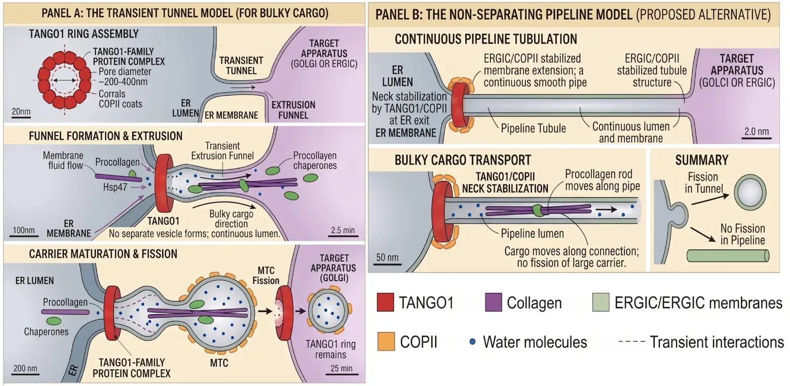
Figure 4. Comparative models of large cargo ER exit and intracellular transport. (Panel A): The Transient Tunnel Model. TANGO1 complexes organize into a defined ring structure (~200–400 nm pore diameter) at the ERES, corralling inner COPII coat components to delineate a wide neck for bulky cargo transit. Recruitment and fusion of ERGIC-derived membranes fluidify the lumen, establishing a directional membrane fluid flow. Procollagen bundles (purple) are bound to Hsp47 chaperones (green ovals) and are extruded through a transient, funnel-like neck into a growing carrier without forming intermediate standard COPII vesicles. The expanding tubule matures into a discrete Mega-Transport Carrier (MTC). Following membrane fission, the MTC detaches, delivering bulky cargo to the Golgi while leaving the TANGO1 ring assembly intact at the ER membrane. (Panel B): The Non-Separating Pipeline Model. TANGO1 and COPII/ERGIC proteins stabilize a direct, permanent or long-lived tubular extension that forms a continuous membrane connection between the ER lumen and the target compartment. The procollagen rods move continuously along the lumen of the stabilized pipe, eliminating the requirement for carrier fission along the conduit.
However, when the phase-separated, fluid procollagen-HSP47 complexes crowd the ERES and bind to TANGO1’s luminal docking domain, they form a dense, restricted protein network that severely confines the space available to the IDR tails. To minimize this localized loss of conformational entropy, the polymer chains spontaneously generate an entropic contraction force, acting like a compressed microscopic spring that pulls back to reclaim their disordered freedom. Because the TANGO1 scaffold is anchored in the rigid ERES ring, it cannot move backward to relieve this tension; instead, this thermodynamic force is translated into a physical pull on the mobile cargo. The resulting pressure differential acts as a continuous fluid pump or capillary siphon, smoothly drawing the pliable procollagen pool out of the bulk ER lumen and driving its anterograde flow through the tunnel toward the early Golgi network at a rate of 1 nm·s−1 [40].
We hypothesize that, in the tunnel model, COPII coat proteins and their accessory molecules behave and are ultimately sorted differently. These proteins are hijacked and incorporated into a static scaffold. Rather than acting as mobile vehicles, they serve as the stationary part of the architectural nozzle, keeping the mouth of the intercompartmental pipeline open and anchoring, stabilizing, and gatekeeping locally. They remain at the periphery of the ERES in a state of suspended polymerization, preventing a complete cage from forming and ensuring that no fission event can close the tunnel while the procollagen pool flows through it.
4.3. Gating the Tunnel: The Dual-Helix Lipid Diffusion Barrier
While forming a direct intercompartmental channel solves the logistical volume deficit of bulk procollagen trafficking, it introduces a profound topological crisis: the potential loss of organelle identity through unregulated lateral lipid mixing. The ER membrane is thin and enriched with lipids like phosphatidylcholine, while downstream Golgi compartments are packed with rigid sphingolipids and cholesterol [80,81,82]. Left unchecked, lateral diffusion across an open membrane tube would cause these lipids and untargeted membrane proteins to mix and dilute each other. This biophysical dilemma is elegantly resolved by an unusual structural feature within TANGO1’s core architecture [49]. Rather than utilizing a conventional single transmembrane span, TANGO1 inserts into the bilayer via a specialized dual-helix membrane domain. This domain consists of one true transmembrane-spanning helix paired with an adjacent hydrophobic intramembrane loop that penetrates only the cytoplasmic leaflet, anchoring horizontally without crossing to the luminal side. Upon oligomerization into the macro-structural 200 nm ring, these specialized dual helices pack tightly side-by-side at the highly curved neck of the budding carrier, aligning like a dense row of structural pylons [50].
Biophysical reconstitution and mathematical modeling have shown that this rigid, proteinaceous ring forces an extreme local constriction and structural compression on the surrounding lipids, creating a potent lipid diffusion barrier [40]. By functioning as a physical molecular collar, this structure establishes a strict segregation zone [49] that permits the unimpeded anterograde flow of the luminal, fluid procollagen condensate through the open center of the tunnel while completely arresting the lateral, inter-organelle diffusion of resident membrane lipids and proteins. Consequently, the cell can maintain a continuous, high-volume shipping pipeline without sacrificing the distinct biochemical boundaries that define the early secretory pathway.
4.4. Golgi Processing: Fluid-State Transition and the Maturation Paradigm
Upon flowing through the TANGO1-gated transient intercompartmental tunnel and crossing the dual-helix lipid diffusion barrier, the pliable procollagen pool enters the early Golgi architecture. Rather than acting as a simple passive conduit or as a recipient of isolated packages from detached vesicles, the Golgi apparatus functions as a highly coordinated, continuous processing line. Within this organelle, the incoming fluid cargo undergoes a carefully timed transition, with sequential carbohydrate remodeling, as it shifts from its protective, fluid-droplet phase inside the ER to highly concentrated, parallel macromolecular crystalline arrays. This phase not only completes the molecule's chemical maturation but also provides classic historical evidence of how bulk cargo transitions through the Golgi stack.
Historically, the intracellular trafficking of procollagen was the critical catalyst that resolved the long-standing debate between the static vesicular transport and dynamic cisternal maturation models of the Golgi apparatus. Under the traditional vesicular view, Golgi cisternae were thought to be stationary compartments, with cargo packed into small COPI vesicles that hopped from one cisterna to the next, and these vesicles were too small to accommodate procollagen molecules.
Pioneering work using three-dimensional electron microscopy and live-cell imaging showed that procollagen molecules never leave the Golgi lumen during anterograde transport. Instead, the cargo is transported via a cisternal progression-maturation model [83]. New cis-cisternae are continually generated at the proximal face of the Golgi stack by homotypic fusion and coalescence of incoming cargo-filled membranes originating from the downstream end of the transient tunnel-fed ERGIC pipeline [83]. The entire membrane cisterna then physically moves forward through the stack like a conveyor belt. As it progresses, the cisterna gradually shifts its biochemical identity because resident enzymes are continuously recycled backward into preceding cisternae via retrograde COPI vesicles, ensuring the large cargo moves forward without ever leaving the main luminal envelope [84].
As the procollagen pool progresses through the maturing cis-, medial-, and trans-cisternae [83], its carbohydrate structures become remodeled by an ordered relay of these retrograde-recycled resident enzymes. The core oligosaccharide chains previously added to the non-helical $$N$$- and $$C$$-terminal propeptides within the ER lumen are systematically targeted. Golgi mannosidase removes the excess mannose residues, allowing subsequent medial and trans glycosyltransferases to attach N-acetylglucosamine, galactose, and terminal sialic acid residues [83]. Concurrently, the carbohydrate chains attached to specific hydroxylysine residues within the central triple-helical domain are completed [83]. This precise, sequential addition of carbohydrates establishes the highly specific surface chemical signature required for downstream ECM signaling and collagen-receptor interactions.
4.5. The Hydrophobic Transition: Lateral Packing and Crystalline Bundling
Within the flattened lumen of the medial- and trans-Golgi cisternae, procollagen undergoes a physical transition. While inside the ER lumen, the molecule is maintained in a highly fluid, dynamic biomolecular condensate to prevent premature structural stacking; the changing internal environment of the Golgi stack triggers a deliberate crystallization. This transition is driven by a progressive luminal acidification gradient, which decreases from near neutral in the ER, to a pH of 6.5–6.7 in the cis-Golgi, 6.3–6.5 in the medial Golgi, to ~6.0 in the Trans-Golgi Network (TGN) [85,86]. As the pH drops toward the isoelectric point of the procollagen terminal domains, individual triple helices would lose their relative repulsive surface charges. This localized environmental shift [40,79] overcomes the protective fluid properties of the initial ER condensate [34], causing the mature triple helices to phase-separate into a rigid, structured state that condenses laterally into electron-dense procollagen bundles within maturing Golgi cisternae [83,87].
The procollagen bundle’s journey through the intracellular secretory pathway culminates at the TGN. Functioning as the central logistics and sorting hub of the eukaryotic cell, the TGN is dynamically compartmentalized to separate specialized cargo destined for lysosomes, the plasma membrane, or regulated extracellular secretion [88,89]. When laterally condensed, electron-dense arrays of parallel procollagen rods arrive at this terminal station, the cell faces a geometric packaging constraint that mirrors the initial size paradox encountered at the ERES. At the TGN, cargo exit typically relies on clathrin coats and adaptor protein complexes (e.g., AP-1) that polymerize into rigid polyhedral cages with strict diameter constraints (50–150 nm) [90], which are insufficient to capture the procollagen bundles [91]. Consequently, the TGN bypasses conventional coat-driven budding, engaging kinesin motors and Rab6-mediated membrane tubulation to pull giant, non-clathrin tubulo-vesicular mega-carriers out of specialized TGN exit domains [92,93].
5. Phase IV—Exocytosis and Extracellular Maturation
At the cell surface, wide-aperture exocytosis of dense, laterally aligned procollagen arrays is driven by Rab8a/Rab11-recruited exocyst complexes [93], working alongside peripheral soluble N-ethylmaleimide-sensitive factor attachment protein receptors (SNAREs) (VAMP7, Syntaxin 4, and SNAP-23) to sustain dilated fusion pores that prevent cargo shearing [94,95]. Once extruded intact into the extracellular space, procollagen molecules undergo targeted enzymatic processing by zinc-dependent metalloproteinases, thereby triggering self-assembly. Specifically, procollagen C-proteinase, enhanced by PCPE-1, and procollagen N-proteinases proteolytically clip the terminal globular propeptides [96,97,98,99,100]. Stripping these protective hydrophilic caps causes an instantaneous drop in solubility within the aqueous environment, creating a strong thermodynamic driver that promotes spontaneous self-association and fibrillogenesis of the hydrophobic rigid rods [101].
To convert these fragile non-covalent polymer assemblies into a high-tensile matrix that can withstand physiological mechanical stress, the extracellular matrix initiates enzyme-catalyzed cross-linking [102,103]. Copper-dependent lysyl oxidase oxidatively deaminates specific lysine and hydroxylysine residues within the non-helical telopeptide regions into reactive aldehydes (allysine/hydroxyallysine) [104,105,106]. These aldehydes spontaneously condense with adjacent amino acid residues across precisely aligned strands, generating divalent intermediates that mature into permanent, trivalent pyrrole and pyridinoline cross-links [104,107]. By covalently locking the adjacent triple helices together, this final extracellular maturation phase solves the tissue-scale size paradox, transforming fluid cellular exports into a resilient supramolecular matrix scaffold.
6. Open Questions
Based on the current literature on procollagen export, the fluid procollagen condensate in the ER, and the TANGO1-mediated transient tunnel framework, several profound biochemical, structural, and physiological questions remain.
-
Directed Cargo Flow and Condensate Clearance Dynamics: Although recent biophysical frameworks, such as the entropic contraction model proposed by Bunel et al. [40], elegantly describe how entropic forces pull the fluid procollagen/HSP47 condensate pool through the transient tunnel, the precise mechanochemical trigger that initiates this directional momentum remains unknown. In addition, once the fluid droplet crosses the TANGO1 boundary, it is unclear whether the condensate clears the lumen completely or whether a residual fraction remains at the exit site to preserve channel geometry and membrane stability. Mapping the physical parameters that govern the volumetric throughput, viscous drag, and complete clearance of a phase-separated biomolecular droplet across the TANGO1 ring structure represents a vital, unmapped frontier in live-cell biophysics.
-
Distal Tunnel Resolution. Transient Scission Versus Perennial Continuities: While TANGO1 remains strictly localized to the ERES boundary, the distal fate of the inter-compartmental tunnel that bridges the ER and ERGIC remains a pivotal topological question. It remains unresolved whether this membrane pipeline undergoes dynamic, cycle-by-cycle scission driven by specific fission machinery once the phase-separated condensate pool translocates, or whether it persists as a long-term, perennial structural continuity. In hyper-active matrix-secreting cells such as myofibroblasts, maintaining a persistent, non-cleaved membrane bridge would drastically reduce the kinetic cost of repeated coat assembly and scission; however, the mechanisms that would stabilize such an open, multi-compartmental conduit against lipid mixing and organellar collapse remain uncharacterized.
-
Can transient tunnels and the non-separating pipelines co-exist? Transient tunnels and non-separating pipelines could coexist, and this remains open to debate. This may depend on cargo load, cell type, and secretory demand. Under basal physiological conditions with moderate collagen synthesis, discrete transient tunnels could open, translocate collagen bundles, and resolve via scission, avoiding compromise of organellar compartmentalization and preventing leakage of ER-resident chaperones (BiP, PDI, HSP47). Under hyper-secretory physiological regimes (e.g., active osteoblasts depositing bone matrix, or dermal fibroblasts during active wound repair), high local procollagen flux could keep the transient tunnel open indefinitely, effectively converting the connection into a quasi-permanent non-separating pipeline. Recent studies also indicate that ERES are morphologically and functionally heterogeneous [108]. Subpopulations enriched in specific TANGO1 isoforms (TANGO1-Long vs. TANGO1-Short) and distinct tethering partners (cTAGE5, Sec12, NRZ complex) may form specialized high-throughput “pipeline” conduits at central juxtanuclear ERES, while peripheral ERES operate via discrete, transient budding/tunneling events. For a continuous pipeline to co-exist without compromising the biochemically distinct identities of the ER and ERGIC/Golgi, TANGO1 must form an organized proteinaceous fence/diffusion barrier (via its transmembrane and intramembrane helices) [49]. This barrier would allow directional bulk cargo translocation while preventing homogenization of ER-resident proteins and lipids. To directly test the difference between a transient tunnel (which cycles through opening, scission/fission, and closure) and a continuous pipeline (a sustained, persistent conduit), using continuous loss photobleaching of a diffusible luminal reporter may demonstrate the answer. The transient tunnel fundamentally depends on scission/fission machinery to terminate the bridge, whereas a continuous pipeline does not. In the transient model, fluorescence depletion should be negligible or occur in stochastic, stepwise drops corresponding to discrete, time-limited opening and closing/fission cycles. A non-separating pipeline would cause rapid, steady, uninterrupted depletion of fluorescence because the lumen remains continuously open.
-
Intercompartmental Continuity and Cisternal Maturation Dynamics: A major spatial paradox arises when reconciling the TANGO1-mediated transient tunnel model with the classic cisternal maturation paradigm governing post-ER transport. The classical maturation framework dictates that ERGIC elements homotypically coalesce at the proximal face of the Golgi stack to continuously generate new cis-cisternae. However, if the transient tunnel acts as a direct, continuous membrane pipeline connecting the ER to the ERGIC interface, the mechanical hand-off of bulk cargo into a forward-progressing Golgi cisterna remains topologically unresolved. Specifically, it is unknown how this continuous pipeline transfers large procollagen condensates into a standalone, maturing cisterna without allowing downstream Golgi lipids and resident processing enzymes to flow retrograde across TANGO1’s dual-helix diffusion barrier. Defining the lipid-gating mechanics at this junction will be essential to unify intercompartmental tunnel transport with Golgi ribbon dynamics.
-
Physical Phase Transitions of Procollagen Beyond the ER: Recent biophysical insights have unmasked the fluid, phase-separated droplet nature of procollagen inside the ER lumen [40]. The precise physical state of the cargo during its progression through the Golgi stack remains unresolved. Historically, static electron micrographs of lateral procollagen bundling in maturing Golgi cisternae have been interpreted as a rigid, solid-like crystalline condensation. However, whether the intra-Golgi environmental shifts, specifically the progressive luminal acidification, changing calcium levels, and altered ion ratios, induce a true liquid-to-solid phase transition or merely compress the liquid condensate into a highly ordered, high-density liquid-crystalline state remains an open biophysical question. Determining the material properties of post-ER procollagen aggregates in vivo will be crucial to understanding how the intra-cisternal environment regulates cargo transport without triggering catastrophic, irreversible intra-Golgi precipitation.
-
Tissue-Specific Machinery and Structural Heterogeneity Across the Collagen Superfamily: While Type I procollagen in interstitial fibroblasts serves as the foundational archetype for the TANGO1/cTAGE5/CUL3 export model, whether these precise components are universally conserved across all twenty-eight human collagen types remains an open question. The collagen superfamily encompasses vast structural diversity, ranging from linear, high-tensile fibrillar collagens (Types I, II, III, V) to sheet-forming basement membrane networks (Type IV) and transmembrane collagens (Types XIII and XVII). Genetic evidence in Drosophila and mammalian models confirms that non-fibrillar collagens, most notably basement membrane procollagen IV, strictly require TANGO1 for ER export [37,109]. Given the comparable molecular lengths (~400 nm for type IV vs. ~300 nm for type I), the spatial barrier imposed by standard COPII vesicles remains identical. However, the presence of flexible interruptions and bulky globular NC1 domains in procollagen IV [110] raises the question of whether ring pore dimensions, gating kinetics, and putative fluid-extrusion dynamics are uniformly conserved or tailored to specific structural classes of collagens. It is currently unknown whether other non-fibrillar or network-forming collagens rely on the identical ~200 nm TANGO1 ring geometry and fluid extrusion mechanics, or if distinct tissues express specialized TANGO1/cTAGE5 isoforms, homologs (such as TALI/MIA2), or uncharacterized tissue-specific chaperones to build cargo-customized export architectures. Systematic mapping of ERES remodeling across ECM-secreting cell types, including chondrocytes, endothelial cells, and epithelial lineages, will be essential for establishing whether a single unified export paradigm governs the entire collagen superfamily.
-
Regulation of Sec31 De-ubiquitination and ERES Recycling Kinetics: The localized monoubiquitination of the outer COPII coat subunit Sec31 by the CUL3/KLHL12 E3 ubiquitin ligase complex is a well-established vital biochemical brake that delays carrier closure to accommodate bulky procollagen; the complementary off-switch governing ERES recovery remains entirely unmapped. To return the ERES to its default 60–90 nm parameters for baseline, spherical vesicular traffic, the cell must systematically remove these monoubiquitin handles from Sec31. The specific deubiquitinating enzymes that execute this de-clamping step, along with the spatial and temporal signals that recruit them after cargo departure, represent a fundamental gap in ubiquitin biology and membrane coat dynamics. Identifying these regulatory deubiquitinating enzymes will be crucial to understanding how high-flux secretory cells dynamically reset their exit sites between successive waves of macromolecular export.
-
Elucidating the Post-Golgi Structural Machinery at the Trans-Golgi Network: While the mechanochemical protocols governing the elongation of COPII-derived carriers at the ERES driven by TANGO1 ring assembly, retrograde lipid conscription, and CUL3/KLHL12 monoubiquitination have been extensively characterized, the structural machinery operating at the TGN remains a major black box. Classical post-Golgi export relies on clathrin lattices and heterotrimeric adaptor complexes (e.g., AP-1, GGA proteins), which are thermodynamically and geometrically constrained to generate small, spherical vesicles (~50–150 nm). Consequently, how the TGN deforms its membrane to accommodate procollagen without relying on spherical coat cages remains an unsolved problem in sorting paradigms. Identifying the TGN-localized coat adaptors or motor-driven tubulation machinery that orchestrates post-Golgi mega-carrier formation represents a critical frontier in organelle logistics.
-
Mechanochemical Calcium Sparks as Upstream Triggers for ERES Ubiquitination: Whereas calcium-dependent penta-EF-hand adaptors peflin and ALG2 are well-established as molecular switches that recruit the CUL3/KLHL12 E3 ubiquitin ligase to monoubiquitinate Sec31 and decelerate vesicle closure [61], the upstream signal that triggers localized calcium release at the ERES remains unknown. A compelling mechanochemical hypothesis posits that the physical crowding or phase separation of procollagen condensates within the luminal microenvironment exerts mechanical tension or steric pressure on the adjacent ER membrane. This localized physical force could dynamically gate neighboring luminal ER calcium release channels, such as $${\text{IP}}_{3}$$ receptors or ryanodine receptors, generating microdomain calcium sparks precisely where bulk cargo accumulates. Determining whether cargo-driven membrane tension directly gates localized ion channels to trigger E3 ligase recruitment will be essential for establishing a direct, mechanochemical feedback loop linking intra-luminal procollagen density with cytosolic coat modification kinetics.
-
Mechanotransduction and Outside-In Signaling Loops in Pathogenic Matrix Export: Although activated myofibroblasts upregulate TANGO1 to sustain high-flux procollagen secretion during fibrotic progression [111], it is unknown whether mechanical forces retroactively control intracellular export machinery, and this remains a major unmapped frontier. A compelling mechanobiological hypothesis posits that increased ECM stiffness engages cell-surface mechanosensors, such as $${\beta }_{1}$$ integrins and focal adhesion complexes, to trigger transcriptional mechanotransduction cascades (e.g., via YAP/TAZ or MRTF-A translocation). This outside-in mechanical signaling could directly drive the transcriptional upregulation or ERES stabilization of TANGO1 and cTAGE5. Elucidating whether ECM stiffening initiates a pathogenic mechanochemical feed-forward loop in which a rigid matrix forces the cell to amplify its export machinery, thereby driving further collagen deposition, would be vital to establishing new therapeutic windows in progressive fibrosis. If mechanosensory feedback from a stiffened ECM directly tunes ERES capacity and TANGO1 conduit dynamics, this would uncover a previously unrecognized mechanically regulated checkpoint at the organelle level. Conceptually, this transforms the secretory apparatus from a passive downstream conduit into an active driver of the fibrotic vicious cycle. From a drug development perspective, targeting the molecular transducers that relay cytoskeletal tension to ERES assemblies offers an attractive therapeutic window: it could selectively silence hyper-secreting myofibroblasts trapped in rigid scar tissue while sparing basal ECM turnover in compliant, healthy organs.
-
Macro-Condensate Clearance and ER-phagy as Quality Control Safety Valves: While structurally compromised procollagen monomers that fail luminal quality control are retrotranslocated across the ER membrane and destroyed via the ubiquitin-proteasome system (ERAD) [112], the physical dimensions of phase-separated procollagen condensates present a severe proteostatic paradox. The barrel-shaped pore of the 20S proteasome can only thread narrow, unfolded single polypeptide chains. Consequently, standard ERAD machinery cannot disassemble or retrotranslocate a multi-protein liquid macro-condensate comprising interlinked procollagen trimers, HSP47 chaperones, and associated luminal factors. This topological bottleneck suggests that the cell must engage specialized selective autophagy pathways to clear aberrant condensates. Identifying the specific ER-phagy receptors that recognize stalled condensates, and the mechanochemical signals that trigger autophagic engulfment of entire condensate-containing ER subdomains, remains a critical, unmapped frontier in procollagen quality control and osteogenesis imperfecta pathology.
-
The Bioenergetics and Colloidal Physics of Post-ER Phase Transitions: Despite the fact that the initial procollagen fluid phase inside the ER lumen serves as a vital protective strategy to prevent premature fibril aggregation [40], the biophysical mechanisms governing its subsequent transition into densely packed intra-Golgi arrays remain largely unmapped. Shifting from a highly dynamic, fluid condensate into a compressed, parallel crystalline bundle without triggering intra-luminal precipitation or irreversible protein clumping represents an extraordinary feat of soft-matter biophysics. Although progressive acidification and shifts in Ca2+/K2+ ion ratios across the cis- to trans-Golgi stack alter surface charges and hydration shells, identifying the specific thermodynamic off-switches or solvent modifiers that safely regulate this colloidal phase transition in vivo remains a fundamental open question in matrix biology.
7. Conclusions
The chronological exploration of the procollagen molecule highlights an extraordinary evolutionary adaptation in cellular logistics. To accommodate a structural cargo whose physical dimensions completely defy the geometric constraints of classic vesicular trafficking, the eukaryotic cell fundamentally rewires its standard secretory protocols. Through a highly synchronized interplay of transmembrane bridging, physical ring corralling, targeted membrane recruitment, and precise biochemical delay, the cell dynamically remodels default 60–90 nm spherical COPII vesicles into expansive, pleiomorphic mega-carriers and transient tunnels. This complex intracellular pipeline operates in direct continuity with highly specific extracellular processing events, such as propeptide cleavage and lysyl oxidase-mediated cross-linking, to transform individual, fluid trimeric chains into the resilient supramolecular networks that support metazoan life.
Despite major breakthroughs in identifying key molecular organizers such as TANGO1, cTAGE5, and the CUL3/KLHL12 complex, several fundamental questions remain unanswered, as discussed above. Resolving these remaining biochemical details will do more than just fill gaps in basic cell biology; it will provide the specific blueprints needed to advance clinical therapeutics. As our understanding of the procollagen export machinery matures from a static inventory of components to a dynamic, physiological timeline, we could unlock the ability to design highly targeted interventions. Uncoupling cargo selection from carrier growth or modulating localized ERES assembly kinetics offers a sophisticated pathway to selectively brake the hyper-secretory state of activated myofibroblasts. Ultimately, targeting this specialized transport window offers a promising paradigm for arresting the progression of devastating fibrotic conditions or alleviating severe genetic connective tissue pathologies, translating fundamental trafficking insights into tangible clinical horizons.
Statement of the Use of Generative AI and AI-Assisted Technologies in the Writing Process
During the preparation of this manuscript, C.M.A. used Gemini 3 Pro to generate theoretical ideas and Grammarly to edit the manuscript for grammar.
Ethics Statement
Not applicable.
Informed Consent Statement
Not applicable.
Data Availability Statement
No new data were generated for this review.
Funding
This research received no external funding.
Declaration of Competing Interests
The author declares no known competing financial interests or personal relationships that could have appeared to influence the work reported in this paper.
References
-
Fidler AL, Boudko SP, Rokas A, Hudson BG. The triple helix of collagens—An ancient protein structure that enabled animal multicellularity and tissue evolution. J. Cell Sci. 2018, 131, jcs203950. DOI:10.1242/jcs.203950 [Google Scholar]
-
Dzamba BJ, DeSimone DW. Extracellular Matrix (ECM) and the Sculpting of Embryonic Tissues. Curr. Top. Dev. Biol. 2018, 130, 245–274. DOI:10.1016/bs.ctdb.2018.03.006 [Google Scholar]
-
Exposito JY, Valcourt U, Cluzel C, Lethias C. The fibrillar collagen family. Int. J. Mol. Sci. 2010, 11, 407–426. DOI:10.3390/ijms11020407 [Google Scholar]
-
Sivalingam DS, Vien I, Kuo CK. Collagen Cross-Linking in Tendons: The Role of Lysyl Oxidase and Posttranslational Modifications in Injury, Aging, and Congenital Disorders. Annu. Rev. Biomed. Eng. 2026, 28, 463–489. DOI:10.1146/annurev-bioeng-110824-033814 [Google Scholar]
-
Padidela R, Hendrickx G, Mortier G, Mäkitie O. The pathogenesis and intersecting mechanisms of monogenic bone mass disorders. Nat. Rev. Endocrinol. 2026, 22, 490–506. DOI:10.1038/s41574-026-01260-x [Google Scholar]
-
Canty-Laird EG, Gupta HS, Birch HL. The Impact of Ageing on Fibrillar Collagens. Subcell. Biochem. 2026, 113, 377–412. DOI:10.1007/978-3-032-05273-5_12 [Google Scholar]
-
Theocharidis G, Connelly JT. Minor collagens of the skin with not so minor functions. J. Anat. 2019, 235, 418–429. DOI:10.1111/joa.12584 [Google Scholar]
-
Deshmukh SN, Dive AM, Moharil R, Munde P. Enigmatic insight into collagen. J. Oral. Maxillofac. Pathol. 2016, 20, 276–283. DOI:10.4103/0973-029x.185932 [Google Scholar]
-
Szarek P, Ruberti JW. Collagen Mechanics. Subcell. Biochem. 2026, 113, 277–342. DOI:10.1007/978-3-032-05273-5_10 [Google Scholar]
-
Seco FC, Amador S, Conzuelo F, Baptista AC, Morgado L, Pina AS. Molecular versatility of polyproline II helices: From natural proteins to biomimetic materials. J. Mater. Chem. B 2026, 14, 1764–1782. DOI:10.1039/d5tb01841b [Google Scholar]
-
Gordon MK, Hahn RA. Collagens. Cell Tissue Res. 2010, 339, 247–257. DOI:10.1007/s00441-009-0844-4 [Google Scholar]
-
Joshi A, Mondal B, Basak T. Molecular foundations of collagen triple helical assembly: The central role of prolyl-4-hydroxylation. Biochem. J. 2026, 483, 71–87. DOI:10.1042/bcj20253467 [Google Scholar]
-
Jensen D, Schekman R. COPII-mediated vesicle formation at a glance. J. Cell Sci. 2011, 124, 1–4. DOI:10.1242/jcs.069773 [Google Scholar]
-
Downes KW, Zanetti G. Mechanisms of COPII coat assembly and cargo recognition in the secretory pathway. Nat. Rev. Mol. Cell Biol. 2025, 26, 910–925. DOI:10.1038/s41580-025-00839-y [Google Scholar]
-
Jin L, Pahuja KB, Wickliffe KE, Gorur A, Baumgartel C, Schekman R, et al. Ubiquitin-dependent regulation of COPII coat size and function. Nature 2012, 482, 495–500. DOI:10.1038/nature10822 [Google Scholar]
-
Kotan IE, Sartori S, Bose R, Bukau B, Kramer G. SRP orchestrates protein biogenesis beyond initial ER membrane targeting. Nat. Commun. 2026, 17, 5316. DOI:10.1038/s41467-026-74404-2 [Google Scholar]
-
Chang J, Garva R, Pickard A, Yeung CC, Mallikarjun V, Swift J, et al. Circadian control of the secretory pathway maintains collagen homeostasis. Nat. Cell Biol. 2020, 22, 74–86. DOI:10.1038/s41556-019-0441-z [Google Scholar]
-
Stefanovic L, Longo L, Zhang Y, Stefanovic B. Characterization of binding of LARP6 to the 5’ stem-loop of collagen mRNAs: Implications for synthesis of type I collagen. RNA Biol. 2014, 11, 1386–1401. DOI:10.1080/15476286.2014.996467 [Google Scholar]
-
Lipp J, Dobberstein B, Haeuptle MT. Signal recognition particle arrests elongation of nascent secretory and membrane proteins at multiple sites in a transient manner. J. Biol. Chem. 1987, 262, 1680–1684. DOI:10.1016/S0021-9258(19)75691-9 [Google Scholar]
-
Liaci AM, Steigenberger B, Telles de Souza PC, Tamara S, Gröllers-Mulderij M, Ogrissek P, et al. Structure of the human signal peptidase complex reveals the determinants for signal peptide cleavage. Mol. Cell 2021, 81, 3934–3948.e3911. DOI:10.1016/j.molcel.2021.07.031 [Google Scholar]
-
Staab-Weijnitz CA, Merl-Pham J, Hennen E, Onursal C, Cabeza-Boeddinghaus N, Kandhari K, et al. Prolyl-3-hydroxylase 1 is a central regulator of collagen post-translational modifications and the collagen biosynthetic network. J. Biol. Chem. 2026, 302, 111422. DOI:10.1016/j.jbc.2026.111422 [Google Scholar]
-
Rahman MM, Sulu R, Adediran B, Tu H, Salo AM, Murthy S, et al. Binding Differences of the Peptide-Substrate-Binding Domain of Collagen Prolyl 4-Hydroxylases I and II for Proline- and Hydroxyproline-Rich Peptides. Proteins 2025, 93, 1732–1746. DOI:10.1002/prot.26839 [Google Scholar]
-
Szarka A, Lőrincz T. The role of ascorbate in protein folding. Protoplasma 2014, 251, 489–497. DOI:10.1007/s00709-013-0560-5 [Google Scholar]
-
Ghosh S, Pramanick S, Kumar D, Mondal B, Basak T. Structural and Mechanistic Insights into the Catalytic Versatility of Lysyl Hydroxylases in Collagen Post-Translational Modifications. J. Biol. Chem. 2026, 302, 113259. DOI:10.1016/j.jbc.2026.113259 [Google Scholar]
-
Casas-Flórez D, Ortega-García R, Sanz-Benito P, Monterroso B, Sanz-Aparicio J, González B. The structure of human glucosidase PGGHG reveals a very specific active site accessible through a flat surface for collagen approximation. Int. J. Biol. Macromol. 2026, 345, 150556. DOI:10.1016/j.ijbiomac.2026.150556 [Google Scholar]
-
Ghosh S, Kumar D, Mondal B, Basak T. Enzymatic craftsmanship in collagen glycosylation. Nat. Commun. 2025, 16, 7252. DOI:10.1038/s41467-025-62768-w [Google Scholar]
-
Malone JP, Alvares K, Veis A. Structure and assembly of the heterotrimeric and homotrimeric C-propeptides of type I collagen: Significance of the alpha2(I) chain. Biochemistry 2005, 44, 15269–15279. DOI:10.1021/bi0508338 [Google Scholar]
-
Ishikawa Y, Bächinger HP. A molecular ensemble in the rER for procollagen maturation. Biochim. Biophys. Acta 2013, 1833, 2479–2491. DOI:10.1016/j.bbamcr.2013.04.008 [Google Scholar]
-
Hartmann J, Zacharias M. Mechanism of collagen folding propagation studied by Molecular Dynamics simulations. PLoS Comput. Biol. 2021, 17, e1009079. DOI:10.1371/journal.pcbi.1009079 [Google Scholar]
-
Acevedo-Jake AM, Jalan AA, Hartgerink JD. Comparative NMR analysis of collagen triple helix organization from N- to C-termini. Biomacromolecules 2015, 16, 145–155. DOI:10.1021/bm501281a [Google Scholar]
-
Buevich AV, Silva T, Brodsky B, Baum J. Transformation of the mechanism of triple-helix peptide folding in the absence of a C-terminal nucleation domain and its implications for mutations in collagen disorders. J. Biol. Chem. 2004, 279, 46890–46895. DOI:10.1074/jbc.M407061200 [Google Scholar]
-
Bächinger HP, Boudko SP. Mysteries of the collagen triple helix. Matrix Biol. 2025, 137, 12–18. DOI:10.1016/j.matbio.2025.02.003 [Google Scholar]
-
Omari S, Makareeva E, Gorrell L, Jarnik M, Lippincott-Schwartz J, Leikin S. Mechanisms of procollagen and HSP47 sorting during ER-to-Golgi trafficking. Matrix Biol. 2020, 93, 79–94. DOI:10.1016/j.matbio.2020.06.002 [Google Scholar]
-
Bhattacharyya S, Wojnacki J, Brouwers N, Malhotra V. Procollagen 1 assembles into phase-separated condensates in the endoplasmic reticulum. J. Cell Biol. 2026, 225, e202603129. DOI:10.1083/jcb.202603129 [Google Scholar]
-
Artlett CM, Connolly LM. TANGO1 Dances to Export of Procollagen from the Endoplasmic Reticulum. Fibrosis 2023, 1, 10008. DOI:10.35534/fibrosis.2023.10008 [Google Scholar]
-
Raote I, Chabanon M, Walani N, Arroyo M, Garcia-Parajo MF, Malhotra V, et al. A physical mechanism of TANGO1-mediated bulky cargo export. eLife 2020, 9, e59426. DOI:10.7554/eLife.59426 [Google Scholar]
-
Saito K, Chen M, Bard F, Chen S, Zhou H, Woodley D, et al. TANGO1 facilitates cargo loading at endoplasmic reticulum exit sites. Cell 2009, 136, 891–902. DOI:10.1016/j.cell.2008.12.025 [Google Scholar]
-
Malhotra V, Erlmann P. The pathway of collagen secretion. Ann. Rev. Cell Dev. Biol. 2015, 31, 109–124. DOI:10.1146/annurev-cellbio-100913-013002 [Google Scholar]
-
Dion AS, Myers JC. COOH-terminal propeptides of the major human procollagens. Structural, functional and genetic comparisons. J. Mol. Biol. 1987, 193, 127–143. DOI:10.1016/0022-2836(87)90632-2 [Google Scholar]
-
Bunel L, Pincet L, Malhotra V, Raote I, Pincet F. A model for collagen secretion by intercompartmental continuities. Proc. Natl. Acad. Sci. USA 2024, 121, e2310404120. DOI:10.1073/pnas.2310404120 [Google Scholar]
-
Cai H, Sasikumar P, Little G, Bihan D, Hamaia SW, Zhou A, et al. Identification of HSP47 Binding Site on Native Collagen and Its Implications for the Development of HSP47 Inhibitors. Biomolecules 2021, 11, 983. DOI:10.3390/biom11070983 [Google Scholar]
-
Ito S, Nagata K. Quality Control of Procollagen in Cells. Ann. Rev. Biochem. 2021, 90, 631–658. DOI:10.1146/annurev-biochem-013118-111603 [Google Scholar]
-
Köhler A, Mörgelin M, Gebauer JM, Öcal S, Imhof T, Koch M, et al. New specific HSP47 functions in collagen subfamily chaperoning. FASEB J. 2020, 34, 12040–12052. DOI:10.1096/fj.202000570R [Google Scholar]
-
Ishikawa Y, Ito S, Nagata K, Sakai LY, Bachinger HP. Intracellular mechanisms of molecular recognition and sorting for transport of large extracellular matrix molecules. Proc. Natl. Acad. Sci. USA 2016, 113, E6036–E6044. DOI:10.1073/pnas.1609571113 [Google Scholar]
-
Yuan L, Kenny SJ, Hemmati J, Xu K, Schekman R. TANGO1 and SEC12 are copackaged with procollagen I to facilitate the generation of large COPII carriers. Proc. Natl. Acad. Sci. USA 2018, 115, E12255–E12264. DOI:10.1073/pnas.1814810115 [Google Scholar]
-
Syx D, Ishikawa Y, Gebauer J, Boudko SP, Guillemyn B, Van Damme T, et al. Aberrant binding of mutant HSP47 affects posttranslational modification of type I collagen and leads to osteogenesis imperfecta. PLoS Genet. 2021, 17, e1009339. DOI:10.1371/journal.pgen.1009339 [Google Scholar]
-
Khan ES, Däinghaus T. HSP47 in human diseases: Navigating pathophysiology, diagnosis and therapy. Clin. Transl. Med. 2024, 14, e1755. DOI:10.1002/ctm2.1755 [Google Scholar]
-
Raote I, Saxena S, Campelo F, Malhotra V. TANGO1 marshals the early secretory pathway for cargo export. Biochim. Biophys. Acta Biomembr. 2021, 1863, 183700. DOI:10.1016/j.bbamem.2021.183700 [Google Scholar]
-
Raote I, Ernst AM, Campelo F, Rothman JE, Pincet F, Malhotra V. TANGO1 membrane helices create a lipid diffusion barrier at curved membranes. eLife 2020, 9, e57822. DOI:10.7554/eLife.57822 [Google Scholar]
-
Raote I, Ortega Bellido M, Pirozzi M, Zhang C, Melville D, Parashuraman S, et al. TANGO1 assembles into rings around COPII coats at ER exit sites. J. Cell Biol. 2017, 216, 901–909. DOI:10.1083/jcb.201608080 [Google Scholar]
-
Saito K, Yamashiro K, Ichikawa Y, Erlmann P, Kontani K, Malhotra V, et al. cTAGE5 mediates collagen secretion through interaction with TANGO1 at endoplasmic reticulum exit sites. Mol. Biol. Cell 2011, 22, 2301–2308. DOI:10.1091/mbc.E11-02-0143 [Google Scholar]
-
Ma W, Goldberg J. TANGO1/cTAGE5 receptor as a polyvalent template for assembly of large COPII coats. Proc. Natl. Acad. Sci. USA 2016, 113, 10061–10066. DOI:10.1073/pnas.1605916113 [Google Scholar]
-
Maeda M, Saito K, Katada T. Distinct isoform-specific complexes of TANGO1 cooperatively facilitate collagen secretion from the endoplasmic reticulum. Mol. Biol. Cell 2016, 27, 2688–2696. DOI:10.1091/mbc.E16-03-0196 [Google Scholar]
-
Bi X, Corpina RA, Goldberg J. Structure of the Sec23/24-Sar1 pre-budding complex of the COPII vesicle coat. Nature 2002, 419, 271–277. DOI:10.1038/nature01040 [Google Scholar]
-
Maeda M, Katada T, Saito K. TANGO1 recruits Sec16 to coordinately organize ER exit sites for efficient secretion. J. Cell Biol. 2017, 216, 1731–1743. DOI:10.1083/jcb.201703084 [Google Scholar]
-
Supek F, Madden DT, Hamamoto S, Orci L, Schekman R. Sec16p potentiates the action of COPII proteins to bud transport vesicles. J. Cell Biol. 2002, 158, 1029–1038. DOI:10.1083/jcb.200207053 [Google Scholar]
-
Miller EA, Barlowe C. Regulation of coat assembly—Sorting things out at the ER. Curr. Opin. Cell Biol. 2010, 22, 447–453. DOI:10.1016/j.ceb.2010.04.003 [Google Scholar]
-
Saito K, Maeda M, Katada T. Regulation of the Sar1 GTPase Cycle Is Necessary for Large Cargo Secretion from the Endoplasmic Reticulum. Front. Cell Dev. Biol. 2017, 5, 75. DOI:10.3389/fcell.2017.00075 [Google Scholar]
-
Kurokawa K, Suda Y, Nakano A. Sar1 localizes at the rims of COPII-coated membranes in vivo. J. Cell Sci. 2016, 129, 3231–3237. DOI:10.1242/jcs.189423 [Google Scholar]
-
Sato K. COPII coat assembly and selective export from the endoplasmic reticulum. J. Biochem. 2004, 136, 755–760. DOI:10.1093/jb/mvh184 [Google Scholar]
-
Sargeant J, Seiler DK, Costain T, Madreiter-Sokolowski CT, Gordon DE, Peden AA, et al. ALG-2 and peflin regulate COPII targeting and secretion in response to calcium signaling. J. Biol. Chem. 2021, 297, 101393. DOI:10.1016/j.jbc.2021.101393 [Google Scholar]
-
Kanadome T, Shibata H, Kuwata K, Takahara T, Maki M. The calcium-binding protein ALG-2 promotes endoplasmic reticulum exit site localization and polymerization of Trk-fused gene (TFG) protein. FEBS J. 2017, 284, 56–76. DOI:10.1111/febs.13949 [Google Scholar]
-
Held A, Lapka J, Sargeant J, Hojanazarova J, Shaheen A, Galindo S, et al. Steady-state regulation of COPII-dependent secretory cargo sorting by inositol trisphosphate receptors, calcium, and penta EF hand proteins. J. Biol. Chem. 2023, 299, 105471. DOI:10.1016/j.jbc.2023.105471 [Google Scholar]
-
Malhotra V. COPII Vesicles Get Supersized by Ubiquitin. Cell 2012, 149, 20–21. DOI:10.1016/j.cell.2012.03.006 [Google Scholar]
-
McGourty CA, Akopian D, Walsh C, Gorur A, Werner A, Schekman R, et al. Regulation of the CUL3 Ubiquitin Ligase by a Calcium-Dependent Co-adaptor. Cell 2016, 167, 525–538.e514. DOI:10.1016/j.cell.2016.09.026 [Google Scholar]
-
Venditti R, Scanu T, Santoro M, Di Tullio G, Spaar A, Gaibisso R, et al. Sedlin controls the ER export of procollagen by regulating the Sar1 cycle. Science 2012, 337, 1668–1672. DOI:10.1126/science.1224947 [Google Scholar]
-
Moretti T, Kim K, Tuladhar A, Kim J. KLHL12 can form large COPII structures in the absence of CUL3 neddylation. Molec Biol. Cell 2023, 34, br4. DOI:10.1091/mbc.E22-08-0383 [Google Scholar]
-
Omari S, Makareeva E, Roberts-Pilgrim A, Mirigian L, Jarnik M, Ott C, et al. Noncanonical autophagy at ER exit sites regulates procollagen turnover. Proc. Natl. Acad. Sci. USA 2018, 115, E10099–E10108. DOI:10.1073/pnas.1814552115 [Google Scholar]
-
Santos AJ, Raote I, Scarpa M, Brouwers N, Malhotra V. TANGO1 recruits ERGIC membranes to the endoplasmic reticulum for procollagen export. eLife 2015, 4, e10982. DOI:10.7554/eLife.10982 [Google Scholar]
-
Saito K, Yamashiro K, Shimazu N, Tanabe T, Kontani K, Katada T. Concentration of Sec12 at ER exit sites via interaction with cTAGE5 is required for collagen export. J. Cell Biol. 2014, 206, 751–762. DOI:10.1083/jcb.201312062 [Google Scholar]
-
Raote I, Ortega-Bellido M, Santos AJ, Foresti O, Zhang C, Garcia-Parajo MF, et al. TANGO1 builds a machine for collagen export by recruiting and spatially organizing COPII, tethers and membranes. eLife 2018, 7, e32723. DOI:10.7554/eLife.32723 [Google Scholar]
-
Aoki T, Ichimura S, Itoh A, Kuramoto M, Shinkawa T, Isobe T, et al. Identification of the neuroblastoma-amplified gene product as a component of the syntaxin 18 complex implicated in Golgi-to-endoplasmic reticulum retrograde transport. Mol. Biol. Cell 2009, 20, 2639–2649. DOI:10.1091/mbc.e08-11-1104 [Google Scholar]
-
Hirata T, Choudhary D, Nguyen Q, Bisnett BJ, Soderblom EJ, Knapik EW, et al. Site-specific glycosylation of Sec24D and myoferlin recruit ERGIC to ER exit sites for collagen trafficking. Nat. Commun. 2026, 17, 6415. DOI:10.1038/s41467-026-73027-x [Google Scholar]
-
Peiffer R, Laverdeur E, Gaigneaux A, Boumahd Y, Gullo C, Rademaker G, et al. Targeting myoferlin in ER/Golgi vesicle trafficking reprograms pancreatic cancer-associated fibroblasts. EMBO J. 2025, 44, 6425–6465. DOI:10.1038/s44318-025-00570-6 [Google Scholar]
-
Johnson A, Bhattacharya N, Hanna M, Pennington JG, Schuh AL, Wang L, et al. TFG clusters COPII-coated transport carriers and promotes early secretory pathway organization. EMBO J. 2015, 34, 811–827. DOI:10.15252/embj.201489032 [Google Scholar]
-
Bogus SM, Wegeng WR, Ruiz M, Chavez SR, Cheung SN, Noori KSM, et al. A hollow TFG condensate spatially compartmentalizes the early secretory pathway. Nat. Commun. 2025, 16, 3715. DOI:10.1038/s41467-025-59118-1 [Google Scholar]
-
Yuan H, Xu X, Gao J, Cao Y, Jin C, Ding K, et al. A novel branched galactomannoglucan from Acorus tatarinowii alleviates liver fibrosis via dual suppression of the transport of collagen and TGF-β/Smad signaling. Carbohydr. Polym. 2026, 382, 125270. DOI:10.1016/j.carbpol.2026.125270 [Google Scholar]
-
Hanna MGt, Block S, Frankel EB, Hou F, Johnson A, Yuan L, et al. TFG facilitates outer coat disassembly on COPII transport carriers to promote tethering and fusion with ER-Golgi intermediate compartments. Proc. Natl. Acad. Sci. USA 2017, 114, E7707–E7716. DOI:10.1073/pnas.1709120114 [Google Scholar]
-
Paroutis P, Touret N, Grinstein S. The pH of the secretory pathway: Measurement, determinants, and regulation. Physiology 2004, 19, 207–215. DOI:10.1152/physiol.00005.2004 [Google Scholar]
-
van Meer G, Voelker DR, Feigenson GW. Membrane lipids: Where they are and how they behave. Nat. Rev. Mol. Cell Biol. 2008, 9, 112–124. DOI:10.1038/nrm2330 [Google Scholar]
-
Harayama T, Riezman H. Understanding the diversity of membrane lipid composition. Nat. Rev. Mol. Cell Biol. 2018, 19, 281–296. DOI:10.1038/nrm.2017.138 [Google Scholar]
-
Bigay J, Antonny B. Curvature, lipid packing, and electrostatics of membrane organelles: Defining cellular territories in determining specificity. Dev. Cell 2012, 23, 886–895. DOI:10.1016/j.devcel.2012.10.009 [Google Scholar]
-
Bonfanti L, Mironov AA, Jr., Martínez-Menárguez JA, Martella O, Fusella A, Baldassarre M, et al. Procollagen traverses the Golgi stack without leaving the lumen of cisternae: Evidence for cisternal maturation. Cell 1998, 95, 993–1003. DOI:10.1016/s0092-8674(00)81723-7 [Google Scholar]
-
Martinez-Menárguez JA, Prekeris R, Oorschot VM, Scheller R, Slot JW, Geuze HJ, et al. Peri-Golgi vesicles contain retrograde but not anterograde proteins consistent with the cisternal progression model of intra-Golgi transport. J. Cell Biol. 2001, 155, 1213–1224. DOI:10.1083/jcb.200108029 [Google Scholar]
-
Kellokumpu S. Golgi pH, Ion and Redox Homeostasis: How Much Do They Really Matter? Front. Cell Dev. Biol. 2019, 7, 93. DOI:10.3389/fcell.2019.00093 [Google Scholar]
-
Schapiro FB, Grinstein S. Determinants of the pH of the Golgi complex. J. Biol. Chem. 2000, 275, 21025–21032. DOI:10.1074/jbc.M002386200 [Google Scholar]
-
Weinstock M, Leblond CP. Formation of collagen. Fed. Proc. 1974, 33, 1205–1218. Available online: https://pubmed.ncbi.nlm.nih.gov/4134092/ (accessed on 17 July 2026).
-
Nakano A. Redefining the entrance and exit of the Golgi apparatus. Curr. Opin. Cell Biol. 2026, 99, 102622. DOI:10.1016/j.ceb.2026.102622 [Google Scholar]
-
Watson ET, Wegeng WR, Aravani S, Ernst AM, von Blume J. Mechanistic insights into cargo sorting and export from the Golgi apparatus. Nat. Rev. Mol. Cell Biol. 2025, 26, 940–956. DOI:10.1038/s41580-025-00907-3 [Google Scholar]
-
Kirchhausen T. Three ways to make a vesicle. Nat. Rev. Mol. Cell Biol. 2000, 1, 187–198. DOI:10.1038/35043117 [Google Scholar]
-
Polishchuk RS, Polishchuk EV, Marra P, Alberti S, Buccione R, Luini A, et al. Correlative light-electron microscopy reveals the tubular-saccular ultrastructure of carriers operating between Golgi apparatus and plasma membrane. J. Cell Biol. 2000, 148, 45–58. DOI:10.1083/jcb.148.1.45 [Google Scholar]
-
Luini A, Mironov AA, Polishchuk EV, Polishchuk RS. Morphogenesis of post-Golgi transport carriers. Histochem. Cell Biol. 2008, 129, 153–161. DOI:10.1007/s00418-007-0365-8 [Google Scholar]
-
Wakana Y, Campelo F. The PKD-Dependent Biogenesis of TGN-to-Plasma Membrane Transport Carriers. Cells 2021, 10, 1618. DOI:10.3390/cells10071618 [Google Scholar]
-
Vardjan N, Jorgacevski J, Zorec R. Fusion pores, SNAREs, and exocytosis. Neuroscientist 2013, 19, 160–174. DOI:10.1177/1073858412461691 [Google Scholar]
-
Rao SK, Huynh C, Proux-Gillardeaux V, Galli T, Andrews NW. Identification of SNAREs involved in synaptotagmin VII-regulated lysosomal exocytosis. J. Biol. Chem. 2004, 279, 20471–20479. DOI:10.1074/jbc.M400798200 [Google Scholar]
-
Kessler E, Takahara K, Biniaminov L, Brusel M, Greenspan DS. Bone morphogenetic protein-1: The type I procollagen C-proteinase. Science 1996, 271, 360–362. DOI:10.1126/science.271.5247.360 [Google Scholar]
-
Hojima Y, van der Rest M, Prockop DJ. Type I procollagen carboxyl-terminal proteinase from chick embryo tendons. Purification and characterization. J. Biol. Chem. 1985, 260, 15996–16003. DOI:10.1016/S0021-9258(17)36357-3 [Google Scholar]
-
Moali C, Font B, Ruggiero F, Eichenberger D, Rousselle P, François V, et al. Substrate-specific modulation of a multisubstrate proteinase. C-terminal processing of fibrillar procollagens is the only BMP-1-dependent activity to be enhanced by PCPE-1. J. Biol. Chem. 2005, 280, 24188–24194. DOI:10.1074/jbc.M501486200 [Google Scholar]
-
Colige A, Li SW, Sieron AL, Nusgens BV, Prockop DJ, Lapière CM. cDNA cloning and expression of bovine procollagen I N-proteinase: A new member of the superfamily of zinc-metalloproteinases with binding sites for cells and other matrix components. Proc. Natl. Acad. Sci. USA 1997, 94, 2374–2379. DOI:10.1073/pnas.94.6.2374 [Google Scholar]
-
Colige A, Vandenberghe I, Thiry M, Lambert CA, Van Beeumen J, Li SW, et al. Cloning and characterization of ADAMTS-14, a novel ADAMTS displaying high homology with ADAMTS-2 and ADAMTS-3. J. Biol. Chem. 2002, 277, 5756–5766. DOI:10.1074/jbc.M105601200 [Google Scholar]
-
Kadler KE, Hojima Y, Prockop DJ. Assembly of collagen fibrils de novo by cleavage of the type I pC-collagen with procollagen C-proteinase. Assay of critical concentration demonstrates that collagen self-assembly is a classical example of an entropy-driven process. J. Biol. Chem. 1987, 262, 15696–15701. DOI:10.1016/S0021-9258(18)47783-6 [Google Scholar]
-
Kadler KE, Holmes DF, Trotter JA, Chapman JA. Collagen fibril formation. Biochem. J. 1996, 316 Pt 1, 1–11. DOI:10.1042/bj3160001 [Google Scholar]
-
Bailey AJ, Peach CM, Fowler LJ. Chemistry of the collagen cross-links. Isolation and characterization of two intermediate intermolecular cross-links in collagen. Biochem. J. 1970, 117, 819–831. DOI:10.1042/bj1170819 [Google Scholar]
-
Eyre DR, Paz MA, Gallop PM. Cross-linking in collagen and elastin. Ann. Rev. Biochem. 1984, 53, 717–748. DOI:10.1146/annurev.bi.53.070184.003441 [Google Scholar]
-
Siegel RC. Lysyl oxidase. Int. Rev. Connect. Tissue Res. 1979, 8, 73–118. DOI:10.1016/b978-0-12-363708-6.50009-6 [Google Scholar]
-
Kagan HM, Li W. Lysyl oxidase: Properties, specificity, and biological roles inside and outside of the cell. J. Cell Biochem. 2003, 88, 660–672. DOI:10.1002/jcb.10413 [Google Scholar]
-
Robins SP. Biochemistry and functional significance of collagen cross-linking. Biochem. Soc. Trans. 2007, 35, 849–852. DOI:10.1042/bst0350849 [Google Scholar]
-
Sree S, Parkkinen I, Their A, Airavaara M, Jokitalo E. Morphological Heterogeneity of the Endoplasmic Reticulum within Neurons and Its Implications in Neurodegeneration. Cells 2021, 10, 970. DOI:10.3390/cells10050970 [Google Scholar]
-
Liu M, Feng Z, Ke H, Liu Y, Sun T, Dai J, et al. Tango1 spatially organizes ER exit sites to control ER export. J. Cell Biol. 2017, 216, 1035–1049. DOI:10.1083/jcb.201611088 [Google Scholar]
-
Casino P, Gozalbo-Rovira R, Rodríguez-Díaz J, Banerjee S, Boutaud A, Rubio V, et al. Structures of collagen IV globular domains: Insight into associated pathologies, folding and network assembly. IUCrJ 2018, 5, 765–779. DOI:10.1107/s2052252518012459 [Google Scholar]
-
Connolly LM, McFalls CM, McMahon IG, Bhat AM, Artlett CM. Caspase 1 Enhances Transport and Golgi Organization Protein 1 Expression to Promote Procollagen Export From the Endoplasmic Reticulum in Systemic Sclerosis Contributing to Fibrosis. Arthritis Rheum. 2023, 75, 1831–1841. DOI:10.1002/art.42535 [Google Scholar]
-
Shen W, Zhang Z, Ma J, Lu D, Lyu L. The Ubiquitin Proteasome System and Skin Fibrosis. Mol. Diagn. Ther. 2021, 25, 29–40. DOI:10.1007/s40291-020-00509-z [Google Scholar]