
Immune Cell States and Maladaptive Tissue Niches in Idiopathic Pulmonary Fibrosis: From Mechanism to Precision Therapy
Immune Cell States and Maladaptive Tissue Niches in Idiopathic Pulmonary Fibrosis: From Mechanism to Precision Therapy
Received: 23 August 2026 Revised: 12 October 2026 Accepted: 21 October 2026 Published: 29 October 2026

© 2026 The authors. This is an open access article under the Creative Commons Attribution 4.0 International License (https://creativecommons.org/licenses/by/4.0/).
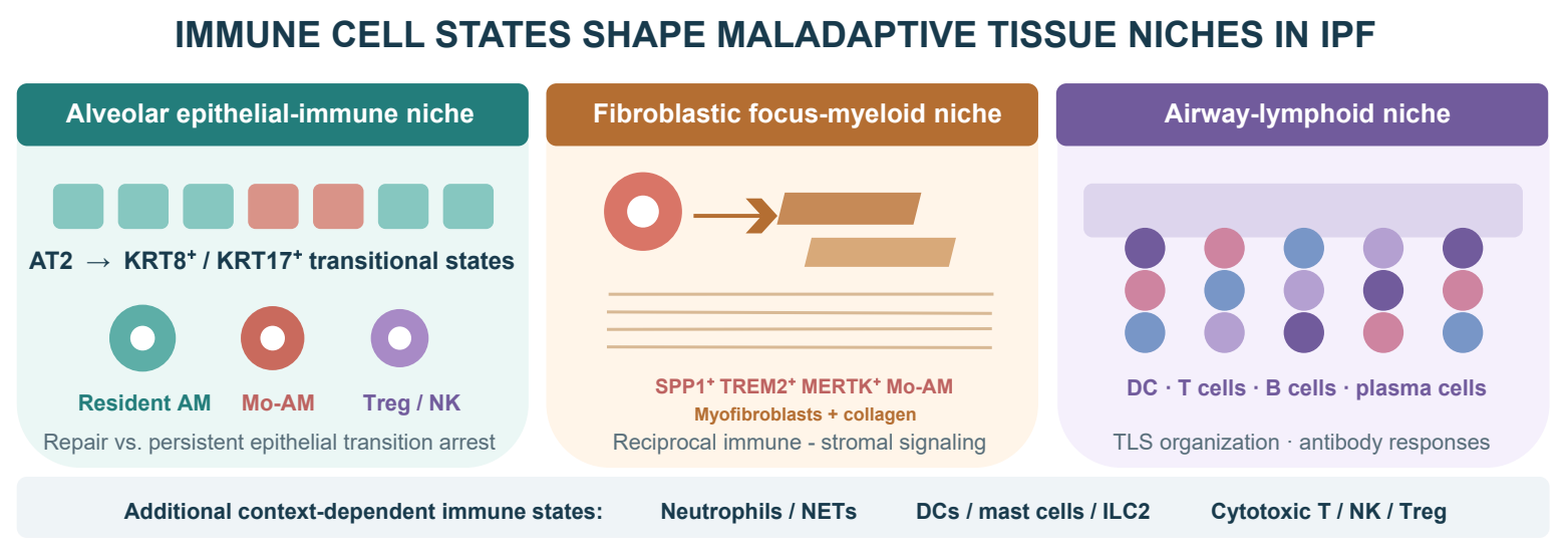
Graphical Abstract
1. Introduction: Resolving the Immune Paradox in IPF
Idiopathic pulmonary fibrosis (IPF) is a progressive, age-associated interstitial lung disease characterized by a usual interstitial pneumonia pattern, failed alveolar regeneration, and relentless matrix remodeling [1]. Nintedanib and pirfenidone slow loss of lung function, and the preferential phosphodiesterase 4B inhibitor nerandomilast also reduced forced vital capacity (FVC) decline and received US approval in 2025 [1,2,3]. None of these therapies restores normal alveolar architecture. A central biological problem therefore remains unresolved: how recurrent epithelial injury is converted into a self-sustaining multicellular niche that continues to promote fibrosis after the initiating insult is no longer evident.
The PANTHER-IPF trial provides the essential clinical boundary for interpreting immunity in IPF. Prednisone, azathioprine, and N-acetylcysteine increased death and hospitalization, demonstrating that broad immunosuppression is both ineffective and harmful [4]. This finding steered away from the earlier concept of IPF as a chronic inflammatory alveolitis. It did not test whether a defined immune-cell state, acting in a defined lung compartment and at a defined stage, contributes to disease. The same distinction is biologically important because macrophages and lymphocytes can support antimicrobial defense, clearance of damaged cells, epithelial regeneration, and fibrosis resolution while other states of the same lineages sustain tissue injury.
Recent technologies make this distinction experimentally tractable. Single-cell RNA sequencing resolves disease-associated macrophages, monocytes, dendritic cells, lymphocytes, and structural cells that are obscured by bulk analysis [5,6,7]. Spatial transcriptomics and multiplexed imaging then place these states beside KRT8- or KRT17-high transitional epithelium, fibroblastic foci, remodeled vessels, honeycomb airspaces, and tertiary lymphoid structures [8,9,10,11]. These maps define anatomically plausible interactions, although proximity and ligand-receptor inference remain hypotheses until signal direction and functional necessity are tested.
Building on our earlier concise review of immune dysregulation in IPF [12] and the broader framework advanced by Shenderov and colleagues [13], the present review focuses on the mechanistic questions that now determine translation. First, immune identity must be resolved beyond a lineage name or an M1/M2 label by defining ontogeny, state, metabolism, and niche. Second, immune function must be assigned to a disease stage because the same response can limit early injury and prolong established fibrosis if it fails to terminate. Third, evidence must be separated into reproducible human association, model-specific causation, and clinical efficacy. We use this framework to identify the immune circuits with the strongest support, explain why promising early programs failed in later trials, and define the measurements needed for a precision immune-directed therapy.
2. Scope and Evidence Framework
This focused narrative review emphasizes primary studies of human IPF tissue, bronchoalveolar lavage (BAL), blood, and clinically annotated cohorts; single-cell or spatial datasets with tissue validation; lineage-tracing and cell-selective perturbation studies; randomized or prospective interventions; and current clinical guidance. Literature published through 31 July 2026 was considered. Studies of other fibrotic interstitial lung diseases were included only when they clarified a mechanism directly relevant to IPF. The review was designed to synthesize mechanism and translational evidence, not to provide a systematic search, pooled effect estimate, or formal risk-of-bias analysis.
We apply three evidence levels throughout. Human association includes a reproducible difference in abundance, state, localization, biomarker level, or clinical outcome; it retains the possibility of confounding and reverse causation. Experimental causation requires a change in fibrosis after cell-selective depletion, transfer, lineage manipulation, or pathway perturbation in a defined model, with interpretation constrained by species, injury type, and intervention timing. Clinical evidence requires testing in patients, with randomized controlled data carrying the greatest weight. This hierarchy is particularly important in IPF because explants usually represent advanced disease, BAL samples are only accessible compartments, and bleomycin combines acute inflammation with partially resolving fibrosis in young animals.
3. From Inflammation to Maladaptive Immunity
3.1. Immune Cells as Participants in a Failed Repair Program
Normal alveolar repair requires a transient and coordinated immune response. Tissue-resident alveolar macrophages clear apoptotic cells and excess surfactant, recruited monocytes expand the myeloid compartment after injury, neutrophils control selected microbial threats, and lymphocytes contribute to host defense and resolution. These functions are coupled to AT2-cell activation, passage through a KRT8-high transitional state, AT1 differentiation, vascular recovery, and matrix turnover [13,14,15,16,17]. Successful repair therefore requires both injury removal and active termination of immune and mesenchymal responses.
In IPF, this program occurs in an aged and genetically susceptible lung in which AT2 cells accumulate stress and transitional-state features, basement membranes become discontinuous, fibroblasts acquire apoptosis-resistant matrix-producing states, and normal alveolar geometry is progressively lost [5,6,7,8,9,10,11,14,15,17]. Immune cells respond to this abnormal niche and are reprogrammed by it. A small number of macrophages positioned beside transitional epithelium or a fibroblastic focus may therefore have greater biological effect than a diffuse increase in total leukocytes, whereas abundant cells in a terminal honeycomb space may represent adaptation to tissue destruction. Cell number alone is an inadequate measure of mechanism (Figure 1).
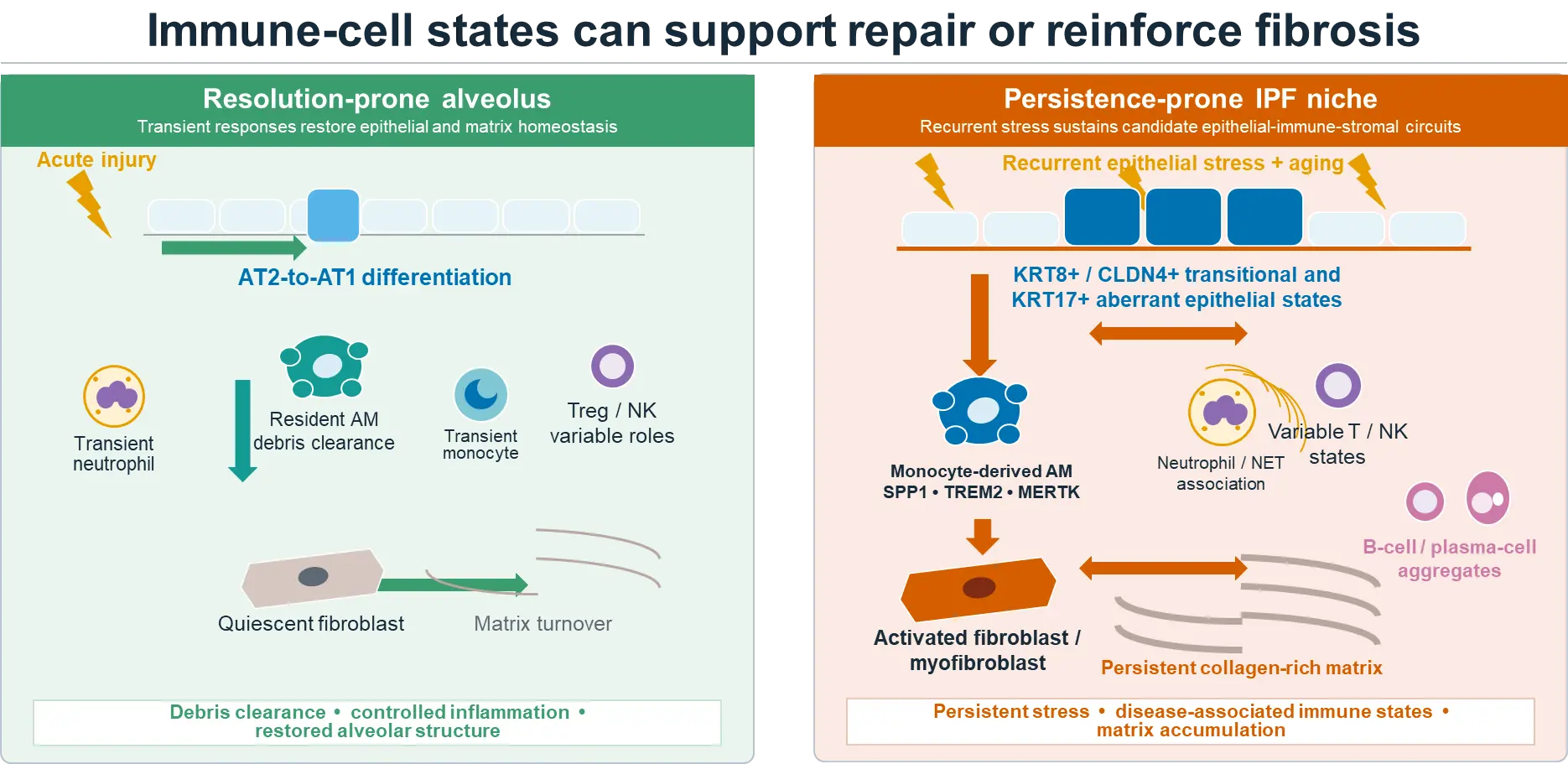
Figure 1. Immune-cell states can support repair or reinforce fibrosis. In a resolution-prone alveolus, coordinated epithelial differentiation, debris clearance, constrained inflammation, and matrix turnover support restoration of structure. In IPF, recurrent epithelial stress is associated with persistent KRT8+/CLDN4+ transitional and KRT17+ aberrant epithelial states, monocyte-derived SPP1+/TREM2+/MERTK+ alveolar macrophages, other disease-associated immune states, activated fibroblasts and collagen-rich matrix. Arrows denote a conceptual synthesis of proposed reciprocal influences instead of proof that every interaction is causal in human IPF. Immune effects vary with origin, compartment, molecular state, and disease stage.
The niche is not simply a passive location for immune infiltration. Repeated epithelial damage releases alarmins, including IL-33 and IL-1 family signals, while apoptotic bodies, oxidized surfactant lipids, and basement-membrane fragments provide persistent inputs to recruited macrophages and other leukocytes. Injured epithelial cells and activated fibroblasts supply chemokines, growth factors, and survival cues. In parallel, matrix stiffening and disrupted alveolar architecture alter cell adhesion, mechanotransduction, oxygenation, and access to clearance pathways [8,9,10,11,14,15,18,19,20,21,22,23]. These local pressures can select for persistent, lipid-handling and lysosome-rich macrophage states and perpetuate fibroblast-immune feedback [12,24]. The resulting immune phenotype therefore reflects not only ontogeny or circulating inflammatory signals but also the biochemical and mechanical properties of the diseased local tissue [8,9,10,11,14,15,18,19,20,21,22,23].
This repair-centered model also separates inflammation from fibrosis. Type I interferons, tumor necrosis factor, or IL-1 can intensify epithelial injury, yet selected inflammatory signals also recruit antimicrobial cells and initiate later repair [25,26]. Type 2 and wound-healing programs can reduce acute damage while promoting fibroblast activation when they persist [27,28]. Thus, the relevant defect in IPF may be failure to complete and terminate a normally useful response. General immune suppression removes protective functions without necessarily interrupting the epithelial-stromal circuit that maintains fibrosis.
3.2. Four Coordinates for Interpreting an Immune-Cell Finding
Four coordinates organize an immune-cell finding in IPF. Origin separates long-lived tissue-resident cells from recruited bone-marrow-derived cells. State defines the transcriptional, epigenetic, metabolic, and functional program acquired in the diseased lung. Space identifies the relevant compartment, including preserved alveolus, transitional epithelial region, fibroblastic focus, bronchiolized or honeycomb structure, vessel, and lymphoid aggregate. Time separates initiating injury, evolving fibrosis, established disease, acute exacerbation, and resolution. A therapeutic hypothesis becomes testable only when all four coordinates are specified.
This framework prevents several common overinterpretations. CD206, CD163, SPP1, TREM2, or MERTK expression can define part of a lung macrophage state but leaves lineage, function, and permanence unresolved. Transcript abundance alone does not measure active protein, receptor occupancy, or signal direction. Cross-sectional human tissue is unable to determine whether a state preceded fibrosis, and mouse and human cell labels are not interchangeable. The strongest mechanism therefore combines lineage-resolved perturbation, human tissue localization, direct functional assays, and longitudinal clinical association, followed by confirmation that intervention changes a meaningful patient outcome.
4. The Myeloid Compartment: The Best-Supported Immune Axis
4.1. Alveolar Macrophage Ontogeny Changes the Interpretation
Alveolar macrophages are often analyzed as a single population, although ontogeny strongly influences their response to injury. In the healthy lung, embryonically derived tissue-resident alveolar macrophages (TR-AMs) self-maintain in the airspace and support surfactant catabolism, efferocytosis, and immune restraint [29,30,31]. Injury recruits circulating monocytes that enter the lung, pass through interstitial states, and can differentiate into monocyte-derived alveolar macrophages (Mo-AMs) [32]. Recruited cells progressively acquire conventional alveolar macrophage markers, so endpoint phenotyping can obscure their origin; fate mapping remains the most reliable approach in experimental models [12,21,33,34].
Lineage-resolved bleomycin studies provide the clearest causal evidence for an immune population in lung fibrosis [21,34]. Mo-AMs expanded after injury, persisted after the acute response, and were required for maximal fibrosis, whereas depletion of TR-AMs did not produce the same effect [34]. In asbestos-induced fibrosis, a spatially restricted niche at bronchoalveolar duct junctions maintained Mo-AMs through M-CSF/M-CSFR signaling, and interruption of this pathway reduced macrophage persistence and fibrosis [21]. Earlier studies linked Ly6C-high monocyte recruitment to generation of profibrotic macrophages [33]. Together, these experiments identify recruited origin, local survival, and persistence as linked determinants of pathogenic activity.
Human IPF data are consistent with this model but provide less lineage resolution. Across single-cell datasets, FABP4-high homeostatic alveolar macrophages decrease as FCN1-high monocyte-like cells and SPP1-high, MERTK-positive, lipid- and lysosome-rich macrophages expand [7,18,22]. Similar states localize to remodeled epithelial and fibroblast-rich regions [8,9,10,11]. These findings support a conserved disease-associated macrophage program while leaving the origin and reversibility of individual SPP1- or TREM2-positive cells unresolved. Until origin can be inferred directly from genetic, epigenetic, or somatic-mutation information, human studies should assign state and location more confidently than ontogeny.
4.2. SPP1, TREM2, MERTK, Galectin-3, and the Lipid-Phagolysosomal Program
A recurrent macrophage program in IPF includes SPP1 (osteopontin), TREM2, MERTK, GPNMB, LGALS3 (galectin-3), APOE, cathepsins, and lipid-handling genes [18,22]. Related states occur in fibrotic liver, kidney, muscle, and heart, suggesting a conserved adaptation to apoptotic debris, matrix fragments, oxidized lipids, and altered tissue mechanics instead of an IPF-specific lineage [19,20,23]. In the alveolus, this program can increase survival, phagolysosomal capacity, lipid processing, and matrix interaction. These functions might support early debris clearance, but sustained activation can stabilize a macrophage population that continues to deliver growth factors, proteases, and matrix-active mediators after the acute injury has passed.
SPP1 illustrates the difference between a state marker and a defined effector. SPP1-high/MERTK-positive macrophages expand in IPF and localize near myofibroblasts [22]. Osteopontin can regulate adhesion, migration, survival, and matrix signaling through integrins and CD44, whereas MERTK supports efferocytosis and macrophage survival. A recent study proposed MERTK-SPP1-SRC-TKS5 signaling as a route to macrophage-to-myofibroblast transdifferentiation in bleomycin injury [35]. The signaling axis is mechanistically provocative, but mesenchymal-marker acquisition alone does not show that macrophages become a quantitatively important collagen-producing lineage in human IPF. Rigorous fate mapping and direct matrix-production measurements remain necessary.
TREM2 has stronger convergent support as a regulator of macrophage persistence. TREM2 is increased in macrophages from human IPF lungs, and genetic deletion, knockdown, soluble receptor, or antibody blockade reduced macrophage survival, profibrotic activity, and fibrosis in experimental systems [24]. These findings place TREM2 upstream of a lipid-adaptive survival program rather than defining it as a simple polarization marker. They also predict a stage-dependent therapeutic window: TREM2-mediated phagocytosis and lipid handling could aid early clearance, whereas the same pathway can maintain pathogenic Mo-AMs during established fibrosis [24]. Efficacy and safety therefore need to be tested after fibrosis is present and in models that measure antimicrobial and epithelial consequences.
Galectin-3 integrates glycan recognition, TGF-β signaling, fibroblast activation, and epithelial injury and is produced by macrophages as well as structural cells [36]. Its enrichment in disease-associated macrophages made inhaled inhibition attractive. TD139/GB0139 reached alveolar macrophages and pharmacodynamically reduced cell surface galectin-3 and circulating fibrosis-associated markers in an early study [37], but the later GALACTIC-1 trial failed to improve FVC and showed greater FVC decline with GB0139 than with placebo [37,38]. The sequence is instructive: cell delivery and proximal target engagement established pharmacology, whereas the randomized efficacy study rejected clinical benefit for that dose, population, and treatment context.
4.3. Macrophage-Fibroblast and Macrophage-Epithelial Circuits
Macrophage effects are executed through multicellular circuits. The CCL18 loop provides a long-standing human example: alveolar macrophage-derived CCL18 increases fibroblast collagen production, and collagen-conditioned macrophages further increase CCL18 [39]. Contemporary single-cell and spatial studies extend this concept to reciprocal networks involving osteopontin, M-CSF, platelet-derived growth factors, TGF-β activation, IL-1 family cytokines, chemokines, matrix fragments, and altered tissue mechanics. Fibroblasts provide survival and positional cues; macrophages provide factors that reinforce fibroblast persistence and matrix remodeling. The niche can therefore maintain both cell states even when no single soluble mediator is dominant.
A parallel circuit links macrophages to failed epithelial regeneration. Injury-associated macrophage signals promote entry of AT2-derived cells into a KRT8-high damage-associated transient progenitor state [14]. This state normally resolves as cells complete AT1 differentiation, but related KRT8- and KRT17-high epithelial populations persist in fibrotic mouse and human lungs [15,17]. Multiplexed imaging places CD206-high macrophages beside transitional alveolar basal-intermediate cells across remodeling stages [11]. These findings support a macrophage-epithelial repair niche, although signal direction remains unresolved. Macrophages may prolong epithelial transition; transitional cells may also recruit and reprogram macrophages; and both may be maintained by matrix stiffness, basement-membrane injury, or fibroblast-derived signals.
4.4. Circulating Monocytes, Interstitial Macrophages, MDSC-Like Cells, and Fibrocytes
Peripheral blood provides a scalable but indirect measure of lung immunity. Higher absolute monocyte counts reproducibly associate with mortality and disease progression in IPF [16]. Circulating fibrocytes also increase in a subset of patients and associate with poor outcome [40]. These signals are clinically accessible and may capture systemic bone marrow activation, but neither is specific to IPF, and neither shows that reducing the measured population improves disease. Their most immediate value is prognostic stratification and selection of patients for paired blood-lung studies.
Myeloid-derived suppressor cell (MDSC)-like populations are increased in the blood of some patients and correlate with disease severity [41]. Their suppressive activity offers a mechanism linking systemic myeloid activation with impaired adaptive immunity, yet human definitions vary, and granulocytic or monocytic MDSC-like cells overlap with conventional populations. Ex vivo suppression establishes function in the assay, not a causal lung mechanism. Prospective studies ought to determine whether these cells track a specific tissue macrophage state, predict progression independently of disease severity, and identify response to a pathway-directed intervention.
Interstitial macrophages remain less resolved than alveolar macrophages. Perivascular, peribronchial, and alveolar-interstitial populations could have distinct developmental origins and functions, while BAL under-samples all three and tissue dissociation can blur their boundaries [21,32]. Spatial proteomics, paired BAL-tissue analysis, and lineage-distinguishing reference mapping are needed to determine whether an interstitial macrophage state provides a target that is distinct from the Mo-AM axis.
4.5. Why M1/M2 Terminology Is Inadequate
The M1/M2 framework is useful for describing responses to defined in vitro stimuli but is poorly matched to macrophages in fibrotic lung [12,13]. IPF macrophages coexpress wound-repair, lipid-metabolic, inflammatory, efferocytic, and matrix-remodeling modules. CD206 or CD163 expression alone is insufficient to assign IL-4 driven alternative activation, and an anti-inflammatory macrophage can still be profibrotic by supporting fibroblast survival or matrix turnover. Mechanistic descriptions must therefore specify cellular origin, tissue compartment, measured function, and molecular program, such as a persistent alveolar macrophage derived from monocytes and expressing high levels of SPP1 and TREM2, instead of relying on a binary polarization label.
5. Other Innate Immune Cells: Amplifiers, Sentinels, and Context-Dependent Regulators
5.1. Neutrophils and Extracellular Traps
Neutrophils are prominent in acute exacerbation and in some patients with advanced physiologic impairment, whereas their contribution to slowly progressive IPF is less established than the macrophage axis. Baseline BAL neutrophilia predicted early mortality in a well-characterized cohort [42]. Occult infection, aspiration, epithelial barrier failure, smoking, and advanced remodeling can all contribute to this association, so chronic neutrophil depletion should not be inferred as beneficial. The key unresolved question is whether a stable neutrophil-high endotype exists independently of these confounders.
Experimental studies identify plausible neutrophil effectors. Neutrophil elastase-deficient mice developed less bleomycin fibrosis and showed reduced TGF-β activation despite a substantial inflammatory response [43], and elastase can directly promote fibroblast proliferation and myofibroblast differentiation [44]. Neutrophil extracellular traps (NETs) activate human lung fibroblasts in vitro, and NET-associated pathways are detected in fibrotic interstitial lung disease [45,46]. NETs can concentrate proteases, histones, coagulation factors, and potential autoantigens, but commonly used NET markers also occur during neutrophil necrosis. Tissue studies need orthogonal markers, and intervention studies need to test treatment after fibrosis is established.
The therapeutic window is narrow because neutrophil antimicrobial function is indispensable in an older population vulnerable to respiratory infection. Targeting a defined effector, e.g., elastase, pathologic NET formation, or a tissue-retention signal, is more plausible than reducing neutrophil number. Biomarker enrichment may also be essential: stable neutrophil-low IPF, neutrophil-high progressive disease, and acute exacerbation are unlikely to share the same dependence on this pathway.
5.2. Dendritic Cells
Dendritic cells (DCs) connect innate sensing with T-cell activation and tolerance. CD1c/BDCA1-positive conventional DCs and other antigen-presenting populations accumulate in fibrotic human lung and lymphoid aggregates [47]. DC perturbation alters inflammation and fibrosis in bleomycin models [48], but older depletion approaches also affected monocytes or macrophages. Newer single-cell taxonomies separate conventional DC1, conventional DC2, activated migratory, and plasmacytoid states, making the earlier literature difficult to map to a specific causal population.
DCs could present epithelial or matrix neoantigens, support helper or cytotoxic T-cell responses, and produce chemokines that organize tertiary lymphoid structures. These mechanisms remain plausible but have only just begun to emerge in IPF. A decisive study would link a spatially localized DC state to a clonally expanded lymphocyte response, identify the cognate antigen, and perturb the interaction in a human-relevant system. Until then, DC accumulation is evidence of local immune organization, not proof of a uniform autoimmune cause.
DCs may also promote fibrosis independently of antigen presentation. A type I interferon induced, epiregulin (EREG) expressing DC3 state activated fibroblast EGFR and NOTCH dependent matrix production; delayed epiregulin neutralization reduced bleomycin induced lung fibrosis and lowered matrix markers in one IPF lung explant, although the prevalence of this circuit in IPF remains unknown [49].
5.3. Mast Cells and Eosinophils
Mast cells increase in fibrotic lung and can localize beside fibroblasts. Tryptase, chymase, histamine, leukotrienes, TGF-β, and other mast-cell mediators can alter epithelial permeability, fibroblast proliferation, matrix processing, vascular tone, and symptoms [50,51]. Human tissue and coculture studies support bidirectional mast-cell-fibroblast activation [51]. Nintedanib also inhibits mast-cell survival and activation at pharmacologically relevant concentrations [52], although the quantitative contribution of this effect to its clinical antifibrotic activity is unknown.
Evidence for eosinophils is more limited. Mild BAL eosinophilia occurs in IPF, whereas marked eosinophilia justifies evaluation for an alternative diagnosis. In experimental fibrosis, eosinophils can provide type 2 cytokines, lipid mediators, proteases, and TGF-β, but cell-selective human evidence remains sparse [12,13]. Mast cells and eosinophils therefore seem to be most relevant in a type 2-high, airway-remodeled, or comorbid allergic endotype as opposed to across all IPF.
5.4. Innate Lymphoid Cells
Group 2 innate lymphoid cells (ILC2s) respond to epithelial IL-25, IL-33, and thymic stromal lymphopoietin and produce IL-5, IL-13, amphiregulin, and other repair-associated mediators. IL-25 and ILC2 abundance were increased in human IPF samples, and IL-25-driven ILC2 responses promoted collagen deposition in mice [27]. Zhang et al. identified NRP1 as a TGF-β1 induced regulator of lung ILC2s that promotes ST2 expression and IL-5 and IL-13 production. Genetic or pharmacologic NRP1 inhibition attenuated bleomycin induced fibrosis, but its relevance to human IPF remains insufficiently established [28]. This pathway provides a direct connection between epithelial stress and type 2 immunity without antigen-specific T cells. The same mediators can support epithelial repair or fibroblast activation depending on timing and niche, and rare ILC2s are difficult to distinguish from activated T cells in dissociated tissue.
ILC2s also exhibit regulatory plasticity. Although conventional resting or type 2-activated ILC2s are not constitutive major sources of IL-10, an inducible IL-10-producing ILC2 state has been described in experimental lung inflammation. Its generation depends on activation context. For example, IL-2 and retinoic acid can promote IL-10 expression, whereas stimulation sufficient for IL-5/IL-13 secretion does not necessarily generate IL-10-producing cells [53,54]. This phenotype can limit excessive inflammatory responses, including eosinophil accumulation, in nonfibrotic lung models. It is therefore plausible that local cues in injured or fibrotic lungs govern a balance between profibrotic IL-5/IL-13-associated and IL-10-associated ILC2 functions. However, neither the abundance of IL-10-producing ILC2s nor a direct protective effect of ILC2-derived IL-10 on established human IPF fibrosis has been demonstrated.
Group 1 and group 3 innate lymphoid cells are even less resolved. Their production of interferon-γ, IL-17, or IL-22 predicts both protective and injurious functions. Protein-level localization, exclusion of conventional lymphocytes, and cell-selective perturbation are required before these populations can be assigned a mechanism or therapeutic priority in IPF.
6. T Lymphocytes: Function Depends on Phenotype, Antigen, and Stage
6.1. CD4 T-Cell Polarization Is a Continuum, Not a Set of Fixed Bins
T cells occupy the IPF interstitium, perivascular regions, remodeled airways, and lymphoid aggregates. Blood and tissue studies show altered activation, costimulation, clonality, and differentiation, although the direction and magnitude vary with compartment, age, disease stage, treatment, and acute exacerbation status [55,56,57,58,59,60]. Classical helper subsets remain a useful vocabulary, but human T cells frequently coexpress programs and change state within tissue.
Not only secreted cytokines, CD4 T cells regulate lung immunity also through antigen-specific recognition, costimulation, direct cell contact, and control of other immune populations. Through cognate interactions with dendritic cells and B cells, CD4 T cells can sustain local antigen presentation, B-cell maturation, class switching, and antibody responses within tertiary lymphoid structures. Similarly, CD4 T-cell interactions with macrophages and epithelial cells may influence cell recruitment, survival, and the persistence or resolution of injury. Tissue residency, clonal expansion, and exhaustion or senescence-related programs therefore can be as important as conventional Th1, Th2, or Th17 labels when interpreting an IPF T-cell state [55,56,57,58,59,60,61,62,63,64,65].
These non-cytokine functions also raise distinct mechanistic questions. A locally expanded CD4 T-cell clone could reflect recognition of microbial, self, or injury-associated antigens. Nevertheless, clonal expansion alone does not establish pathogenic specificity. T-cell receptor sequencing paired with antigen discovery, spatial contact analysis, and functional coculture or perturbation is needed to determine whether CD4 T cells organize a protective response, promote persistent antibody-producing niches, or reinforce pathologic macrophage-stromal circuits.
Th1-associated interferon-γ restrains fibroblasts and type 2 responses in several preclinical systems, yet systemic interferon-γ-1b did not improve survival in IPF [66]. IL-4 and IL-13 promote reparative macrophage programs and fibroblast activation in many models, while randomized inhibition of IL-13 alone or combined IL-4/IL-13 signaling failed to reduce lung-function decline in unselected IPF [67,68]. These discordant results are mechanistically informative: pathway activity in an injury model cannot define drug delivery, timing, or pathway dominance in established human disease.
The IL-1/IL-23/IL-17 axis has similar context dependence. IL-17A was required for maximal fibrosis after bleomycin or IL-1β challenge and linked inflammasome-driven injury to neutrophil recruitment and fibroblast activation [25,26]. IL-17-related signals are present in selected human IPF tissues and blood populations, but the cellular source and magnitude are inconsistent. The current evidence supports IL-17 as an amplifier in inflammatory models and possibly in a measurable patient endotype; routine IL-17 blockade in IPF remains unsupported and carries a substantial infection concern.
6.2. Regulatory T Cells Are Stage-Dependent But Not Simply Protective or Pathogenic
Regulatory T cells (Tregs) provide a clear example of state- and stage-dependent function. Impaired circulating CD4+CD25+FOXP3+ Treg activity was associated with disease severity in one human study [59], whereas other studies found increased activated Treg proportions in advanced disease. In bleomycin models, transfer, depletion, and timing produce divergent outcomes, including exacerbation of fibrosis by selected Treg populations [69]. These results are biologically compatible if early injury-control and late profibrotic Treg states are separated.
Early after injury, Tregs can restrain effector responses, support macrophage efferocytosis, and provide IL-10-dependent repair signals. During established fibrosis, selected Tregs could produce TGF-β, support type 2 macrophages, suppress antifibrotic effector cells, or lose function in the aged niche. A Tff1-expressing Treg state reduced injury and fibrosis in mice [70]. Independently, aged bone marrow increased persistent profibrotic macrophages and weakened a Treg-IL-10 resolution axis, whereas young marrow improved resolution [71]. These studies identify functional heterogeneity and marrow-lung communication without defining the dominant human Treg state.
Treg studies should therefore move beyond FOXP3 counts. Required measurements include thymic versus peripheral origin, suppressive capacity, IL-10, TGF-β and/or amphiregulin production, stability, antigen specificity, tissue residency, spatial relation to macrophage-epithelial niches, and longitudinal association with progression. Global expansion or depletion would ignore the functions that the therapeutic strategy is intended to preserve.
6.3. Cytotoxic CD4 and CD8 T Cells
Loss of CD28 on circulating CD4 T cells is associated with poor IPF outcomes [57]. CD28-null cytotoxic T cells expand in some patients, express granzymes and inflammatory mediators, and promote fibrosis in a humanized mouse system [58]. Chronic antigen stimulation, viral exposure, immunosenescence, and repeated tissue injury could all generate this state. The association with severity is reproducible, whereas the antigen and target cell remain unknown.
CD8 T cells increase in IPF tissue and can occupy epithelium-associated regions [55]. Perforin, granzymes, Fas ligand, and profibrotic cytokines provide routes to epithelial injury, while cytotoxic T cells can also eliminate infected, damaged, or senescent cells. CD8 tissue-resident memory T cells were required for maximal fibrosis in a recent bleomycin study [56]. Translation requires identification of the antigen and target cell because tissue residence alone leaves specificity unresolved, and depletion during acute injury is different from reprogramming a persistent population in chronic disease. This line of concept has been tested in a very recent study, in which immunopeptidomic profiling identified fibrosis associated MHC class I peptides linked to SPP1+TREM2+ macrophages and myofibroblasts. Peptide vaccination reduced bleomycin induced fibrosis, and MAF116–124 specific human cytotoxic T cells killed IPF derived myofibroblasts and IL-4 polarized macrophages ex vivo, although endogenous responses to this antigen in IPF remain unproven [72].
Checkpoint effects appear highly context dependent. In immunocompetent human CTLA4 knock-in mice with established repetitive bleomycin fibrosis, late ipilimumab treatment enhanced CD8 T-cell clearance of senescent cells, expanded AT2 cells, and accelerated fibrosis resolution; CTLA4 expressing CD8 T cells also localized near fibroblastic foci in human IPF. Conversely, CTLA4 blockade worsened fibrosis in the earlier immunodeficient CD28-null T-cell transfer model, indicating that immune context, treatment timing, target-cell identity, and pneumonitis risk must be resolved before clinical translation [58,73].
6.4. Gamma-Delta T Cells and Natural Killer Cells
Gamma-delta T cells respond rapidly to stress ligands and can produce interferon-γ, IL-17, or growth factors without conventional antigen presentation. An interferon-γ-producing gamma-delta population suppressed Th17 responses and bleomycin fibrosis [74], whereas other contexts favor IL-17-producing cells with an opposite predicted effect. Human IPF data remain too limited to assign a dominant function.
Natural killer (NK) cells illustrate how the target-cell state can reverse interpretation. Earlier NK-cell depletion or transfer did not materially alter bleomycin fibrosis [75]. In contrast, a 2026 study identified HLA-E/NKG2A signaling that protected senescent HAS1-positive fibroblasts from NK-cell killing in IPF; NKG2A blockade restored patient NK-cell activity ex vivo, and NK-cell or checkpoint-directed approaches promoted resolution in experimental systems [76]. The newer work provides a coherent senescent-fibroblast clearance mechanism. It also requires controlled clinical testing because global NK activation could injure stressed epithelium or alter antiviral defense.
7. B Cells, Plasma Cells, Tertiary Lymphoid Structures, and Autoantibodies
7.1. Local B-Cell Organization and Systemic Signals
B cells and plasma cells are heterogeneously distributed in IPF. Lymphocyte aggregates and tertiary lymphoid structures (TLSs) occur in a substantial subset and accumulate with disease progression [60]. TLSs provide an architecture for antigen presentation, T-B collaboration, somatic diversification, and local antibody production, but they can also organize protective responses. Germinal-center activity, receptor clonality, and location relative to active remodeling are therefore more informative than aggregate number alone.
Circulating B-cell activating factor (BAFF/BLyS) is increased in some patients and associates with altered B-cell differentiation and clinical severity [65]. CXCL13, which recruits CXCR5-positive B cells and T follicular helper cells, is elevated in plasma and lung and has prognostic value [64]. Abnormal B-cell receptor signaling, autoreactive IgA, and altered circulating T follicular helper and regulatory B-cell populations provide additional evidence of B-lineage dysregulation [61,62,63]. None is present in every patient or is specific to IPF; together, they define a candidate B-cell-active endotype.
7.2. Autoantibodies: Pathogenic Subset, Biomarker, or Epiphenomenon?
Autoantibodies against heat-shock protein 70, vimentin, and other epithelial or matrix-associated antigens associate with poor outcome or progression in subsets of patients [77,78,79]. Immune-complex formation, Fc-receptor signaling, complement activation, and direct effects on stressed cells provide plausible mechanisms. Tissue destruction can also expose intracellular antigens and generate autoantibodies as a secondary event. Longitudinal appearance before progression and transfer or neutralization experiments are needed to resolve directionality.
A depletion study in bleomycin fibrosis separated plasma cells from the broader B-cell compartment: plasma-cell depletion was protective, whereas CD20 B-cell depletion was not [80]. Because mature plasma cells lack CD20, the result points to antibody-secreting cells or their products rather than all B cells in that model. It also shows why rituximab-like therapy may fail to test the strongest B-lineage mechanism. Uncontrolled studies of plasma exchange, rituximab, and intravenous immunoglobulin during autoantibody-associated acute exacerbation generated signals that require randomized clinical trial confirmation [81].
Table 1. Evidence synthesis by cell population.
|
Immune Population or State |
Principal Human Evidence |
Strongest Experimental Evidence |
Balanced Interpretation |
|---|---|---|---|
|
Homeostatic FABP4-high alveolar macrophage |
Relatively enriched in control or less-remodeled regions |
Resident macrophages are dispensable for, or may restrain, fibrosis in lineage-resolved models |
Preserve homeostatic function; abundance alone does not prove protection |
|
FCN1-high monocyte and SPP1/TREM2/MERTK-high macrophage |
Reproducible enrichment and localization near remodeled epithelium and fibroblasts |
Mo-AM depletion, M-CSFR interruption, and TREM2 loss/blockade reduce fibrosis. |
Best-supported immune-cell axis; human origin remains inferred, and timing must be defined |
|
Circulating monocyte/MDSC-like cell |
Counts or phenotypes associate with severity and mortality |
Monocyte recruitment and differentiation alter fibrosis in mice |
Useful prognostic/endotyping candidates; not yet predictive treatment biomarkers |
|
Neutrophil/NET-high state |
Lavage neutrophilia predicts poor outcome; NET signals in fibrotic ILD |
Elastase loss and NET-pathway perturbation reduce fibroblast activation/fibrosis |
Likely amplifier in a subset; preserve antimicrobial function and target a defined effector |
|
Dendritic cell |
Enrichment in fibrotic tissue and lymphoid aggregates |
Broad DC perturbation changes bleomycin responses |
Antigen-presentation dependent and independent roles plausible but cell-state causality remains weak. |
|
Mast cell/ILC2/type 2 innate state |
Increased cells or mediators in subsets of IPF |
Mast-cell mediators and IL-25-ILC2 signaling promote model fibrosis |
Context-dependent amplifier; randomized type 2 cytokine blockade has disappointed |
|
Treg |
Altered number and function correlate with severity, inconsistently |
Protection or harm varies with state and timing |
Functional and stage heterogeneity makes global expansion/depletion unattractive. |
|
Cytotoxic CD4/CD8 T cell |
CD28-null and tissue cytotoxic states associate with poor outcome |
Transfer or depletion changes fibrosis in selected models; CTLA4-dependent senescent-cell clearance improves lung fibrosis |
Antigen and target-cell identity are partially resolved; checkpoint manipulation may help or harm |
|
NK cell |
Inhibitory NKG2A and fibroblast HLA-E define a potential senescence niche |
NK-cell/checkpoint approaches clear senescent fibroblasts in recent models |
Compelling resolution mechanism with human ex vivo support; clinical efficacy and safety remain untested |
|
B cell/plasma cell/TLS state |
BAFF, CXCL13, TLSs, BCR abnormalities, and autoantibodies in subsets |
Plasma-cell, but not CD20-cell, depletion protected in one model |
Supports an autoimmune-like endotype, not universal autoimmunity |
A rational B-lineage trial would prospectively define autoantibody identity and titer, BAFF/CXCL13 level, plasmablast or plasma-cell signature, TLS maturity, and target engagement. Chronic progression and acute exacerbation ought to be analyzed separately, with infection risk built into eligibility and stopping rules. The data support an autoimmune-like circuit in a measurable subset of IPF, not classification of IPF as a uniform autoimmune disease (Table 1).
8. Spatial Immune Niches: Where Cell States Become Mechanisms
8.1. Remodeling Is Geographically Heterogeneous
A maladaptive niche forms when the normally transient signals of repair become self-reinforcing. For example, failure of AT2-to-AT1 differentiation prolongs epithelial stress and alarmin release; the accompanying accumulation of debris and altered surfactant may reinforce macrophage lipid-adaptive survival programs. Persistent macrophages and activated fibroblasts then exchange growth factors, chemokines, osteopontin, and matrix-remodeling signals. Basement-membrane disruption and increasing matrix stiffness can further impede epithelial restoration and sustain fibroblast activation. These interactions create reciprocal selection pressures that retain particular immune states even after the initiating injury has diminished [8,9,10,11,14,15,18,21,22,82,83,84]. This proposed feed-forward model is grounded in converging spatial and experimental findings, but the precise sequence and necessity of each interaction remain to be established in human IPF.
The IPF lung contains relatively preserved alveoli, early septal thickening, fibroblastic foci, dense scar, airway-centered metaplasia, honeycomb cysts, vascular remodeling, and lymphoid aggregates within the same organ. Dissociation-based single-cell analysis removes this geography and preferentially loses fragile epithelial cells, granulocytes, and matrix-embedded populations. Spatial analysis is therefore necessary to determine which cells can interact and whether a candidate state tracks an active remodeling front or an end-stage compartment.
Recent spatial maps show that immune states follow regional remodeling. Image-based transcriptomics across control and fibrotic lungs identified a progression from early epithelial dysregulation to later macrophage-state changes as distal airspaces remodeled [10]. Other studies defined pathologic epithelial, mesenchymal, endothelial, and immune neighborhoods [8,9], and multiplexed imaging placed CD206-high macrophages beside transitional alveolar basal-intermediate cells across remodeling stages [11]. Human IPF is therefore organized into multiple recurrent niches instead of one uniform fibrotic microenvironment.
Spatial information strengthens the mechanism in two ways. Direct proximity makes a proposed circuit anatomically possible, and regional progression can separate an early event from a later adaptation. In the Vannan spatial atlas, epithelial abnormalities preceded the strongest macrophage-state changes along the inferred remodeling gradient [10], supporting epithelial failure as an upstream pressure that reprograms macrophages. The same data allow macrophage feedback during later progression. Inferred gradients are not longitudinal fate maps, so signal direction still requires perturbation.
8.2. Three Recurring Immune-Structural Neighborhoods
The first recurrent neighborhood is an alveolar transition niche containing stressed AT2 cells, KRT8-high transitional cells, macrophages, endothelial cells, and fibroblasts (Figure 2) IL-1 family sigaling, TGF-β activation, osteopontin, altered lipid handling, basement-membrane injury, and matrix stiffness can influence whether epithelial differentiation completes or remains arrested. Normal repair and fibrosis can use similar intermediate states; persistence and failed exit distinguish the pathologic niche [14,15,17,83,84].
The second is a fibroblastic focus-macrophage niche (Figure 2) SPP1-high/MERTK-positive/TREM2-positive macrophages localize near activated fibroblasts and collagen-rich matrix. Candidate reciprocal signals include M-CSF, CCL2-family chemokines, CCL18, osteopontin-integrin interactions, galectin-3, growth factors, and matrix-derived danger signals [21,22,24,39]. Alveolar fibroblast lineages also coordinate inflammatory recruitment and remodeling in experimental studies [82], supporting a bidirectional mechanism in which stromal cells impose part of the macrophage state that later sustains the stromal niche.
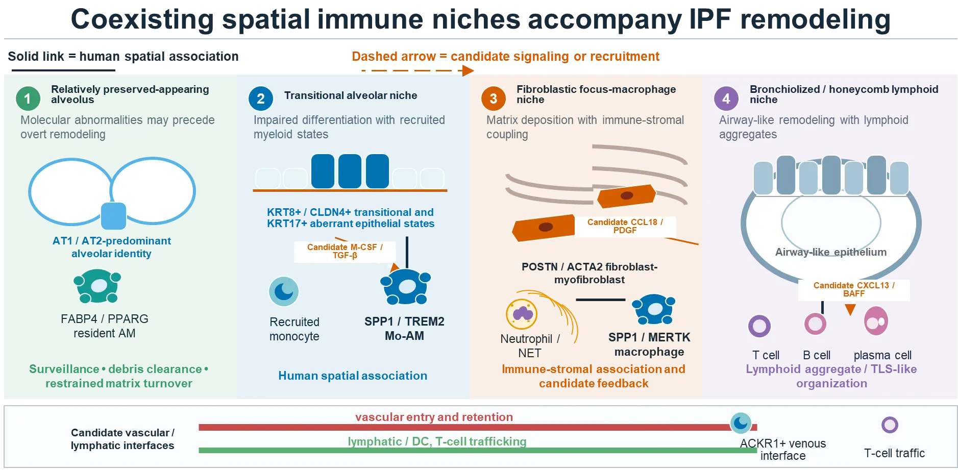
Figure 2. Coexisting spatial immune niches accompany IPF remodeling. The schematic organizes representative spatial states reported in fibrotic human lung, ranging from relatively preserved-appearing alveolar regions to transitional epithelial-myeloid niches, fibroblastic focus-macrophage niches, and bronchiolized or honeycomb regions containing lymphoid aggregates. The arrangement is conceptual and does not establish a longitudinal sequence; multiple states may coexist within the same lung, and relatively preserved-appearing tissue may already be molecularly abnormal. Solid links denote reported human spatial associations. Dashed arrows denote candidate signaling or recruitment mechanisms requiring functional validation.
The third is an airway-lymphoid niche within bronchiolized and honeycomb regions (Figure 2) Secretory or basal-like epithelium, antigen-presenting cells, B cells, plasma cells, T cells, and fibroblasts coexist with altered mucus and microbial exposure. TLS maturation and local autoantibody production may be concentrated in this compartment. These airway-like structures can remain immunologically active, although dense end-stage honeycomb regions may be less reversible than an expanding subpleural remodeling front [60,61,62,63,64,65].
Vascular and lymphatic compartments intersect all three neighborhoods. Endothelial injury regulates leukocyte entry, coagulation, and oxygen gradients, while lymphatic remodeling changes antigen and fluid drainage [85,86,87,88]. These interfaces remain less defined than epithelial and fibroblast compartments and can be incorporated into future immune-niche studies.
8.3. What Spatial Data Still Cannot Tell Us
Current spatial data retain important limitations. Most studies use explants from advanced IPF, inferred trajectories are not direct time series, and sampling or preservation varies across regions. Platform resolution ranges from multicellular spots to near-single-cell measurements, and targeted imaging panels could omit a decisive ligand, receptor, or activation marker. Computational cell-cell communication combines expression with proximity while leaving ligand availability, receptor occupancy, and downstream activity unmeasured [89].
The next step is a perturbable spatial map. Serial clinical samples, regional imaging, rapid-autopsy or transplant tissue, spatial proteomics, immune-receptor clonality, and matrix composition can be integrated with precision-cut lung slices, immune-containing organoids, or perfused ex vivo systems. The goal is to identify a reversible interaction in a drug-accessible region and then show that disrupting it changes epithelial repair or matrix production. A larger atlas without functional testing will add description but may not advance therapy.
9. Aging, Immunosenescence, and Systemic Control of Lung Immunity
Age is the strongest demographic risk factor for IPF and alters every immune compartment. Hematopoietic stem cells become myeloid biased; monocytes and macrophages change metabolic and inflammatory responses; macrophage self-renewal and efferocytosis decline; naive T- and B-cell pools contract; clonally expanded cytotoxic cells accumulate; and senescent structural cells express ligands that modify immune surveillance [13,71,76,90]. These changes interact with telomere dysfunction, mitochondrial stress, impaired proteostasis, and an aged extracellular matrix.
Two complementary studies separate hematopoietic aging from the aged lung niche. An aged pulmonary microenvironment imposed a dysfunctional alveolar macrophage response even when macrophage age was not the only variable [90]. Conversely, aged bone marrow worsened fibrosis by supplying persistent monocyte-derived macrophages and weakening a Treg IL-10 resolution circuit; young marrow improved resolution [71]. IPF immunosenescence therefore reflects both cell history and the tissue environment in which the cell is placed.
This interaction explains coexisting immune hyperactivity and incompetence. CD28-null T cells can be cytotoxic while the host has weak vaccine responses; macrophages can retain inflammatory or fibrogenic programs while clearing microbes and apoptotic cells inefficiently; and NK cells can be present yet unable to eliminate HLA-E-positive senescent fibroblasts [76]. Therapeutic strategies should therefore restore a defined missing function rather than stimulate immunity globally. Examples include NKG2A blockade to restore NK-cell surveillance of senescent fibroblasts and, more provisionally, macrophage-directed or pro-resolving interventions that restore efferocytosis and inflammatory resolution [58,73,76,91,92].
Blood monocyte count, leukocyte gene expression, T-cell differentiation, autoantibody profiles, and circulating chemokines may capture parts of this marrow-lung axis [16,64,93,94]. Clinical use requires prospective validation beyond age and baseline disease severity and, critically, demonstration that a biomarker predicts response to a mechanism-directed therapy. Prognosis and target dependence are different properties; a marker of rapid decline does not automatically identify the pathway to inhibit.
10. Microbiome, Infection, and Immune Tone
The distal lung is continuously exposed to inhaled and aspirated microbial products. In IPF, altered anatomy, impaired mucociliary clearance, reflux, antibiotic exposure, immunosenescence, and epithelial barrier failure can change microbial burden and composition. Higher bacterial burden and selected taxa associate with progression, and experimental studies support microbiota-dependent inflammatory signaling [95,96]. Specific microbial products, like outer membrane vesicles from expanded Bacteroides and Prevotella, induced IL-17B, which cooperated with TNF-α to induce epithelial neutrophil recruiting and Th17 promoting programs, whereas microbiota depletion or Il17b deficiency reduced fibrosis [97]; whether this circuit operates in human IPF remains unproven. These findings provide a plausible source of chronic pattern-recognition input to macrophages, neutrophils, and epithelium.
Directionality remains difficult to establish because progressive fibrosis itself impairs clearance and changes the microbial niche. Low-biomass sequencing is vulnerable to contamination, and microbial DNA alone does not equate to viable infection. In the randomized CleanUP-IPF trial, addition of co-trimoxazole or doxycycline did not reduce nonelective respiratory hospitalization or death and increased adverse events [98]. The result argues against routine broad antimicrobial therapy for stable IPF. A future microbiome-directed strategy would require a defined organism or host-response biomarker, a mechanism-specific intervention, and direct assessment of resistance and ecological disruption.
Latent or recurrent viruses could also shape cytotoxic and innate states, but the human evidence remains inconsistent [99,100,101]. Infection becomes a central safety variable whenever an immune pathway is targeted because the same macrophage, neutrophil, or lymphocyte function that sustains fibrosis may protect an older patient from pneumonia. Trials must prospectively capture vaccination, microbiologic surveillance, exacerbation adjudication, and infection-related stopping rules.
11. Therapeutic Translation: What Failed, What Was Learned, and What Should Change
11.1. Broad Immunosuppression Failed Because the Hypothesis and Intervention Were Mismatched
PANTHER-IPF established that nonspecific suppression with prednisone and azathioprine is harmful [1,4]. The regimen altered many leukocyte and structural-cell functions, increased infection and other toxicities, and lacked a validated profibrotic target. The trial therefore argues against broad immunosuppression as an IPF strategy while leaving state-selective immune mechanisms to be tested independently.
More selective programs also failed. Carlumab did not slow IPF and paradoxically increased circulating CCL2, consistent with incomplete or compensatory target control [102]. Dual IL-4/IL-13 neutralization with SAR156597 missed its primary efficacy objective [68], and lebrikizumab achieved IL-13 pharmacodynamic activity without reducing FVC decline [67]. These trials weaken the hypothesis that one chemokine or type 2 cytokine is a dominant driver in unselected established IPF. A biomarker-defined subgroup remains possible, but it now requires prospective evidence rather than post hoc explanation.
11.2. Encouraging Biology Is Not the Same as Confirmed Efficacy
Recombinant pentraxin-2 (PRM-151; zinpentraxin alfa) was developed to alter monocyte, macrophage, and fibrocyte differentiation. A randomized phase II study showed a smaller FVC decline and better preservation of six-minute walk distance over 28 weeks [103]. The phase III STARSCAPE trial, however, was terminated after a prespecified futility analysis and showed no benefit over placebo [104]. This sequence replaces the earlier efficacy narrative: pentraxin-2 remains mechanistically informative, but zinpentraxin alfa lacks confirmed clinical efficacy in IPF.
The galectin-3 program followed a similar trajectory. Inhaled TD139/GB0139 reached alveolar macrophages and showed short-term target engagement [37]. In the 52-week GALACTIC-1 phase IIb trial, GB0139 did not slow FVC decline; the least-squares mean decline was greater with GB0139 than with placebo, and development for IPF was discontinued [37,38]. Early lung delivery and biomarker movement were therefore real pharmacologic observations, but they did not predict clinical benefit.
Nerandomilast provides an important contemporary contrast. This preferential PDE4B inhibitor has anti-inflammatory and antifibrotic activity but was developed against a defined molecular mechanism as opposed to as broad immunosuppression [105]. In FIBRONEER-IPF, nerandomilast reduced FVC decline over 52 weeks and subsequently received FDA approval for adults with IPF [2,3]. This result validates neither immune-cell targeting in general nor a single-cell mechanism; it shows that a pathway with immune and structural-cell effects can succeed when dose, population, endpoint, and clinical development are aligned (Table 2).
Table 2. Lessons from immune-directed clinical studies.
|
Intervention and Immune Rationale |
Study Signal |
Interpretation for Future Development |
|---|---|---|
|
Prednisone + azathioprine + N-acetylcysteine; broad suppression |
Increased death and hospitalization in PANTHER-IPF |
Broad immunosuppression is harmful and should not be used for IPF outside another indication |
|
Carlumab; neutralize CCL2-dependent monocyte recruitment |
No efficacy; paradoxical rise in total CCL2 |
Confirm durable tissue target control and anticipate ligand compensation |
|
SAR156597; dual IL-4/IL-13 neutralization |
Primary efficacy objective not met |
Type 2 cytokines are not a universal dominant driver in established, unselected IPF |
|
Lebrikizumab; IL-13 neutralization |
Pharmacodynamic activity without reduced FVC decline |
Target engagement is necessary but not sufficient; endotype or pathway redundancy may matter |
|
Recombinant pentraxin-2; alter monocyte/fibrocyte differentiation |
Phase II signal not confirmed; phase III STARSCAPE stopped for futility |
A positive early study requires phase III confirmation before efficacy is inferred |
|
Inhaled TD139/GB0139; inhibit galectin-3 in alveolar macrophage/stromal circuits |
Early lung target engagement; phase IIb GALACTIC-1 failed and FVC decline favored placebo |
Proximal pharmacology did not predict patient benefit; dose, state dependence, and pathway redundancy remain limiting |
|
Autoantibody-targeted plasma exchange/rituximab/IVIG |
Uncontrolled acute-exacerbation signal |
Investigational; needs randomized, biomarker-selected evaluation |
|
NKG2A blockade or NK-cell therapy; clear senescent fibroblasts |
Human ex vivo target engagement and experimental resolution |
High-priority translational hypothesis; infection and epithelial cytotoxicity require monitoring |
|
Nerandomilast; preferential PDE4B inhibition with immune- and structural-cell effects |
Reduced 52-week FVC decline in FIBRONEER-IPF; FDA-approved for adult IPF in 2025 |
A defined pathway with mixed anti-inflammatory and antifibrotic effects can succeed without broad immune suppression |
11.3. A Precision-Immunology Framework
Before a large clinical trial, an immune-directed IPF program needs to answer six linked questions (Figure 3).
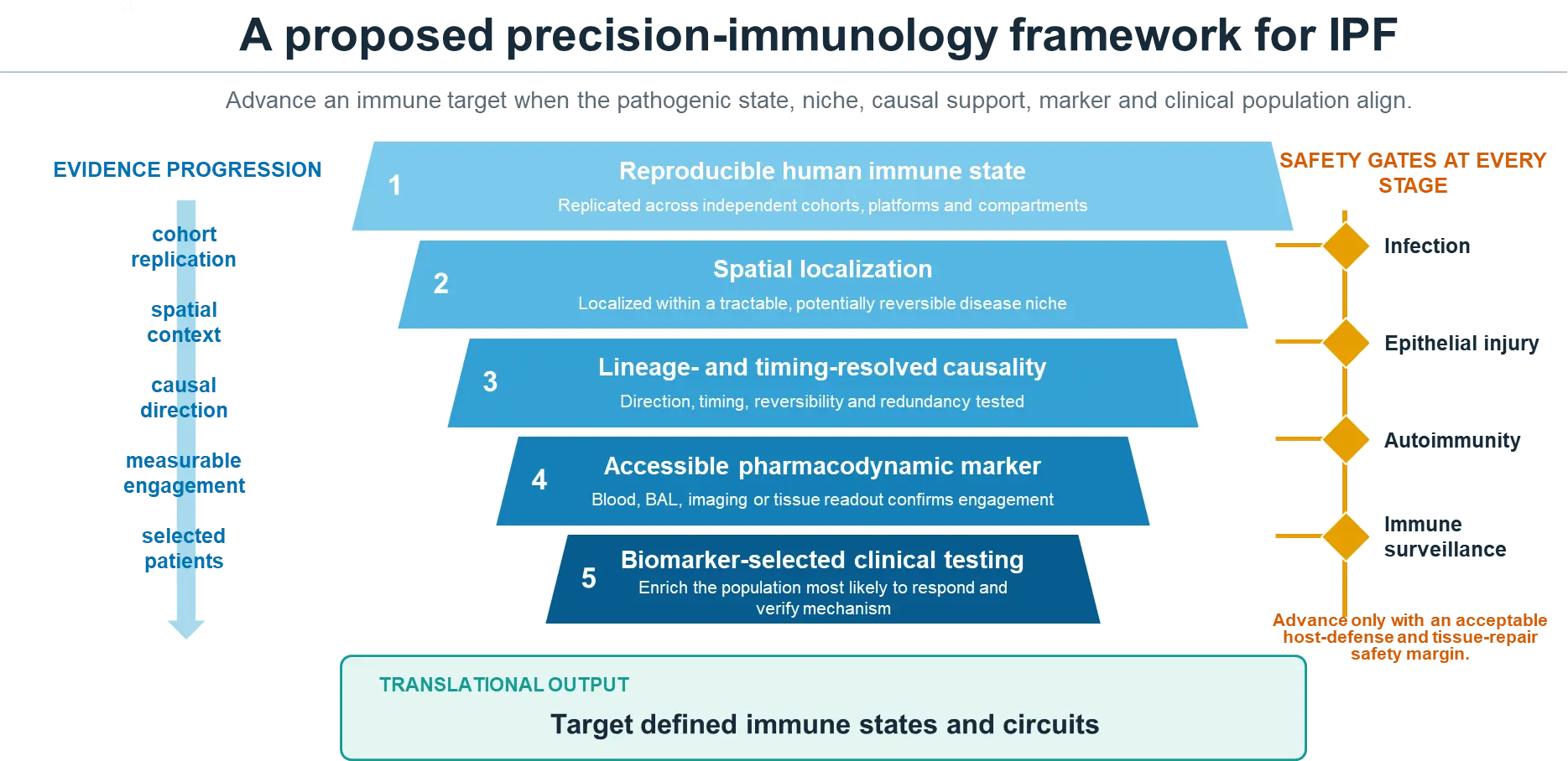
Figure 3. A proposed precision-immunology framework for IPF. The framework prioritizes immune targets supported by reproducible human cell states, spatial localization, lineage- and timing-resolved causal evidence, accessible pharmacodynamic markers, and biomarker-selected clinical testing. Infection, epithelial injury and repair, autoimmunity and immune-surveillance risks should be assessed at every stage.
-
Which cell state and function are being targeted? Define lineage, state program, and required effector instead of naming a broad cell class.
-
Where does the mechanism operate? Demonstrate the state in an anatomically relevant and drug-accessible lung niche.
-
When does the state become harmful? Test treatment after fibrosis is established and define the intended clinical stage.
-
Which patients carry the circuit? Prespecify an enrichment biomarker, such as a macrophage-state signature, plasma-cell/autoantibody endotype, neutrophil/NET phenotype, or senescent-fibroblast/NK-checkpoint signal.
-
Was the target engaged in the lung? Link exposure to BAL, tissue, imaging, or a validated proximal pharmacodynamic measurement in the relevant compartment.
-
Which protective functions may be lost? Measure infection, epithelial injury, autoimmunity, immune surveillance, and acute exacerbation as mechanism-based safety outcomes.
Combination therapy is likely, but each component needs a defined role. An antifibrotic can reduce stromal amplification while a state-selective immune therapy removes a sustaining signal. A regenerative intervention can fail if a hostile macrophage or matrix niche persists, whereas macrophage reprogramming may have little effect after alveolar architecture is irreversibly destroyed. Sequential or stage-adapted therapy could therefore be more effective than simultaneous broad blockade, and the order ought to be tested experimentally.
12. Outstanding Questions and a Research Roadmap
-
Can human macrophage origin be resolved directly? Somatic mutations, mitochondrial variants, DNA methylation, chromatin accessibility, transplant chimerism, and longitudinal sampling could distinguish recruited from resident lineages without assuming mouse marker equivalence [106,107,108].
-
Which immune state is a driver and which is an adaptation? Perturbation after a state is established, especially in a human multicellular system, is more informative than preventing its initial appearance during acute model injury.
-
What defines a reversible niche? Regional imaging and spatial biomarkers should separate active remodeling fronts from dense scar. A therapeutic response likely needs to be measured regionally because whole-lung averages dilute local biological change.
-
Which antigens organize adaptive immunity? Immunopeptidomics has begun to identify fibrosis associated HLA class I ligands and candidate therapeutic epitopes [72], but it has not established which antigens are spontaneously recognized by clonally expanded T or B cells in IPF. Paired T-cell and B-cell receptor sequencing, HLA typing, antigen discovery, and spatial mapping are required to determine whether clonal responses recognize self, microbe, or injury-associated antigens.
-
Can blood predict lung target activity? Monocyte, chemokine, autoantibody, and T-cell signatures require paired lung validation and prospective testing as predictive biomarkers, not only as prognostic markers.
-
How could age and comorbidity shape experimental models? Older animals, repetitive epithelial injury, human precision-cut lung slices, and systems incorporating infection or aspiration are more likely than prophylactic bleomycin in young mice to expose therapeutic liabilities.
-
Can immunity be reprogrammed instead of depleted? Restoring efferocytosis, metabolic fitness, IL-10-mediated resolution, or senescent-cell surveillance may preserve host defense, but each proposed benefit requires direct functional testing.
-
How do approved antifibrotics alter immune niches? Nintedanib, pirfenidone, and nerandomilast act across structural and immune pathways [105,109,110]. Background therapy may change state abundance, target dependence, pharmacodynamics, and combination safety and should be incorporated into experimental and clinical sampling.
Several reporting standards would make these questions answerable across studies. Human reports should specify tissue region, disease stage, treatment, acute-exacerbation status, smoking history, infection assessment, and sample-processing effects. Experimental studies should report intervention timing relative to injury, verify cell selectivity, include both sexes and aged animals where feasible, and measure epithelial repair and host defense together with collagen. Ligand-receptor and trajectory analyses should be labeled as predictions and followed by orthogonal validation. Negative results are especially valuable when they define a failed stage, compartment, or endotype.
13. Conclusions
The immune system in IPF is neither the sole engine of disease nor an incidental bystander. It consists of spatially and temporally organized cell states embedded within failed alveolar regeneration and persistent stromal remodeling. The most coherent evidence implicates recruited and durable macrophage states that couple lipid-lysosomal adaptation and survival to epithelial and fibroblast signaling. Neutrophils, dendritic cells, mast cells, innate lymphoid cells, T cells, NK cells, B cells, and plasma cells contribute with greater heterogeneity and, in several settings, opposite effects during injury and repair.
This accumulated evidence indicates both the harm of broad immunosuppression and the continuing importance of immune biology. The therapeutic unit should be a defined pathogenic state or intercellular circuit that is present in a measurable patient subgroup, operates in a reversible lung region, and can be interrupted without sacrificing antimicrobial defense or tissue repair. Human spatial evidence, lineage- and timing-resolved perturbation, lung target-engagement measurements, and biomarker-integrated trials now provide a practical path to test that model.
Statement of the Use of Generative AI and AI-Assisted Technologies in the Writing Process
During preparation of this manuscript, the authors used ChatGPT to assist with literature searching, language wording, and illustration designing. The authors take full responsibility for the accuracy and integrity of the final manuscript.
Author Contributions
Conceptualization, G.L.; writing and editing, K.-Y.T. and G.L.
Ethics Statement
Not applicable.
Informed Consent Statement
Not applicable.
Data Availability Statement
Not applicable.
Funding
NIH R01HL183230, R01AI170913 and R42HL170898.
Declaration of Competing Interest
The authors declare that they have no known competing financial interests or personal relationships that could have appeared to influence the work reported in this paper.
References
- Raghu G, Remy-Jardin M, Richeldi L, Thomson CC, Inoue Y, Johkoh T, et al. Idiopathic Pulmonary Fibrosis (an Update) and Progressive Pulmonary Fibrosis in Adults: An Official ATS/ERS/JRS/ALAT Clinical Practice Guideline. Am. J. Respir. Crit. Care Med. 2022, 205, e18–e47. DOI:10.1164/rccm.202202-0399ST [Google Scholar]
- Richeldi L, Azuma A, Cottin V, Kreuter M, Maher TM, Martinez FJ, et al. Nerandomilast in Patients with Idiopathic Pulmonary Fibrosis. N. Engl. J. Med. 2025, 392, 2193–2202. DOI:10.1056/NEJMoa2414108 [Google Scholar]
- U.S. Food and Drug Administration. FDA Approves Drug to Treat Idiopathic Pulmonary Fibrosis. Available online: https://www.fda.gov/drugs/news-events-human-drugs/fda-approves-drug-treat-idiopathic-pulmonary-fibrosis (accessed on 11 August 2026).
- The Idiopathic Pulmonary Fibrosis Clinical Research Network, Raghu G, Anstrom KJ, King TE, Jr., Lasky JA, Martinez FJ. Prednisone, Azathioprine, and N-Acetylcysteine for Pulmonary Fibrosis. N. Engl. J. Med. 2012, 366, 1968–1977. DOI:10.1056/NEJMoa1113354 [Google Scholar]
- Adams TS, Schupp JC, Poli S, Ayaub EA, Neumark N, Ahangari F, et al. Single-cell RNA-seq reveals ectopic and aberrant lung-resident cell populations in idiopathic pulmonary fibrosis. Sci. Adv. 2020, 6, eaba1983. DOI:10.1126/sciadv.aba1983 [Google Scholar]
- Habermann AC, Gutierrez AJ, Bui LT, Yahn SL, Winters NI, Calvi CL, et al. Single-cell RNA sequencing reveals profibrotic roles of distinct epithelial and mesenchymal lineages in pulmonary fibrosis. Sci. Adv. 2020, 6, eaba1972. DOI:10.1126/sciadv.aba1972 [Google Scholar]
- Reyfman PA, Walter JM, Joshi N, Anekalla KR, McQuattie-Pimentel AC, Chiu S, et al. Single-Cell Transcriptomic Analysis of Human Lung Provides Insights into the Pathobiology of Pulmonary Fibrosis. Am. J. Respir. Crit. Care Med. 2019, 199, 1517–1536. DOI:10.1164/rccm.201712-2410OC [Google Scholar]
- Franzén L, Olsson Lindvall M, Hühn M, Ptasinski V, Setyo L, Keith BP, et al. Mapping spatially resolved transcriptomes in human and mouse pulmonary fibrosis. Nat. Genet. 2024, 56, 1725–1736. DOI:10.1038/s41588-024-01819-2 [Google Scholar]
- Mayr CH, Santacruz D, Jarosch S, Bleck M, Dalton J, McNabola A, et al. Spatial transcriptomic characterization of pathologic niches in IPF. Sci. Adv. 2024, 10, eadl5473. DOI:10.1126/sciadv.adl5473 [Google Scholar]
- Vannan A, Lyu R, Williams AL, Negretti NM, Mee ED, Hirsh J, et al. Spatial transcriptomics identifies molecular niche dysregulation associated with distal lung remodeling in pulmonary fibrosis. Nat. Genet. 2025, 57, 647–658. DOI:10.1038/s41588-025-02080-x [Google Scholar]
- Weeratunga P, Hunter B, Sergeant M, Bull J, Clelland C, Denney L, et al. Temporo-spatial cellular atlas of the regenerating alveolar niche in idiopathic pulmonary fibrosis. Nat. Commun. 2025, 16, 7150. DOI:10.1038/s41467-025-61880-1 [Google Scholar]
- Liu H, Cui H, Liu G. The Intersection between Immune System and Idiopathic Pulmonary Fibrosis—A Concise Review. Fibrosis 2025, 3, 10004. DOI:10.70322/fibrosis.2025.10004 [Google Scholar]
- Shenderov K, Collins SL, Powell JD, Horton MR. Immune dysregulation as a driver of idiopathic pulmonary fibrosis. J. Clin. Investig. 2021, 131, e143226. DOI:10.1172/JCI143226 [Google Scholar]
- Choi J, Park JE, Tsagkogeorga G, Yanagita M, Koo BK, Han N, et al. Inflammatory Signals Induce AT2 Cell-Derived Damage-Associated Transient Progenitors that Mediate Alveolar Regeneration. Cell Stem Cell 2020, 27, 366–382.e7. DOI:10.1016/j.stem.2020.06.020 [Google Scholar]
- Jiang P, Gil de Rubio R, Hrycaj SM, Gurczynski SJ, Riemondy KA, Moore BB, et al. Ineffectual Type 2–to–Type 1 Alveolar Epithelial Cell Differentiation in Idiopathic Pulmonary Fibrosis: Persistence of the KRT8hi Transitional State. Am. J. Respir. Crit. Care Med. 2020, 201, 1443–1447. DOI:10.1164/rccm.201909-1726LE [Google Scholar]
- Scott MKD, Quinn K, Li Q, Carroll R, Warsinske H, Vallania F, et al. Increased monocyte count as a cellular biomarker for poor outcomes in fibrotic diseases: A retrospective, multicentre cohort study. Lancet Respir. Med. 2019, 7, 497–508. DOI:10.1016/S2213-2600(18)30508-3 [Google Scholar]
- Strunz M, Simon LM, Ansari M, Kathiriya JJ, Angelidis I, Mayr CH, et al. Alveolar regeneration through a Krt8+ transitional stem cell state that persists in human lung fibrosis. Nat. Commun. 2020, 11, 3559. DOI:10.1038/s41467-020-17358-3 [Google Scholar]
- Aran D, Looney AP, Liu L, Wu E, Fong V, Hsu A, et al. Reference-based analysis of lung single-cell sequencing reveals a transitional profibrotic macrophage. Nat. Immunol. 2019, 20, 163–172. DOI:10.1038/s41590-018-0276-y [Google Scholar]
- Fabre T, Barron AMS, Christensen SM, Asano S, Bound K, Lech MP, et al. Identification of a broadly fibrogenic macrophage subset induced by type 3 inflammation. Sci. Immunol. 2023, 8, eadd8945. DOI:10.1126/sciimmunol.add8945 [Google Scholar]
- Jaitin DA, Adlung L, Thaiss CA, Weiner A, Li B, Descamps H, et al. Lipid-Associated Macrophages Control Metabolic Homeostasis in a Trem2-Dependent Manner. Cell 2019, 178, 686–698.e14. DOI:10.1016/j.cell.2019.05.054 [Google Scholar]
- Joshi N, Watanabe S, Verma R, Jablonski RP, Chen CI, Cheresh P, et al. A spatially restricted fibrotic niche in pulmonary fibrosis is sustained by M-CSF/M-CSFR signalling in monocyte-derived alveolar macrophages. Eur. Respir. J. 2020, 55, 1900646. DOI:10.1183/13993003.00646-2019 [Google Scholar]
- Morse C, Tabib T, Sembrat J, Buschur KL, Bittar HT, Valenzi E, et al. Proliferating SPP1/MERTK-expressing macrophages in idiopathic pulmonary fibrosis. Eur. Respir. J. 2019, 54, 1802441. DOI:10.1183/13993003.02441-2018 [Google Scholar]
- Ramachandran P, Dobie R, Wilson-Kanamori JR, Dora EF, Henderson BEP, Luu NT, et al. Resolving the fibrotic niche of human liver cirrhosis at single-cell level. Nature 2019, 575, 512–518. DOI:10.1038/s41586-019-1631-3 [Google Scholar]
- Cui H, Banerjee S, Xie N, Hussain M, Jaiswal A, Liu H, et al. TREM2 promotes lung fibrosis via controlling alveolar macrophage survival and pro-fibrotic activity. Nat. Commun. 2025, 16, 1761. DOI:10.1038/s41467-025-57024-0 [Google Scholar]
- Gasse P, Riteau N, Vacher R, Michel ML, Fautrel A, di Padova F, et al. IL-1 and IL-23 Mediate Early IL-17A Production in Pulmonary Inflammation Leading to Late Fibrosis. PLoS ONE 2011, 6, e23185. DOI:10.1371/journal.pone.0023185 [Google Scholar]
- Wilson MS, Madala SK, Ramalingam TR, Gochuico BR, Rosas IO, Cheever AW, et al. Bleomycin and IL-1β–mediated pulmonary fibrosis is IL-17A dependent. J. Exp. Med. 2010, 207, 535–552. DOI:10.1084/jem.20092121 [Google Scholar]
- Hams E, Armstrong ME, Barlow JL, Saunders SP, Schwartz C, Cooke G, et al. IL-25 and type 2 innate lymphoid cells induce pulmonary fibrosis. Proc. Natl. Acad. Sci. USA 2014, 111, 367–372. DOI:10.1073/pnas.1315854111 [Google Scholar]
- Zhang J, Qiu J, Zhou W, Cao J, Hu X, Mi W, et al. Neuropilin-1 mediates lung tissue-specific control of ILC2 function in type 2 immunity. Nat. Immunol. 2022, 23, 237–250. DOI:10.1038/s41590-021-01097-8 [Google Scholar]
- Guilliams M, De Kleer I, Henri S, Post S, Vanhoutte L, De Prijck S, et al. Alveolar macrophages develop from fetal monocytes that differentiate into long-lived cells in the first week of life via GM-CSF. J. Exp. Med. 2013, 210, 1977–1992. DOI:10.1084/jem.20131199 [Google Scholar]
- Hashimoto D, Chow A, Noizat C, Teo P, Beasley MB, Leboeuf M, et al. Tissue-Resident Macrophages Self-Maintain Locally throughout Adult Life with Minimal Contribution from Circulating Monocytes. Immunity 2013, 38, 792–804. DOI:10.1016/j.immuni.2013.04.004 [Google Scholar]
- Hussell T, Bell TJ. Alveolar macrophages: Plasticity in a tissue-specific context. Nat. Rev. Immunol. 2014, 14, 81–93. DOI:10.1038/nri3600 [Google Scholar]
- Aegerter H, Lambrecht BN, Jakubzick CV. Biology of lung macrophages in health and disease. Immunity 2022, 55, 1564–1580. DOI:10.1016/j.immuni.2022.08.010 [Google Scholar]
- Gibbons MA, MacKinnon AC, Ramachandran P, Dhaliwal K, Duffin R, Phythian-Adams AT, et al. Ly6Chi Monocytes Direct Alternatively Activated Profibrotic Macrophage Regulation of Lung Fibrosis. Am. J. Respir. Crit. Care Med. 2011, 184, 569–581. DOI:10.1164/rccm.201010-1719OC [Google Scholar]
- Misharin AV, Morales-Nebreda L, Reyfman PA, Cuda CM, Walter JM, McQuattie-Pimentel AC, et al. Monocyte-derived alveolar macrophages drive lung fibrosis and persist in the lung over the life span. J. Exp. Med. 2017, 214, 2387–2404. DOI:10.1084/jem.20162152 [Google Scholar]
- Wei Y, Guo H, Yang X, Tang XX. Macrophage‐to‐Myofibroblast Transdifferentiation Contributes to Pulmonary Fibrosis via the MERTK‐SPP1‐SRC‐TKS5 Signaling Axis. Adv. Sci. 2026, 13, e75620. DOI:10.1002/advs.75620 [Google Scholar]
- MacKinnon AC, Gibbons MA, Farnworth SL, Leffler H, Nilsson UJ, Delaine T, et al. Regulation of Transforming Growth Factor-β1–driven Lung Fibrosis by Galectin-3. Am. J. Respir. Crit. Care Med. 2012, 185, 537–546. DOI:10.1164/rccm.201106-0965OC [Google Scholar]
- Hirani N, MacKinnon AC, Nicol L, Ford P, Schambye H, Pedersen A, et al. Target inhibition of galectin-3 by inhaled TD139 in patients with idiopathic pulmonary fibrosis. Eur. Respir. J. 2021, 57, 2002559. DOI:10.1183/13993003.02559-2020 [Google Scholar]
- European Union Clinical Trials Register. GALACTIC-1: A Randomized Phase 2b Study of Inhaled GB0139 in Idiopathic Pulmonary Fibrosis. Available online: https://www.clinicaltrialsregister.eu/ctr-search/trial/2018-002664-73/results (accessed on 11 August 2026).
- Prasse A, Pechkovsky DV, Toews GB, Jungraithmayr W, Kollert F, Goldmann T, et al. A Vicious Circle of Alveolar Macrophages and Fibroblasts Perpetuates Pulmonary Fibrosis via CCL18. Am. J. Respir. Crit. Care Med. 2006, 173, 781–792. DOI:10.1164/rccm.200509-1518OC [Google Scholar]
- Moeller A, Gilpin SE, Ask K, Cox G, Cook D, Gauldie J, et al. Circulating Fibrocytes Are an Indicator of Poor Prognosis in Idiopathic Pulmonary Fibrosis. Am. J. Respir. Crit. Care Med. 2009, 179, 588–594. DOI:10.1164/rccm.200810-1534OC [Google Scholar]
- Fernandez IE, Greiffo FR, Frankenberger M, Bandres J, Heinzelmann K, Neurohr C, et al. Peripheral blood myeloid-derived suppressor cells reflect disease status in idiopathic pulmonary fibrosis. Eur. Respir. J. 2016, 48, 1171–1183. DOI:10.1183/13993003.01826-2015 [Google Scholar]
- Kinder BW, Brown KK, Schwarz MI, Ix JH, Kervitsky A, King TE. Baseline BAL Neutrophilia Predicts Early Mortality in Idiopathic Pulmonary Fibrosis. Chest 2008, 133, 226–232. DOI:10.1378/chest.07-1948 [Google Scholar]
- Chua F, Dunsmore SE, Clingen PH, Mutsaers SE, Shapiro SD, Segal AW, et al. Mice Lacking Neutrophil Elastase Are Resistant to Bleomycin-Induced Pulmonary Fibrosis. Am. J. Pathol. 2007, 170, 65–74. DOI:10.2353/ajpath.2007.060352 [Google Scholar]
- Gregory AD, Kliment CR, Metz HE, Kim KH, Kargl J, Agostini BA, et al. Neutrophil elastase promotes myofibroblast differentiation in lung fibrosis. J. Leukoc. Biol. 2015, 98, 143–152. DOI:10.1189/jlb.3HI1014-493R [Google Scholar]
- Chrysanthopoulou A, Mitroulis I, Apostolidou E, Arelaki S, Mikroulis D, Konstantinidis T, et al. Neutrophil extracellular traps promote differentiation and function of fibroblasts. J. Pathol. 2014, 233, 294–307. DOI:10.1002/path.4359 [Google Scholar]
- Khawaja AA, Chong DLW, Sahota J, Mikolasch TA, Pericleous C, Ripoll VM, et al. Identification of a Novel HIF-1α-αMβ2 Integrin-NET Axis in Fibrotic Interstitial Lung Disease. Front. Immunol. 2020, 11, 2190. DOI:10.3389/fimmu.2020.02190 [Google Scholar]
- Marchal-Sommé J, Uzunhan Y, Marchand-Adam S, Kambouchner M, Valeyre D, Crestani B, et al. Dendritic Cells Accumulate in Human Fibrotic Interstitial Lung Disease. Am. J. Respir. Crit. Care Med. 2007, 176, 1007–1014. DOI:10.1164/rccm.200609-1347OC [Google Scholar]
- Bantsimba-Malanda C, Marchal-Sommé J, Goven D, Freynet O, Michel L, Crestani B, et al. A Role for Dendritic Cells in Bleomycin-induced Pulmonary Fibrosis in Mice? Am. J. Respir. Crit. Care Med. 2010, 182, 385–395. DOI:10.1164/rccm.200907-1164OC [Google Scholar]
- Odell ID, Steach H, Gauld SB, Reinke-Breen L, Karman J, Carr TL, et al. Epiregulin is a dendritic cell–derived EGFR ligand that maintains skin and lung fibrosis. Sci. Immunol. 2022, 7, eabq6691. DOI:10.1126/sciimmunol.abq6691 [Google Scholar]
- Shimbori C, Upagupta C, Bellaye PS, Ayaub EA, Sato S, Yanagihara T, et al. Mechanical stress-induced mast cell degranulation activates TGF-β1 signalling pathway in pulmonary fibrosis. Thorax 2019, 74, 455–465. DOI:10.1136/thoraxjnl-2018-211516 [Google Scholar]
- Wygrecka M, Dahal BK, Kosanovic D, Petersen F, Taborski B, von Gerlach S, et al. Mast Cells and Fibroblasts Work in Concert to Aggravate Pulmonary Fibrosis. Am. J. Pathol. 2013, 182, 2094–2108. DOI:10.1016/j.ajpath.2013.02.013 [Google Scholar]
- Overed-Sayer C, Miranda E, Dunmore R, Liarte Marin E, Beloki L, Rassl D, et al. Inhibition of mast cells: A novel mechanism by which nintedanib may elicit anti-fibrotic effects. Thorax 2020, 75, 754–763. DOI:10.1136/thoraxjnl-2019-214000 [Google Scholar]
- Morita H, Kubo T, Rückert B, Ravindran A, Soyka MB, Rinaldi AO, et al. Induction of human regulatory innate lymphoid cells from group 2 innate lymphoid cells by retinoic acid. J. Allergy Clin. Immunol. 2019, 143, 2190–2201.e9. DOI:10.1016/j.jaci.2018.12.1018 [Google Scholar]
- Seehus CR, Kadavallore A, Torre BDL, Yeckes AR, Wang Y, Tang J, et al. Alternative activation generates IL-10 producing type 2 innate lymphoid cells. Nat. Commun. 2017, 8, 1900. DOI:10.1038/s41467-017-02023-z [Google Scholar]
- Daniil Z, Kitsanta P, Kapotsis G, Mathioudaki M, Kollintza A, Karatza M, et al. CD8+ T lymphocytes in lung tissue from patients with idiopathic pulmonary fibrosis. Respir. Res. 2005, 6, 81. DOI:10.1186/1465-9921-6-81 [Google Scholar]
- Feng X, Yu F, He XL, Cheng PP, Niu Q, Zhao LQ, et al. CD8+ tissue-resident memory T cells are essential in bleomycin-induced pulmonary fibrosis. Am. J. Physiol. Cell Physiol. 2024, 327, C1178–C1191. DOI:10.1152/ajpcell.00368.2024 [Google Scholar]
- Gilani SR, Vuga LJ, Lindell KO, Gibson KF, Xue J, Kaminski N, et al. CD28 Down-Regulation on Circulating CD4 T-Cells Is Associated with Poor Prognoses of Patients with Idiopathic Pulmonary Fibrosis. PLoS ONE 2010, 5, e8959. DOI:10.1371/journal.pone.0008959 [Google Scholar]
- Habiel DM, Espindola MS, Kitson C, Azzara AV, Coelho AL, Stripp B, et al. Characterization of CD28null T cells in idiopathic pulmonary fibrosis. Mucosal Immunol. 2019, 12, 212–222. DOI:10.1038/s41385-018-0082-8 [Google Scholar]
- Kotsianidis I, Nakou E, Bouchliou I, Tzouvelekis A, Spanoudakis E, Steiropoulos P, et al. Global Impairment of CD4+CD25+FOXP3+ Regulatory T Cells in Idiopathic Pulmonary Fibrosis. Am. J. Respir. Crit. Care Med. 2009, 179, 1121–1130. DOI:10.1164/rccm.200812-1936OC [Google Scholar]
- Todd NW, Scheraga RG, Galvin JR, Iacono AT, Britt EJ, Luzina IG, et al. Lymphocyte aggregates persist and accumulate in the lungs of patients with idiopathic pulmonary fibrosis. J. Inflamm. Res. 2013, 6, 63–70. DOI:10.2147/JIR.S40673 [Google Scholar]
- Asai Y, Chiba H, Nishikiori H, Kamekura R, Yabe H, Kondo S, et al. Aberrant populations of circulating T follicular helper cells and regulatory B cells underlying idiopathic pulmonary fibrosis. Respir. Res. 2019, 20, 244. DOI:10.1186/s12931-019-1216-6 [Google Scholar]
- Heukels P, van Hulst JAC, van Nimwegen M, Boorsma CE, Melgert BN, von der Thusen JH, et al. Enhanced Bruton’s tyrosine kinase in B-cells and autoreactive IgA in patients with idiopathic pulmonary fibrosis. Respir. Res. 2019, 20, 232. DOI:10.1186/s12931-019-1195-7 [Google Scholar]
- Neys SFH, Heukels P, van Hulst JAC, Rip J, Wijsenbeek MS, Hendriks RW, et al. Aberrant B Cell Receptor Signaling in Naïve B Cells from Patients with Idiopathic Pulmonary Fibrosis. Cells 2021, 10, 1321. DOI:10.3390/cells10061321 [Google Scholar]
- Vuga LJ, Tedrow JR, Pandit KV, Tan J, Kass DJ, Xue J, et al. C-X-C Motif Chemokine 13 (CXCL13) is a Prognostic Biomarker of Idiopathic Pulmonary Fibrosis. Am. J. Respir. Crit. Care Med. 2014, 189, 966–974. DOI:10.1164/rccm.201309-1592OC [Google Scholar]
- Xue J, Kass DJ, Bon J, Vuga L, Tan J, Csizmadia E, et al. Plasma B Lymphocyte Stimulator and B Cell Differentiation in Idiopathic Pulmonary Fibrosis Patients. J. Immunol. 2013, 191, 2089–2095. DOI:10.4049/jimmunol.1203476 [Google Scholar]
- King TE, Albera C, Bradford WZ, Costabel U, Hormel P, Lancaster L, et al. Effect of interferon gamma-1b on survival in patients with idiopathic pulmonary fibrosis (INSPIRE): A multicentre, randomised, placebo-controlled trial. Lancet 2009, 374, 222–228. DOI:10.1016/S0140-6736(09)60551-1 [Google Scholar]
- Maher TM, Costabel U, Glassberg MK, Kondoh Y, Ogura T, Scholand MB, et al. Phase 2 trial to assess lebrikizumab in patients with idiopathic pulmonary fibrosis. Eur. Respir. J. 2021, 57, 1902442. DOI:10.1183/13993003.02442-2019 [Google Scholar]
- Raghu G, Richeldi L, Crestani B, Wung P, Bejuit R, Esperet C, et al. SAR156597 in idiopathic pulmonary fibrosis: A phase 2 placebo-controlled study (DRI11772). Eur. Respir. J. 2018, 52, 1801130. DOI:10.1183/13993003.01130-2018 [Google Scholar]
- Birjandi SZ, Palchevskiy V, Xue YY, Nunez S, Kern R, Weigt SS, et al. CD4+CD25hiFoxp3+ Cells Exacerbate Bleomycin-Induced Pulmonary Fibrosis. Am. J. Pathol. 2016, 186, 2008–2020. DOI:10.1016/j.ajpath.2016.03.020 [Google Scholar]
- Okamoto M, Kuratani A, Okuzaki D, Kamiyama N, Kobayashi T, Sasai M, et al. Tff1-expressing Tregs in lung prevent exacerbation of Bleomycin-induced pulmonary fibrosis. Front. Immunol. 2024, 15, 1440918. DOI:10.3389/fimmu.2024.1440918 [Google Scholar]
- Farhat A, Radhouani M, Deckert F, Zahalka S, Pimenov L, Fokina A, et al. An aging bone marrow exacerbates lung fibrosis by fueling profibrotic macrophage persistence. Sci. Immunol. 2025, 10, eadk5041. DOI:10.1126/sciimmunol.adk5041 [Google Scholar]
- Bai Z, Lan T, Hong W, Que H, Zhu M, Xiao X, et al. Immunopeptidome profiling in pulmonary fibrosis provides a platform for identifying therapeutic targets. Nat. Immunol. 2026, 27, 923–936. DOI:10.1038/s41590-026-02501-x [Google Scholar]
- Yadav S, Anbalagan M, Khatun S, Prabhakaran D, Matsunaga Y, Manges J, et al. Reactivation of CTLA4-expressing T cells accelerates resolution of lung fibrosis in a humanized mouse model. J. Clin. Investig. 2025, 135, e181775. DOI:10.1172/JCI181775 [Google Scholar]
- Segawa S, Goto D, Iizuka A, Kaneko S, Yokosawa M, Kondo Y, et al. The regulatory role of interferon-γ producing gamma delta T cells via the suppression of T helper 17 cell activity in bleomycin-induced pulmonary fibrosis. Clin. Exp. Immunol. 2016, 185, 348–360. DOI:10.1111/cei.12802 [Google Scholar]
- Monnier J, Zabel BA. Anti-Asialo GM1 NK Cell Depleting Antibody Does Not Alter the Development of Bleomycin Induced Pulmonary Fibrosis. PLoS ONE 2014, 9, e99350. DOI:10.1371/journal.pone.0099350 [Google Scholar]
- Merkt W, Rodon L, Deicher FS, Freitag M, Claus M, Lister R, et al. Natural killer cell immunotherapy reverses lung fibrosis by eliminating senescent fibroblasts. Sci. Transl. Med. 2026, 18, eadq5442. DOI:10.1126/scitranslmed.adq5442 [Google Scholar]
- Kahloon RA, Xue J, Bhargava A, Csizmadia E, Otterbein L, Kass DJ, et al. Patients with Idiopathic Pulmonary Fibrosis with Antibodies to Heat Shock Protein 70 Have Poor Prognoses. Am. J. Respir. Crit. Care Med. 2013, 187, 768–775. DOI:10.1164/rccm.201203-0506OC [Google Scholar]
- Koether K, Besnard V, Sandig H, Carruthers A, Miranda E, Grootenboer-Mignot S, et al. Autoantibodies are associated with disease progression in idiopathic pulmonary fibrosis. Eur. Respir. J. 2023, 61, 2102381. DOI:10.1183/13993003.02381-2021 [Google Scholar]
- Li FJ, Surolia R, Li H, Wang Z, Kulkarni T, Liu G, et al. Autoimmunity to Vimentin Is Associated with Outcomes of Patients with Idiopathic Pulmonary Fibrosis. J. Immunol. 2017, 199, 1596–1605. DOI:10.4049/jimmunol.1700473 [Google Scholar]
- Prêle CM, Miles T, Pearce DR, O’Donoghue RJ, Grainge C, Barrett L, et al. Plasma cell but not CD20-mediated B-cell depletion protects from bleomycin-induced lung fibrosis. Eur. Respir. J. 2022, 60, 2101469. DOI:10.1183/13993003.01469-2021 [Google Scholar]
- Donahoe M, Valentine VG, Chien N, Gibson KF, Raval JS, Saul M, et al. Autoantibody-Targeted Treatments for Acute Exacerbations of Idiopathic Pulmonary Fibrosis. PLoS ONE 2015, 10, e0127771. DOI:10.1371/journal.pone.0127771 [Google Scholar]
- Tsukui T, Wolters PJ, Sheppard D. Alveolar fibroblast lineage orchestrates lung inflammation and fibrosis. Nature 2024, 631, 627–634. DOI:10.1038/s41586-024-07660-1 [Google Scholar]
- Wang F, Ting C, Riemondy KA, Douglas M, Foster K, Patel N, et al. Regulation of epithelial transitional states in murine and human pulmonary fibrosis. J. Clin. Investig. 2023, 133, e165612. DOI:10.1172/JCI165612 [Google Scholar]
- Wu H, Yu Y, Huang H, Hu Y, Fu S, Wang Z, et al. Progressive Pulmonary Fibrosis Is Caused by Elevated Mechanical Tension on Alveolar Stem Cells. Cell 2020, 180, 107–121.e17. DOI:10.1016/j.cell.2019.11.027 [Google Scholar]
- Schupp JC, Adams TS, Cosme C, Raredon MSB, Yuan Y, Omote N, et al. Integrated Single-Cell Atlas of Endothelial Cells of the Human Lung. Circulation 2021, 144, 286–302. DOI:10.1161/CIRCULATIONAHA.120.052318 [Google Scholar]
- Bian F, Lan YW, Zhao S, Deng Z, Shukla S, Acharya A, et al. Lung endothelial cells regulate pulmonary fibrosis through FOXF1/R-Ras signaling. Nat. Commun. 2023, 14, 2560. DOI:10.1038/s41467-023-38177-2 [Google Scholar]
- El-Chemaly S, Malide D, Zudaire E, Ikeda Y, Weinberg BA, Pacheco-Rodriguez G, et al. Abnormal lymphangiogenesis in idiopathic pulmonary fibrosis with insights into cellular and molecular mechanisms. Proc. Natl. Acad. Sci. USA 2009, 106, 3958–3963. DOI:10.1073/pnas.0813368106 [Google Scholar]
- Baluk P, Naikawadi RP, Kim S, Rodriguez F, Choi D, Hong YK, et al. Lymphatic Proliferation Ameliorates Pulmonary Fibrosis after Lung Injury. Am. J. Pathol. 2020, 190, 2355–2375. DOI:10.1016/j.ajpath.2020.08.018 [Google Scholar]
- Dimitrov D, Türei D, Garrido-Rodriguez M, Burmedi PL, Nagai JS, Boys C, et al. Comparison of methods and resources for cell-cell communication inference from single-cell RNA-Seq data. Nat. Commun. 2022, 13, 3224. DOI:10.1038/s41467-022-30755-0 [Google Scholar]
- McQuattie-Pimentel AC, Ren Z, Joshi N, Watanabe S, Stoeger T, Chi M, et al. The lung microenvironment shapes a dysfunctional response of alveolar macrophages in aging. J. Clin. Investig. 2021, 131, e140299. DOI:10.1172/JCI140299 [Google Scholar]
- Warheit-Niemi HI, Edwards SJ, SenGupta S, Parent CA, Zhou X, O’Dwyer DN, et al. Fibrotic lung disease inhibits immune responses to staphylococcal pneumonia via impaired neutrophil and macrophage function. JCI Insight 2022, 7, e152690. DOI:10.1172/jci.insight.152690 [Google Scholar]
- Morimoto K, Janssen WJ, Terada M. Defective efferocytosis by alveolar macrophages in IPF patients. Respir. Med. 2012, 106, 1800–1803. DOI:10.1016/j.rmed.2012.08.020 [Google Scholar]
- Herazo-Maya JD, Noth I, Duncan SR, Kim S, Ma SF, Tseng GC, et al. Peripheral Blood Mononuclear Cell Gene Expression Profiles Predict Poor Outcome in Idiopathic Pulmonary Fibrosis. Sci. Transl. Med. 2013, 5, 205ra136. DOI:10.1126/scitranslmed.3005964 [Google Scholar]
- Herazo-Maya JD, Sun J, Molyneaux PL, Li Q, Villalba JA, Tzouvelekis A, et al. Validation of a 52-gene risk profile for outcome prediction in patients with idiopathic pulmonary fibrosis: An international, multicentre, cohort study. Lancet Respir. Med. 2017, 5, 857–868. DOI:10.1016/S2213-2600(17)30349-1 [Google Scholar]
- Han MK, Zhou Y, Murray S, Tayob N, Noth I, Lama VN, et al. Lung microbiome and disease progression in idiopathic pulmonary fibrosis: An analysis of the COMET study. Lancet Respir. Med. 2014, 2, 548–556. DOI:10.1016/S2213-2600(14)70069-4 [Google Scholar]
- O’Dwyer DN, Ashley SL, Gurczynski SJ, Xia M, Wilke C, Falkowski NR, et al. Lung Microbiota Contribute to Pulmonary Inflammation and Disease Progression in Pulmonary Fibrosis. Am. J. Respir. Crit. Care Med. 2019, 199, 1127–1138. DOI:10.1164/rccm.201809-1650OC [Google Scholar]
- Yang D, Chen X, Wang J, Lou Q, Lou Y, Li L, et al. Dysregulated Lung Commensal Bacteria Drive Interleukin-17B Production to Promote Pulmonary Fibrosis through Their Outer Membrane Vesicles. Immunity 2019, 50, 692–706.e7. DOI:10.1016/j.immuni.2019.02.001 [Google Scholar]
- Martinez FJ, Yow E, Flaherty KR, Snyder LD, Durheim MT, Wisniewski SR, et al. Effect of Antimicrobial Therapy on Respiratory Hospitalization or Death in Adults With Idiopathic Pulmonary Fibrosis. JAMA 2021, 325, 1841. DOI:10.1001/jama.2021.4956 [Google Scholar]
- Le Hingrat Q, Ghanem M, Cazes A, Visseaux B, Collin G, Descamps D, et al. No association between human herpesvirus or herpesvirus saimiri and idiopathic pulmonary fibrosis. ERJ Open Res. 2020, 6, 00243-2020. DOI:10.1183/23120541.00243-2020 [Google Scholar]
- Tang YW, Johnson JE, Browning PJ, Cruz-Gervis RA, Davis A, Graham BS, et al. Herpesvirus DNA Is Consistently Detected in Lungs of Patients with Idiopathic Pulmonary Fibrosis. J. Clin. Microbiol. 2003, 41, 2633–2640. DOI:10.1128/JCM.41.6.2633-2640.2003 [Google Scholar]
- Wootton SC, Kim DS, Kondoh Y, Chen E, Lee JS, Song JW, et al. Viral Infection in Acute Exacerbation of Idiopathic Pulmonary Fibrosis. Am. J. Respir. Crit. Care Med. 2011, 183, 1698–1702. DOI:10.1164/rccm.201010-1752OC [Google Scholar]
- Raghu G, Martinez FJ, Brown KK, Costabel U, Cottin V, Wells AU, et al. CC-chemokine ligand 2 inhibition in idiopathic pulmonary fibrosis: A phase 2 trial of carlumab. Eur. Respir. J. 2015, 46, 1740–1750. DOI:10.1183/13993003.01558-2014 [Google Scholar]
- Raghu G, van den Blink B, Hamblin MJ, Brown AW, Golden JA, Ho LA, et al. Effect of Recombinant Human Pentraxin 2 vs Placebo on Change in Forced Vital Capacity in Patients With Idiopathic Pulmonary Fibrosis. JAMA 2018, 319, 2299. DOI:10.1001/jama.2018.6129 [Google Scholar]
- Richeldi L, Schiffman C, Behr J, Inoue Y, Corte TJ, Cottin V, et al. Zinpentraxin Alfa for Idiopathic Pulmonary Fibrosis: The Randomized Phase III STARSCAPE Trial. Am. J. Respir. Crit. Care Med. 2024, 209, 1132–1140. DOI:10.1164/rccm.202401-0116OC [Google Scholar]
- Herrmann FE, Hesslinger C, Wollin L, Nickolaus P. BI 1015550 is a PDE4B Inhibitor and a Clinical Drug Candidate for the Oral Treatment of Idiopathic Pulmonary Fibrosis. Front. Pharmacol. 2022, 13, 838449. DOI:10.3389/fphar.2022.838449 [Google Scholar]
- Ludwig LS, Lareau CA, Ulirsch JC, Christian E, Muus C, Li LH, et al. Lineage Tracing in Humans Enabled by Mitochondrial Mutations and Single-Cell Genomics. Cell 2019, 176, 1325–1339.e22. DOI:10.1016/j.cell.2019.01.022 [Google Scholar]
- Byrne AJ, Powell JE, O’Sullivan BJ, Ogger PP, Hoffland A, Cook J, et al. Dynamics of human monocytes and airway macrophages during healthy aging and after transplant. J. Exp. Med. 2020, 217, e20191236. DOI:10.1084/jem.20191236 [Google Scholar]
- Ogger PP, Albers GJ, Hewitt RJ, O’Sullivan BJ, Powell JE, Calamita E, et al. Itaconate controls the severity of pulmonary fibrosis. Sci. Immunol. 2020, 5, eabc1884. DOI:10.1126/sciimmunol.abc1884 [Google Scholar]
- Inomata M, Kamio K, Azuma A, Matsuda K, Kokuho N, Miura Y, et al. Pirfenidone inhibits fibrocyte accumulation in the lungs in bleomycin-induced murine pulmonary fibrosis. Respir. Res. 2014, 15, 16. DOI:10.1186/1465-9921-15-16 [Google Scholar]
- Wollin L, Maillet I, Quesniaux V, Holweg A, Ryffel B. Antifibrotic and Anti-inflammatory Activity of the Tyrosine Kinase Inhibitor Nintedanib in Experimental Models of Lung Fibrosis. J. Pharmacol. Exp. Ther. 2014, 349, 209–220. DOI:10.1124/jpet.113.208223 [Google Scholar]