
An Anti-Aging Tissue-Made Cosme-Nutraceutical Product: Effectiveness and Safety by an In Vitro and In Vivo Study
An Anti-Aging Tissue-Made Cosme-Nutraceutical Product: Effectiveness and Safety by an In Vitro and In Vivo Study
Pierfrancesco Morganti 1,2,* Marco Palombo 3 Giuseppe Fabrizi 4 Giovanna Donnarumma 5 Xinghua Gao 2,6 Wladimir E. Yudin 7
Received: 10 October 2025 Revised: 24 February 2026 Accepted: 07 July 2026 Published: 06 August 2026

© 2026 The authors. This is an open access article under the Creative Commons Attribution 4.0 International License (https://creativecommons.org/licenses/by/4.0/).
1. Introduction
The effective and safety products, obtainable from the beauty and food industry by the use of innovative ingredients and vehicles, have increased the global demand for cosmeceuticals and nutraceuticals, with particular attention reserved to the so-called pro-aging and anti-aging products, science-oriented [1,2,3]. Consequently, consumers are looking for personal care products and diet supplements that are capable of avoiding the aggressive surgical external procedures [4], would have the capacity to range the so-called Beauty from Within (BW) [5,6]. BW, is the combined use of the same ingredients applied on the skin by cosmetics and taken by the oral route by diet supplements, as reported by numerous studies [7,8,9,10,11]. The majority of consumers, in fact, wishing to maintain their young appearance as long as possible, are searching for specialized pro-ageing cosmetic products (Cosmeceuticals) and diet supplements (nutraceuticals) which, considered “actives” because well studied and formulated, are supposed to slow down wrinkles and fine lines. However, these specialized cosme-nutraceuticals have to be controlled for their effectiveness and safety by methods globally recognized as scientifically correct [1,2,3,4,5,6,7,8]. The proposed formulation, made by a water-soluble tissue (tissue-carrier) used as a vehicle embedded by selected active ingredients (activated-tissue), might represent the first cosmetic product of a new generation of innovative cosme-nutraceuticals [5,6,7,8,9,10,11].
Aims
According to our previously used experimental studies [9,10,11,12,13,14], the aim of this paper has been to verify by in vitro and in vivo methods the effectiveness, safety, and supposed mechanism of action of an innovative cosme-nutraceutical formulation. However it is to underline that Chitin nanodibriks as well as the Nanolignin have been carefully selected and controlled for their purity and effectiveness as well as for their capacity to encapsulate the active ingredients.And the results have been reported by our numerous published papers not all reported by the references [8,12,14,15,16] This novel product, made by a biodegradable tissue (tissue-carrier) entrapping pro-aging, anti-aging ingredients encapsulated into a nanochitin-nanolignin complex (activated-tissue) has shown to easily overcross the skin barrier, as reported by our previously published research [9,10,11,12,13,14]. Thus while on the one hand the delivery capacity of the tissue-carrier is reported together with its effectiveness to act as anti-aging agent able to increase the collagen synthesis [9,10,11,17,18,19],on the other hand it has been shown the regenerative activity and the safety of the formulated cosme-nutraceutical [12,13,14,15,16,20,21,22,23,24,25,26,27,28,29,30,31,32,33,34,35,36,37,38,39]. In order to better understand the supposed mechanism of action of the product under study, it has been formulated by the use of the same carrier and active ingredients used in our previous studies. At this purpose, the activated tissue has been compared to its own vehicle, made by a novel biodegradable tissue (tissue-carrier) previously used by our research group for other studies [9,10,11,32,33,34,35]. It is interesting to underline that the proposed new tissue-carrier (vehicle) and the final product embedded by selected active ingredients (activated-tissue-carrier) are both free of preservatives, emulsifiers, fragrances, and other chemical ingredients, normally used as part of the actual emulsions. Therefore, the use of this innovative cosme-nutraceutical seems able to reduce or avoid any kind of allergic and skin sensitizing phenomena together with the waste environmental problems, because it is packed in biodegradable packagings, plastic-free also.
2. Materials and Methods
2.1. Materials
The following materials were tested:
The carrier-tissue, made by fibers of natural polymers such as pullulan and other water-soluble polymers. This tissue, embedded by the Chitin nanofibrils-Nanolignin complex (CN-NL) represents the used-vehicle (tissue-carrier).
The tissue-carrier, activated with its fibers by the CN-NL complex, encapsulating the supposed pro-aging/anti-aging ingredients (Nicotinamide, allantoin, sodium ascorbyl phosphate, and fish-collagen peptides) has been reported as (Activated-carrier-tissue).
It is interesting to underline that the tissue components, such as pullulan, lignin, and chitin, hydrolyzed by the skin enzymes, have shown to possess an interesting antibacterial, antioxidant, and skin repairing activity. Consequently, the carrier-polymers, used as a vehicle, seem to act as an active ingredient also.
2.2. Experimental Procedure
The goal of an anti-aging cosmeceutical would be to restablish a youthful aspect to aged skin, slowing down, as much as possible, wrinkles and fine Lines. At this purpose, our studies have been conducted by means of an Experimental study using in vitro cell cultures of keratinocytes and fibroblasts, and in vivo, 30 volunteer healthy women (58–67 years old) characterized by dry skin and evident signs of skin aging.
The aim of the paper has been to verify the effectiveness and safety of both the vehicle (tissue-carrier) and the activated formulation (activated-tissue) made by a tissue embedded by the selected ingredients. For trying to show the anti-aging effectiveness of the innovative product in the study, it has been to underline and control some of the main causes characterizing the aging phenomena, such as the increased formation of Radical Oxygen Species (ROS) and Radical Nitrogen Species (RNS). These chemicals, in fact, provoke a reduced production of antioxidant compounds, and are among the main causes of the ozidative damages. Moreover, aged skin shows a reduction of Collagen and ATP (adenosine triphosphate) synthesis, fundamental components of the Extra Cellular Matrix (ECM), while the release of the destructive metalloproteinase enzymes and the interleukine-8 (IL-8) result increased. On the other hand, aged people show a reduced skin hydration with an increased Trans Epidermal Water Loss (TEWL) together with a reduced synthesis of superficial skin lipids that are accompanied by a modified keratinocytes and fibroblasts’activity, are among the causes of an increased number of skin’ black spots, fine lines, and wrinkles. Consequently, these are the parameters controlled by keratinocyte and fibroblast cultures and by the control of a group of women characterized by aging phenomena.
2.3. Statistical Analysis
Statistical analysis was performed applying the Student’s t test for paired data by at least three instrumental measurements with p < 0.05, while Clinical ones were submitted to the Wilcoxon signed test (intra-group analysis vs. TO). All the analyses were controlled by the SAS statistical package (SAS Institute Inc., Cary, NC, USA).
2.3.1. In Vitro Methods
Fibroblasts and Keratinocytes Proliferation
According to our previous studies [9,10,12,13], the aged human keratinocytes and fibroblasts were isolated from the skin of volunteer donors of the group. The cell were cultivated in 9 BM medium (Cambrex, Walkersville, MD, USA) with 10% fetal bovine serum at 37 °C and 5% CO2.
Before starting and during the study viability, cytotoxicity, and proliferation of both keratinocytes and fibroblasts were determined by the MTT-test, and the relative cells were used from the second to fifth passage. Three cultures of keratinocytes and/or fibroblasts were considered control, while on other 6 cultures (3+3) were added 10 ng/mL of micronized tissue-carrier and activated-ones embedded by the active ingredients (activated carrier) respectively.
Fibroblasts and keratinocytes (2 × 10⁵ cell/mL) were used and suspesed in alpha-MEM culture medium, and placed in different Petri dishes containing 10% foetal bovine serum (FBF), 100 units/mL penicillin, and 100 Units/mL streptomycyn. To the cultures in study 10 ng/mL of micronized tissue-carrier and activated-tissue-carrier were added and compared to the control.
The obtained results, made by the means of 3 cultures for each group, are reported in Figure 1, showing the medium percent of cell proliferation on the tissue-carrier and the activated tissue with respect to the control value.
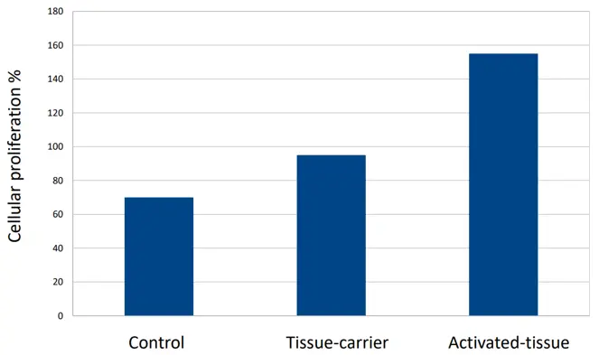
Figure 1. Fibroblast proliferation obtained by the use of the tissue-carrier (vehicle) compared to the control and the same tissue embedded by the selected ingredients (activated tissue). All the p values are highly significant as control and as the tissue-carrier and tissue-carrier activated.
ATP Role and Activity
All the biochemical processes require energy accumulated by ATP (adenosine-triphosphate). Thus ATP was measured on a culture of keratinocytes irradiated with 4 J/cm square UVA + 0.4 cm square UVB (SOL500 lamp, Hönle Group, Munich, Germany) and compared with ATP levels on keratinocytes not irradiated, both added with the tissues in study. As is known, irradiation, dose-dependent, causes a strong reduction of the ATP activity [9].
Of the dish cultures, 6 products in the study received 10 ng/mL of the tested products (3 tissue-carrier and 3 activated carrier, respectively), 24 h before UV irradiation, while 3 served as controls. At this purpose, it’s to underline that all biochemical processes require energy in the form of adenosine triphosphate (ATP), so that ATP activity has been measured on a control-culture of keratinocytes irradiated and not by 4 J/cm2 UVA + 0.4 J UVB (SOL500 lamp, Munich, Germany). All the treated keratinocytes,dose-dependent, have been compared to the ATP level of keratinocytes irradiated and not, preventively treated by the products in the study [9].
The results, obtained by the means of 3 cultures for each tissue (tissue-carrier and activated tissue), were compared to the control and reported in Figure 2 as the %increase of ATP.
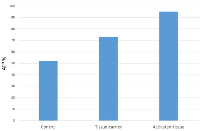
Figure 2. Protective activity of ATP production of keratinocyte cultures irradiated by UVA/UVB and treated by 10 ng/mL of tissue-carrier and activated-carrier and compared with the untreated control. All p values are highly significant (p < 0.005) vs. control as to groups.
IL-8 Release
In the study, micronized particles of tissue-carrier and activated-carrier were introduced in the fresh cell culture medium at 10 ng/mL, added with 100 ng/mL of TNF-alpha, which is able to release IL-8. The positive control was hydrocortisone at 1 μM. After a 24-h incubation at 37 °C and 5% of CO2, the quantity of IL-8 was evaluated by ELISA on the culture’s supernatant. At this purpose, it’s important to remember the interaction activity between ECM and the biological signals. On one hand, these signals are crucial for the normal skin structure function and degeneration, while on the other hand, the cells die in the presence of low oxygen and nutrient concentrations, as well as for high levels of Radical Oxygen Species (ROS) and cytokines. They, in fact, as Tumor Necrosis Factor Alpha (TNF-alpha) and interleukin-8 (IL-8) are all involved in the inflammatory phenomena occurring during the aging process, also [19].
The % of the inhibited release rate of IL-8 stimulated by the TNF-alpha activity has been reported as the mean activity of three cultures of the in-study tissue-carriers and activated-carrier are shown in Figure 3.
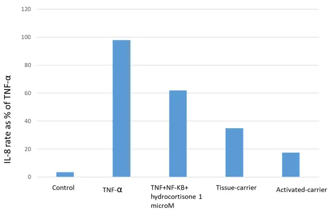
Figure 3. Inhibition of IL-8 release on human keatinocytes stimulated by TNF-α. N = 18. Dose = 10 ng/mL. All p values are highly significant (p < 0.005) as control and the groups.
Metalloproteinase Release (MMP)
During the Skin aging process, activated by ultraviolet light, there is an increased production of ROS and metalloproteinases (MMP) that, degrading the main components of the Extracellular Matrix (ECM), cause a contemporary reduction of collagen type I and IV [19]. It’s important to remember, in fact, that the main mechanism regulating the aging process is based on the balance between the synthesis of new collagen, such as collagen I and IV, followed by the degradation of MMP1.
Human fibroblasts were seeded in sterile flasks with DMEM medium and 10% FBS. After 24 h of incubation, the original medium was replaced with DMEM containing 600 micromoles H2O2 (to induce ROS production and cellular senescence). Thus the cells were incubated for a further 2 h. Soon after the medium was removed and replaced by a fresh one. The cells were cultures for a further 144 h, with medium renewal at a 70 h. Both senescent and normal fibroblasts were then trypsinized, seeded in 96-well microplastics and cultured for another 24 h. After this period of incubation, the culture medium was replaced with DMEM medium containing either the Transforming Growth Factor (TGF-Beta), then the tissue-carriers all at the concentration of 10 ng/mL. The mean results from triplicate cultures of both the tissue-carrier and the activated-carrier groups are presented in Figure 4, showing the percentage reduction in MMP-1 % secretion in senescent fibroblasts following. TGF-beta treatment.
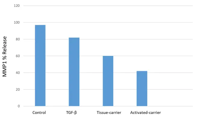
Figure 4. MMP1 release of aged fibroblasts in TGF-β culture treated by tissue-carrier and Activated-carrier. N = 12; dose: 10 ng/mL. All p values are highly significant (p < 0.005) as control and the treated groups.
Collagen Synthesis
Collagen is the main component of the ECM that is necessary to maintain the skin’elasticity and firmness, slows down the appearance of fine Lines and wrinkling, which normally increase during the aging phenomena [18].
Its synthesis was controlled in Fibroblast’ cultures by histochemical methods to verify the resulted variations of Collagen I and IV (by ELISA) and Collagen III (by immunocytochemistry and chemiluminescence), respectively, on samples pre-treated by the in study tissue-carriers (10 ng/mL) and the activated-tissue (10 ng/mL), compared to the untreated control.
The obtained mean results as % increase of the different collagens obtained in fibroblast cultures are reported in Figure 5, Figure 6 and Figure 7.
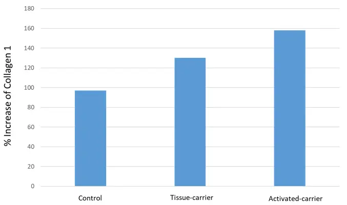
Figure 5. Increase of Collagen I in Fibroblast cultures added with tissue-carrier and activated-carrier compared to the control. N = 9; dose: 10 ng/mL. All p values are highly significant (p < 0.005) as control and the groups.
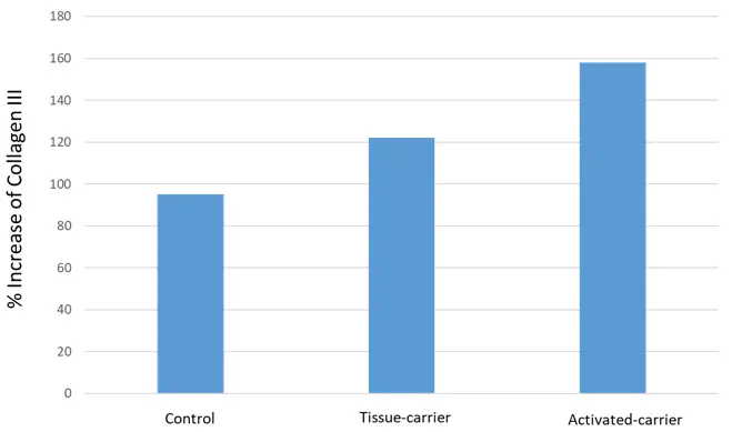
Figure 6. Increase of Collagen III obtained by Fibroblast culture added by tissue-carrier and Activated carrier compared to the control. N = 9; dose: 10 ng/mL. All p values are highly significant (p < 0.005) as control and the groups.
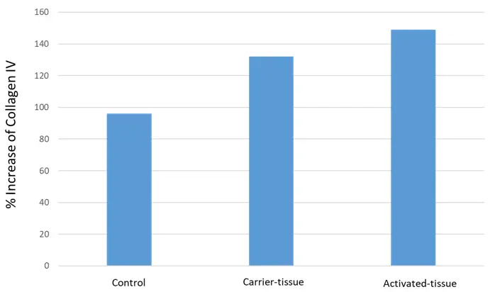
Figure 7. Increase of Collagen IV in Fibroblast culture added with the in study tissues compared to the control. N = 9; dose: 10 ng/mL. All p values are highly significant (p < 0.005) as control and in study tissues.
Antioxidant Activity
Skin redox in healthy people is the balance existing between the environmental oxidizing chemicals and the physiological antioxidant compounds. When a redox imbalance occurs, the so-called oxidative stress appears, manifested by the lipid membrane’s oxidation, which is a fundamental compound for the cellular integrity [19]. Thus, for example, it has been shown that ROS and RNS, as building-up toxic by-products, might be the main cause not only of the peroxidation of polyunsaturated fatty acids (PUFA) present in the cell membrane, such as Linoleic acid, but are also the main oxidative agents for carboydrates and the complex macromolecules, such as DNA and RNA, thus representing the main cause of cell death and many degenerative disorders [19]. Moreover, the oxidative stress, accelerating the degradation of tge ECM components and reducing the collagen synthesis, contributes to the aging phenomena by the appearing of wrinkles and fine Lines [18,19]. The protective and anti-aging effect of both the vehicle (tissue-carrier) and the selected active ingredients (Activated-carrier) was verified by controlling the lipid peroxidation of linoleic acid by the formation of malonil dialdehyde (MDA). Linoleic acid (10 mM) was dissolved in 1 mL of methanol alcohol, dried under nitrogen, and redissolved in 2 mL of phosphate buffer, as previously reported [10]. Lipid peroxidation of linoleic acid of both tissues (10 ng/mL) was induced by adding 10 microliters of fetal bovine serum (FBS) for 15 min at 37 °C and compared to the untreated control. The total antioxidant capacity of in-study tissues was evaluated in vitro by the total peroxyl-radical-trapping parameter (TRAP) method previously used by our group [10].
The control consisted of linoleic acid peroxidated by 10 microliters of AMVN alone for 15 min at 37 °C, and the MDA was detected by a fluorimetric method, as previously reported [10]. The antioxidant capacity of both tissues, verified by the TRAP method, is reported in Figure 8 as the reduction of MDA and the % contemporary increase of the antioxidant capacity.
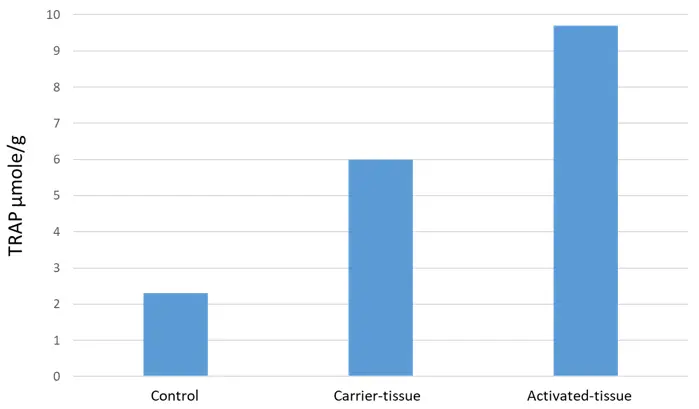
Figure 8. Antioxidant capacity of nanoparticles of Tissue-carrier and Activated-carrier on the peroxidation of linoleic acid. The values are highly significant (p < 0.001).
Citotoxicity
The cytotoxicity of the in-study tissues was performed by the MTT method modified by our group [12,15]. Briefly, both the cells, keratinocytes and fibroblasts, were placed in plates at a density of 1 × 10⁴ cells in 200 microliter of medium and incubated per 24 h to allow the cells to attach. Then the cells in study were exposed to a serial concentration for 48 h at 37 °C. At the end of incubation, 20 microliters of the MTT solution were added, continuing for another 4 h. The medium was then replaced with 100 microliters of dimethyl sulfoxide to dissolve the MTT crystals. The plates were shaken for 10 min. measuring the addorbance at 570 nm. The obtained mean results on keratinocytes and fibroblasts viability are reported in Figure 9.
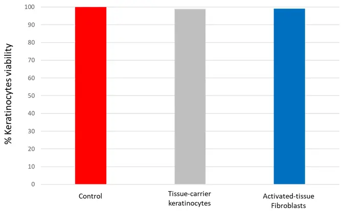
Figure 9. Effect of Tissue-carrier and Activated tissue-carrier on the keratinocytes and fibroblasts viability. All p values are not significant as the control and the groups.
2.3.2. In Vivo Methods
According to our previous studies and methods reported in references [9,10,12,13], 30 voluntary women, preventively selected by plastic surgeons, received a bottle of distilled water and 60 units of the envelooed tissues (15 × 20 cm tissues) (30 tissue-carrier and 30 activated carrier respectively).
All the subjects were instructed to apply every day, in the morning, and for 20 min on their water-cleaned skin, the tissue-carrier on the right cheek, and the tissue-carrier on the left ones. Soon after the face of the treated subjects was wetted, spraying on both cheeks the assigned distilled water. Moreover all were instructed to massage softly all the face by the fingers until the total absorption of both the tissue products was obtained. As a consequence, the skin appeared soft and well moisturized on both cheek surfaces, while all the tissues disappeared, totally absorbed.
During the controls and before starting the treatments and after the acclimatization period (around 20 min), all the subjects have been maintained in a rest position in a room at controlled temperature (Co 24 ± 2) and humidity (RH 50 ± 10%).
The Skin parameters were controlled by the plastic surgery/dermatologist, both de visu and by some known Bioengineering parameters. Skin moisturizing and transepidermal water loss (TEWL) were controlled by the 3C System (Dermotec, San Giovanni Teatino, Italy) [13] while the aged spots were controlled by Chromameter (Minolta CR-300, Konica Minolta, Tokyo, Japan) and the skin firmness and elasticity by Cutometer MPA 580 (Courage & Kazaka, Cologne, Germany). Skin Lines and the depth of wrinkles were valuated de visu by a score method.
The Evaluation of treated and untreated subjects was performed at day 1 baseline (TO), and at week 4 (T28), 8 (T56), and 12 (T84) weeks of treatment.
On the reported figures for the in vivo studies, the right cheek (tissue-carrier) is indicated by the color gray, while the left ones (activated tissue-carrier) is Sky blue.
Skin Hydration and Transepidermal Wter Loss (TEWL)
Skin hydration was controlled by the 3C System methodology [12]. The instrument/(Dermotec, Italy) often used for our group, has separate test parameters. The probes of this computerized instrument collect up to 15 separate readings over a 25 s sampling period. Skin hydration is based on the measurement of dielectric costant of water controlled by the skin capacitance, while TEWL is measured indirectly by two pairs of sensors making use of the Flick’s diffusion law readings. The measurements, automatically averaged, were taken in the area between nose and cheek. The obtained results are reported in Figure 10 and Figure 11.
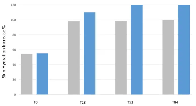
Figure 10. Skin hydration increase of Aged Women treated by Tissue-carrier at different treatment-period tissue-carrier (right cheek) and Activated-tissue (left cheek). N = 30, T = 22 °C, RH 50%.
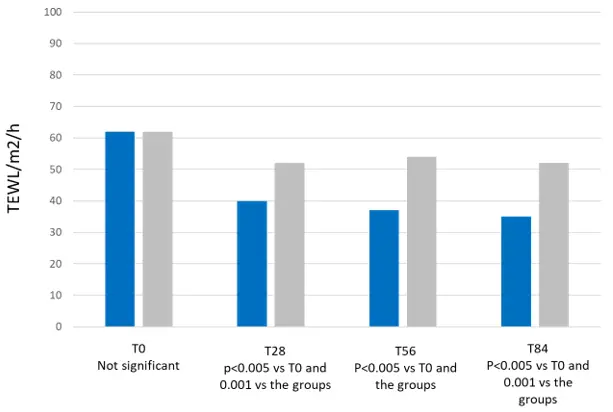
Figure 11. Skin TEWL of aged women treated by tissue-carrier and activated carrier. N = 30, T = 22 °C, RH 50%.
Aged (Black) Spots
The spot’s intensity color was evaluated at day 30 and 60 on different skin areas by the Minolta Chromameter CR-300 (Konica Minolta, Tokyo, Japan). The obtained results are reported in Figure 12.
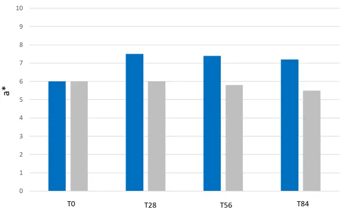
Figure 12. Chromatometer values a* (difference to baseline (T0) after the global treatment on right (Tissue-carrier) and left (activated-carrier)). N = 30, T = 22 °C, RH 50%. All p values are significant vs. the untreated (control) and the carrier.
Skin Firmness and Elasticity
The parameters firmness (RO) and elasticity (R2) were evaluated by Cutometer MPA 580 (Courage & Kazaka, Cologne, Germany). It measuring principle is based on the suction method, where negative pressure deforms the skin mechanically
The obtained results are reported in Figure 13.
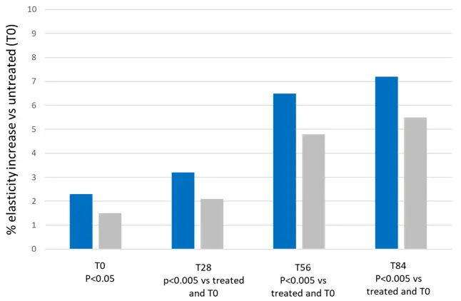
Figure 13. Elasticity increase from baseline of aged skin treated by tissue-carrier and activated-tissue. N = 30, T = 22 °C, RH 50%.
Skin Wrinkles and Lines
Skin wrinkling was evaluated by an expert plastic surgery by an analog scale based on 1–4 Clinical scores from baseline and at 0 (T0), 28 (T28), 56 (T56), and 84 (T84) days of treatment. The scores were: no treatment, 1; few indistinct wrinkles, 2; few distinct wrinkles and lines, 3; Severe distinct swallow wrinkles and lines, 4. The obtained results were reported in Figure 14.
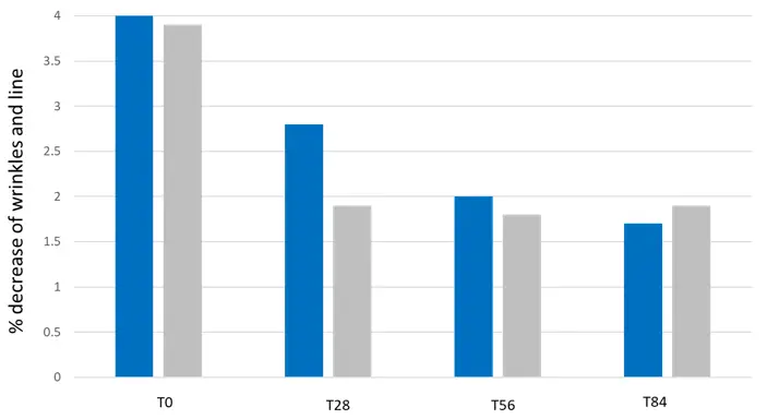
Figure 14. % decrease evaluation of wrinkles and fine lines controlled visually by points by plastic surgeons. N = 30, T = 22 °C, RH 50%. All the values were highly significant (0.005) vs. untreated (T0) and the groups.
3. Obtained Results and Comment
As clearly shown by all the in vitro and in vivo results Reported in Table 1 and Table 2 and Figure 1, Figure 2, Figure 3, Figure 4, Figure 5, Figure 6, Figure 7, Figure 8, Figure 9, Figure 10, Figure 11, Figure 12, Figure 13 and Figure 14, both carrier-tissue and activated-tissue have shown to have an interesting anti-aging effectiveness when applied on aged skin and controlled by the different parameters selected and used. On the one hand, it’s interesting to underline (see Table 1) the effectiveness shown in vitro by both the activated-tissue and the control tissue able to increase the Collagen I, III (Figure 5 and Figure 6) and collagen IV (Figure 7) as well as the capacity they have to neutralize the release of MMP1 (Figure 4) and IL-8 (Figure 3), acting positively on the proliferation and activity of both keratinocytes and fibtoblasts (Figure 1 and Figure 2). Moreover, they have shown to possess an interesting antioxidant activity (Figure 8) showing to be safe and no toxic for both keratinocytes and fibroblasts (Figure 9). On the other hand (see Table 2), both the tissues (activated and not) have shown in vivo to possess an interesting effectiveness on black-spots (Figure 12), being also able to increase skin hydration (Figure 10) firmness and elasticity (Figure 13), contemporary decreasing the TEWL (Figure 11) together with the depth of wrinkles and number of fine Lines the results of which results unusual and probably wrong for one reported data (Figure 14). The evident unusual and unspected effectiveness of the tissue-carrier both in vitro and in vivo is probably due to the antioxidant and skin repairing effectivenss shown by the Chitin nanofibrils—Nanolignin complexes, evidenced by a research studies published by our group also [9,10,11,32,33,34,35] and by the supposed synthesis of Hyaluronic acid obtained from the use of some Chitin polymeric units and recently published [36,37,38,39]. These complexes, in fact, have shown not only to have the capacity to entrap the active ingredients protecting them from the Environment’oxidative phenomena, but to posses also the ability to release them at level of skin layers, at the designed dose and time [11,14,15,20,21,22]. On the other hand, both the tissues seem able to release the ingredients more quickly when compared to the normal emulsions as previously reported [11]. Therefore, these new obtained results seem to support our idea to use the proposed natural tissues as novel vehicles for making innovative Cosmetic and diet supplements,in partial substitution of the normal emulsions. Moreover, it seems interesting to underline the possibilty to realize biodegradable tissues water-soluble, useful to make innovative cosmetics (cosmeceuticals) and diet supplements (nutraceuticals) for obtaining the socalled Beauty from Within also. Thus by this novel technology it will be possible to use the same active ingredients to be applied on the skin by cosmeceuticals and contemporary taken by the oral route for ranging a new Italian Beauty by innovative cosme-nutraceuticals characterized by an interesting effectiveness and safety.
Table 1. In vitro methods on keratinocytes and fibroblasts cultures: Obtained results.
|
Control |
Tissue-Carrier |
Activated-Tissue |
|
|---|---|---|---|
|
Cellular proliferation % of aged fibriblasts |
71.05 ± 0.2 |
87.04 ± 0.2 |
157.02 ± 0.4 |
|
ATP recovery % on irradiated keratinocytes |
54.02 ± 0.15 |
73.02 ± 0.5 |
95 ± 0.3 |
|
IL-8 Inhibition as % of TNF activity |
0.5 ± 0.004 |
38.03 ± 0.8 |
0.18 ± 0.005 |
|
MMP1 release % |
95.5 ± 0.8 |
62.0 ± 0.3 |
43.5 ± 0.23 |
|
Collagen I synthesis increase % |
98.3 ± 0.05 |
124.8 ± 0.2 |
158.07 ± 0.43 |
|
Collagen III synthesjs Increase % |
87.2 ± 0.07 |
121.5 ± 0.3 |
169.0 ± 0.40 |
|
Collagen IV synthesis increase % |
98.1 ± 0.10 |
139.4 ± 0.2 |
148.7 ± 0.51 |
|
% increase of Antioxidant capacit |
22.2 ± 0.05 |
60.0 ± 0.01 |
98.8 ± 0.012 |
|
Cell viability % |
100 |
99.2 ± 0.05 |
99.8 ± 0.06 |
Table 2. In vivo methods: results obtained by treatments on 30 women (V = vehicle (tissue-carrier); AV = Actuvated-tissue)).
|
T28 V |
AV |
T56 V |
AV |
T84 V |
AV |
|
|---|---|---|---|---|---|---|
|
Skin hydration increase % |
59.5 |
65.2 |
59.5 |
72.4 |
60.0 |
71.7 |
|
TEWL reduction % |
48.2 |
66.3 |
49.1 |
68.4 |
49.0 |
69.7 |
|
Aged spots color reduction % |
0 |
25.3 |
0.1 |
24.9 |
0.4 |
24.3 |
|
Elasticity increase % |
20.3 |
32.7 |
49.4 |
55.3 |
52.4 |
71.3 |
|
Wrinkling decrease % |
42.0 |
30.3 |
48.2 |
55.5 |
49.5 |
47.7 |
4. Conclusive Remarks
We hope that these tissues used as novel vehicles might be useful to slow down and possibly avoid not only the allergic and sensitization skin phenomena, but also to notably reduce the consumption of water, energy, and natural raw materials, reducing plastic waste also. Thus the lifestyle and the Environment will surely be ameliorated for the incoming generations [8,9,10,11,14,15,20,21,22,23,24].
The high biocompatibility and bioactivity of Chitin nanofibrils, nanolignin, and the different biosaccharides obtained by the biomass and used to realize both the tissues (carrier-tissue and activated-tissue) result, in fact, fundamental for developing a skin effectiveness and safety’acgivity as shown by this and other research studies, previously published [8,9,10,11,12,13,14,15,16,20,21,22,23,24,26,27,28,29,30,31,32,33,34,35,36,37,38,39,40,41,42,43]. Moreover, it is also to underline the biodegradability of the bamboo-support and the nanotechnology-based biomimetic ingredients and polymers used during the electrospinning technology for giving further eco-compability to the final product [16,29,30,31,32,33,34,35,36,37,38,39]. By these tissues it seems possible to favour the skin regeneration in a quick time without provoking any kind of side effects, being them 100% skin- and evironmentally-friendly, due to the biodegradable packaging used also [16,23,24,25,26,27,28,29,30,31,32,33,34,35,36,38,39,40,41,42,43].
In conclusion, in our opinion and by the use of this innovative product, possessing an interesting pro-aging effectiveness, it would be possible to obtain the supposed 360-degree Beauty and Wellness [16,23,24,25,26,27,28,29,30,31,32,33,34,35,36,38,39,40,41,42,43].
Author Contributions
Writing original draft, P.M. and M.P.; Writing review and editing, P.M., M.P., G.F. and G.D.; Supervision, P.M., M.P., X.G., W.E.Y. and G.D.
Ethics Statement
The study was conducted in accordance with the 2024 revised Declaration of Helsinki while ethical review was waived according to the Cosmetic Regulation No. 1223/2009 and the subsequent modifications.
Informed Consent Statement
Informed consent was obtained from all the subjects involved in the study.
Data Availability Statement
Not applicable.
Funding
This research received no external funding.
Declaration of Competing Interest
The authors declare that they have no known competing financial intesrests or personal relationships that could have appeared to influence the work reported in this paper.
References
- Chermahini SH, Majid FA. Cosmeceutical values, antimicrobial activities and antioxidant properties of cashew leaves extract. Afr. J. Biotechnol. 2011, 10, 14573–14582. DOI:10.5897/AJB11.1873 [Google Scholar]
- Meng J, Pan P. Investigating the effects of cosmeceutical product advertising in beauty‐care decision making. Int. J. Pharm. Healthc. Mark. 2012, 6, 250–266. DOI:10.1108/17506121211259412 [Google Scholar]
- Barbu A, Catană ȘA, Deselnicu DC, Cioca LI, Ioanid A. Factors influencing consumer behavior toward green products: A systematic literature review. Int. J. Environ. Res. Public Health 2022, 19, 16568. DOI:10.3390/ijerph192416568 [Google Scholar]
- Yousefian F, Van Alfen B, Yadlapati S, Goldberg DJ, Gold M. Cosmeceuticals and Other Techniques to Reduce Bruising from Surgical and Cosmetic Procedures. Dermatol. Rev. 2025, 6, e70013. DOI:10.1002/der2.70013 [Google Scholar]
- Wæraas A. Beauty from Within: What bureaucreacid stand for. Am. Rev. Public Adm. 2014, 44, 675–692. DOI:10.1177/0275074013480843 [Google Scholar]
- William Reed. Beauty-from-Within and Beyond: Exploring the Modern Consumer Through the Lens of Clinical Research. 2021. William Reed Report. Available online: https://www.nutraingredients.com/News/Promotional-features/Beauty-from-within-and-beyond-Exploring-the-modern-consumer-through-the-lens-of-clinical-research/ (accessed on 20 January 2026).
- Wisniewski JD, Ellis DL, Lupo MP. Facial Rejuvenation: Combining Cosmeceuticals with Cosmetic Procedures. Cutis 2014, 94, 122–126. Available online: https://cdn.mdedge.com/files/s3fs-public/Document/September-2017/CT094090122.pdf (accessed on 20 July 2025).
- Morganti P. Natural Products Work in Multiple Ways. In Nutritional Cosmetics. Beauty from Within; Tabor A, Blair RM, Eds.; William Andrew Publishing: Cambridge, MA, USA, 2009; pp. 95–111. DOI:10.1016/B978-0-8155-2029-0.50012-0 [Google Scholar]
- Morganti P, Fabrizi G, Palombo M, Ruocco E, Cardillo A, Morganti G, et al. Chitin-nanofibrils: A new active Cosmetic carrier. J. Appl. Cosmetol. 2008, 26, 113–128. Available online: https://www.researchgate.net/publication/228634199_Chitin-nanofibrils_A_new_active_cosmetic_carrier (accessed on 20 January 2026).
- Morganti P, Palombo M, Fabrizi G, Guarneri F, Svolacchia F, Cardillo A, et al. New insights on anti-aging activity of chitin nanofibril-hyaluronan block copolymers entrapping active ingredients: In vitro and in vivo study. J. Appl. Cosmetol. 2013, 31, 1–29. Available online: https://www.scientificeditorial.com/index.php/JAC/article/view/New-Insights-on-Anti-Aging-Activity-of-Chitin-Nanofibril-Hyaluro (accessed on 20 January 2026).
- Morganti P, Fabrizi G, Beatrice BM, Danti S, Azimi B, Gagliardini A, et al. Skin Delivery and Innovative Carrier’s Effectiveness. J. Intern. Med. Res. Rep. 2024, 132, 2–5. DOI:10.47363/JIMRR/2024(3)132 [Google Scholar]
- Morganti P, Del Ciotto P, Fabrizi G, Guarneri F, Cardillo A, Palombo M, et al. Safety and tolerability of Chitin nanofibrils-hyaluronan nanoparticles entrapping Lutein. Note I. Nanoparticles characterization,bioavailability and Biodegradability. SOFW J. 2013, 139, 12–23. Available online: https://www.researchgate.net/publication/236168179_Safety_And_Tolerability_of_Chitin_Nanofibrils-Hyaluronic_Acid_Nanoparticles_Entrapping_Lutein_Note_1_Nanoparticles_Characterization_and_Bioavailability (accessed on 20 January 2026).
- Cardillo A, Morganti P. Fast and non-invasive method for assessing skin hydration. J. Appl. Cosmetol. 1994, 12, 11–16. Available online: https://www.scopus.com/pages/publications/0028357884 (accessed on 20 January 2026).
- Danti S, Trombi L, Fusco A, Azimi B, Lazzeri A, Morganti P, et al. Chitin Nanofibrils and Nanolignin as Functional Agents in Skin Regeneration. Int. J. Mol. Sci. 2019, 20, 2669. DOI:10.3390/ijms20112669 [Google Scholar]
- Morganti P, Morganti G, Coltelli MB. Skin and pollution: The Smart nano-based cosmeceutical tissues to save the Planet’ecosystem. In Nanocosmetics: Fundamentals, Applications and Toxicity; Nanda A, Nanda S, Nguyen TA, Rajendran S, Slimani Y, Eds.; Elsevier: Amsterdam, The Netherland, 2020; pp. 287–304. [Google Scholar]
- Morganti P. Use of Chitin nanofibrils from biomass for an innovative bioeconomy. In Nanofabrication Using Nanomaterials; Ebothe J, Ahmed W, Eds.; One Central Press: Altrincham, UK, 2016; pp. 1–22. [Google Scholar]
- Torres A, Rego L, Martins MS, Ferreira MS, Cruz MT, Sousa E, et al. How to Promote Skin Repair? In-Depth Look at Pharmaceutical and Cosmetic Strategies. Pharmaceuticals 2023, 16, 573. DOI:10.3390/ph16040573 [Google Scholar]
- Bar O, Valiukevičienė S. Skin aging and type I Collagen: A systematic review of interventions with potential collagen-related effects. Cosmetics 2025, 12, 129. DOI:10.3390/cosmetics12040129 [Google Scholar]
- Tortajada-Pérez J, Carranza ADV, Trujillo-del Río C, Collado-Pérez M, Millán JM, García-García G, et al. Lipid Oxidation at the Crossroads: Oxidative Stress and Neurodegeneration Explored in Caenorhabditis elegans. Antioxidants 2025, 14, 78. DOI:10.3390/antiox14010078 [Google Scholar]
- Morganti P, Morganti G, Colao C. Biofunctional Textiles for Aging Skin. Biomedicines 2019, 7, 51. DOI:10.3390/biomedicines7030051 [Google Scholar]
- Morganti P, Morganti G, Coltelli MB. Chitin Nanomaterials and nanocomposites for tissue repair. In Marine-Derived Biomaterials for Tissue Engineering Applications; Choi AH, Ben-Nissan B, Eds.; Springer: Singapore, 2019; pp. 523–544. ISBN 978-981-13-8855-2 [Google Scholar]
- Morganti P, Coltelli MB. Smart Tissues Skin-Friendly as Carriers for Natural Cosme-Nutraceuticals. DNN 2025, 3, DNN.000564. DOI:10.31031/DNN.2025.03.000564 [Google Scholar]
- Morganti P. Chitin-Lignin Complex as New Green Tissue-Carrier for Innovative Pro- Aging Cosme-Nutraceuticals. Assoc. J. Health Sci. 2025, 4, AJHS.000585. DOI:10.31031/AJHS.2025.04.000585 [Google Scholar]
- Morganti P. Nanoparticles and Nanostructures Man-Made or Naturally Recovered: The Biomimetic Activity of Chitin Nanofibrils. J. Nanomater. Mol. Nanotechnol. 2012, 1, 1000101. DOI:10.4172/2324-8777.1000101 [Google Scholar]
- Smirnova NV, Kolbe KA, Dresvyanina EN, Grebennikov SF, Dobrovolskaya IP, Yudin VE, et al. Effect of Chitin Nanofibrils on Biocompatibility and Bioactivity of the Chitosan-Based Composite Film Matrix Intended for Tissue Engineering. Materials 2019, 12, 1874. DOI:10.3390/ma12111874 [Google Scholar]
- Coltelli MB, Gigante V, Panariello L, Aliotta L, Morganti P, Danti S, et al. Chitin nanofibrils in renewable materials for packaging and personal care applications. Adv. Mater. Lett. 2019, 10, 425–430. DOI:10.5185/amlett.2019.2250 [Google Scholar]
- Morganti P. Saving the Environment by Nanotechnology and Waste Raw Materials: Use of Chitin Nanofibrils by EU research projects. J. Appl. Cosmetol. 2013, 31, 89–96. Available online: https://www.researchgate.net/publication/259850050_Saving_the_Environment_by_Nanotechnology_and_Waste_Raw_Materials_Use_of_Chitin_Nanofibrils_by_EU_research_projects(accessed on 20 June 2026).
- Morganti P. Biomimetic Materials Mimicking Nature at the Base of EU Projects. JSRR 2014, 3, 532–544. DOI:10.9734/JSRR/2014/5645 [Google Scholar]
- Morganti P. The Meaning of Nano Dimension Involving Cosmetics: From the Lab to an Industrial Green Process. JSRR 2015, 4, 79–100. DOI:10.9734/JSRR/2015/11182 [Google Scholar]
- Lohani A, Khan S, Morganti P. (Eds.). Nanodermatology: Advances in Theory and Practice; Elsevier: Amsterdam, The Netherlands, 2026. [Google Scholar]
- Morganti P. Beauty and Wellness at 360. J. Appl. Cosmetol. 2010, 28, 13–24. Available online: https://www.researchgate.net/profile/Pierfrancesco-Is-The-Given-Name-Morganti-Is-The-Family-Name/publication/266277899_Beauty_and_Wellness_at_360/links/5457977c0cf2cf516482178d/Beauty-and-Wellness-at-360.pdf (accessed on 20 January 2026).
- Morganti P, Fabrizi G, Palombo P, Palombo M, Guarneri F, Cardillo A, et al. New chitin complexes and their anti-aging activity from inside out. J. Nutr. Health Aging 2012, 16, 242–245. DOI:10.1007/s12603-011-0358-0 [Google Scholar]
- Scialla S. Nanomaterials for skin anti-aging. Recent status and future prospects. Nanodermatology: Advances in Theory and Practice; Elsevier: Amsterdam, The Netherlands, 2026; pp. 347–376. Available online: https://www.sciencedirect.com/science/chapter/edited-volume/abs/pii/B9780443363269000046 (accessed on 20 June 2026).
- Coltelli MB, Morganti P, Castelvetro V, Lazzeri A, Danti S, Benjelloun-Mlayah B, et al. Chitin nanofibrils-nanolignin complexes as carriers for functional molecules for Skin contact applications. Nanomaterials 2025, 12, 1295. DOI:10.3390/nano12081295 [Google Scholar]
- Kodolova-Chukhontseva VV, Dresvyanina EN, Nashchekina YA, Dobrovol’skaya IP, Bystrov SG, Ivan’kova EM, et al. Application of the Composite Fibers Based on Chitosan and Chitin Nanofibrils in Cosmetology. J. Funct. Biomater. 2022, 13, 198. DOI:10.3390/jfb13040198 [Google Scholar]
- Rathod S, Shinde K, Shinde N, Aloorkar N. Cosmeceuticals and nanotechnology in beauty Care products. Res. J. Top. Cosmet. Sci. 2021, 12, 93–101. DOI:10.52711/2321-5844.2021.00013 [Google Scholar]
- Balaji M, Vashishth R, Das U. Plant-based functional foods for healthy aging: Current trends, bioactive compounds, and future perspectives. J. Funct. Foods 2026, 138, 107155. DOI:10.1016/j.jff.2026.107155 [Google Scholar]
- Glass GE. Cosmeceuticals: The principles and practice of skin rejuvenation by nonprescription topical therapy. In Aesthetic Surgery Journal Open Forum; Oxford University Press: New York, NY, USA, 2020; Volume 2. DOI:10.1093/asjof/ojaa038 [Google Scholar]
- Crespi O, Rosset F, Pala V, Sarda C, Accorinti M, Quaglino P, et al. Cosmeceuticals for Anti-Aging: Mechanisms, Clinical Evidence, and Regulatory Insights—A Comprehensive Review. Cosmetics 2025, 12, 209. DOI:10.3390/cosmetics12050209 [Google Scholar]
- Georgescu C, Bartoletti E. Cosmetics, Dermocosmetics and Cosmeceuticals in Aesthetic Medicine: What We Have Never Asked, and Why It Matters. A Clinical Perspective. Aesthetic Medicine. 2026. Available online: https://mattioli1885journals.com/index.php/aestheticmedicine/article/view/18475 (accessed on 20 June 2026).
- Jayakumar R, Chennazhi KP, Srinivasan S, Nair SV, Furuike T, Tamura H. Chitin Scaffolds in Tissue Engineering. Int. J. Mol. Sci. 2011, 12, 1876–1887. DOI:10.3390/ijms12031876 [Google Scholar]
- Amal TR, Radhika Rajasree SR. Incorporation of chitin nanocrystals into fish collagen-gelatin bio-scaffolds: Enhancing functional properties and biomedical applications. Int. J. Biol. Macromol. 2025, 319, 145581. DOI:10.1016/j.ijbiomac.2025.145581 [Google Scholar]
- Abdel-Mohsen AM, Abdel-Rahman RM, Skotnicova K. Recent Advances and Applications of Chitin and Chitosan Hydrogel Scaffolds in Tissue Engineering. Gels 2026, 12, 427. DOI:10.3390/gels12050427 [Google Scholar]