
Case Studies of Acute Care of Ventilatory Pump Failure: A Medicolegal Interpretation of When Conventional Care Is Negligent

Case Studies of Acute Care of Ventilatory Pump Failure: A Medicolegal Interpretation of When Conventional Care Is Negligent
Received: 27 April 2026 Revised: 03 June 2026 Accepted: 01 July 2026 Published: 14 July 2026

© 2026 The authors. This is an open access article under the Creative Commons Attribution 4.0 International License (https://creativecommons.org/licenses/by/4.0/).
1. Introduction
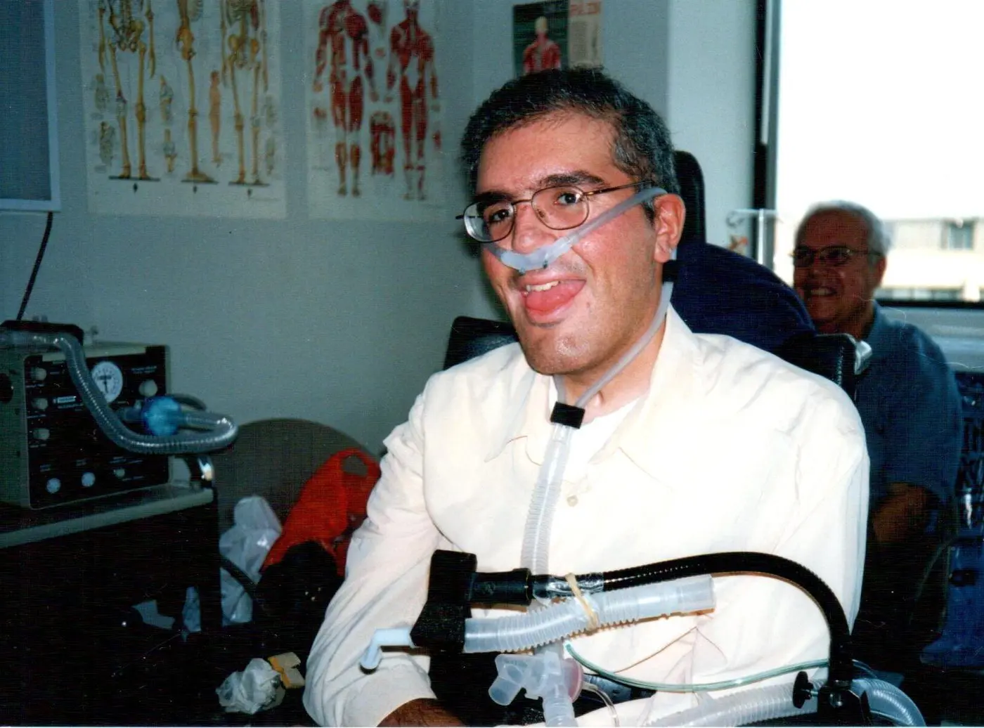
When conventional management results in morbidity and death, is it defensible? This question is central to the respiratory management of patients with ventilatory pump failure (VPF). VPF is a respiratory muscle dysfunction with a variety of causes, including advanced age, certain cancers, and neurological conditions. It causes respiratory symptoms and elevated blood carbon dioxide (CO2) levels initially due to shallow breathing during sleep. Oxygen (O2) is conventionally administered to these patients, rather than ventilatory or cough assistance, to normalize blood O2 and CO2 levels. However, O2 blunts the hypoxic drive to breathe without assisting the weak muscles in ventilating the lungs and coughing effectively [1,2]. This typically causes mildly elevated CO2 levels to soar, often from slightly above the normal value of 44 mm Hg to over 150 mm Hg, resulting in respiratory failure, coma, and standard-of-care intubation (SOCI). Then, because the translaryngeal tube can make ventilator weaning impossible while the patient remains intubated, and because it is conventionally assumed that patients unable to breathe cannot be extubated, two options are usually offered: standard-of-care intubation leading to tracheostomy (SOCIT), with a tracheostomy tube inserted through the neck for ventilatory support (Figure 1), or standard-of-care intubation leading to death (SOCID), consisting of palliative care with O2 and morphine followed by extubation to expected death. A third option is extubation to full noninvasive ventilatory support (NVS) via mouthpieces (Figure 2) or nasal interfaces (Figure 3), including continuous noninvasive ventilatory support (CNVS), as is done in many centers around the world [3], but rarely in the United States. The question is whether conventional management that results in SOCIT or SOCID may constitute malpractice when airway tubes or death are avoidable by using NVS, or whether it is defensible on the grounds that it is conventional management. What about when physicians do not know about NVS and vital mechanical in-exsufflation (MIE) to clear the airways of debris, but are asked to inform themselves, but do not do it? Continuous NVS (CNVS) has been used via simple mouthpieces since it was first described in 1953 [4], and has been used by hundreds to thousands of U.S. patients in the New York Metropolitan area [5], but it requires effort to learn and offers little to no financial incentive.
Further, when inspiratory muscles are weak, so are the expiratory muscles needed to cough effectively to prevent pneumonia and respiratory failure [6]. MIE devices provide deep insufflations followed by rapid exsufflations to compensate for ineffective “cough” flows to prevent pneumonias and permit intubated patients too weak to breathe on their own to be extubated without resort to tracheostomies. It is also used in many centers internationally, but rarely in the U.S. [7,8], where it has been available since 1953 [9].
Thus, NVS and MIE can be used to maintain or return to normal blood O2 and CO2 levels for people with very weak respiratory muscles, but this is rarely done in the U.S. Once patients with weak respiratory muscles become symptomatic, sleep blood gases are abnormal, and with further weakening, daytime blood gases become abnormal, and symptoms progress [10]. These patients often first seek emergency services (ES) for dyspnea triggered by otherwise mild upper respiratory tract infections (URIs). They then often face a dangerous “conventional” pathway. ES trained to treat lung disease with O2, default to it to result in SOCIT or SOCID [3] rather than use NVS and MIE to avoid intubation or offer extubation to NVS and MIE as described, with 99% success for over 250 patients [7,8]. Thus, when may “conventional” care that results in SOCIT or death become legally indefensible and potentially constitute malpractice? We analyze five cases where, instead of supporting the muscles of VPF patients with NVS and MIE, O2 was administered, as for treating lung disease, resulting in SOCIT or death.
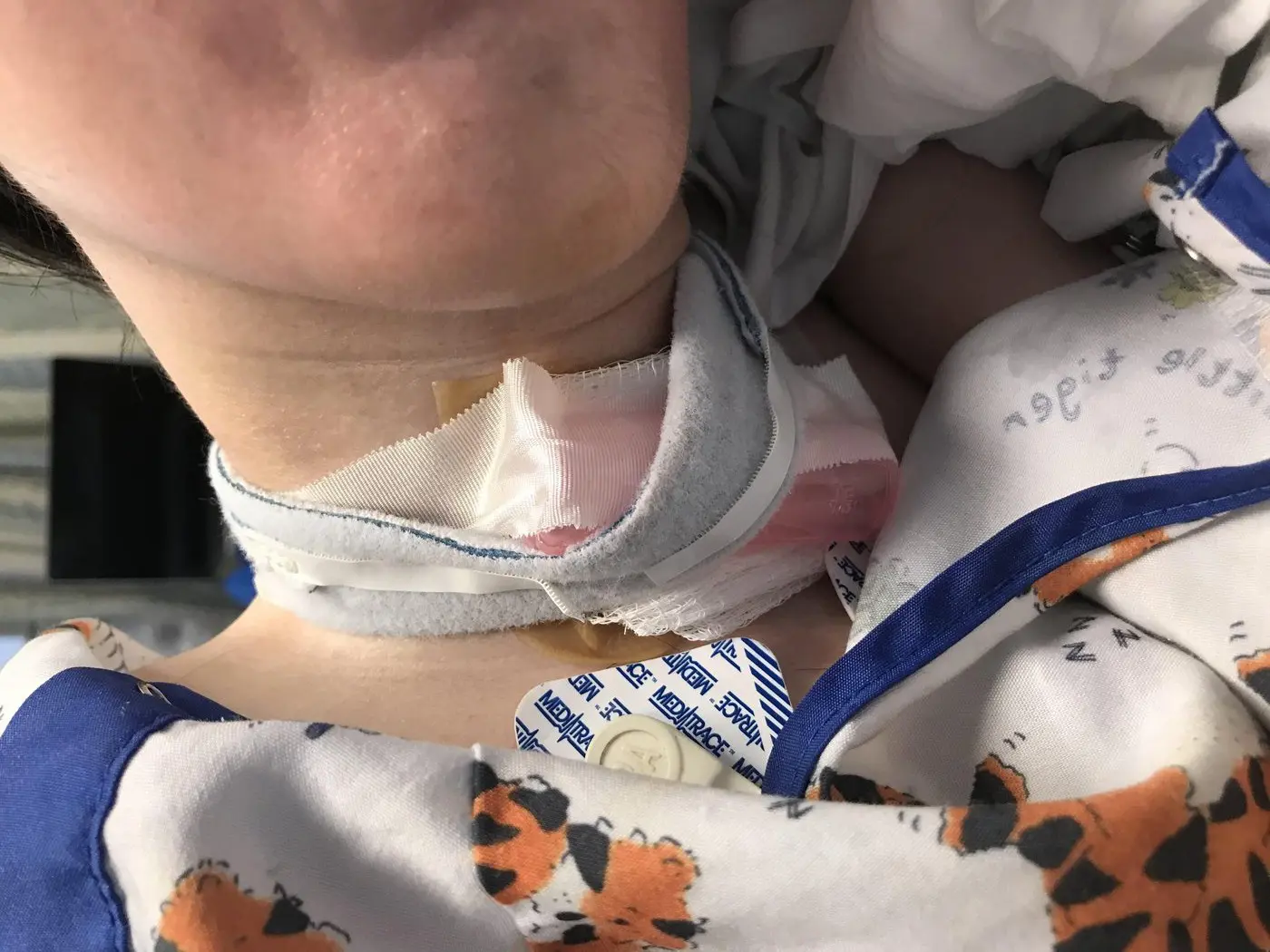
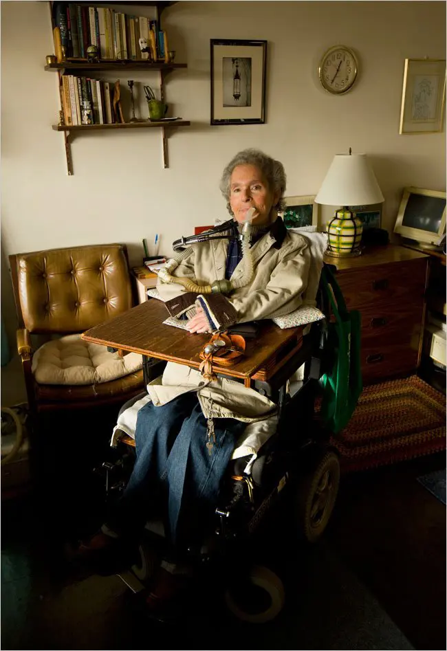
Figure 2. Air delivered via a 15 mm angled mouthpiece as this patient has used continuously for 69 years.
1.1. Current Guidelines: Partial Progress and Persistent Gaps
Widely diffused academic guidelines now warn explicitly that O2 monotherapy can worsen symptomatic hypercapnia in VPF and lead to intubation (SOCI) [11]. Some guidelines emphasize early administration of sleep “noninvasive ventilation” (NIV), but they generally only pertain to low-pressure support (PS) bi-level NIV, which cannot optimally rest muscles or be extended to continuous ventilatory support, that is, CNVS [12,13]. Patients who become dependent on NVS for more than 16 h a day are typically urged to undergo tracheostomies, especially when they seek ES for URIs and get intubated. Instead of using MIE via the tubes to clear the airways, cure the pneumonias, and then extubate to NVS and MIE, they are usually unnecessarily subjected to SOCIT or SOCID, since extubation to NVS and MIE has thus far been reported in only one U.S. center, though many elsewhere [3,7,8]. Conventional acute care algorithms fail to consider the extubation of “unweanable” patients without tracheostomy tubes or palliative deaths. This leaves a gap between what is technically possible and what is typically practiced.
1.2. Methods and Case Selection
The five cases presented in this manuscript are all patients of the senior author (J.R.B.), drawn from his clinical and medicolegal practice at the Center for Noninvasive Mechanical Ventilation, University Hospital, Newark, New Jersey. In each instance, medical records, hospital charts, and—where available—legal depositions and court documents were reviewed to reconstruct the clinical course and the key decision points. Cases were selected because they exemplify distinct, recurring failure modes in the conventional acute management of ventilatory pump failure, not because they constitute a statistically representative sample of all such cases encountered. Readers should note that this is a descriptive case series, not a controlled comparative study, and therefore cannot establish that conventional care always fails or that noninvasive alternatives always succeed. The cases do, however, illustrate patterns of error that the senior author has repeatedly observed across clinical and medicolegal settings and are presented here to assist forensic practitioners, medical examiners, and litigators in recognizing similar scenarios.
The following figures illustrate the clinical difference between conventional tracheostomy mechanical ventilation (TMV) and noninvasive ventilatory support (NVS), the two management pathways central to the cases discussed in this article.
1.3. Case Illustrations with Fully Approved Patient and Next of Kin Consent
Cases 1 through 4 were dependent on motorized wheelchairs, and all had vital capacities (VCs) less than 200 mL, which makes it impossible to survive more than a few minutes to a few hours without full ventilatory support. All litigations were for wrongful death except for Case 2, where, not permitted by New York State law, it was for the parents’ suffering caused by wrongful death.
1.3.1. Case 1—Fatal Discontinuation of CNVS, Oxygen Use, and Failed Extubation
A 28-year-old man with VPF from advanced neuromuscular disease had been dependent on nocturnal NVS since age 13 and was currently dependent on CNVS at 1350 mL delivered ventilator volumes. Due to having unmeasurable cough flows, he used MIE for effective airway clearance to maintain normal blood O2 levels without receiving supplemental O2. Upon presentation to a local hospital ES dyspneic with cold symptoms on 21 October 2025, he was removed from the CNVS; he required around-the-clock ventilatory support for survival using his home portable ventilator, and was placed without consent only on O2. Predictably, this quickly resulted in hypercapnic coma (SOCI). He failed conventional ventilator weaning parameters and spontaneous breathing trials for days while intubated. The treating team was referred to an NVS center that specializes in extubating patients unable to wean from ventilatory support without resort to tracheostomies. His physicians were advised by the NVS center to optimally prepare for extubation using MIE via the airway tube to clear the airways and resolve the pneumonia. They ignored this, then extubated him directly to O2 and low PS, 7 cm H2O noninvasive ventilation (NIV), when he needed 20 cm H2O to support his lung ventilation. The O2 and ineffective NIV resulted in his CO2 soaring to over 150 mm Hg and coma, which could have been prevented by extubation to CNVS and MIE as described [7,8]. With immediate respiratory distress and hypoxia, the O2 was increased to 100%, and his mother was physically prevented from administering MIE to clear his airways. The hospital had no MIE units. Cardiopulmonary arrest resulted, and he died. The death certificate noted that he died from muscular dystrophy when, in fact, he died from preventable respiratory failure had he been extubated back to CNVS and MIE as described [7,8]. His life expectancy should have been to age 40, when such patients usually die from heart failure rather than respiratory failure [14,15].
1.3.2. Root Cause & Medico-Legal Analysis
All unweanable intubated patients conventionally undergo tracheostomies or palliative care deaths. However, this has been reported as unnecessary in at least 21 centers across 19 countries [3]. The patient’s physicians were informed of this but ignored it for their conventionally typical, deadly approach. The parents chose not to press charges for malpractice.
No autopsy was performed in this case. The death certificate attributed the cause of death to muscular dystrophy, which obscures the proximate clinical mechanism: preventable acute hypercapnic respiratory failure following inappropriate removal from life-sustaining CNVS and subsequent extubation to manifestly inadequate ventilatory support. For forensic practitioners, this case illustrates how disease-attributed death certificates can mask iatrogenic causation. A forensic pathologist or medical examiner reviewing such a case should seek the hospital’s ventilator logs, extubation orders, blood gas records, and nursing notes to determine whether the clinical death was caused or hastened by a management failure rather than by the natural progression of an underlying lethal disease, which pure VPF need not be. The treating team’s assumption that the disease was “inevitably fatal” ignored this reality and did not resolve the medicolegal question of whether death occurred prematurely due to substandard care. Here, the proximate mechanism of death was self-evident: a patient with no measurable independent breathing capacity was removed from continuous ventilatory support and then extubated to manifestly inadequate settings. Just as a fatal gunshot wound to the heart does not require an autopsy to establish causation, the mechanism here needed no post-mortem confirmation. An autopsy would have served mainly to formally exclude any competing cause and to document the chain of events for the record—a useful but not indispensable step, since the operative cause was already apparent from the clinical facts.
1.3.3. Case 2—Anoxic Injury, Recovery on CNVS, and Fatal Removal from CNVS
A 35-year-old man, CNVS dependent for 19 years with severe VPF, had survived anoxic brain injury when he, too, was extubated to inadequate NIV and O2 at age 15, then re-intubated and transferred to an NVS center to be extubated to CNVS and MIE without resort to tracheotomy. After extubation, he initially weaned to sleep NVS for 1 year but, weakening with disease progression, required CNVS for 19 years. He maintained normal cognitive function and life in the community during this time. After 19 years of CNVS dependence, he presented to a hospital ES for abdominal pain without respiratory symptoms despite CNVS dependence. The triage nurses reported that his portable life-support ventilator was only a “CPAP” or “BiPAP machine”. Hospital staff removed him from it despite his protestations, not realizing he depended on it for survival, citing policy against “non-hospital equipment”. He suffered immediate respiratory arrest. Apneic, his father attempted to reconnect him to his ventilator, but both parents were forcibly removed under guard for 3 h. Clinicians then typically attempted a minimal 4 cm PS NIV coupled with 100% O2, but he remained unarousable. When the guards went to supper, his parents returned to find him deceased. His abdominal pain was never worked up. In the subsequent malpractice defense, three “expert” physicians claimed they would have done, and would again do, the same: use low-PS NIV with O2 and, if still in distress, intubate. They ignored or did not understand that he had been using a portable ventilator for full support for 19 years and had not presented to ES for any respiratory complaints or need to be removed from his life-support ventilator.
1.3.4. Root Cause & Medico-Legal Analysis
The typical critical error was misclassifying a life-sustaining ventilator as a “CPAP” or “BiPAP” unit that is legally not one and wasn’t being used. The ES staff clearly thought that only invasive airway tubes could be used for “ventilatory support”. The defense argued that the patient and parents had “refused intubation”, but they, unlike the physicians, knew that his ventilator was providing NVS, and he certainly should not have needed to be intubated. The argument was legally incoherent, yet three expert witnesses for the defense said they would have done the same. The removal of that support precipitated the arrest and subsequent successful litigation. Ironically, the parents indicated that they would drop charges if the physicians apologized, which they refused to do. The case was resolved through litigation; settlement terms are confidential and are therefore not reported here.
No autopsy was performed. Death was attributed to respiratory failure in the context of chronic ventilatory dependence. The forensic significance of this case centers on the precipitating event: removal of a documented life-support device by hospital staff who misidentified it as a CPAP unit. For the purposes of death certification and medicolegal analysis, the cause of death would more precisely be characterized as acute respiratory arrest precipitated by the removal of life-sustaining noninvasive ventilatory support. A forensic examiner would need to reconstruct the sequence: triage documentation at the time of presentation, the moment the ventilator was removed, the immediate respiratory arrest, the three-hour period during which the parents were excluded, and the circumstances in which the patient was found deceased. These facts are pivotal for establishing both the cause of death and liability, and they would not be ascertainable from the death certificate alone. The absence of an autopsy in a case of potential iatrogenic death is not a neutral administrative decision. It represents a significant forensic gap: an autopsy could not have shown what the ventilator did or failed to do; it could only have excluded other causes of death. Here, no such alternative cause existed—the respiratory arrest followed immediately upon removal of the life-support device—so the cause was clear without post-mortem examination, and an autopsy would have added only formal confirmation.
1.3.5. Case 3—Refusal to Consider Extubation to CNVS and Prolonged Trach Ventilation (TMV)
A 24-year-old man with VPF due to muscular dystrophy presented with symptoms of pneumonia and ineffective airway clearance due to poor cough flows. He underwent SOCI in the local hospital ES. He was then deemed “unweanable” in critical care by the usual weaning indices, as for Cases 1 and 2. His family provided the physicians with publications describing the successful extubations of over 250 unweanable VPF patients to CNVS and MIE, but they ignored this. THE FAMILY then requested transfer to an NVS unit for extubation. The treatment team refused to request information or consider a transfer, stating that their management was “standard”. They extubated him to O2 and low-PS NIV, and he also immediately arrested. This caused severe anoxic encephalopathy. Unable to further cooperate, he underwent emergency tracheotomy and spent two months, unresponsive, in a chronic nursing ventilator facility, dependent on TMV, before dying due to tube complications.
1.3.6. Root Cause & Medico-Legal Analysis
The hospital provided conventional care, resulting in SOCIT. They denied the patient access to extubation to CNVS and MIE despite it being reported to be 99% successful for avoiding tracheotomy and death in such cases [7,8]. Ignoring feasible, peer-reviewed, evidence-based alternatives presented by the patient’s family transformed a clinical judgment call into a failure of informed consent and reasonable care. While the malpractice suit was successful, the settlement was only for $200,000, which was a little more than sufficient to pay for the 2 months of internment in the nursing ventilator unit, which today would cost about $1500 per day.
The patient survived for two months on tracheostomy mechanical ventilation before dying from tube complications. Whether an autopsy was performed at the time of eventual death is not documented in the records reviewed. For forensic pathologists, the challenge in this case lies in attributing long-term morbidity and eventual death to the original management failure. The proximate cause of death (tube complication) is clinically distinct from the index error (refusal to attempt extubation to CNVS + MIE that results in the tube being unnecessarily placed), yet the chain of causation is traceable. Courts in comparable cases have permitted “loss of chance” and “increased risk of harm” theories to bridge this type of causal gap. Accurate death certification should reflect this chain wherever the facts support it.
1.3.7. Case 4—Misuse of Bi-Level NIV and O2 for Amyotrophic Lateral Sclerosis (ALS)
A 44-year-old man had ALS with VPF. Following a prior episode of O2-induced CO2 narcosis, a pulmonologist discontinued the O2 but prescribed nocturnal bi-level NIV at a PS of 9 cm H2O for less than half of what he needed to maintain normal blood gases or survive once muscles weakened further, for him to need CNVS. So, he remained symptomatic from severe CO₂ retention day and night. One night, his oronasal interface, through which he received the NVS, shifted from being tightly over his nose and mouth. This created an insufflation leak, causing CO2 to rise and leading to respiratory arrest, without triggering the ventilator’s low-pressure alarm, as the oronasal interface still lay loosely against his skin. With inadequate inspiratory pressures and volumes and the device relying on flow algorithms designed for sleep apnea, no alarm sounded, and he was found deceased in the morning, despite his ventilator working normally.
1.3.8. Root Cause & Medico-Legal Analysis
The treating physicians did not attempt to normalize the patient’s CO2 levels, day or night, with adequate NVS settings. Adequate settings would have preserved a normal ventilatory (hypoxic and hypercapnic) drive with arousals in response to even small increases in CO2, prompting arousals to seek assistance. The prescription of 9 cm H2O was grossly inadequate for someone with severely paralyzed inspiratory muscles. The fatal outcome reflected the combined effects of the absence of NVS and a major air leak caused by insufficient interface fixation. As described in published NVS protocols, maintaining normal blood gases often requires CNVS for such patients, not only sleep NVS. Because the family was never informed that the death was attributable to inadequate settings and poor interface retention, rather than to device malfunction, they litigated against the ventilator manufacturer despite no evidence of ventilator dysfunction. At the time of this writing, the case remains active.
Whether an autopsy was performed was not specified in the records reviewed; given the active litigation, this information may be subject to legal confidentiality. From a forensic pathology standpoint, death in this scenario—an unresponsive patient found deceased in the morning with the ventilator still operational—should prompt systematic investigation to exclude primary cardiac events, aspiration, and device malfunction before attributing death to hypoventilation caused by inadequate settings and ventilator use overall. The death certificate attribution and the adequacy of the ventilator alarm configuration (specifically, whether the oronasal interface leak was detectable by the device’s flow-based algorithm) are central forensic questions. Pathologists reviewing such cases should obtain the ventilator event log, interface fitting records, prescribed pressure settings, and all prior blood gas values to evaluate whether the prescribed settings were demonstrably inadequate to sustain life. Suboptimal treatment of chronic hypercapnia that leads to depression of ventilatory drive can be malpractice.
1.3.9. Case 5—Unrecognized VPF, Unnecessary Tracheostomy, and Late Decannulation to CNVS
A 32-year-old Florida man, a full-time employed PhD clinical psychologist with mild, generalized muscle weakness, was asymptomatic except for being unable to run and getting a bit short of breath lying on his back due to diaphragm weakness. He lived independently in the community and required no personal assistance, but developed a colonic abscess. He underwent surgery to evacuate it. Post-operatively, he failed four extubation attempts to oxygen and low-PS BiPAP NIV, and remained ventilator-unweanable largely because of the presence of the intubation tube. He subsequently underwent a tracheostomy. Over the next six months, he was subjected to daily weaning trials, during which he experienced repeated episodes of respiratory distress while staff questioned him about “why he did not want to breathe”, and he remained continuously dependent on TMV. He was then transferred to a New Jersey hospital, closer to his family, with the expectation of lifelong placement in a nursing ventilator unit and ongoing TMV. At the NJ receiving hospital, critical care physicians there also asked why he did not “want to breathe”, despite being able to speak, and did not recognize that his difficulty weaning from TMV was largely due to the tracheostomy tube itself and a low VC. Several days before the planned transfer to the nursing facility, his mother learned about the NJ NVS center and arranged the transfer. He was immediately decannulated to CNVS and MIE upon arrival, and once the tube was out, he rapidly weaned to sleep-only nasal NVS. He returned to full-time employment and filed a lawsuit against the Florida physicians. The case was successful at trial but was then overturned on appeal to a superior court. He continued to work and live alone using sleep NVS for the last 23 years instead of a lifelong institutionalization and TMV dependence at an estimated cost of approximately $440,000 per year to taxpayers for nursing home institutionalization or 16 h per day of home nursing at $75 per hour.
1.3.10. Root Cause & Medico-Legal Analysis
The treating teams interpreted persistent ventilator dependence as “lack of effort” rather than tube-exacerbated ventilator dependence, and failed to extubate him to CNVS and MIE despite the fact that he was a highly functioning patient with only a weak diaphragm. Following decannulation, he returned to full-time employment in Florida and has required only sleep-NVS over the last 23 years, rather than institutionalization, which would have already cost taxpayers over $10 million.
Loss of Chance and Disproportionate Harm: Resorting to tracheotomy and prolonged TMV, without exploring feasible noninvasive strategies or referral to a specialized center for them, converted a transient postoperative vulnerability into a plan for lifelong institutionalization, personal financial ruin, loss of quality of life, and a disaster for taxpayers. The subsequent decannulation and rapid weaning to NVS underscore the magnitude of the lost opportunity attributable to the original, uninformed physician management. The clinical trajectories and outcomes of the five cases are summarized in Table 1.
Table 1. Summary of Case Outcomes.
|
Case |
VC Before Ventilatory Support Was Terminated Without Consent * |
Outcome |
|---|---|---|
|
1 |
<200 mL |
Death (hypercapnic coma, cardiopulmonary arrest) |
|
2 |
<200 mL |
Death (respiratory arrest after life-support ventilator removed) |
|
3 |
<200 mL |
Anoxic encephalopathy → tracheostomy → death |
|
4 |
<200 mL |
Death (found unresponsive; ventilator alarm not triggered) |
|
5 |
Mild weakness pre-operatively; VC fell only after intubation |
Unnecessary tracheostomy → later decannulated to NVS |
* VC less than 200 mL indicates that survival is possible only with continuous ventilatory support.
2. Discussion
These five cases, four of which resulted in litigation and the other an equally valid case of malpractice, exemplify how conventional respiratory management of VPF predictably results in SOCIT/SOCID, with the former, SOCIT, resulting in ongoing nursing care for tracheal suctioning that costs approximately $500,000 per year in 2026 dollars for nursing and medical complications [16,17]. The critical failures were not diagnostic mysteries; they were specific choices to ignore addressing the pathophysiology of VPF, for which NVS and MIE were needed rather than O2 and low-PS NIV, resulting in tracheostomy tubes or death.
While normally, legal cases would be supported by autopsy results, the physicians assumed muscular dystrophies are inevitably fatal, not realizing that up to CNVS can be provided via noninvasive interfaces, and that many patients have been using this for over 60 years and staying free from respiratory complications. Indeed, it has been demonstrated that even total paralysis of all except eye muscles does not have to result in death, with many such patients surviving 30 to over 65 years since leaving Iron Lungs in the 1950s [3,10,18,19]. In 1 year, four of Bach’s muscular dystrophy patients were removed from their life-sustaining ventilators by the hospital ESs to die in their local hospitals. No autopsies were performed since there were no competing causes of death. This practice of forgoing autopsy in VPF-related hospital deaths warrants scrutiny from the forensic community, even though it was removal from life-sustaining ventilatory support that was the obvious cause of the deaths. The assumption that the absence of a competing cause of death makes autopsy unnecessary overlooks the medicolegal purpose of the autopsy: not only to establish cause of death, but to document the mechanism and circumstances of dying in a manner that can support or refute allegations of substandard care. Where iatrogenic mismanagement is plausible, forensic pathologists and medical examiners should consider whether autopsy—or, at a minimum, toxicological analysis and blood gas sampling—is warranted. Accurate cause-of-death certification that reflects the true mechanism (e.g., “acute hypercapnic respiratory failure precipitated by removal of life-sustaining noninvasive ventilatory support” rather than “muscular dystrophy”) serves both the historical record and the interests of justice.
Clinically, several specific mechanisms of failure emerge from these cases:
-
Failure to Recognize VPF and CNVS as Life Support: In cases 1–3, hospital staff did not appreciate that patients presenting with portable ventilators could be dependent on ventilatory support. Their ventilators were mislabeled as CPAP and BiPAP units, which are not legal ventilators. Removal from CNVS in triage becomes a lethal error rather than a clinical adjustment.
-
Oxygen as the Trigger for Acute Decompensation: For all five cases, O2 was inappropriately administered without prior assessment of PaCO2 and without the realization that it was not addressing the issues that were causing the hypoxia, that is, hypoventilation and/or airway secretions for which NVS and MIE are required. The O2 was the catalyst for the SOCIT/SOCID pathway. This phenomenon has been quantified and reported for hundreds of VPF patients [2].
-
Ineffective Airway Clearance and Use of MIE: In none of the cases was MIE used to clear the airways to prevent hypoxia or to reverse it. The decreases in O2 levels were treated with O2 delivery, which did not address the hypoventilation and retained secretions causing the hypoxia. This is despite 73 years of evidence of the importance of MIE in maintaining airway clearance for VPF NVS users, and 39 years of its successful use in facilitating extubation of ventilator-unweanable patients to CNVS [5,7,8]. Airway suctioning alone is often inadequate, whether via the upper airways or airway tubes, because up to 92% of the time suctioning catheters fail to enter and clear the left airways [20]. Since breathing and coughing are vital bodily functions for survival, in the expert opinion of the authors, withholding MIE from patients known to require it may be regarded as functionally analogous to withholding standard pharmacological therapy for a treatable infection.
-
The “Unweanable” Myth: All five patients were described as unweanable, largely because of the use of airway tubes. However, in cases 2 and 5, successful weaning occurred only after tube removal, consistent with the experience of more than 250 similarly labeled patients who, once extubated, returned to the same level of noninvasive support they had used before hospitalization if any at all [7,8]. Patients with VCs as low as 5% of normal, with little ability to breathe any air into their lungs while intubated or tracheostomized, have been able to wean from CNVS to part-time NVS after tube removal. By failing to seek expert input or to implement CNVS/MIE protocols, whether through lack of awareness or omission, treating teams effectively convert manageable, often transient episodes of respiratory failure into permanent tracheostomy dependence or death.
2.1. Real-World Barriers and Limitations of This Analysis
The preceding case analyses should be interpreted in light of important real-world constraints that shape the medicolegal standard of care applicable to any given clinical setting. The authors acknowledge that the use of CNVS and MIE as described here represents a level of specialised practice developed over decades at a small number of expert centres. Several factors may complicate the direct application of these standards across all clinical environments, and forensic practitioners must account for them in any specific case analysis.
Equipment availability is uneven. MIE devices are not universally present in community hospitals, rural emergency departments, or acute care wards. In settings where MIE is genuinely unavailable, although it can usually be procured overnight in most cases, the failure to use it may not independently constitute negligence—although the failure to transfer the patient to a centre where it is available may raise separate concerns.
Trained personnel are required. Effective titration of CNVS pressures and the use of MIE for extubation require specialised competencies that are not part of standard respiratory therapy or critical care curricula in most institutions. Community hospitals and general intensive care units may lack staff with this expertise, which represents a systemic gap affecting what a “reasonably prudent clinician” in that setting could be expected to do independently.
Published success rates must be contextualised. The 99% extubation success rate cited for over 250 patients derives from the authors’ own highly specialised centre, among the most experienced globally for this indication. These figures should not be assumed to apply universally without adjustment for institutional volume, case mix, and staffing experience.
The legal standard of care is not defined by what is achievable at an expert centre. In most jurisdictions, malpractice liability attaches when a clinician fails to provide the care that a reasonably prudent clinician with similar training and resources, in a comparable practice environment, would have provided under the same circumstances. The medicolegal analysis for a VPF patient managed at a large academic medical centre with established NVS experience, therefore, differs substantially from that for a patient managed in a rural community emergency department. Forensic experts and litigators must perform a careful, case-specific assessment of the resources and expertise genuinely available within the particular institution and geographic setting. These considerations do not negate the core argument of this article—that awareness of feasible noninvasive alternatives is itself a professional obligation, and that clinicians specifically informed of those options bear a heightened standard of accountability—but they are essential context for any rigorous medicolegal analysis.
2.2. Medico-Legal and Ethical Analysis: When Conventional Care Becomes Negligent
Hospital teams conventionally justify O2 supplementation, low-PS bi-level NIV, and early tracheostomy as “standard” care mainly because these practices are familiar, can be appropriate for patients with lung disease, and were reinforced during their training. Yet few physicians have ever seen the many hundreds of post-polio survivors who left Iron Lungs for mouthpiece NVS and have lived over 60 years doing so without tracheotomies. Nor have they seen the now 23 formerly infants with Werdnig–Hoffman disease who became nasal CNVS-dependent from as young as 3 months of age and are now 20 to 32 years old without tracheostomy tubes. These CNVS-dependent patients retain only trace eye movements and absolutely no VC or any ability to inhale any air at all [17].
Seen against these long-term outcomes, continued adherence to conventional pathways is at odds with the emerging legal standard of “reasonable medical care”, despite many international clinical practice guidelines that promote the conventional SOCIT/D.
The American Law Institute’s recent restatement proposes that malpractice should be judged not by what is merely customary, but by what a reasonably careful clinician would do in light of current medical knowledge and feasible alternatives [21,22]. Under this framework, the central question is not whether a pattern of care is widespread, but whether it is reasonable given what is published, practically available, and with them specifically informed about it (Box 1).
In three of the five cases, patients who had been noninvasively managed were shifted to invasive pathways that culminated in death. In all five cases, tracheostomy was presented as “inevitable”, while the alternatives, MIE and CNVS, were not discussed by the doctors. This raises concerns about the validity of consent. Consent for tracheostomy obtained without a clear, honest explanation of safer, evidence-based noninvasive options cannot be regarded as fully informed. A review of the literature and clinical experience from reference centers suggests that patients and families rarely, if ever, choose tracheostomy when they understand that noninvasive strategies can offer comparable or superior outcomes for survival and quality of life [18].
Box 1. Litigator’s Summary.
When Reviewing Charts for VPF Patients Who Suffered Injury or Death, Look for These Specific Red Flags:
-
-
Was O2 administered without attempting to achieve normal blood O2 and CO2 levels by using NVS and MIE?
-
-
Was the CO2 known before administering O2 or measured while using it?
-
-
Were NVS ventilator settings used or only CPAP and low PS bi-level?
-
-
Was MIE used at effective settings (that is, 50 to 60 cm H2O) via upper airway noninvasive interfaces or 60 to 70 cm H2O via airway tubes?
-
-
If extubated, was O2 avoided; was the SpO2 normal without O2 administration before and after extubation; was MIE used via the tube to clear secretions at optimal pressures; and was extubation to NVS settings with someone present to use MIE for all ambient air O2 desaturations below 95% for 1 or 2 days post-extubation? In other words, was SOCIT/SOCID avoided by using NVS and MIE? [7,8].
2.3. Disincentives for Paradigm Shift to Humane Noninvasive Management
Americans spent about $7.8 billion on medical care in 1970 for roughly half the current population. In 2025, $5.6 trillion was spent. The federal debt has now climbed to $40 trillion. For a patient using TMV solely because of respiratory muscle weakness, qualifying for Medicaid typically requires losing most personal assets so that ongoing costs can be shifted to taxpayers via Medicaid and added to the federal debt. The financial consequences of unnecessary TMV can often be avoided. However, in a context in which many stakeholders benefit financially from invasive management, the fact that tracheostomies can actually impede the recovery of autonomous breathing for patients with weak muscles is easily overlooked.
3. Conclusions
The cases and data reviewed here converge on a stark conclusion: for cognitively intact patients with VPF from any cause, continued reliance on “conventional” pathways that result in tracheotomy or death no longer represents a necessary or benign approach. It has become a predictable source of preventable morbidity, mortality, and expense. Tracheostomy or death need not be the default endpoints of VPF. When evidence-based noninvasive protocols are implemented, prolonged survival [10], fewer hospitalizations [10,22], and, in many cases, sustained community living and employment can be anticipated over decades [18]. Thus, legally, conventional pathways may no longer be considered defensible in themselves in light of feasible alternatives, particularly when clinicians have been specifically informed of those alternatives and chosen not to engage with them. The emerging “reasonable medical care” standard articulated by the American Law Institute explicitly shifts malpractice analysis away from the customary and toward what a prudent clinician should do given the available evidence. In the VPF context, that evidence now includes that O2 and low PS bi-level NIV are not alternatives to NVS and MIE; that extubation to CNVS and MIE can be performed safely, even for patients too weak to breathe at all; and that survival and quality-of-life outcomes are attainable for CNVS users. Similarly, when families present published CNVS and MIE protocols, and request transfer or consultation with established experts, refusal to seek such expertise becomes a denial of feasible, desirable, evidence-based alternatives, precisely the sort of omission that can support a “loss of chance” theory in malpractice litigation. As noted, ethically, these cases also reveal a profound deficit in informed consent. Tracheostomy is rarely, if ever, a chosen path when there are other alternatives, except when framed as “inevitable” or “the only safe option”. When consideration of CNVS and MIE is omitted or actively dismissed, the patients’ decisions are necessarily uninformed. Thus, litigation can focus not only on the technical aspects of care but also on the integrity of the consent process itself.
In the expert opinion of the authors, it may now be legally indefensible for clinicians and institutions to persist in SOCIT and SOCID pathways without considering, offering, or clearly documenting sound clinical reasons for not using CNVS and MIE. As courts, regulators, and guidelines continue to move away from custom-based defenses and toward evidence-anchored standards, such omissions are more likely to be framed as breaches of duty, with resulting exposure to civil liability, regulatory sanctions, and professional discipline. Beyond liability, they also represent a failure of stewardship over finite health-care resources. Billions of dollars are consumed each year to sustain TMV users in nursing institutionalization when noninvasive community management would cost a fraction of that amount: A cost analysis of continuous TMV versus CNVS in Duchenne muscular dystrophy found institutionalized TMV users cost approximately $237,000 per year, compared with as little as $9800 per year for CNVS users managed at home without nursing assistance [17]. For patients with VPF who could safely be managed with CNVS and MIE, this is the only standard that should plausibly be defended in the clinic, in the courtroom, and in the court of public judgment.
Statement of the Use of Generative AI and AI-Assisted Technologies in the Writing Process
During the preparation of this manuscript, the authors used Grammarly only for grammar, spelling, and language clarity checks. After using this tool, the authors reviewed and edited the content as needed and took full responsibility for the content of the published article.
Author Contributions
Conceptualization, J.R.B., F.R.D.Q. and N.V.S.J.; Methodology, F.R.D.Q., N.V.S.J. and J.R.B.; Software, not applicable; Validation, J.R.B., M.K., F.R.D.Q. and N.V.S.J.; Formal Analysis, F.R.D.Q., N.V.S.J. and J.R.B.; Investigation, J.R.B., F.R.D.Q. and N.V.S.J.; Resources, J.R.B.; Data Curation, J.R.B. and F.R.D.Q.; Writing—Original Draft Preparation, F.R.D.Q. and N.V.S.J.; Writing—Review & Editing, J.R.B., M.K., F.R.D.Q. and N.V.S.J.; Visualization, J.R.B.; Supervision, J.R.B. and M.K.; Project Administration, F.R.D.Q.; Funding Acquisition, not applicable. All authors have read and agreed to the published version of the manuscript.
Ethics Statement
Ethical review and approval were waived for this study because it is a retrospective descriptive case series and medicolegal analysis based on previously treated cases, medical records, hospital charts, and, where available, legal documents. The manuscript did not involve any prospective intervention, experimental procedure, randomization, or interaction with human participants for research purposes. Patient and/or next-of-kin consent for publication of the case information and related clinical illustrations was obtained where applicable.
Informed Consent Statement
Written informed consent for publication was obtained from the patients and/or their next of kin, as applicable, for the case information and clinical illustrations included in this manuscript. All case descriptions were prepared with attention to confidentiality and are presented only to the extent necessary for the clinical and medicolegal analysis.
Data Availability Statement
The data supporting this article are derived from medical records, hospital charts, legal documents, and clinical case materials reviewed by the authors. These data are not publicly available because they contain confidential patient information and medicolegal materials. Further details may be available from the corresponding author upon reasonable request, subject to applicable privacy, ethical, legal, and institutional restrictions.
Funding
This research received no external funding.
Declaration of Competing Interest
The authors declare that they have no known competing financial interests or personal relationships that could have appeared to influence the work reported in this paper.
References
- Gay PC, Edmonds LC. Severe hypercapnia after low-flow oxygen therapy in patients with neuromuscular disease and diaphragmatic dysfunction. Mayo Clin. Proc. 1995, 70, 327–330. DOI:10.4065/70.4.327 [Google Scholar]
- Chiou M, Bach JR, Saporito LR, Albert O. Quantitation of oxygen induced hypercapnia in respiratory pump failure. Rev. Port. de Pneumol. 2016, 22, 262–265. DOI:10.1016/j.rppnen.2016.03.005 [Google Scholar]
- Goncalves MR, Bach JR, Ishikawa Y, Saporito L, Winck JC. Continuous noninvasive ventilatory support outcomes for neuromuscular disease: A multicenter data collaboration. Pulmonology 2021, 27, 509–517. DOI:10.1016/j.pulmoe.2021.06.007 [Google Scholar]
- Affeldt JE. Round Table Conference on Poliomyelitis Equipment; 28–29 May 1953; Roosevelt Hotel, New York City; National Foundation for Infantile Paralysis-March of Dimes, Inc.: White Plains, NY, USA, 1953. [Google Scholar]
- Bach JR, Alba AS, Saporito LR. Intermittent positive pressure ventilation via the mouth as an alternative to tracheostomy for 257 ventilator users. Chest 1993, 103, 174–182. DOI:10.1378/chest.103.1.174 [Google Scholar]
- Gonçalves MR, Bach JR. Mechanical insufflation-exsufflation improves outcomes for neuromuscular disease patients with respiratory tract infections: A step in the right direction. Am. J. Phys. Med. Rehabil. 2005, 84, 89–91. DOI:10.1097/01.PHM.0000151946.66303.8C [Google Scholar]
- Bach JR, Gonçalves MR, Hamdani I, Winck JC. Extubation of unweanable patients with neuromuscular weakness: A new management paradigm. Chest 2010, 137, 1033–1039. DOI:10.1378/chest.09-2144 [Google Scholar]
- Bach JR, Sinquee D, Saporito LR, Botticello AL. Efficacy of mechanical insufflation-exsufflation in extubating unweanable subjects with restrictive pulmonary disorders. Respir Care 2015, 60, 477–483. DOI:10.4187/respcare.03584 [Google Scholar]
- Bach JR. Update and perspective on noninvasive respiratory muscle aids. Part 2: The expiratory aids. Chest 1994, 105, 1538–1544. DOI:10.1378/chest.105.5.1538 [Google Scholar]
- Bach JR. Noninvasive respiratory management of patients with neuromuscular disease. Ann. Rehabil. Med. 2017, 41, 519. DOI:10.5535/arm.2017.41.4.519 [Google Scholar]
- Bushby K, Finkel R, Birnkrant DJ, Case LE, Clemens PR, Cripe L, et al. Diagnosis and management of Duchenne muscular dystrophy, part 2: Implementation of multidisciplinary care. Lancet Neurol. 2010, 9, 177–189. DOI:10.1016/S1474-4422(09)70272-8 [Google Scholar]
- Bach JR. Changing trends in the management of end-stage respiratory muscle failure in neuromuscular disease: Current recommendations of an international consensus. Am. J. Phys. Med. Rehabil. 2013, 92, 267–277. DOI:10.1097/PHM.0b013e31826edcf1 [Google Scholar]
- Bach JR. Update and Perspectives on Noninvasive Respiratory Muscle Aids. Part 1: The Inspiratory Aids. Chest 1994, 105, 1230–1240. DOI:10.1378/chest.105.4.1230 [Google Scholar]
- Ishikawa Y, Miura T, Ishikawa Y, Aoyagi T, Ogata H, Hamada S, et al. Duchenne muscular dystrophy: Survival by cardio-respiratory interventions. Neuromuscul. Disord. 2011, 21, 47–51. DOI:10.1016/j.nmd.2010.09.006 [Google Scholar]
- Bach JR, Gonçalves MR. Compendium of Interventions for the Noninvasive Management of Ventilatory Pump Failure: For Neuromuscular Diseases, Spinal Cord Injury, Morbid Obesity, and Critical Care Neuromyopathies, 2nd ed.; Lightning Press: La Vergne, TN, USA, 2023; p. 706. ISBN 978-3-70-2401450 [Google Scholar]
- Bach JR, Rajaraman R, Ballanger F, Tzeng AC, Ishikawa Y, Kulessa R, et al. Neuromuscular ventilatory insufficiency: The effect of home mechanical ventilator use vs. oxygen therapy on pneumonia and hospitalization rates. Am. J. Phys. Med. Rehabil. 1998, 77, 8–19. DOI:10.1097/00002060-199801000-00003 [Google Scholar]
- Bach JR, Tran J, Durante S. Cost and physician effort analysis of invasive vs. noninvasive respiratory management of Duchenne muscular dystrophy. Am. J. Phys. Med. Rehabil. 2015, 94, 474–482. DOI:10.1097/PHM.0000000000000228 [Google Scholar]
- Bach JR. Quality of life, ethical, and palliative issues. In A Compendium of Interventions for the Noninvasive Management of Ventilatory Pump Failure: For Humane Management of Neuromuscular Diseases, Spinal Cord Injury, Morbid Obesity, Chest Wall Deformity, Critical Care Neuromyopathy, and Other Neurological and Pulmonary Disorders, 1st ed.; Bach JR, Gonçalves MR, Eds.; Second Issue; Universal Edition: Vienna, Austria, 2023; pp. 641–667. [Google Scholar]
- Bach JR, Saporito LR, Weiss W. Spinal muscular atrophy type 1 survival without new pharmacotherapies: Two treatment paradigms. Am. J. Phys. Med. Rehabil. 2024, 103, 233–237. DOI:10.1097/PHM.0000000000002354 [Google Scholar]
- Fishburn MJ, Marino RJ, Ditunno JF. Atelectasis and pneumonia in acute spinal cord injury. Arch. Phys. Med. Rehabil. 1990, 71, 197–200. [Google Scholar]
- Aaron DG, Robertson CT, King LP, Sage WM. A New Legal Standard for Medical Malpractice. JAMA 2025, 333, 1161–1165. DOI:10.1001/jama.2025.0097 [Google Scholar]
- American Law Institute. Restatement of the Law Third, Torts: Medical Malpractice; Engstrom NF, Green MD, Hall MA, Reporters; American Law Institute: Philadelphia, PA, USA, 2024. [Google Scholar]