
NINJ1: From an Adhesion Molecule to an Executor of Plasma Membrane Rupture
NINJ1: From an Adhesion Molecule to an Executor of Plasma Membrane Rupture
Received: 21 May 2026 Revised: 05 June 2026 Accepted: 29 June 2026 Published: 13 July 2026

© 2026 The authors. This is an open access article under the Creative Commons Attribution 4.0 International License (https://creativecommons.org/licenses/by/4.0/).
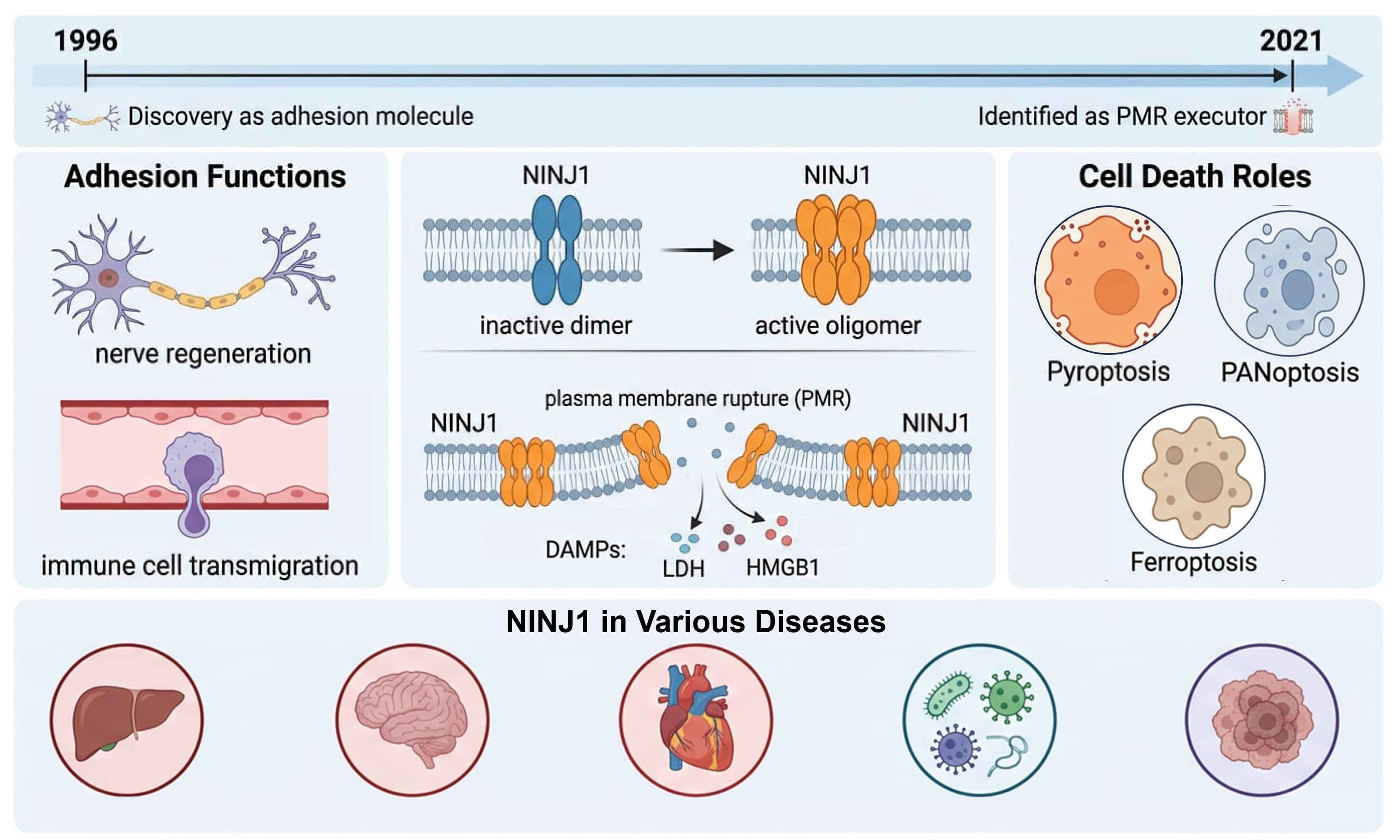
Graphical Abstract
1. Introduction
Cell death is a fundamental biological process for multicellular organisms to maintain tissue homeostasis, defend against pathogen invasion, and eliminate aberrant cells. Among the various forms of programmed cell death, lytic cell death is characterized by terminal features of plasma membrane rupture and release of intracellular contents. The released damage-associated molecular patterns (DAMPs) can activate innate immune responses, driving the amplification and propagation of inflammatory reactions [1,2]. Pyroptosis, necroptosis, and ferroptosis all fall within the category of lytic cell death, playing dual-edged roles in infection defense and disease pathogenesis [3,4]. For a long time, PMR was generally considered a passive event caused by osmotic imbalance during cell death. For example, in studies of pyroptosis, the classic model includes two steps: first, Gasdermin D (GSDMD) forms small pores in the plasma membrane, leading to IL-1β release and non-selective ion flow; subsequently, osmotic swelling triggers PMR, releasing large-molecular-weight DAMPs such as HMGB1 and LDH [5], where the second step of PMR was thought not to require specialized molecular executors.
NINJ1 was first identified by Araki and Milbrandt in 1996 through differential hybridization screening from a rat sciatic nerve injury model. This protein is a 16 kDa cell surface molecule with two transmembrane regions, both the N-terminus and C-terminus, located extracellularly. Its extracellular domain contains an evolutionarily conserved region of approximately 26 amino acids that mediates homophilic adhesion and promotes axonal extension of dorsal root ganglion neurons in vitro [6]. In the two decades following its initial discovery, functional studies of NINJ1 gradually expanded to multiple fields including neuroinflammation, immune cell migration, and tumor biology: Ifergan et al. found that NINJ1 mediates monocyte transmigration across the blood-brain barrier, closely associated with multiple sclerosis and experimental autoimmune encephalomyelitis (EAE) [7]; Yang et al. revealed the dual function of NINJ1 in p53-dependent tumorigenesis [8]; additionally, roles of NINJ1 in Toll-like receptor 4 signaling [9], atherosclerosis [10], endothelial cell protection [11], osteoclast development [12], and cellular senescence [13,14] have been successively reported. However, during this period, NINJ1 was always regarded as a relatively marginal adhesion/inflammation-related molecule and did not attract widespread attention.
In 2021, Kayagaki et al. identified NINJ1 as an active executor of PMR through a forward genetic screen, a discovery that fundamentally altered the core paradigm of cell death biology [15]. They first demonstrated that PMR is an active, protein-mediated process rather than a passive osmotic lysis event, changing the understanding of terminal membrane rupture in cell death biology. Second, they redefined NINJ1’s function from a simple adhesion molecule to a terminal executor of cell death, expanding research on this molecule from adhesion, migration, and inflammation-related functions to cell death. This review mainly summarizes the current research status of NINJ1’s structure, activation regulation, disease association, and therapeutic targeting, and analyzes key gaps remaining in the current research field.
2. Structural Basis of NINJ1-Mediated Plasma Membrane Rupture
2.1. NINJ1 Forms an Auto-Inhibited Dimer Under Steady-State Conditions
As a protein residing on the plasma membrane, how is NINJ1’s PMR activity inhibited under steady-state conditions? In 2025, Pourmal et al. used cryo-electron microscopy (cryo-EM) to resolve the three-dimensional structure of inactive NINJ1 [16], revealing an elegant “auto-inhibition” mechanism. In the resting state, NINJ1 forms a face-to-face homodimer with a three-helix conformation, where transmembrane helix 1 (TM1) does not undergo kinking. The inactive dimer sequesters the membrane rupture-inducing hydrophilic surface of NINJ1 within the dimer interface while masking the binding site for TM1 kinking in adjacent activated NINJ1 molecules. To validate this mechanism, researchers performed mutational experiments: mutations that disrupt dimer stability led to spontaneous NINJ1 activation and cell death, whereas mutations that enhance dimer stability inhibited NINJ1 activity. Mutations that disrupt stability also promoted spontaneous TM1 kinking, a structural hallmark of NINJ1 activation. Thus, NINJ1 mutually inhibits itself in a trans-dimeric form to prevent unplanned PMR and cell death (Figure 1A). Activation requires a series of structural changes: dimer dissociation, TM1 kinking, and rearrangement of amphipathic helices—acting like a molecular switch to transition NINJ1 from a steady state to an active state.
2.2. Active State: Amphipathic Filaments and Three PMR Models
How does activated NINJ1 assemble into a membrane-rupturing structure? Several independent structural studies in recent years have provided three complementary yet distinct models. Degen et al. combined ultra-high-resolution microscopy, cryo-EM, mutational analysis, and molecular dynamics simulations to propose an atomic model of NINJ1-mediated membrane rupture [17]. In living cells, NINJ1 exists as a monomer on the cell membrane, with α1 and α2 helices in the extracellular region and α3 and α4 helices inserted into the membrane. Upon cell death, the amphipathic α1 and α2 helices insert into the membrane to form a knotted conformation, connecting adjacent protomers to form large oligomers (Figure 1B). Cryo-electron microscopy structures reveal that these NINJ1 fibers adopt a tightly packed, fence-like array of transmembrane α-helices. The assembled larger oligomers stabilize the size and shape of damaged membranes by covering their edges, facilitating the release of DAMPs and other intracellular contents into the extracellular environment (Figure 1C). David et al., through a series of structural biology experiments and cell imaging, demonstrated that NINJ1 monomers can oligomerize and insert into the cell membrane to excise a corresponding membrane fragment, proposing a “cookie-cutting” mechanism for cell lysis—where NINJ1 oligomerization punches holes in the plasma membrane and releases cookie-shaped disk-like membrane structures [18] (Figure 1D). Sahoo et al., using cryo-EM to study the structural properties of NINJ1 and NINJ2, proposed a different model: when cells initiate lytic death, they begin to swell, stretching the plasma membrane. This promotes NINJ1 dimer formation, and a series of dimers aggregate into long chains that bind to the membrane like a zipper. The lateral tension of the swollen membrane pulls on both sides of the zipper, weakening interactions along the central line. When tension exceeds a threshold, the zipper splits open, forming pores in the membrane. If NINJ1 subunits fully cross-link during zipper formation, a NINJ1 ring structure with a central pore forms on the plasma membrane; if not fully cross-linked, NINJ1 subunits continue extending to form filaments that wrap around membrane fragments due to hydrophobic interactions between NINJ1’s hydrophobic side and the membrane fragments. The entire PMR mechanism depends on the physical strength of plasma membrane expansion [19] (Figure 1E). Although these models differ in detail, they converge on a consensus: the active state of NINJ1 is an amphipathic linear polymer whose hydrophilic surface repels lipids and stabilizes membrane edges, ultimately achieving membrane rupture. These models help understand NINJ1-mediated PMR to varying degrees. Currently, the conditions triggering NINJ1’s transition from inactive to active states remain to be further explored.
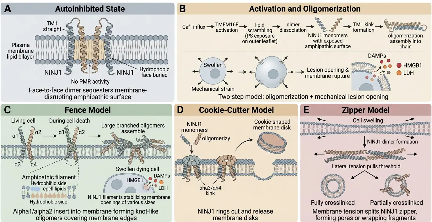
Figure 1. Structural mechanism of NINJ1-mediated plasma membrane rupture: (A) Autoinhibited State. In the resting state, NINJ1 forms face-to-face homodimers, adopting a triple-helical conformation, with the transmembrane helix 1 (TM1) remaining in a straight state. The dimer seals the exposed hydrophobic surface induced by membrane rupture at the interface, while masking the binding sites of adjacent activated NINJ1 molecules that have kinked TM1, thereby achieving self-inhibition. In this state, the cell has no PMR activity. Mutations that disrupt the stability of the dimer lead to spontaneous activation of NINJ1 and cell death, while mutations that enhance the stability of the dimer inhibit NINJ1 activity. (B) Activation and Oligomerization. The activation of NINJ1 follows a two-step model. Step 1 (oligomerization): Ca2+ influx and other signals destabilize and dissociate the autoinhibited dimer, causing NINJ1 monomers to expose the amphiphilic surface, forming a kink (kink) through TM1 to undergo oligomerization assembly. Step 2 (lesion opening): Cell expansion and external mechanical strain drive the final opening of membrane damage, resulting in plasma membrane rupture and release of cytoplasmic contents. (C) “Fence” Model. In living cells, NINJ1 exists as monomers on the cell membrane, with the α1 and α2 helices located on the extracellular side, and the α3 and α4 helices inserted into the membrane. Upon cell death, these helices insert into the membrane, driving oligomerization into branched fibrils. These fibrils form a tightly packed, fence-like array that stabilizes the edges of membrane lesions. The amphipathic architecture of the fibril (hydrophobic faces inward, hydrophilic faces outward) creates a conduit for the release of DAMPs and cytosolic components. (D) “Cookie-Cutter” Model. The NINJ1 monomer undergoes oligomerization and inserts into the cell membrane, forming a ring-like oligomer, “cutting” out corresponding disc-shaped membrane sheets on the plasma membrane, and the released NINJ1 ring wraps the lipid components and releases them to the extracellular environment, leading to loss of membrane area and PMR. Each subunit’s α3/α4 transmembrane helix has crucial kinks that are essential for the formation of the ring structure and the membrane cutting activity. (E) “Zipper” Model. When the cell initiates lytic cell death and begins to expand, the plasma membrane is stretched, promoting NINJ1 to form dimers, and a series of dimers polymerize to form a long chain, binding to the membrane to form a structure similar to a zipper. The lateral tension of the expanded plasma membrane pulls from both sides of the zipper, and when the tension exceeds a certain threshold, the zipper splits and opens, forming holes in the membrane. If all NINJ1 subunits are cross-linked when forming the zipper structure, a NINJ1 ring with a hole in the middle is formed on the plasma membrane; if not fully cross-linked, the NINJ1 subunits continue to extend to form filaments.
3. Activation Signals and Regulatory Mechanisms of NINJ1
The activation trigger mechanism of NINJ1 is one of the most active research directions. Dondelinger et al. found that NINJ1 is a key regulator of PMR and DAMPs release in various forms of regulated necrosis (ferroptosis, parthanatos, H2O2-induced necrosis, and secondary necrosis), and its function is independent of pore-forming molecules such as MLKL, GSDMD, and GSDME [20]. NINJ1 oligomerization is induced by cell swelling, but reactive oxygen species (ROS) are neither sufficient nor necessary, indicating an additional unknown activation mechanism upstream of cell swelling-induced oligomerization. Addressing this gap, a recent preprint by the Borges team proposes a critical role for calcium influx: calcium activates the phospholipid scramblase TMEM16F, driving membrane lipid rearrangement and thereby triggering NINJ1 activation [21]. Lipid rearrangement refers to the translocation of phosphatidylserine (PS) from the inner to the outer leaflet of the membrane, altering membrane lipid asymmetry and local mechanical properties. The same team further demonstrated that extracellular ATP-induced NINJ1-mediated cell lysis via the P2X7R receptor similarly depends on calcium influx and TMEM16F lipid rearrangement, and that this process is independent of inflammasomes, pannexin, and GSDMD. Notably, the sources and regulation of calcium signaling in macrophages are highly complex, involving multiple levels, including epigenetics and calcium channel expression regulation [22], suggesting that NINJ1 activation thresholds may differ significantly across different inflammatory contexts. Based on these findings, a separate preprint has hypothesized a “two-step activation” model: Signal 1 (calcium influx, lipid rearrangement) triggers NINJ1 auto-inhibited dimer dissociation and oligomerization; Signal 2 (cell swelling) drives membrane damage [23]. Importantly, although NINJ2 can oligomerize with kinetics similar to NINJ1, it fails to execute PMR upon cell swelling—a contrast that, as demonstrated by Sahoo et al., supports the specificity of Step 2 in the two-step model [19] (Figure 1B). This model decomposes PMR execution into two regulatable steps, providing multiple potential targets for therapeutic intervention: blocking oligomerization in Step 1 or inhibiting membrane damage in Step 2.
Zhu et al. identified NINJ1 as the key mediator of mechanostrain-induced plasma membrane rupture (PMR), demonstrating that its expression level inversely correlates with membrane rupture thresholds [24]. However, NINJ1 alone is insufficient to achieve complete membrane disintegration during pyroptosis and requires additional mechanical force. This reveals a synergistic regulatory model where chemical cues and biophysical forces converge on NINJ1 to fine-tune PMR sensitivity. Beyond this direct mechanism, mechanical tension also sensitizes cells to ferroptosis via iron metabolism reprogramming [25]. Thus, in high-stress microenvironments, mechanical forces may coordinately drive lytic cell death by concurrently lowering the threshold for NINJ1-mediated PMR and activating ferroptosis pathways. Moreover, mechanical signals broadly influence transcriptional regulation [26,27,28,29], suggesting that the mechanical microenvironment may indirectly modulate NINJ1 expression, thereby dictating tissue-specific PMR susceptibility.
4. Role of NINJ1 in Different Cell Death Pathways
4.1. Pyroptosis
Pyroptosis is a form of programmed cell death mediated by the Gasdermin family of proteins (e.g., GSDMD, GSDME) [30]. Upon inflammasome activation, caspases-1/4/5/11 cleave GSDMD to release its N-terminal domain (GSDMD-NT), which forms pores on the cell membrane, releasing pro-inflammatory cytokines such as interleukin-1β (IL-1β) and triggering inflammatory responses [31]. After GSDMD pore formation, NINJ1 is activated: its N-terminal helix inserts into the plasma membrane and oligomerizes, leading to complete plasma membrane rupture and promoting the release of large-molecular-weight DAMPs (e.g., HMGB1, LDH) [5]. NINJ1 deletion does not affect GSDMD pore formation or IL-1β release but significantly reduces leakage of HMGB1 and other large molecules, alleviating inflammatory damage [9]. Moreover, NINJ1 deletion only blocks PMR rather than cell death, indicating that NINJ1-mediated PMR is a downstream event after GSDMD executes pyroptosis, with its core role being to promote the release of inflammatory contents. This functional division has been validated by multiple studies (Figure 2A). Septins have been confirmed to promote macrophage pyroptosis by regulating GSDMD cleavage and NINJ1-mediated PMR [32]; studies on how pore diameter dynamics control cell volume expansion during pyroptosis further suggest precise spatiotemporal coordination between GSDMD pores and NINJ1-mediated PMR [33]. Importantly, GSDMD pore formation does not always lead to PMR—under sub-lethal stimulation, GSDMD can form pores to release cytokines without triggering NINJ1-dependent PMR, allowing cells to repair membrane pores and survive. Only under sustained or strong stimulation do downstream signals (osmotic swelling, calcium influx, etc.) reach the threshold for triggering NINJ1 oligomerization. This threshold effect may be an important mechanism for the body to finely regulate inflammation intensity.
4.2. Post-Apoptotic Lysis
Apoptosis is a caspase-mediated programmed cell death executed via extrinsic (death receptor/caspase-8) and intrinsic (BAK/BAX-MOMP-caspase-9) pathways, resulting in characteristic morphological changes and the formation of apoptotic bodies [1]. When clearance is blocked, uncleared apoptotic cells can undergo secondary necrosis, releasing DAMPs and LDH via NINJ1-dependent PMR [34]. In GSDME-positive cells, caspase-3-activated GSDME can synergize with NINJ1 to promote PMR. NINJ1 executes PMR by sensing membrane curvature changes and exposed phosphatidylserine caused by post-apoptotic cell swelling, without affecting early membrane blebbing during apoptosis—a mechanism conserved across multiple cell death pathways [35].
4.3. PANoptosis
PANoptosis is a novel inflammatory programmed cell death pathway characterized by the synergistic action of pyroptosis, apoptosis, and necroptosis [36]. Its core is the assembly of a PANoptosome complex, which integrates key components of multiple cell death pathways, including ASC, caspases-1, RIPK1, RIPK3, and caspase-8. Recent studies show that NINJ1 plays a unique and critical role in PANoptosis: it independently mediates plasma membrane rupture and the release of inflammatory content via oligomerization, a process that is independent of traditional effector molecules (GSDMD, GSDME, MLKL). Experiments confirm that individual inhibition of MLKL, caspase-1, or caspase-3 has limited effects on NINJ1 oligomerization, but combined inhibition significantly blocks this process, indicating that PANoptosis regulates NINJ1 by integrating multiple signaling pathways [37]. Han et al. found that under infection and heat stress conditions, NINJ1 is a key executor of PANoptosis, independently releasing inflammatory molecules and DAMPs without GSDMD, GSDME, or MLKL. In a heat stress mouse model, NINJ1 deletion significantly reduced mortality [38]. Xu et al. found that influenza A virus (IAV) infection upregulates NINJ1 expression and mediates PANoptosis-induced cell lysis; NINJ1 deletion alleviated IAV-induced lung injury and mortality [39]. Additionally, the ZBP1-NINJ1 axis has been confirmed to play a key role in ethanol-induced PANoptosis and alcohol-related liver disease [40,41] (Figure 2B). These studies collectively indicate that NINJ1 may be a shared terminal PMR executor across pathways, making it an ideal target for limiting PANoptosis-related inflammatory damage.
4.4. Ferroptosis
Ferroptosis is an iron-dependent regulated cell death characterized by abnormally increased lipid peroxidation in cell membranes, regulated by multiple metabolic pathways including antioxidant systems (defects in GPX4 and FSP1 function), iron metabolism (Fe2+ promoting lipid peroxidation via Fenton reaction), and cysteine metabolism (system Xc− transporter inhibition reducing GSH synthesis) [42]. The role of NINJ1 in ferroptosis is controversial: on one hand, studies show that NINJ1 regulates PMR downstream of phospholipid peroxidation in GPX4 inhibitor-induced ferroptosis, promoting DAMPs release from ferroptotic cells [20]. For example, Ramos et al. reported that NINJ1 oligomerizes during ferroptosis, and Ninj1 deficiency protects macrophages and fibroblasts from ferroptosis-related PMR. NINJ1 is not essential for upstream steps of ferroptosis but is critical for early loss of membrane integrity, and DAMPs release [43]. Recently, some studies have found that cell mechanical state may be an important factor in determining ferroptosis sensitivity. Mechanical stress increases free iron and sensitizes ferroptosis through NCOA4-mediated ferritinophagy [25,44]. Considering that NINJ1 is also affected by mechanical forces, mechanical stress may represent a candidate convergence point between upstream iron-metabolic drivers and downstream PMR execution—though this linkage requires direct testing. On the other hand, some studies found that NINJ1 knockout increases xCT expression and stability, promoting cystine uptake and elevating CoA and GSH concentrations, thereby protecting cancer cells from xCT inhibitor-induced ferroptosis. Notably, glycine—known to inhibit classical NINJ1-mediated membrane rupture—has no effect on ferroptosis [45] (Figure 2C). This phenomenon implies that NINJ1 has two functional roles in ferroptosis: classical PMR execution and non-classical metabolic regulation, possibly mediated by different molecular interfaces or regulatory circuits. This discovery expands the understanding of NINJ1’s functional diversity, but the structural basis and relative contributions of these two roles in vivo remain to be explored.
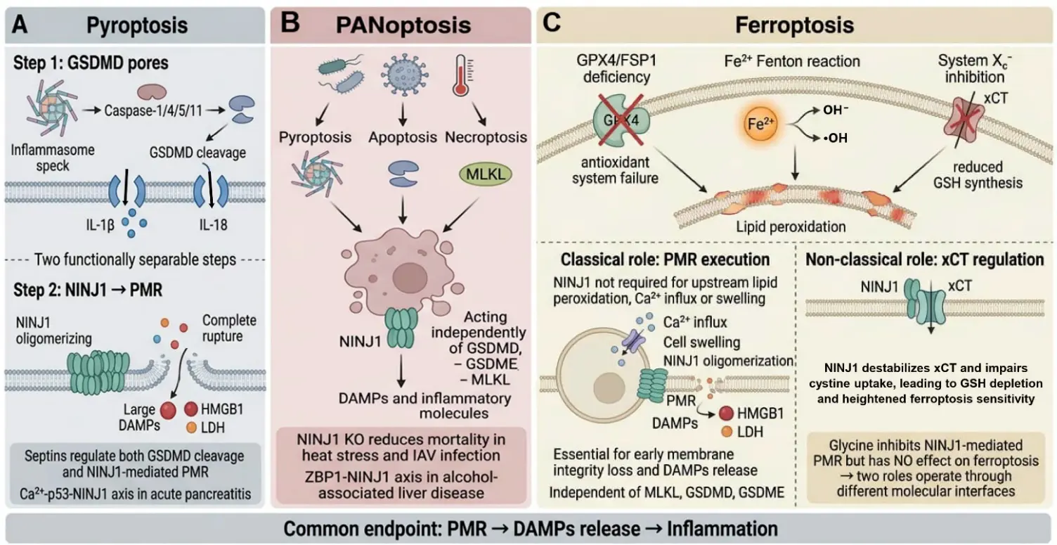
Figure 2. The role of NINJ1 in cell death. (A) Pyroptosis. The PMR mediated by NINJ1 is the downstream event after GSDMD performs pyroptosis. After the activation of the inflammasome, Caspase-1/4/5/11 cleaves GSDMD, and its N-terminal fragment forms small pores on the plasma membrane, mediating the release of small molecule cytokines such as IL-1β and IL-18 and non-selective ionic flow. NINJ1 then oligomerizes to execute the final plasma membrane rupture, resulting in the release of large molecule DAMPs (HMGB1, LDH) to the extracellular space. (B) PANoptosis. Pyroptosis, necroptosis, and apoptotic pathways are simultaneously activated under conditions such as infection (bacteria, viruses) and heat stress. NINJ1 acts as the final executor to release inflammatory molecules and DAMPs. (C) Ferroptosis. Ferroptosis is a form of iron-dependent regulated cell death, with its core feature being the abnormal increase in lipid peroxidation of the cell membrane, regulated by multiple metabolic pathways. NINJ1 can regulate the PMR downstream of phospholipid peroxidation in ferroptosis induced by GPX4 inhibitors, promoting the release of DAMPs by ferroptotic cells. NINJ1 is not essential for the upstream steps of ferroptosis, but is crucial for the early loss of membrane integrity and the release of DAMPs. NINJ1 knockout can increase the expression and stability of xCT, thereby promoting cystine input and increasing CoA and GSH concentrations, protecting cancer cells from ferroptosis induced by xCT inhibitors.
5. Role of NINJ1 in Various Diseases
5.1. Inflammatory Diseases and Targeted Therapy
Since NINJ1-mediated PMR is a key step in DAMPs release and inflammation amplification, inhibiting NINJ1 may be a critical strategy to alleviate cell death-related inflammation. In 2023, Kayagaki et al. reported the development of an anti-NINJ1 monoclonal antibody in Nature. This antibody blocks NINJ1 oligomerization and PMR. In multiple liver cell PMR models induced by TNF plus d-galactosamine, concanavalin A, Jo2 anti-Fas agonist antibody, or ischemia-reperfusion injury, NINJ1 inhibition or Ninj1 deletion improved tissue damage, reduced serum LDH, transaminases, and DAMPs levels, and decreased neutrophil infiltration [46]. This was the first demonstration in animal models that targeting the PMR execution step can produce clear therapeutic benefits.
In the cardiovascular field, NINJ1 plays a multifaceted role. Wu et al. found that NINJ1 enhances macrophage infiltration and promotes abdominal aortic aneurysm (AAA) development by blocking the TLR4-ANXA2 interaction in AAA formation [47]. Consistent with its pathogenic role in AAA, NINJ1 has also been identified as a potential anti-inflammatory target for thoracic aortic dissection [48]. Jeon et al. reported that soluble NINJ1 (the shed extracellular segment from the cell surface) has anti-inflammatory activity, ameliorating atherosclerosis by competitively inhibiting the adhesive function of membrane-bound NINJ1 [10]. This seemingly contradictory phenomenon suggests that the membrane-bound and soluble forms of NINJ1 may have opposite biological effects, which is important for designing therapeutic strategies—simply blocking NINJ1 function may have different or even opposite effects in different cardiovascular pathologies.
During organ ischemia/reperfusion (I/R) injury, NINJ1 is governed by tissue-specific post-translational modifications. In the kidney, TRIM72-mediated ubiquitination at K111 normally targets NINJ1 for degradation; however, AKI downregulates TRIM72, thereby stabilizing NINJ1 and driving PMR, HMGB1 release, and fibrosis via MMT and NETs [49]. In the liver, Ubc9-mediated SUMOylation at K103 inhibits NINJ1 membrane localization, while hepatic I/R reduces Ubc9 levels, permitting NINJ1 translocation and DAMP-driven NF-κB activation [50]. Notably, NINJ1 also interacts with Dusp1 in the liver to regulate macrophage and neutrophil recruitment during I/R [51]. These parallel yet distinct pathways establish NINJ1 as a convergent effector of I/R injury, with its precise impact dictated by tissue-specific regulatory networks.
In traumatic brain injury, NINJ1-mediated PMR of pyroptotic endothelial cells exacerbates blood-brain barrier disruption caused by neutrophil extracellular traps (NETs) [52]. In acute pancreatitis, the calcium-p53-NINJ1 signaling axis mediates acinar cell PMR [53]. These findings indicate that the NINJ1-PMR axis is a key link in tissue damage in various inflammatory diseases (Figure 3). Consequently, targeting NINJ1 represents a promising therapeutic avenue. Emerging therapeutic strategies include anti-NINJ1 antibodies [46], sNINJ1-mimetic peptides (ML56/PN12) that suppress macrophage pro-inflammatory gene expression [54], and muscimol (a GABA-A agonist) that blocks NINJ1 oligomerization [55].
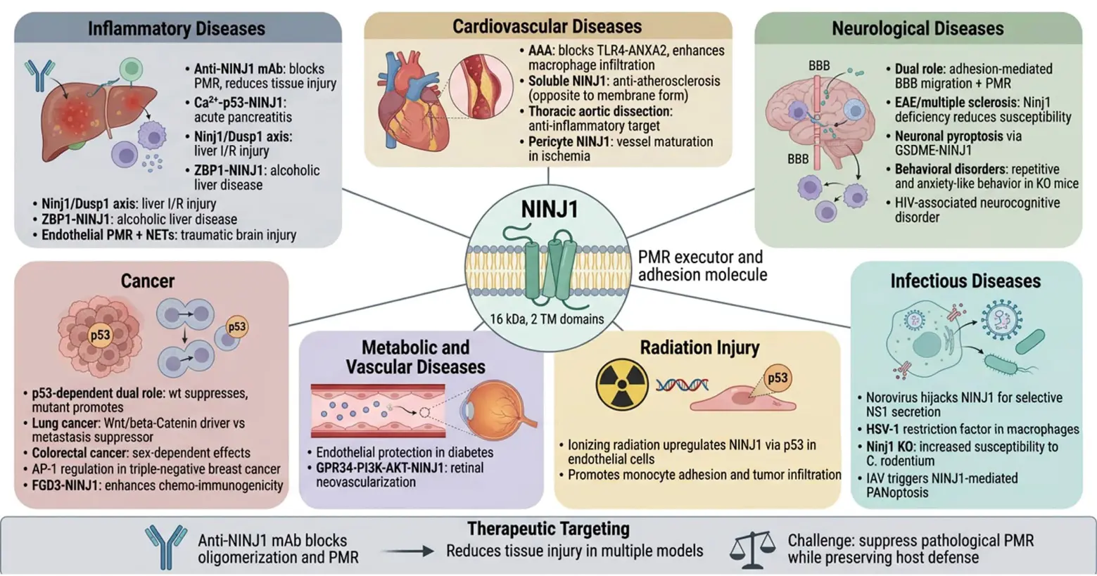
Figure 3. The multifaceted roles and therapeutic potential of NINJ1 in various diseases. The NINJ1 protein possesses the dual identities of a PMR executor and an adhesion molecule. It plays a role in various disease types such as inflammatory diseases, cardiovascular diseases, neurological diseases, metabolic and vascular diseases, infectious diseases, tumors and radiation injuries. Anti-NINJ1 monoclonal antibodies can prevent NINJ1 oligomerization and PMR, and alleviate tissue damage in various animal models. In the future, NINJ1 may serve as a therapeutic target for multiple diseases, but the key lies in how to inhibit pathological PMR while retaining its defensive function.
5.2. Neurological Diseases
NINJ1 also plays dual roles as an adhesion molecule and PMR executor in the nervous system. In multiple sclerosis and EAE models, NINJ1 is considered a key molecule for myeloid cell transmigration across the blood-brain barrier [7], and Ninj1 deficiency attenuates EAE clinical symptoms and susceptibility [56]. This function is mainly mediated by NINJ1’s homophilic adhesion activity, which parallels ICAM-1/Src signaling: ICAM-1 ligation activates Src kinases that phosphorylate ICAM-1 at Tyr518, promoting clustering and leukocyte transmigration [57]. Although no study has directly demonstrated NINJ1-ICAM-1/Src signaling cross-talk, the co-involvement of both molecules in leukocyte recruitment raises the hypothesis that they may function cooperatively, which warrants future investigation. However, under traumatic brain injury and neuroinflammation conditions, NINJ1’s PMR function may also participate: pyroptotic endothelial cells release DAMPs via NINJ1-mediated PMR, further exacerbating blood-brain barrier disruption and inflammatory infiltration [52]. Thus, in neurological diseases, NINJ1 may participate in pathology through both adhesion and PMR mechanisms, with the relative contribution of each mechanism varying by disease type and stage.
At the behavioral level, Ninj1-knockout mice exhibit repetitive and anxiety-like behaviors [58], suggesting that NINJ1 may have undefined roles in neural development or synaptic function. In HIV-associated neurocognitive disorders, caspase-cleaved Gasdermin E-induced neuronal pyroptosis is also associated with NINJ1-related PMR mechanisms [59]. These findings indicate that NINJ1’s function in the nervous system is far more complex than initially thought and warrants further in-depth study.
5.3. Infectious Diseases
NINJ1’s role in infection defense was noted when it was found to be involved in lytic cell death. Ninj1-deficient mice are more susceptible to Citrobacter rodentium infection, indicating the important function of PMR in antibacterial host defense. Mechanistically, NINJ1 utilizes its evolutionarily conserved extracellular domain for oligomerization, thereby executing PMR. This result was also validated in a study by Elisabet Bjanes et al.: targeted mutations in the Card19locus were found to be associated with impaired NINJ1 expression, defective cell lysis, and increased susceptibility to Yersinia infection [60]. With deepening research, new roles of NINJ1 in infectious diseases have been gradually discovered. In 2025, Song et al. reported that murine norovirus (MNoV) uses NINJ1 to selectively secrete its intracellular viral protein NS1 [61]. During infection, NINJ1 is recruited to viral replication sites, forming oligomerized punctate structures that directly interact with NS1, enabling selective binding and secretion of NS1. This discovery reveals a new role for NINJ1 in protein secretion. Additionally, NINJ1 has been reported as a restriction factor for HSV-1 in mouse macrophages [62], indicating its important function in host antiviral defense. These findings suggest that NINJ1’s biological functions extend beyond PMR execution—it may regulate multiple aspects of membrane dynamics: from pore formation to membrane fragment release and selective protein secretion.
5.4. Metabolic, Vascular-Related Diseases, and Radiation Injury
Beyond the major diseases mentioned above, NINJ1 is also implicated in other pathologies. In diabetes, NINJ1 function blockade protects endothelial cells from high-glucose-induced damage [11]. In hindlimb ischemia models, pericyte-specific NINJ1 deletion weakens vessel maturation and blood flow recovery [63]. In retinal diseases, ganglion cell-derived LysoPS induces retinal neovascularization by activating the microglial GPR34-PI3K-AKT-NINJ1 axis [64]. In hemorrhagic and hemolytic conditions, hemolysis releases free heme as a potent DAMP that activates TLR4 on monocytes and endothelial cells [65]. Heme oxygenase degrades heme into biliverdin, carbon monoxide, and iron; biliverdin has been linked to cerebral vasospasm, microthrombus, and neuronal apoptosis after subarachnoid hemorrhage [66]. Whether NINJ1-PMR synergizes with heme-driven pathology to exacerbate vasospasm remains to be determined. In radiation injury, ionizing radiation upregulates endothelial cell NINJ1 expression via the p53 pathway, promoting monocyte adhesion and tumor infiltration [14,67]; whether NINJ1 further amplifies post-radiation inflammation via PMR-mediated DAMPs release remains to be determined.
5.5. Tumors
NINJ1 exhibits dual functions (tumor-suppressive and tumor-promoting) in tumors. In 2017, Yang et al. found that NINJ1 has dual functions in p53-dependent tumorigenesis: in cells with wild-type p53, NINJ1 deletion inhibits cell proliferation; in cells with mutant p53, NINJ1 deletion promotes tumor progression by increasing mutant p53 expression [8]. In lung cancer, NINJ1’s role is also contradictory: Hyun et al. found that NINJ1 drives lung tumor formation and progression via the Wnt/β-catenin signaling pathway [68], whereas Jang et al. reported that NINJ1 suppresses metastatic properties in lung cancer cells by inhibiting intracellular signaling pathways [69]. In colorectal cancer, Ninj1 deficiency differentially mitigates chemically induced tumorigenesis with sex differences [70,71]. AP-1 has been identified as an upstream regulator of NINJ1 in TNFα-mediated triple-negative breast cancer progression [72]. A recent study found that FGD3 enhances chemotherapy efficacy and immunogenicity in breast cancer by mediating lytic cell death, revealing the potential of cell death and NINJ1-related pathways in tumor immunity [73]. Overall, NINJ1’s function in tumors highly depends on genetic background, tumor type, and microenvironmental conditions, and it cannot be simply classified as a tumor promoter or suppressor.
6. Summary and Outlook
Research on NINJ1 has made significant progress in recent years, but many limitations and gaps remain. Currently, most experiments on NINJ1-mediated PMR use macrophages, and NINJ1’s expression levels, activation thresholds, and functional dependencies may vary significantly among different cell types. Little is known about NINJ1’s activation thresholds and functional roles in non-immune cells (epithelial cells, endothelial cells, neurons, cardiomyocytes, etc.). The discovery of SIGLEC12 in necroptosis further indicates that PMR execution mechanisms are more diverse than initially thought [74]. Whether NINJ1, SIGLEC12, and other potential PMR executors coordinate, compete, or complement each other is an urgent question to address.
Since NINJ1 mediates plasma membrane rupture in lytic cell death, a natural follow-up question is: does NINJ1 participate in other physiological processes dependent on membrane remodeling? Cell division involves a series of membrane dynamic events, including nuclear envelope breakdown (NEBD) in early mitosis and plasma membrane constriction/separation during cytokinesis—all essentially involving membrane disassembly and reconstruction. To explore NINJ1’s potential role in these physiological membrane remodeling processes, we knocked down NINJ1 expression via RNA interference and systematically analyzed cell cycle distribution changes using flow cytometry. Results showed that NINJ1 downregulation had no significant effect on cell cycle progression. This preliminary observation suggests that NINJ1 may not play a key role in plasma membrane remodeling and nuclear envelope disassembly/reconstruction during cell division, although further validation with more rigorous experimental approaches is warranted. Mechanistically, NINJ1 activation may require specific pathological signal cascades, whereas NEBD during mitosis is driven by CDK1-Cyclin B kinase complex-mediated lamin phosphorylation, belonging to a completely different molecular regulatory system. Thus, although NINJ1 has potent membrane rupture activity, its function is strictly process-specific, activated only in specific signal environments related to cell death, rather than acting as a universal membrane remodeling factor in all membrane structure change-related biological processes.
The fence model of Degen et al., the “cookie-cutting” model of David et al., and the “zipper” model of Sahoo et al. converge on the core mechanism but differ in PMR details. These models have not been fully unified, possibly because cryo-EM structures primarily derive from in vitro reconstitution systems or detergent-treated samples, which differ significantly from in situ membrane environments. Additionally, the polymorphism of active-state structures suggests that NINJ1 may adopt different assembly strategies depending on membrane composition and tension. There is currently a lack of real-time dynamic observation of NINJ1’s entire process (from dimer dissociation to filament formation and membrane fragment release) on intact cell membranes. Future developments in in situ cryo-electron tomography (cryo-ET) and live-cell super-resolution imaging may answer this question.
Moreover, the understanding of NINJ1 activation signals remains incomplete. Although calcium pathways and cell swelling have been proposed as key signals for NINJ1 activation based on preprint findings [21,23], several critical questions remain: What signal triggers dimer dissociation? Is TM1 kinking actively driven or passively responsive to membrane environmental changes? How do changes in membrane lipid composition precisely regulate NINJ1 conformational transitions? Do different forms of cell death share the same activation mechanism? How mechanical forces affect NINJ1 conformational transitions and oligomerization, and the synergy between mechanical and chemical signals—all require in-depth study. Recent studies have revealed that mechanical signals can regulate gene expression through chromatin remodeling complexes and transcription factor pathways [26], raising the possibility that the mechanical microenvironment may modulate NINJ1 expression and thereby influence lytic cell death thresholds. Hemodynamic flow represents one such physiologically relevant signal; disturbed shear stress upregulates endothelial ICAM-1 to promote atherogenesis [75]. Given that NINJ1 and MMP9 are independent predictors of plaque vulnerability [76], it is imperative to investigate whether flow dynamics further govern NINJ1-mediated PMR by modulating endothelial membrane tension and death thresholds.
Currently, disease research on NINJ1 mainly relies on mouse models, and species-specific discoveries like SIGLEC12 suggest possible important differences in PMR mechanisms between mice and humans. Most disease studies use global or myeloid-specific Ninj1-knockout mice, making it difficult to distinguish NINJ1’s contributions in different cell types.
At the therapeutic translation level, anti-NINJ1 antibodies perform well in acute liver injury models, but their long-term safety and efficacy in chronic inflammatory diseases remain unclear. Some studies show that NINJ1-mediated PMR plays protective roles in host defense; thus, how to inhibit pathological PMR while preserving its physiological functions is a question worth considering. Strategies to address these issues may include developing tissue-specific delivery systems to limit the scope of NINJ1 inhibitors; using different targets in the two-step activation model for refined intervention; or developing conditionally activated NINJ1 modulators that act only in pathological microenvironments. Furthermore, NINJ1 presents a novel conceptual target for CNS delivery. FUS leverages BBB disruption for drug transport [77,78], and NINJ1 participates in endothelial barrier disruption through both adhesion and PMR. It is tempting to speculate that NINJ1 modulation could synergize with FUS to optimize brain tumor penetration, though this speculative yet intriguing avenue warrants future investigation.
From its initial discovery as a nerve injury-induced adhesion molecule in 1996 to its confirmation as an active executor of PMR in lytic cell death in 2021, and subsequent significant advances in structural analysis, activation regulation, cross-pathway functions, and disease associations, NINJ1 research has established a relatively complete knowledge framework. However, many questions remain: the complete signaling pathway for NINJ1 activation, functional heterogeneity in different tissues and cell types, coordination with other PMR executors, and safety windows for therapeutic targeting—all will be key directions for future research. Deepening NINJ1 research will not only advance understanding of basic mechanisms of lytic cell death but also provide new therapeutic strategies for inflammatory diseases, neurodegenerative diseases, tumors, and more. With advances in structural biology, single-cell omics, and targeted drug development, NINJ1 research will bring more breakthroughs to basic research and clinical translation in cell death and inflammation biology in the future.
Statement of the Use of Generative AI and AI-Assisted Technologies in the Writing Process
During the preparation of this manuscript, the author used Qoder (Alibaba) in order to polish the text, improve grammar, and enhance the clarity of schematic diagrams. This tool was not used to generate scientific concepts, data, or references. After using this tool, the author reviewed and edited the content as needed and takes full responsibility for the content of the published article.
Acknowledgments
The authors would like to thank their colleagues for the insightful academic discussions and valuable suggestions, which greatly facilitated the conceptualization and drafting of this review.
Author Contributions
Conceptualization, H.L. and L.C.; Writing—Original Draft Preparation, H.L.; Writing—Review & Editing, H.L., Y.S. and L.C.; Figure Preparation, H.L.; Supervision, L.C.; Funding Acquisition, Y.S. and L.C. All authors have read and agreed to the published version of the manuscript.
Ethics Statement
Not applicable.
Informed Consent Statement
Not applicable.
Data Availability Statement
This is a Review article with no original data generated; all analyzed data are from previously published studies in the References.
Funding
This work was supported by grants from National Natural Science Foundation of China (82373525 and 82173465, L.C.; 82504352, Y.S.); Outstanding Youth Fund of Jiangsu Province (BK20240039, L.C.); Shanghai Science and Technology Committee Project (24ZR1414300, Y.S.); China Postdoctoral Science Foundation project (2025M771452, Y.S.).
Declaration of Competing Interest
The authors declare that they have no known competing financial interests or personal relationships that could have appeared to influence the work reported in this paper.
References
- Galluzzi L, Vitale I, Aaronson SA, Abrams JM, Adam D, Agostinis P, et al. Molecular mechanisms of cell death: Recommendations of the Nomenclature Committee on Cell Death 2018. Cell Death Differ 2018, 25, 486–541. DOI:10.1038/s41418-017-0012-4 [Google Scholar]
- Pandey A, Shen C, Feng S, Man SM. Cell biology of inflammasome activation. Trends Cell Biol. 2021, 31, 924–939. DOI:10.1016/j.tcb.2021.06.010 [Google Scholar]
- Ai Y, Meng Y, Yan B, Zhou Q, Wang X. The biochemical pathways of apoptotic, necroptotic, pyroptotic, and ferroptotic cell death. Mol. Cell 2024, 84, 170–179. DOI:10.1016/j.molcel.2023.11.040 [Google Scholar]
- Newton K, Dixit VM, Kayagaki N. Dying cells fan the flames of inflammation. Science 2021, 374, 1076–1080. DOI:10.1126/science.abi5934 [Google Scholar]
- Broz P. Pyroptosis: Molecular mechanisms and roles in disease. Cell Res. 2025, 35, 334–344. DOI:10.1038/s41422-025-01107-6 [Google Scholar]
- Araki T, Milbrandt J. Ninjurin, a Novel Adhesion Molecule, Is Induced by Nerve Injury and Promotes Axonal Growth. Neuron 1996, 17, 353–361. DOI:10.1016/s0896-6273(00)80166-x [Google Scholar]
- Ifergan I, Kebir H, Terouz S, Alvarez JI, Lécuyer M, Gendron S, et al. Role of ninjurin‐1 in the migration of myeloid cells to central nervous system inflammatory lesions. Ann. Neurol. 2011, 70, 751–763. DOI:10.1002/ana.22519 [Google Scholar]
- Yang HJ, Zhang J, Yan W, Cho SJ, Lucchesi C, Chen M, et al. Ninjurin 1 has two opposing functions in tumorigenesis in a p53-dependent manner. Proc. Natl. Acad. Sci. USA 2017, 114, 11500–11505. DOI:10.1073/pnas.1711814114 [Google Scholar]
- Jennewein C, Sowa R, Faber AC, Dildey M, von Knethen A, Meybohm P, et al. Contribution of Ninjurin1 to Toll-Like Receptor 4 Signaling and Systemic Inflammation. Am. J. Respir. Cell Mol. Biol. 2015, 53, 656–663. DOI:10.1165/rcmb.2014-0354OC [Google Scholar]
- Jeon S, Kim TK, Jeong SJ, Jung IH, Kim N, Lee MN, et al. Anti-Inflammatory Actions of Soluble Ninjurin-1 Ameliorate Atherosclerosis. Circulation 2020, 142, 1736–1751. DOI:10.1161/circulationaha.120.046907 [Google Scholar]
- Wang X, Qin J, Zhang X, Peng Z, Ye K, Wu X, et al. Functional blocking of Ninjurin1 as a strategy for protecting endothelial cells in diabetes mellitus. Clin. Sci. 2018, 132, 213–229. DOI:10.1042/cs20171273 [Google Scholar]
- Bae SJ, Shin MW, Son T, Lee HS, Chae JS, Jeon S, et al. Ninjurin1 positively regulates osteoclast development by enhancing the survival of prefusion osteoclasts. Exp. Mol. Med. 2019, 51, 1–16. DOI:10.1038/s12276-018-0201-3 [Google Scholar]
- Toyama T, Sasaki Y, Horimoto M, Iyoda K, Yakushijin T, Ohkawa K, et al. Ninjurin1 increases p21 expression and induces cellular senescence in human hepatoma cells. J. Hepatol. 2004, 41, 637–643. DOI:10.1016/j.jhep.2004.06.027 [Google Scholar]
- Cho SJ, Rossi A, Jung YS, Yan W, Liu G, Zhang J, et al. Ninjurin1, a target of p53, regulates p53 expression and p53-dependent cell survival, senescence, and radiation-induced mortality. Proc. Natl. Acad. Sci. USA 2013, 110, 9362–9367. DOI:10.1073/pnas.1221242110 [Google Scholar]
- Kayagaki N, Kornfeld OS, Lee BL, Stowe IB, O’Rourke K, Li Q, et al. NINJ1 mediates plasma membrane rupture during lytic cell death. Nature 2021, 591, 131–136. DOI:10.1038/s41586-021-03218-7 [Google Scholar]
- Pourmal S, Truong ME, Johnson MC, Yang Y, Zhou L, Alegre K, et al. Autoinhibition of dimeric NINJ1 prevents plasma membrane rupture. Nature 2025, 637, 446–452. DOI:10.1038/s41586-024-08273-4 [Google Scholar]
- Degen M, Santos JC, Pluhackova K, Cebrero G, Ramos S, Jankevicius G, et al. Structural basis of NINJ1-mediated plasma membrane rupture in cell death. Nature 2023, 618, 1065–1071. DOI:10.1038/s41586-023-05991-z [Google Scholar]
- David L, Borges JP, Hollingsworth LR, Volchuk A, Jansen I, Garlick E, et al. NINJ1 mediates plasma membrane rupture by cutting and releasing membrane disks. Cell 2024, 187, 2224–2235.e16. DOI:10.1016/j.cell.2024.03.008 [Google Scholar]
- Sahoo B, Mou Z, Liu W, Dubyak G, Dai X. How NINJ1 mediates plasma membrane rupture and why NINJ2 cannot. Cell 2025, 188, 292–302.e11. DOI:10.1016/j.cell.2024.11.021 [Google Scholar]
- Dondelinger Y, Priem D, Huyghe J, Delanghe T, Vandenabeele P, Bertrand MJM. NINJ1 is activated by cell swelling to regulate plasma membrane permeabilization during regulated necrosis. Cell Death Dis. 2023, 14, 755. DOI:10.1038/s41419-023-06284-z [Google Scholar]
- Borges JP, Wang Y, David L, Volchuk A, Martins B, Cai R, et al. NINJ1 is activated by calcium-driven plasma membrane lipid scrambling during lytic cell death. bioRxiv 2024, 2024-10. DOI:10.1101/2024.10.23.619800 [Google Scholar]
- Ji M, Liang H, Dong S, Guo Y, Lin Y, Zhang H, et al. The SWI/SNF chromatin-remodeling subunit DPF2 regulates macrophage inflammation in intestinal injury via the CACNA1D-mediated MAPK pathway. Proc. Natl. Acad. Sci. USA 2025, 122, e2518762122. DOI:10.1073/pnas.2518762122 [Google Scholar]
- Hartenian E, Bernard EM, Ammirati G, Leloup HB, Mari SA, Degen M, et al. Membrane Tension Drives Opening of NINJ1 Lesions in Dying Cells. bioRxiv 2024. DOI:10.1101/2024.10.29.620849 [Google Scholar]
- Zhu Y, Xiao F, Wang Y, Wang Y, Li J, Zhong D, et al. NINJ1 regulates plasma membrane fragility under mechanical strain. Nature 2025, 644, 1088–1096. DOI:10.1038/s41586-025-09222-5 [Google Scholar]
- Luo C, Liang H, Ji M, Ye C, Lin Y, Guo Y, et al. Autophagy induced by mechanical stress sensitizes cells to ferroptosis by NCOA4-FTH1 axis. Autophagy 2025, 21, 1263–1282. DOI:10.1080/15548627.2025.2469129 [Google Scholar]
- Chang L, Azzolin L, Di Biagio D, Zanconato F, Battilana G, Lucon Xiccato R, et al. The SWI/SNF complex is a mechanoregulated inhibitor of YAP and TAZ. Nature 2018, 563, 265–269. DOI:10.1038/s41586-018-0658-1 [Google Scholar]
- Ji M, Chen D, Shu Y, Dong S, Zhang Z, Zheng H, et al. The role of mechano-regulated YAP/TAZ in erectile dysfunction. Nat. Commun. 2023, 14, 3758. DOI:10.1038/s41467-023-39009-z [Google Scholar]
- Zheng L, Luo C, Yang N, Pei H, Ji M, Shu Y, et al. Ionizing radiation-induced long noncoding RNA CRYBG3 regulates YAP/TAZ through mechanotransduction. Cell Death Dis. 2022, 13, 209. DOI:10.1038/s41419-022-04650-x [Google Scholar]
- Ji M, Dong S, Lu S, Liang H, Lin Y, Luo C, et al. Mechano-YAP/TAZ-regulated smooth muscle cells are an important source of Wnt signalling for gut regeneration. Clin. Transl. Med. 2024, 14, e70005. DOI:10.1002/ctm2.70005 [Google Scholar]
- Kayagaki N, Dixit VM. Rescue from a fiery death: A therapeutic endeavor. Science 2019, 366, 688–689. DOI:10.1126/science.aaw1177 [Google Scholar]
- Kayagaki N, Stowe IB, Lee BL, O’Rourke K, Anderson K, Warming S, et al. Caspase-11 cleaves gasdermin D for non-canonical inflammasome signalling. Nature 2015, 526, 666–671. DOI:10.1038/nature15541 [Google Scholar]
- Brokatzky D, Gomes MC, Robertin S, Albino C, Miles SL, Mostowy S. Septins promote macrophage pyroptosis by regulating gasdermin D cleavage and ninjurin-1-mediated plasma membrane rupture. Cell Chem. Biol. 2024, 31, 1518–1528.e6. DOI:10.1016/j.chembiol.2024.07.003 [Google Scholar]
- Bastien E, Duprez G, Delanoë-Ayari H, Leloup H, Rivière C, Petrilli V, et al. Pore size dynamics control complex volume swelling in pyroptosis. Proc. Natl. Acad. Sci. USA 2026, 123, e2508022123. DOI:10.1073/pnas.2508022123 [Google Scholar]
- Sachet M, Liang YY, Oehler R. The immune response to secondary necrotic cells. Apoptosis 2017, 22, 1189–1204. DOI:10.1007/s10495-017-1413-z [Google Scholar]
- Yang JY, Luo CH, Wang KB, Tu XY, Xiao YY, Ou YT, et al. Unraveling the mechanisms of NINJ1-mediated plasma membrane rupture in lytic cell death and related diseases. Int. J. Biol. Macromol. 2025, 309, 143165. DOI:10.1016/j.ijbiomac.2025.143165 [Google Scholar]
- Wang Y, Kanneganti TD. From pyroptosis, apoptosis and necroptosis to PANoptosis: A mechanistic compendium of programmed cell death pathways. Comput. Struct. Biotechnol. J. 2021, 19, 4641–4657. DOI:10.1016/j.csbj.2021.07.038 [Google Scholar]
- Zhou X, Yu X, Wan C, Li F, Wang Y, Zhang K, et al. NINJ1 Regulates Platelet Activation and PANoptosis in Septic Disseminated Intravascular Coagulation. Int. J. Mol. Sci. 2023, 24, 4168. DOI:10.3390/ijms24044168 [Google Scholar]
- Han JH, Karki R, Malireddi RKS, Mall R, Sarkar R, Sharma BR, et al. NINJ1 mediates inflammatory cell death, PANoptosis, and lethality during infection conditions and heat stress. Nat. Commun. 2024, 15, 1739. DOI:10.1038/s41467-024-45466-x [Google Scholar]
- Xu Y, Zheng Y, Liu Y, Wei C, Ren J, Zuo W, et al. Ninjurin-1 mediates cell lysis and detrimental inflammation of PANoptosis during influenza A virus infection. Sig Transduct. Target. Ther. 2025, 10, 307. DOI:10.1038/s41392-025-02391-9 [Google Scholar]
- Qin Q, Chen W, King CD, Kumar SP, El Farran CA, Vogel P, et al. Identification of an IRF–ZBP1–caspase-8–NINJ1 axis in driving PANoptosis and pathology during alcohol-associated liver disease. Proc. Natl. Acad. Sci. USA 2025, 122, e2525296122. DOI:10.1073/pnas.2525296122 [Google Scholar]
- Zhang Z, Lu S, Shu Y, Zhang S, Guo Y, Lin Y, et al. Chromatin remodeling in pericentral hepatocytes modulates MASH through CYP450 activity. J. Hepatol. 2026. DOI:10.1016/j.jhep.2026.03.035 [Google Scholar]
- Jiang X, Stockwell BR, Conrad M. Ferroptosis: Mechanisms, biology and role in disease. Nat. Rev. Mol. Cell Biol. 2021, 22, 266–282. DOI:10.1038/s41580-020-00324-8 [Google Scholar]
- Ramos S, Hartenian E, Santos JC, Walch P, Broz P. NINJ1 induces plasma membrane rupture and release of damage-associated molecular pattern molecules during ferroptosis. EMBO J. 2024, 43, 1164–1186. DOI:10.1038/s44318-024-00055-y [Google Scholar]
- Zhang H, Lin Y, Ji M, Guo Y, Liang H, Kang K, et al. PABPC3 drives ovarian cancer metastasis and drug sensitivity by downregulating CLDN1 expression. Cell Death Dis. 2025, 16, 840. DOI:10.1038/s41419-025-08151-5 [Google Scholar]
- Chen SY, Wu J, Chen Y, Wang YE, Setayeshpour Y, Federico C, et al. NINJ1 regulates ferroptosis via xCT antiporter interaction and CoA modulation. Cell Death Dis. 2024, 15, 755. DOI:10.1038/s41419-024-07135-1 [Google Scholar]
- Kayagaki N, Stowe IB, Alegre K, Deshpande I, Wu S, Lin Z, et al. Inhibiting membrane rupture with NINJ1 antibodies limits tissue injury. Nature 2023, 618, 1072–1077. DOI:10.1038/s41586-023-06191-5 [Google Scholar]
- Wu Z, Xu Z, Pu H, Ding A, Hu J, Lei J, et al. NINJ1 Facilitates Abdominal Aortic Aneurysm Formation via Blocking TLR4‐ANXA2 Interaction and Enhancing Macrophage Infiltration. Adv. Sci. 2024, 11, e2306237. DOI:10.1002/advs.202306237 [Google Scholar]
- Sheng Y, Wu L, Chang Y, Liu W, Tao M, Chen X, et al. Tomo-seq identifies NINJ1 as a potential target for anti-inflammatory strategy in thoracic aortic dissection. BMC Med. 2023, 21, 396. DOI:10.1186/s12916-023-03077-1 [Google Scholar]
- Ye K, Lin S, Chen C, Chen Z, Lin K, Li G, et al. NINJ1 ubiquitination by TRIM72 protects against plasma membrane rupture and AKI-CKD progression. Cell Death Differ. 2026, 1–14. DOI:10.1038/s41418-026-01746-1 [Google Scholar]
- Huang K, Xu L, Na S, Xu Y, Liu Q, Ye S, et al. Ubc9‐mediated SUMOylation of Ninj1 alleviates inflammatory responses in hepatic ischaemia/reperfusion injury. Clin. Transl. Med. 2026, 16, e70677. DOI:10.1002/ctm2.70677 [Google Scholar]
- Hu Y, Zhan F, Wang Y, Wang D, Lu H, Wu C, et al. The Ninj1/Dusp1 Axis Contributes to Liver Ischemia Reperfusion Injury by Regulating Macrophage Activation and Neutrophil Infiltration. Cell. Mol. Gastroenterol. Hepatol. 2023, 15, 1071–1084. DOI:10.1016/j.jcmgh.2023.01.008 [Google Scholar]
- Zheng XB, Wang X, Gao SQ, Gao CC, Li T, Han YL, et al. NINJ1-mediated plasma membrane rupture of pyroptotic endothelial cells exacerbates blood-brain barrier destruction caused by neutrophil extracellular traps in traumatic brain injury. Cell Death Discov. 2025, 11, 69. DOI:10.1038/s41420-025-02350-x [Google Scholar]
- Lee C, Xin G, Li F, Wan C, Yu X, Feng L, et al. Calcium/P53/Ninjurin 1 Signaling Mediates Plasma Membrane Rupture of Acinar Cells in Severe Acute Pancreatitis. Int. J. Mol. Sci. 2023, 24, 11554. DOI:10.3390/ijms241411554 [Google Scholar]
- Mahib MMR. Ninjurin1 in cardiovascular and vascular biology: From molecular mechanisms to therapeutic opportunities. Clin. Transl. Med. 2026, 16, e70646. DOI:10.1002/ctm2.70646 [Google Scholar]
- den Hartigh AB, Loomis WP, Anderson MJ, Frølund B, Fink SL. Muscimol inhibits plasma membrane rupture and ninjurin-1(NINJ1) oligomerization during pyroptosis. Commun. Biol. 2023, 6, 1010. DOI:10.1038/s42003-023-05354-4 [Google Scholar]
- Ahn BJ, Le H, Shin MW, Bae SJ, Lee EJ, Wee HJ, et al. Ninjurin1 Deficiency Attenuates Susceptibility of Experimental Autoimmune Encephalomyelitis in Mice. J. Biol. Chem. 2014, 289, 3328–3338. DOI:10.1074/jbc.M113.498212 [Google Scholar]
- Liu G, Vogel SM, Gao X, Javaid K, Hu G, Danilov SM, et al. Src Phosphorylation of Endothelial Cell Surface Intercellular Adhesion Molecule-1 Mediates Neutrophil Adhesion and Contributes to the Mechanism of Lung Inflammation. Arterioscler. Thromb. Vasc. Biol. 2011, 31, 1342–1350. DOI:10.1161/atvbaha.110.222208 [Google Scholar]
- Le H, Ahn BJ, Lee HS, Shin A, Chae S, Lee SY, et al. Disruption of Ninjurin1 Leads to Repetitive and Anxiety-Like Behaviors in Mice. Mol. Neurobiol. 2017, 54, 7353–7368. DOI:10.1007/s12035-016-0207-6 [Google Scholar]
- Fernandes JP, Branton WG, Cohen EA, Koopman G, Kondova I, Gelman BB, et al. Caspase cleavage of gasdermin E causes neuronal pyroptosis in HIV-associated neurocognitive disorder. Brain 2024, 147, 717–734. DOI:10.1093/brain/awad375 [Google Scholar]
- Bjanes E, Sillas RG, Matsuda R, Demarco B, Fettrelet T, DeLaney AA, et al. Genetic targeting of Card19 is linked to disrupted NINJ1 expression, impaired cell lysis, and increased susceptibility to Yersinia infection. PLoS Pathog. 2021, 17, e1009967. DOI:10.1371/journal.ppat.1009967 [Google Scholar]
- Song J, Zhang L, Moon S, Fang A, Wang G, Gheshm N, et al. Norovirus co-opts NINJ1 for selective protein secretion. Sci. Adv. 2025, 11, eadu7985. DOI:10.1126/sciadv.adu7985 [Google Scholar]
- Hartenian E, Agustoni M, Broz P. NINJ1 blocks HSV-1 entry into macrophages to impact viral replication and immunity. EMBO Rep. 2025, 27, 69–88. DOI:10.1038/s44319-025-00638-8 [Google Scholar]
- Minoshima A, Kabara M, Matsuki M, Yoshida Y, Kano K, Tomita Y, et al. Pericyte-Specific Ninjurin1 Deletion Attenuates Vessel Maturation and Blood Flow Recovery in Hind Limb Ischemia. Arterioscler. Thromb. Vasc. Biol. 2018, 38, 2358–2370. DOI:10.1161/atvbaha.118.311375 [Google Scholar]
- Chen L, Zhang H, Zhang Y, Li X, Wang M, Shen Y, et al. Ganglion cell-derived LysoPS induces retinal neovascularisation by activating the microglial GPR34-PI3K-AKT-NINJ1 axis. J. Neuroinflamm. 2024, 21, 278. DOI:10.1186/s12974-024-03265-7 [Google Scholar]
- Bozza MT, Jeney V. Pro-inflammatory Actions of Heme and Other Hemoglobin-Derived DAMPs. Front. Immunol. 2020, 11, 1323. DOI:10.3389/fimmu.2020.01323 [Google Scholar]
- Hasanpour-Segherlou Z, Hutchinson H, Xu H, Amini S, Klaas E, Martinez ME, et al. Role of biliverdin reductase, a heme degradation pathway enzyme, in the development of vasospasm after subarachnoid hemorrhage. J. Neurointerv. Surg. 2026, 18, 1042–1048. DOI:10.1136/jnis-2025-023108 [Google Scholar]
- Kang JH, Woo JK, Jang YS, Oh SH. Radiation Potentiates Monocyte Infiltration into Tumors by Ninjurin1 Expression in Endothelial Cells. Cells 2020, 9, 1086. DOI:10.3390/cells9051086 [Google Scholar]
- Hyun SY, Min HY, Lee HJ, Cho J, Boo HJ, Noh M, et al. Ninjurin1 drives lung tumor formation and progression by potentiating Wnt/β-Catenin signaling through Frizzled2-LRP6 assembly. J. Exp. Clin. Cancer Res. 2022, 41, 133. DOI:10.1186/s13046-022-02323-3 [Google Scholar]
- Jang Y, Kang J, Woo JK, Kim HM, Hwang J, Lee S, et al. Ninjurin1 suppresses metastatic property of lung cancer cells through inhibition of interleukin 6 signaling pathway. Int. J. Cancer 2016, 139, 383–395. DOI:10.1002/ijc.30021 [Google Scholar]
- Song C, Kim N, Nam RH, Choi SI, Jang JY, Kim EH, et al. Ninjurin1 deficiency differentially mitigates colorectal cancer induced by azoxymethane and dextran sulfate sodium in male and female mice. Int. J. Cancer 2025, 156, 826–839. DOI:10.1002/ijc.35225 [Google Scholar]
- Shu Y, Jin X, Ji M, Zhang Z, Wang X, Liang H, et al. Ku70 Binding to YAP Alters PARP1 Ubiquitination to Regulate Genome Stability and Tumorigenesis. Cancer Res. 2024, 84, 2836–2855. DOI:10.1158/0008-5472.Can-23-4034 [Google Scholar]
- Qiao Y, He H, Jonsson P, Sinha I, Zhao C, Dahlman-Wright K. AP-1 Is a Key Regulator of Proinflammatory Cytokine TNFα-mediated Triple-negative Breast Cancer Progression. J. Biol. Chem. 2016, 291, 5068–5079. DOI:10.1074/jbc.M115.702571 [Google Scholar]
- Zhu J, Dai X, Ghosh S, Wei E, Mao C, Jiang Q, et al. FGD3 mediates lytic cell death, enhancing efficacy and immunogenicity of chemotherapy agents in breast cancer. J. Exp. Clin. Cancer Res. 2025, 44, 299. DOI:10.1186/s13046-025-03559-5 [Google Scholar]
- Noh H, Hashem Z, Boms E, Najafov A. SIGLEC12 mediates plasma membrane rupture during necroptotic cell death. Nature 2026, 649, 460–466. DOI:10.1038/s41586-025-09741-1 [Google Scholar]
- Chiu JJ, Chien S. Effects of Disturbed Flow on Vascular Endothelium: Pathophysiological Basis and Clinical Perspectives. Physiol. Rev. 2011, 91, 327–387. DOI:10.1152/physrev.00047.2009 [Google Scholar]
- Wei XL, Da X, Zhang YG, Li ZA, Liu BJ, Yan RF, et al. NINJ1 and MMP9: Potential biomarkers for intracranial atherosclerosis plaque vulnerability. Front. Neurol. 2025, 16, 1552948. DOI:10.3389/fneur.2025.1552948 [Google Scholar]
- Mehkri Y, Woodford S, Pierre K, Dagra A, Hernandez J, Reza Hosseini Siyanaki M, et al. Focused Delivery of Chemotherapy to Augment Surgical Management of Brain Tumors. Curr. Oncol. 2022, 29, 8846–8861. DOI:10.3390/curroncol29110696 [Google Scholar]
- Durham PG, Butnariu A, Alghorazi R, Pinton G, Krishna V, Dayton PA. Current clinical investigations of focused ultrasound blood-brain barrier disruption: A review. Neurotherapeutics 2024, 21, e00352. DOI:10.1016/j.neurot.2024.e00352 [Google Scholar]