
AI-Driven Prediction and Prevention of Primary Graft Dysfunction After Heart Transplantation

AI-Driven Prediction and Prevention of Primary Graft Dysfunction After Heart Transplantation

Received: 17 June 2026 Revised: 10 September 2026 Accepted: 20 October 2026 Published: 02 November 2026

© 2026 The authors. This is an open access article under the Creative Commons Attribution 4.0 International License (https://creativecommons.org/licenses/by/4.0/).
1. Introduction
Heart transplantation remains the gold standard and definitive medical therapy for end-stage heart failure. Its success and outcomes are largely dependent on the vulnerability of the donor allograft to injury during procurement, preservation, and implantation. Primary graft dysfunction (PGD) is defined as ventricular dysfunction of the cardiac allograft occurring within 24 h of transplantation in the absence of an identifiable secondary cause (e.g., hyperacute rejection, pulmonary hypertension, or surgical complication). It is classified by ventricular involvement (left, right, or biventricular) and graded as mild, moderate, or severe [1]. We use PGD throughout, defined per international society for heart and lung transplantation (ISHLT) consensus as graft dysfunction within 24 h of transplantation without a discernible secondary cause. Where cited studies used “primary graft failure” (PGF) or different endpoints (e.g., 30-day outcomes), we retain their original term and specify the endpoint, so differences in outcome definition remain transparent. It is a paramount early complication, representing a leading cause of 30-day mortality. According to the most recent systematic reviews and meta-analyses, pooled incidences of mild, moderate, and severe left ventricular PGD were 3.5%, 6.6%, and 7.7%, respectively, and severe PGD was associated with 1-year mortality in up to 41% of individuals [2]. It is crucial to highlight the rising incidence of severe PGD in the modern transplant era, which has been related to the potential for longer ischemic time and both medically complex recipients and donors [3].
The pathophysiology of PGD is multifactorial, encompassing donor brain death related catecholamine storm, recipient inflammatory response, surgical factors, and ischemia-reperfusion injury (IRI) [4,5,6]. At the subcellular level, IRI triggers the cascade of reactive oxygen species overproduction, damage-associated molecular pattern (DAMP) release, mitochondrial dysfunction, and activation of innate immune pathways [5]. The clinical severity of PGD is closely intertwined with a surge in post-reperfusion proinflammatory cytokines such as IL-6, IL-8, IL-10, and IL-18 [6]. The most recent metabolomic study has shown that failure to restore cellular energy metabolism, reflected by coordinated upstream substrate accumulation, as well as a distinct temporal pattern of succinate elevation, is associated with severe PGD [7]. Despite the growing understanding, clinical prediction of PGD remains limited. The 2024 ISHLT Consensus Conference on Graft Dysfunction underlines major limitations in existing risk-stratification tools, resulting in poor outcomes in predicting PGD [1]. This gap between clinical prediction ability and biological complexity highlights the major challenge that AI and machine-based learning may be able to adequately address.
This focused review provides novel insights into the organizational aspects of the transplant framework, enabling systematic integration of emerging biomarkers and computational tools across all four phases of the transplant process. These include (1) pre-transplant recipient and donor evaluation, (2) organ preservation and transport, (3) intraoperative decision assistance, and (4) early postoperative surveillance. By synthesizing existing evidence, we hope to contribute to a concise roadmap for integrating AI into a comprehensive PGD risk-mitigation strategy. Throughout, we distinguish established clinical evidence from proposed AI applications that remain conceptual and require prospective validation.
2. Methods
This focused (narrative) review was conducted to synthesize evidence at the intersection of artificial intelligence, molecular biomarkers, and primary graft dysfunction (PGD) after heart transplantation. We searched PubMed/MEDLINE, Embase, and the Cochrane Library from database inception to [Month/Year], and supplemented electronic searches by hand-searching reference lists of included articles and relevant society consensus statements. Search terms combined controlled vocabulary and free-text keywords across three concept blocks: (i) ‘heart transplantation’ AND (ii) ‘primary graft dysfunction’ OR ‘primary graft failure’ AND (iii) ‘artificial intelligence’ OR ‘machine learning’ OR ‘deep learning’ OR ‘metabolomics’ OR ‘proteomics’ OR ‘donor-derived cell-free DNA’ OR ‘machine perfusion’. We restricted results to English-language, human studies. Titles and abstracts were screened for relevance, and full texts were reviewed for studies reporting PGD prediction, molecular biomarkers, organ-preservation platforms, or AI/ML methods applicable to any phase of the transplant pathway. Given the deliberately broad and emerging nature of the topic, we did not apply formal risk-of-bias scoring or perform quantitative synthesis; we therefore describe this work as a focused narrative review rather than a systematic review, and we acknowledge that non-systematic selection carries a risk of selection bias.
3. The Failure of Traditional Prediction: From RADIAL to the Modern Era
The first dedicated PGD prediction tool was the RADIAL score, introduced by Segovia et al. (originally derived to predict primary graft failure, an endpoint predating the current standardized ISHLT PGD definition) [8]. They identified six multivariate risk factors: right atrial pressure (RAP) ≥ 10 mm Hg, recipient age ≥ 60 years, diabetes mellitus, inotrope dependence, donor age ≥ 30 years, and length of ischemic time ≥ 240 min. Interestingly, later validation studies showed it to be inadequate in current practice. In a retrospective, observational study, encompassing 2746 individuals across 10 centers in North America and Europe, the RADIAL score had an Area under the receiver-operating-characteristic curve (AUC) of 0.53 (95% CI 0.48–0.58), suggesting performance no better than chance in a contemporary cohort [3]. Another study focusing on patients bridged to transplant with continuous flow left ventricular assist device (LVAD) showed that the RADIAL score did not predict sufficiently the risk of severe PGD [9]. A different scoring system, the PREDICTA score, was established based on UK national data [10]. It showed a C-statistic of 0.704 compared to the RADIAL score, indicating superiority in the analyzed cohort. The included variables were recipient diabetes mellitus, donor age, implant time, bypass time >180 min, and preoperative mechanical circulatory support (short-term ventricular assist devices/extracorporeal membrane oxygenation (ECMO)) [10]. However, its application has not been validated outside the UK population, and its dependence on intraoperative variables markedly limits applicability for pre-transplant decision making. An approach based on logistic regression with limited number of prearranged binary variables sets a major limitation of these traditional scores. This strategy cannot capture the complex donor-recipient interplay, non-linear interactions, and high-dimensional feature spaces that define PGD biology. The modern risk factors have been identified by the International Consortium on PGD and include total ischemic time (OR 1.20 per additional hour), durable LVAD support (OR 1.77), and acute preoperative dialysis (OR 2.41), which markedly differ from those included in RADIAL [3]. The ABCD clinical risk score revealed that risk factors for mild and moderate PGD were markedly different from those for severe PGD, indicating differences in several pathophysiological pathways [11]. The aforementioned inconsistencies provide the explanation for AI-machine learning approaches that may provide advanced, non-linear associations across high-dimensional datasets.
3.1. Phase 1: The AI Enhanced Donor and Recipient Evaluation
Utilizing machine learning for PGD prediction represents a fundamental change from hypothesis-driven variable selection to data-driven pattern recognition. A non-linear artificial neural network model developed from the ISHLT Heart Transplant Registry predicted 30-day primary graft failure (PGF), an endpoint defined more broadly than the 24-h PGD that this review focuses on and that overlaps with, but is not identical to, PGD. This distinction should be borne in mind when comparing its performance with PGD-specific models [12]. Based on 64,964 participants, the incidence of PGF within one month was 3.7%, with a C-index of 0.70 using 33 of 77 risk variables. Sex, ischemia, the underlying heart failure had the greatest effect on predicted PGF, whereas kidney function had a lower impact, emphasizing how machine learning can impact currently used clinical assumptions [12]. Another recent study selected 8008 PGD individuals from the United Network for Organ Sharing (UNOS) database and applied five multi-layer perceptron machine learning models for comparison, achieving an area under the receiver operating characteristic curve of 0.868 and substantially outperforming all conventional scores [13]. Moreover, they identified the top twenty most relevant interactions and features between donors and recipients, showing computational tools as a promising application in PGD prediction. These effects are invisible to additive regression-based scoring systems and reflect the biological reality that PGD risk emerges from the specific pairing of a given donor with a given recipient [13]. We emphasize that such outputs are intended as clinical decision support, not automated organ acceptance: a higher predicted PGD probability quantifies risk to inform the transplant team but does not itself dictate acceptance or decline, particularly for highly urgent recipients or marginal-donor situations where proceeding may remain the correct decision. Kampaktsis et al. showed that for 1-year post transplant mortality, the ML algorithms (AUC 0.689) outperformed the IMPACT score (AUC 0.569), solidifying ML’s continuous superiority across transplant outcomes [14]. Reported discrimination metrics are not directly comparable across these studies, as they were derived in different populations (e.g., registry vs. single-center cohorts) and predicted different endpoints and time horizons (e.g., 30-day PGF, severe PGD, 1-year mortality), the values should therefore be read as within-study performance rather than head-to-head comparisons.
Currently available machine learning models rely on registry-derived clinical variables, whereas molecular biomarkers offer significant improvement. A study by Giangreco et al. identified through microvesicle proteomics that pre-transplant plasma kallikrein (KLKB1) was the most predictive factor for severe PGD, achieving Area under the receiver-operating-characteristic curve (AUROC) of 0.72 [15]. Of particular importance is the negative predictive value of 0.92–0.96, which could potentially identify low-risk recipients adequate for less-than-robust donors. The KLKB1-inotrope classifier, in particular, outperformed existing composite scores by more than 50 percent. Another study utilized untargeted plasma metabolomics, suggesting that preoperative metabolic profiles may distinguish individuals who can develop severe PGD in the future [16]. Alterations in branched chain amino-acids, tryptophan, and arachidonic acid metabolism mirror the recipient’s inflammatory state prior to transplant. Currently, existing risk scores do not include molecular biomarkers and rely solely on clinical and hemodynamic variables. It translates that proteomic and metabolomic data represent a completely untapped feature space that ML frameworks are uniquely positioned to combine with traditional predictors. No study has so far combined proteomic, metabolomic and clinical data into a single ML framework, remaining a critical gap.
Regarding the advanced donor heart imaging, the assessment remains mostly subjective depending on echocardiography and clinical judgment. The donor left ventricular global longitudinal strain (GLS) for PGD prediction achieved AUC of 0.889 at a cutoff of −11.5%, substantially outperforming both RADIAL and conventional EF [17]. In contrast, conventional donor assessment depends on a subjective visual estimate of EF with poor inter-observer reproducibility, which was not a significant predictor of PGD in multivariate analysis [17]. Global longitudinal strain is able to detect subclinical subendocardial dysfunction before the development of overt systolic impairment, a sensitivity that EF is fundamentally lacking [18]. AI-enhanced donor assessment through automated strain analysis using deep learning is a near-term opportunity.
3.2. Phase 2: AI-Guided Viability Assessment and Organ Preservation
Donor heart preservation is a critical determinant of success following heart transplant, and machine perfusion technologies have drastically transformed this landscape. They not only provide superior organ protection but also enable real-time assessment of donor heart viability. Machine perfusion platforms such as hypothermic oxygenated perfusion (HOPE) and ex situ heart perfusion (ESHP) generate continuous multiparametric data (e.g., perfusate lactate kinetics, troponin release curves, hemodynamic waveforms, oxygen consumption, microvascular flow signals), however, clinical decisions continue to be threshold-based rules (e.g., “is lactate clearing?”). This approach is inadequate: Truby et al. found that neither end-of-run lactate nor troponin I was significantly associated with PGD during ESHP, whereas complex metabolomic signatures such as long-chain acylcarnitine accumulation and branched-chain amino acid depletion kinetics, were significant predictors of moderate-severe PGD [19]. Additionally, Saemann et al. found that myocardial microvascular flow changes during hypothermic perfusion predicted Donation-after-Circulatory-Death (DCD) heart contractility (R2 up to 0.90), whereas lactate alone was not a significant predictor [20,21]. The data required to predict graft failure is already present on these perfusion circuits, but it is multivariate, time-varying, and nonlinear, exceeding human capabilities for threshold-based interpretation, representing clearly the domain where ML excels. Algorithms such as recurrent neural networks could take in the full temporal trajectory of HOPE- and ESHP-derived perfusate metabolomics, microvascular flow waveforms, and contractile parameters to generate a real-time composite viability score that quantifies the probability of PGD before the point of no return at implantation. The following trials show the growing clinical evidence for these perfusion modalities and the data infrastructure on which such AI-driven viability scoring could be built. It should be emphasized that this distinction is relevant, as the perfusion trials and metabolomic analyses described above constitute demonstrated evidence, whereas the recurrent-neural-network viability score proposed here is conceptual. To our knowledge, no such algorithm has yet been developed, trained, or validated on machine-perfusion data in heart transplantation, and its clinical utility remains conceptual.
A randomized, controlled, open-label multicenter clinical trial showed that HOPE reduced PGD by 61% compared with static cold storage [22]. This was a landmark study demonstrating a reduction in PGD following heart transplant. These findings were confirmed by a metanalysis in which HOPE markedly reduced severe PGD compared to static cold storage (0.27 [0.10–0.63]; high certainty), whereas normothermic machine perfusion was non-inferior and did not show superiority in reducing PGD [23]. ESHP has been created to secure safe and extended preservation of cardiac allografts. This technology allows continuous monitoring during transportation, with myocardial viability defined by a progressive decline in lactate clearance in perfusate [24]. A recent study demonstrated that ESHP was related to improved survival for ≥6-h preservation, attenuating the adverse effect of prolonged preservation [25]. For DCD heart transplantation, ESHP allowed an increase in donor pool preserving survival rates and rejection episodes comparable to outcomes from donation-after-brain-death [26]. It is important to mention the perfusate analytics as an AI substrate, where machine perfusion creates continuous biochemical and physiological data streams (perfusate flow rates, temperature, pH, pressure, and lactate), suitable for machine-based learning viability scoring system. A metabolomic profiling of ex situ normothermic perfusion showed accelerated depletion of non-esterified fatty acids, branched-chain amino acids leucine/isoleucine, ketones, 3-hydroxybutyrate, a pattern consistent with subclinical and clinical allograft dysfunction [19]. Importantly, alterations in arginine, leucine/isoleucine, C16-OH/C14-DC, C12:1-OH/C10:1-DC were related to increased odds of moderate to severe PGD, whereas neither end-of-run troponin I nor lactate, i.e., the current critical standard, was associated with PGD [19]. In another study, severe PGD was associated with post-reperfusion plasma metabolomics changes such as broad acylcarnitine accumulation, alterations in nitrogen and amino acid handling, and selective disturbances in tricarboxylic acid cycle intermediates [7]. Interestingly, succinate showed the most pronounced divergence overtime, indicating failure to restore central energy metabolism in severe PGD. These discoveries provide the foundation for the AI-based viability scoring system, which could potentially combine real-time hemodynamic parameters, perfusate metabolomics, and echocardiogram data into a continuous risk analysis during organ transportation. These system implementations would not only enhance multidisciplinary team dynamics but could provide new objective viability metrics to lead the decision-making process. The ultimate, and most speculative, long-term goal is a closed-loop system in which AI algorithms could continuously optimize and adjust perfusion parameters (oxygenation, temperature, perfusate composition, flow) to restore organ function. Although, this system is aspirational, it could increase the number of transplantable organs, while reducing the PGD risk. However, future prospective validation and regulatory approval before any clinical use.
3.3. Phase 3: Intraoperative Decision Reinforcement
The intraoperative period is a fundamental time that remains not fully explored for PGD prevention. There are numerous modifiable factors that are independently related to PGD. These include, but are not limited to, blood product transfusion volume, implant time, bypass time, cardioplegia strategy. Prolonged cardiopulmonary bypass and total ischemic times are consistently associated with severe PGD and early mortality, likely through amplified systemic inflammation, greater transfusion requirements, and worsened ischemia–reperfusion injury. Prior/redo sternotomy similarly increases bypass time, intraoperative blood use, and the risk of severe PGD and early death. These factors are recognized but are currently managed by static, threshold-based judgment rather than dynamic, integrated risk estimation, the gap an intraoperative AI tool could address. One potential implication of AI is integrating real-time hemodynamic assessment, surgical timeline data, and laboratory values for dynamic risk evaluation. For instance, an algorithm could notify and flag when transfusion requirements and cumulative ischemic time exceed the threshold related with elevated PGD risk, necessitating mechanical circulatory support (MCS). No intraoperative AI system for PGD has been developed or tested; the following is a proposal extrapolated from analogous systems successfully implemented in adjacent fields. These include hypotension-prediction system in non-cardiac surgery and natural language processing approaches applied to unstructured notes, showing a promising enhancement for future model development [27].
3.4. Phase 4: Early Postoperative Surveillance and Prompt Diagnosis
The first 72 h post-surgery are the most critical, where recognition of PGD manifestation is crucial to guide escalation of care. Studies have shown that severe PGD was related to poor clinical outcome [6]. Based on the analysis, recipient and donor plasma cytokine profiles did not predict left ventricular graft dysfunction, however, severe PGD was related to a surge in post-reperfusion intragraft and recipient plasma proinflammatory cytokines (IL-6, IL-8, IL-10, IL-18). Another study measured plasma concentrations in plasma samples of individuals pre and post heart transplant [28]. Based on the K-nearest neighbor (KNN) network classifier analysis IL-10, IL-6, sIL-6Rα, IL-1RA, IL-16, sVEGFR-1, IGFBP-1, HGF and sHer-2 were identified as the strongest signals promptly after transplant, whereas osteopontin, sICAM-1, IGFBP-1, SCGF-ß, HGF, sVCAM-1 and MIF were enhanced in organ storage solutions [28]. These stage-specific signatures may provide the basis for an AI-driven early warning system that can detect the inflammatory trajectory toward PGD before any clinical deterioration is evident. This early-warning system is a proposal, as the biomarker data above are observational and not yet part of any validated PGD model.
Donor-derived cell-free DNA (dd-cfDNA) is a robust biomarker used to detected both acute cellular rejection and antibody-mediated rejection, given its high negative predictive value of 97% [29,30]. Based on the retrospective analysis, individuals with elevated dd-cfDNA (≥0.20%), in the absence of histological rejection, showed a near 5-fold increased risk of mortality, and a 3-fold risk of PGD, as opposed to those with negative dd-cfDNA [31]. Based on the Surveillance HeartCare Outcomes Registry, those heart transplant recipients with positive gene expression profiling and positive dd-cfDNA conferred a 3-fold increase in the 30 day incidence of cardiovascular death or graft dysfunction [32]. While dd-cfDNA has emerged as a potent noninvasive biomarker for rejection surveillance, its utility for PGD detection is intrinsically limited. In the immediate post-reperfusion period (median 5.36%) dd-cfDNA levels are massively elevated due to ischemia-reperfusion injury and surgical trauma, and do not reach a stable interpretable baseline until approximately postoperative day 10 [33,34]. Existing commercial tests have been validated only for 8–55 days after transplantation [30,35]. This poses a critical “signal-to-noise” problem: within the 24-h window of PGD manifestation, dd-cfDNA cannot reliably distinguish pathological graft failure from the expected necrotic burden of transplantation itself. Whether more sophisticated computational approaches like ML-based analysis of dd-cfDNA fragment length patterns, GC content, or decay kinetics instead of absolute levels could extract a PGD-specific signal out of this noisy early window remains an open but speculative question that deserves dedicated investigation [34,36]. Accordingly, any role for dd-cfDNA in early PGD detection should be regarded as hypothesis-generating rather than clinically established, and current evidence does not support its use for PGD diagnosis within the first postoperative days.
Another promising area of research would be an incorporation of an AI-based surveillance system into the data collected during the postoperative intensive care unit (ICU) recovery. By utilizing continuous data of hemodynamics, laboratory values, echocardiographic assessment, and ventilator parameters, we could consecutively update PGD risk scores, allowing earlier intervention with targeted anti-inflammatory medications, inotropic support, or mechanical circulatory support. This ICU surveillance model is likewise conceptual and has not been built or validated for PGD after heart transplantation.
According to the ISHLT Consensus Conference, vasoplegia usually co-exists with PGD and their relationship remains poorly understood. This might be an appropriate challenge for machine-based learning patter recognition to differentiate overlapping hemodynamic phenotypes.
4. An Integrated Framework for the AI-Enhanced PGD Prevention Pipeline
Based on the presented evidence from all four phases, we suggest the AI-enhanced PGD prevention pipeline as an integrated framework. The novel and critical innovation of this framework is based on continuity, where each step proceeds into the next, generating a cumulative, real-time risk profile (Figure 1). Additional information is available in Supplementary Table S1.
- (1)
-
Donor assessment: machine-based learning donor evaluation incorporating clinical variables, laboratory values, echocardiogram findings, and donor biomarkers to create a donor organ quality score
- (2)
-
Donor-recipient matching: machine-based learning algorithms, which include recipient molecular risk profiles, modeling non-linear donor recipient interplay
- (3)
-
Organ preservation optimization: real time metabolomic monitoring, dynamic variability scoring, closed-loop perfusion parameter augmentation
- (4)
-
Intraoperative decision guidance: incorporation of hemodynamics, transfusion report, surgical timeline to direct prophylactic interventions
- (5)
-
Postoperative surveillance: early recognition of PGD based on multimodal AI assessment (hemodynamics, laboratory values, cardiovascular imagining, biomarkers such as dd-cfDNA, cytokines)
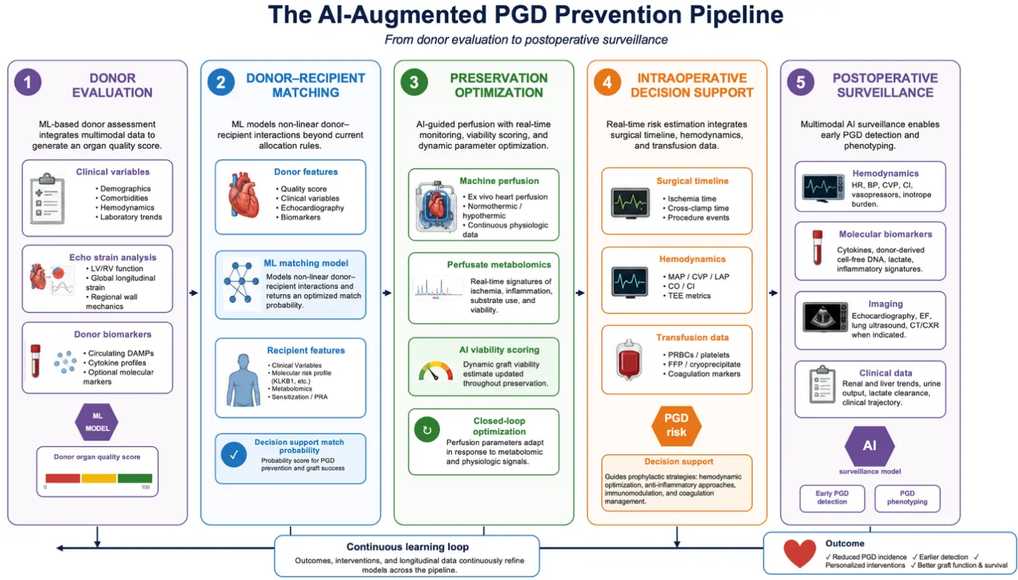
Figure 1. The AI-Augmented PGD Prevention Pipeline. Abbreviations: LV/RV: Left ventricle/right ventricle, PRA: Panel reactive antibody, MAP: Mean arterial pressure, CVP: Central venous pressure, LAP: left atrial pressure, CI/CO: Cardiac index/cardiac output, TEE: Transesophageal echocardiography, PRBCs: packed red blood cells, FFP: fresh frozen plasma, HR: Heart rate, BP: blood pressure, EF: ejection fraction, CT: computed tomography, CXR: chest X-ray.
5. Limitations and Challenges to Clinical Translation
It is important to highlight several limitations of this study. First, most cited models were developed and internally validated within single registries (e.g., UNOS, ISHLT); external and prospective validation is largely lacking, and performance often degrades on external data. Second, real-world integration is challenging—donor offers are assessed under time pressure and incomplete data, intraoperative/ICU settings demand low latency, and alerts must be actionable to avoid alarm fatigue. Third, the pipeline presumes interoperable, standardized data (variable definitions, strain conventions, perfusate assays, dd-cfDNA thresholds) that do not yet exist across centers; the absence of a shared PGD data standard is itself rate-limiting. Finally, regulatory approval, prospective safety evaluation, and clear clinical accountability are prerequisites for deployment. We therefore regard this framework as a research agenda, not a deployment-ready tool.
Current models are evaluated almost solely on discrimination (AUROC/C-statistic), which is insufficient on its own. Future validation should also report calibration and clinical utility (e.g., net-benefit analysis) with transparent reporting per standards such as TRIPOD-AI. Additional model-specific risks include limited explainability of “black-box” predictions (undermining clinician trust and accountability in allocation), algorithmic fairness (registry underrepresentation may encode bias into donor–recipient matching), and data drift (evolving donor profiles, preservation technology, and immunosuppression require ongoing recalibration, not one-time validation). Finally, the multi-omics evidence remains early, derived from small, mostly single-center studies without external validation, with no integrated proteomic–metabolomic–clinical model yet identifying candidate features for future frameworks rather than deployable biomarkers.
6. Conclusions and Future Directions
PGD remains one of the most consequential early complications following heart transplantation. Despite years of clinical experience and numerous scoring systems, reliable prediction of PGD remains poor, reflecting crucial mismatch between the biological complexity and low-dimensional tool application. Machine learning algorithms capable of modeling non-linear interactions across high-dimensional feature spaces have already demonstrated superior discrimination over traditional scores, with AUROCs approaching 0.87 in registry-based cohorts. The confluence of machine learning, multi-omics biomarker discovery, and cutting-edge organ preservation technologies provides a transformative chance to bridge this gap. Our proposed integrated framework for the AI-enhanced PGD prevention pipeline offers novel, unique mapping computational decision support to every step of the transplant process, where risk may be modified and quantified. We use “prevention” in an aggregate, pipeline-level sense: most components described here provide risk prediction, phenotyping, monitoring, or decision support, whereas direct evidence that AI itself prevents PGD remains limited and is currently strongest for organ preservation technologies such as machine perfusion. It is important to distinguish that some of AI applications described in this manuscript address risk prediction, phenotyping, monitoring, and decision support. However, direct evidence that AI itself prevents PGD remains limited, with the clearest preventive signal arising from AI-adjacent organ preservation technologies such as machine perfusion rather than from predictive algorithms. Translation of this framework from concept to clinical setting will require broad data collection combining recipient, donor, preservation, intra- and postoperative variables within shared computational software. Because PGD remains a leading cause of early post heart transplant mortality, future prospective, randomized controlled, multi-center studies are warranted to further validate proposed algorithms and to incorporate them into clinical practice.
Supplementary Materials
The following supporting information can be found at: https://www.sciepublish.com/article/pii/1301, Table S1: Specific Parameters for the AI-Enhanced PGD Prevention Pipeline.
Author Contributions
Conceptualization, A.W.; Methodology, A.W.; Software, A.W.; Validation, A.W, D.S.; Formal Analysis, A.W.; Investigation, A.W, D.S.; Resources, A.W.; Data Curation, A.W.; Writing—Original Draft Preparation, A.W.; Writing—Review & Editing, A.W. and D.S.; Visualization, A.W.; Supervision, D.S.
Ethics Statement
Not applicable.
Informed Consent Statement
Not applicable.
Data Availability Statement
Data sharing is not applicable to this article.
Funding
No funding.
Declaration of Competing Interest
Authors declare no conflict of interests.
References
-
Kobashigawa J, Zuckermann A, Potena L, Ardehali A, Berman M, Chang PP, et al. Summary of the International Society for Heart and Lung Transplantation (ISHLT) Consensus Conference on Graft Dysfunction within the First 72 hours after Heart Transplantation: A 10-year Update. J. Heart Lung Transplant. 2026, 45, e149–e171. DOI:10.1016/j.healun.2025.12.029 [Google Scholar]
-
Buchan TA, Moayedi Y, Truby LK, Guyatt G, Posada JD, Ross HJ, et al. Incidence and impact of primary graft dysfunction in adult heart transplant recipients: A systematic review and meta-analysis. J. Heart Lung Transplant. 2021, 40, 642–651. DOI:10.1016/j.healun.2021.03.015 [Google Scholar]
-
Moayedi Y, Truby LK, Foroutan F, Han J, Guzman J, Angleitner P, et al. The International Consortium on Primary Graft Dysfunction: Redefining Clinical Risk Factors in the Contemporary Era of Heart Transplantation. J. Card. Fail. 2024, 30, 805–815. DOI:10.1016/j.cardfail.2023.09.018 [Google Scholar]
-
Dugbartey GJ. Cellular and molecular mechanisms of cell damage and cell death in ischemia–reperfusion injury in organ transplantation. Mol. Biol. Rep. 2024, 51, 473. DOI:10.1007/s11033-024-09261-7 [Google Scholar]
-
Silvis MJM, Kaffka Genaamd Dengler SE, Odille CA, Mishra M, Van Der Kaaij NP, Doevendans PA, et al. Damage-Associated Molecular Patterns in Myocardial Infarction and Heart Transplantation: The Road to Translational Success. Front. Immunol. 2020, 11, 599511. DOI:10.3389/fimmu.2020.599511 [Google Scholar]
-
Holmström EJ, Syrjälä SO, Dhaygude K, Tuuminen R, Krebs R, Nykänen A, et al. Severe primary graft dysfunction of the heart transplant is associated with increased plasma and intragraft proinflammatory cytokine expression. J. Heart Lung Transplant. 2023, 42, 807–818. DOI:10.1016/j.healun.2023.01.005 [Google Scholar]
-
Rust CJ, Preston JD, Chakragiri A, Tang A, Randhawa SS, Weinberg J, et al. Plasma Metabolomic Signatures of Mitochondrial Energetic Disruption in Severe Primary Graft Dysfunction After Heart Transplantation. Eur. J. Cardiothorac. Surg. 2026, 68, ezag067. DOI:10.1093/ejcts/ezag067 [Google Scholar]
-
Segovia J, Cosío MDG, Barceló JM, Bueno MG, Pavía PG, Burgos R, et al. RADIAL: A novel primary graft failure risk score in heart transplantation. J. Heart Lung Transplant. 2011, 30, 644–651. DOI:10.1016/j.healun.2011.01.721 [Google Scholar]
-
Truby LK, Takeda K, Topkara VK, Takayama H, Garan AR, Yuzefpolskaya M, et al. Risk of severe primary graft dysfunction in patients bridged to heart transplantation with continuous-flow left ventricular assist devices. J. Heart Lung Transplant. 2018, 37, 1433–1442. DOI:10.1016/j.healun.2018.07.013 [Google Scholar]
-
Avtaar Singh SS, Das De S, Rushton S, Berry C, Al-Attar N. PREDICTA: A Model to Predict Primary Graft Dysfunction After Adult Heart Transplantation in the United Kingdom. J. Card. Fail. 2019, 25, 971–977. DOI:10.1016/j.cardfail.2019.07.009 [Google Scholar]
-
Benck L, Kransdorf EP, Emerson DA, Rushakoff J, Kittleson MM, Klapper EB, et al. Recipient and surgical factors trigger severe primary graft dysfunction after heart transplant. J. Heart Lung Transplant. 2021, 40, 970–980. DOI:10.1016/j.healun.2021.06.002 [Google Scholar]
-
Linse B, Ohlsson M, Stehlik J, Lund LH, Andersson B, Nilsson J. A machine learning model for prediction of 30-day primary graft failure after heart transplantation. Heliyon 2023, 9, e14282. DOI:10.1016/j.heliyon.2023.e14282 [Google Scholar]
-
Ding S, Liang Y, Chang CY, Brown C, Jiang X, Hu X, et al. Discover important donor-recipient risk factors and interactions in heart transplant primary graft dysfunction with machine learning. J. Am. Med. Inform. Assoc. 2025, 32, 1101–1109. DOI:10.1093/jamia/ocaf066 [Google Scholar]
-
Kampaktsis PN, Tzani A, Doulamis IP, Moustakidis S, Drosou A, Diakos N, et al. State-of-the-art machine learning algorithms for the prediction of outcomes after contemporary heart transplantation: Results from the UNOS database. Clin. Transplant. 2021, 35, e14388. DOI:10.1111/ctr.14388 [Google Scholar]
-
Giangreco NP, Lebreton G, Restaino S, Jane Farr M, Zorn E, Colombo PC, et al. Plasma kallikrein predicts primary graft dysfunction after heart transplant. J. Heart Lung Transplant. 2021, 40, 1199–1211. DOI:10.1016/j.healun.2021.07.001 [Google Scholar]
-
Preston JD, Rust CJ, Reed AC, Randhawa SS, Tang A, Rooney KA, et al. Recipient plasma metabolomics as a predictor of heart transplant severe primary graft dysfunction. Am. J. Transplant. 2026, 26, 760–771. DOI:10.1016/j.ajt.2025.12.006 [Google Scholar]
-
Palani H, Balasubramani G. Donor Left Ventricular Function Assessed by Echocardiographic Strain is a Novel Predictor of Primary Graft Failure After Orthotopic Heart Transplantation. J. Cardiothorac. Vasc. Anesth. 2021, 35, 3010–3020. DOI:10.1053/j.jvca.2021.03.018 [Google Scholar]
-
Mihos CG, Liu JE, Anderson KM, Pernetz MA, O’Driscoll JM, Aurigemma GP, et al. Speckle-Tracking Strain Echocardiography for the Assessment of Left Ventricular Structure and Function: A Scientific Statement From the American Heart Association. Circulation 2025, 152, e96–e109. DOI:10.1161/CIR.0000000000001354 [Google Scholar]
-
Truby LK, Kwee LC, Bowles DE, Casalinova S, Ilkayeva O, Muehlbauer MJ, et al. Metabolomic profiling during ex situ normothermic perfusion before heart transplantation defines patterns of substrate utilization and correlates with markers of allograft injury. J. Heart Lung Transplant. 2024, 43, 716–726. DOI:10.1016/j.healun.2023.12.002 [Google Scholar]
-
Saemann L, Kohl M, Veres G, Korkmaz-Icöz S, Großkopf A, Karck M, et al. Prediction Model for Contractile Function of Circulatory Death Donor Hearts Based on Microvascular Flow Shifts During Ex Situ Hypothermic Cardioplegic Machine Perfusion. J. Am. Heart Assoc. 2022, 11, e027146. DOI:10.1161/JAHA.122.027146 [Google Scholar]
-
Saemann L, Wenzel F, Kohl M, Korkmaz-Icöz S, Hoorn F, Loganathan S, et al. Monitoring of perfusion quality and prediction of donor heart function during ex-vivo machine perfusion by myocardial microcirculation versus surrogate parameters. J. Heart Lung Transplant. 2021, 40, 387–391. DOI:10.1016/j.healun.2021.02.013 [Google Scholar]
-
Rega F, Lebreton G, Para M, Michel S, Schramm R, Begot E, et al. Hypothermic oxygenated perfusion of the donor heart in heart transplantation: The short-term outcome from a randomised, controlled, open-label, multicentre clinical trial. Lancet 2024, 404, 670–682. DOI:10.1016/S0140-6736(24)01078-X [Google Scholar]
-
Kim DY, Kang M, Kim J, Choi D, Lee SJ. Machine perfusion across different donor pathways in heart transplantation: A systematic review and network meta-analysis. J. Heart Lung Transplant. 2025, 44, 1920–1932. DOI:10.1016/j.healun.2025.08.013 [Google Scholar]
-
Gaillard M, Krifi H, Akamkam A, Decante B, Vallee A, Guihaire J. Innovative Strategies for Organ Preservation in Heart Transplantation: Uniform Cooling Preservation and Ex-situ Normothermic Perfusion. J. Vis. Exp. 2025, 225, 67882. DOI:10.3791/67882 [Google Scholar]
-
Berg AR, Krishnan A, Heng EE, Choi AY, Zhou A, Alnasir DI, et al. Ex vivo heart perfusion offsets ischemic penalties with ≥6-hour preservation in adult donation-after-brain-death heart transplantation. J. Thorac. Cardiovasc. Surg. 2026, 171, 961–971.e4. DOI:10.1016/j.jtcvs.2025.11.025 [Google Scholar]
-
Tessari C, Lucertini G, Addonizio M, Geatti V, Bacich D, Pradegan N, et al. Ex-Vivo Heart Perfusion Machines in DCD Heart Transplantation Model: The State of Art. Transpl. Int. 2025, 38, 12987. DOI:10.3389/ti.2025.12987 [Google Scholar]
-
Guggilla V, Kang M, Bak MJ, Tran SD, Pawlowski A, Nannapaneni P, et al. Large language models accurately identify immunosuppression in intensive care unit patients. J. Am. Med. Inform. Assoc. 2025, 32, 1888–1898. DOI:10.1093/jamia/ocaf141 [Google Scholar]
-
Ledwoch N, Wiegmann B, Chichelnitskiy E, Wandrer F, Kühne JF, Beushausen K, et al. Identification of distinct secretory patterns and their regulatory networks of ischemia versus reperfusion phases in clinical heart transplantation. Cytokine 2022, 149, 155744. DOI:10.1016/j.cyto.2021.155744 [Google Scholar]
-
Khush KK, Patel J, Pinney S, Kao A, Alharethi R, DePasquale E, et al. Noninvasive detection of graft injury after heart transplant using donor-derived cell-free DNA: A prospective multicenter study. Am. J. Transplant. 2019, 19, 2889–2899. DOI:10.1111/ajt.15339 [Google Scholar]
-
Holzhauser L, DeFilippis EM, Nikolova A, Byku M, Contreras JP, De Marco T, et al. The End of Endomyocardial Biopsy? JACC Heart Fail. 2023, 11, 263–276. DOI:10.1016/j.jchf.2022.11.002 [Google Scholar]
-
Moeller CM, Fernandez Valledor A, Oren D, Rahman S, Baranowska J, Hertz A, et al. Significance of Elevated Donor-Derived Cell-Free DNA in Heart Transplant Recipients With Negative Endomyocardial Biopsies: A Dawn of a New Era. Circ. Heart Fail. 2025, 18, e012787. DOI:10.1161/CIRCHEARTFAILURE.125.012787 [Google Scholar]
-
Teuteberg J, Patel S, Baran DA, Shah P, Sayer G, Nguyen AB, et al. Multimodal molecular testing provides prognostic value for heart transplant recipients. J. Heart Lung Transplant. 2026, 45, 388–400. DOI:10.1016/j.healun.2025.11.013 [Google Scholar]
-
Knüttgen F, Beck J, Dittrich M, Oellerich M, Zittermann A, Schulz U, et al. Graft-derived Cell-free DNA as a Noninvasive Biomarker of Cardiac Allograft Rejection: A Cohort Study on Clinical Validity and Confounding Factors. Transplantation 2022, 106, 615–622. DOI:10.1097/TP.0000000000003725 [Google Scholar]
-
Agbor-Enoh S, Shah P, Tunc I, Hsu S, Russell S, Feller E, et al. Cell-Free DNA to Detect Heart Allograft Acute Rejection. Circulation 2021, 143, 1184–1197. DOI:10.1161/CIRCULATIONAHA.120.049098 [Google Scholar]
-
North PE, Ziegler E, Mahnke DK, Stamm KD, Thomm A, Daft P, et al. Cell-free DNA donor fraction analysis in pediatric and adult heart transplant patients by multiplexed allele-specific quantitative PCR: Validation of a rapid and highly sensitive clinical test for stratification of rejection probability. PLoS ONE 2020, 15, e0227385. DOI:10.1371/journal.pone.0227385 [Google Scholar]
-
Kataria A, Kumar D, Gupta G. Donor-derived Cell-free DNA in Solid-organ Transplant Diagnostics: Indications, Limitations, and Future Directions. Transplantation 2021, 105, 1203–1211. DOI:10.1097/TP.0000000000003651 [Google Scholar]