
Advances in Bidirectional Neural Interaction for Intelligent Upper-Limb Prostheses
Advances in Bidirectional Neural Interaction for Intelligent Upper-Limb Prostheses
Yawen Hu
†
 Lin Zeng
†
Ming Cheng
Li Jiang
*
Lin Zeng
†
Ming Cheng
Li Jiang
*
Received: 08 June 2026 Revised: 14 July 2026 Accepted: 29 July 2026 Published: 10 August 2026

© 2026 The authors. This is an open access article under the Creative Commons Attribution 4.0 International License (https://creativecommons.org/licenses/by/4.0/).
1. Introduction
Upper limb amputation not only leads to a decline in grasping, manipulation, and activities of daily living but also disrupts the native sensory and motor pathways between the hand and the central nervous system (CNS), making it difficult for amputees to achieve natural motor control, tactile perception, and body ownership. Although upper limb prostheses have advanced substantially in mechanical design, actuation capabilities, and sensing performance, their clinical application remains limited by unnatural control, insufficient sensory feedback, and a high cognitive burden. Conventional prostheses mainly rely on unidirectional control, in which users drive prosthetic movements through electromyographic or other biological signals, whereas information generated during interactions between the prosthesis and the environment, such as contact force, slip, temperature, and position, is difficult to effectively relay to the human nervous system. As a result, users remain highly dependent on vision during grasping and manipulation. Therefore, establishing bidirectional neural pathways that support both motor intention output and sensory information input is critical for improving prosthetic performance.
Bidirectional neural interaction in upper-limb prostheses mainly consists of neural motor control and sensory neural feedback. Motor neural control corresponds to the efferent pathway, with the core objective of extracting motor intentions from the CNS, peripheral nerves, or muscle activity and translating them into prosthetic joint movements, grasp patterns, or continuous control commands. Existing approaches mainly include brain-computer interface (BCI) [1] and myoelectric control interface [2]. BCI can directly acquire motor-related information from the CNS and have broad applicability, but they still face challenges in real-time performance and long-term stability. Myoelectric interfaces acquire electromyographic signals through surface or implanted electrodes. Among them, surface electromyography (sEMG) has become the most widely used approach for upper-limb prosthetic control because of its intuitiveness, wearability, and portability [3]. With advances in deep learning [4], electromyographic decomposition [5], and related techniques, myoelectric control is gradually evolving from simple discrete pattern recognition toward multi-degree-of-freedom [6], continuous [7], and adaptive control [8].
Sensory neural feedback corresponds to the afferent pathway and aims to deliver tactile [9], thermal [10,11], and motion status [12] acquired by prosthetic sensors back to users in a form that can be interpreted and integrated by the nervous system. The main approaches include electrical stimulation, vibrotactile stimulation, pressure stimulation, and multimodal feedback. According to the perceptual mapping relationship, sensory feedback strategies can be classified into sensory substitution, modality-matched feedback, and somatotopically matched feedback. Sensory substitution is flexible and easy to integrate, but usually requires training to establish the association between stimulation and perception [13]. Modality-matched feedback transmits information through a sensory channel similar to the original modality, thereby reducing cognitive burden [14]. Somatotopically matched feedback [15], achieved through phantom limb mapping [16], targeted reinnervation, or direct neural stimulation, enables feedback to be perceived as originating from the corresponding region of the missing hand, and is therefore closer to the goal of natural sensory restoration.
This paper reviews bidirectional neural interaction in intelligent upper-limb prostheses. First, brain–computer interfaces, myoelectric interfaces, and their motor intention decoding methods are introduced. The main stimulation modalities and perceptual mapping strategies for sensory feedback are then summarized. Finally, the development trends and key challenges of motor control, sensory feedback, and closed-loop bidirectional neural interaction systems are discussed.
2. Neuromotor Interface
Neuromotor interfaces rely on decoding motor intentions from neural signals and subsequently generating motor commands to actuate external devices in accordance with user intent. According to the acquisition source, neuromotor interfaces primarily include BCI and myoelectric interfaces. BCIs enable the direct acquisition of motor information from the CNS, thereby serving a broader population. Myoelectric interfaces directly reflect neuromuscular activity and exhibit stronger motor relevance, which has led to their widespread application in the field of motor intention decoding. Consequently, BCI and myoelectric interface are each described in detail in the following sections.
2.1. Brain-Computer Interface
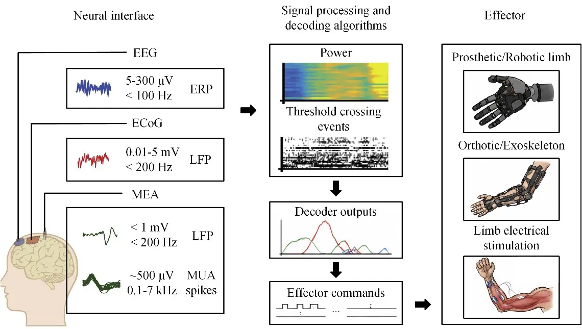
BCI technology establishes direct communication between the brain and external devices. Its core objective is to decode user intent from brain activity and translate it into commands for external device control. As shown in Figure 1, BCI systems utilize three main components: neural interfaces that record from but may also stimulate the brain, signal processing, and decoding algorithms and effectors that evoke movement and may transduce sensory input [17].
According to the signal acquisition method, BCIs can be classified as invasive, semi-invasive, or non-invasive. Invasive BCIs typically employ microelectrode arrays (MEAs) to directly record neuronal action potentials or local field potentials (LFPs), providing high spatial resolution and a superior signal-to-noise ratio (SNR). In recent years, invasive BCIs have achieved remarkable advances in neuroprosthetic control, demonstrating substantial potential for restoring motor function [18]. However, their widespread clinical translation remains limited by the need for craniotomy and the associated risks of surgical trauma, infection, immune responses, and long-term signal instability [19]. As a result, invasive BCIs have not yet been widely adopted in clinical practice and remain in the early stages of clinical translation [20].
Semi-invasive BCIs are primarily represented by electrocorticography (ECoG)-based systems, which record cortical activity using electrode arrays placed on the cortical surface beneath the dura mater. Compared with invasive BCIs, ECoG avoids penetrating brain tissue, thereby reducing surgical invasiveness while preserving relatively high spatial and temporal resolution. Nevertheless, implantation still requires craniotomy, and the associated risks of surgical trauma and infection continue to limit its widespread clinical application [20].
For non-invasive BCIs, electroencephalography (EEG) is commonly used for its high temporal resolution, safety, portability, and low cost [21]. EEG captures brain electrical activity via electrodes placed on the scalp. According to the International 10–20 system, electrodes are standardly placed at specific positions on the scalp. Using the midline from the nasion to the inion as a reference, this distance is divided into 10 equal intervals, which are marked at proportions of 10%, 20%, 20%, 20%, 20%, and 10%. EEG has been extensively investigated for motor intention decoding and has shown considerable potential in assistive communication, motor rehabilitation, and HMI [19]. However, despite its increasing acceptance in clinical research, EEG-based BCIs remain largely confined to research settings and limited clinical applications [22]. Their widespread clinical adoption is still hindered by low SNR and spatial resolution, frequent recalibration requirements, and limited robustness during long-term daily use.
2.1.1. Typical BCI Paradigms
BCI paradigms refer to design patterns used in experiments or applications to elicit and detect specific EEG signals. Classic paradigms include the P300 evoked potential, steady-state visual evoked potential (SSVEP), and motor imagery (MI) [23].
P300-based BCI systems rely on event-related potentials (ERPs). These systems elicit a P300 response through the presentation of specific stimulus sequences and decode user intention by integrating its temporal characteristics with scalp topography. This approach is suited for single-target selection from multiple candidates, such as the P300 speller [24]. In recent years, P300 BCIs have found expanded applications in the field of robotics [25].
SSVEP-based BCI systems rely on EEG responses evoked by periodic visual stimulation at the stimulation frequency and its harmonic frequency components, and have attracted increasing attention in the BCI field due to their high information transfer rate (ITR) and minimal user training, with successful applications in high-speed spelling [26], prosthetic hand [27], and robotic arm [28].
MI-based BCI systems decode user intent by analyzing modulations of sensorimotor rhythms (SMRs) elicited during MI tasks. This paradigm is widely recognized as one of the most established, intuitive, and safe approaches in BCI research. MI has been used in continuous brain-actuated robotic devices such as brain control of robotic arms [29], wheelchairs [30], and mobile robots [31].
2.1.2. EEG-Based Decoding
The typical pipeline for EEG-based motor intention decoding consists of several stages, including signal acquisition, preprocessing, feature extraction, and classification or regression. Due to the dynamic and high complexity of EEG signals, the selection of appropriate features is critical to the performance of EEG decoding models. Feature extraction can be achieved based on time, frequency, time-frequency, and spatial information contained in the signals. Common EEG time-domain features include autoregressive (AR), adaptive autoregression, root mean square (RMS), and integrated EEG (IEEG) [32]. In the frequency domain, EEG signals are typically characterized through spectral analysis, which describes the distribution of signal power across different frequencies. To further capture the non-stationary dynamics of EEG, researchers have introduced time-frequency analysis methods, such as the short-time Fourier transform (STFT) and continuous wavelet transform (CWT), as well as adaptive signal decomposition techniques, such as empirical mode decomposition (EMD) [33]. For spatial-domain analysis, the common spatial pattern (CSP) serves as a classic algorithm that effectively extracts spatial features from MI EEG signals [34]. However, individual feature extraction methods generally capture only specific aspects of EEG signals. For example, time-domain features are insufficient to characterize spectral dynamics, frequency-domain analyses overlook the non-stationary nature of EEG signals, and the effectiveness of spatial features is highly dependent on electrode configuration and inter-subject variability. Therefore, learning robust feature representations that effectively capture the complementary temporal, spectral, and spatial features of EEG signals remains a major challenge for improving motor intention decoding performance.
In the field of EEG analysis, traditional machine learning methods have been widely used for classification and regression tasks. Among these, support vector machines (SVM) and linear discriminant analysis (LDA) are the most commonly used classifiers for MI EEG-based classification tasks [35]. Recently, deep learning has emerged as a promising approach for EEG decoding owing to its powerful capability for automatic feature learning. Among deep learning architectures, convolutional neural networks (CNNs) have been extensively adopted for EEG decoding due to their inherent ability to learn hierarchical feature representations. Recurrent neural networks (RNNs) and long short-term memory (LSTM) networks, by contrast, are well suited for modeling temporal dependencies in EEG signals, thereby capturing their non-stationary temporal dynamics. Graph neural networks (GNNs) represent multi-channel EEG signals as graph-structured data, where electrodes are modeled as nodes, enabling explicit modeling of functional connectivity patterns to improve spatial representation learning. Song et al. subsequently proposed a dynamic graph convolutional neural network (DGCNN) that learns the intrinsic relationships among EEG channels, thereby enabling the extraction of more discriminative features [36].
Although deep learning has reduced the reliance on handcrafted feature engineering, most existing models remain task-specific and require large amounts of labeled data for training, resulting in limited generalization. Recently, the rapid development of self-supervised learning (SSL) and foundation models has shifted EEG decoding from task-specific models toward large-scale pre-trained models. By pre-training on large-scale, multi-task, and multi-subject neural datasets, foundation models learn transferable neural representations that can be efficiently adapted to downstream decoding tasks through minimal fine-tuning [37]. This paradigm has demonstrated significant advantages in cross-subject learning and few-shot learning BCIs, providing a promising new direction for EEG-based motor intention decoding.
2.2. Myoelectric Interface
Myoelectric interfaces are typically classified as invasive or non-invasive depending on how the electrodes contact muscle tissue. Invasive interfaces use needle, fine-wire, or implanted electrodes to record intramuscular electromyogram (iEMG) signals with high SNR. However, their clinical application in HMI remains largely limited to early feasibility studies due to invasiveness, poor long-term stability, and limited portability. In contrast, non-invasive myoelectric interfaces acquire sEMG signals via skin-surface electrodes, offering advantages such as intuitiveness, wearability, safety, low cost, and portability. Consequently, sEMG-based myoelectric interfaces have become one of the most clinically established and widely adopted neuromotor interface technologies, with broad applications in commercial prostheses, rehabilitation robots, exoskeletons, and other HMI systems. Therefore, this section focuses on non-invasive sEMG-based myoelectric interfaces.
2.2.1. sEMG Recordings
From the perspective of acquisition strategies, sEMG recording systems have evolved from single-channel and few-channel configurations to multi-electrode arrays. Early sEMG acquisition predominantly relied on wet Ag/AgCl electrodes, which offer excellent electrical conductivity and are widely used in clinical assessments and experimental studies [38]. However, these electrodes require conductive gel and may cause skin irritation and inflexibility, thereby limiting their suitability for long-term wear. With the development of wearable myoelectric interfaces, dry electrodes and multi-channel acquisition systems have attracted increasing attention. Multi-electrode systems simultaneously record electrical activity from different muscle regions, providing richer spatial information for gesture recognition, continuous motion estimation, and HMI. Further increasing electrode density gives rise to high-density sEMG (HD-sEMG) arrays [39]. High-density arrays capture the distribution and propagation pattern of motor unit action potentials (MUAPs) across the skin surface through denser spatial sampling, providing an important foundation for motor unit (MU) decomposition, neural drive estimation, and the decoding of fine motor intention.
2.2.2. sEMG-Based Decoding
In early sEMG-based motor intention decoding, the complexity of neuromuscular encoding and the lack of a unified analytical framework led to the adoption of data-driven approaches to learn the mapping between sEMG signals and motor outputs [6]. Such methods typically consist of signal preprocessing, sliding window segmentation, feature extraction, and machine learning. Handcrafted features are primarily extracted from the time, frequency, and time-frequency domains. Time-domain features capture information such as sEMG signal amplitude, waveform complexity, and zero-crossing rate. Low computational complexity makes these features well suited for real-time applications. The classic time-domain feature set proposed by Hudgins et al. includes mean absolute value (MAV), mean absolute value slope (MAVS), waveform length (WL), zero crossing (ZC), and slope sign change (SSC) [40]. Tkach et al. demonstrated that appropriate combinations of time-domain features can overcome the impact of electrode location shift and muscle fatigue on classification performance to a certain extent, but this approach is insufficient to fundamentally address robustness issues caused by the non-stationarity of sEMG signals [41]. Frequency-domain features primarily characterize the distribution of sEMG signal energy across the frequency spectrum, including mean and median frequency, as well as power spectral features. Although they can reflect muscle fatigue and variations in contraction state, they are limited in their ability to capture the temporal dynamics of frequency components. Phinyomark et al. compared a variety of time- and frequency-domain features and reported that most time-domain features exhibit strong redundancy. In contrast, frequency-domain features show relatively weak class separability, thereby limiting their applicability in complex dynamic motion recognition tasks [42]. To further characterize the transient changes of non-stationary sEMG signals, time-frequency analysis methods such as the STFT, wavelet transform (WT), wavelet packet transform (WPT), Wigner-Ville distribution (WVD), and Choi-Williams distribution (CWD) have been gradually introduced into sEMG-based motor intention decoding research. Time-frequency analysis methods preserve both temporal and spectral information, making them more suitable for extracting sEMG features during dynamic movements. However, their relatively high computational complexity may increase computational latency and limit their deployment in real-time control applications. These limitations have motivated the development of data-driven methods capable of automatically learning more discriminative feature representations from sEMG signals.
With the development of deep learning, researchers have begun leveraging neural networks to automatically learn movement-related features from sEMG time series, multi-channel signals, or sEMG images. Compared with traditional handcrafted features and shallow machine learning models, deep learning models have stronger nonlinear representation capability and can better capture the complex mapping between sEMG signals and motor intentions. Common architectures include CNNs, RNNs, LSTM networks, encoder-decoders, attention mechanisms, and Transformers [4,43,44,45]. In recent years, large-scale approaches have gradually emerged as new trends in sEMG-based motion decoding. Kaifosh et al. used an sEMG wristband to collect large-scale data and achieved three tasks: wrist-controlled cursor tracking, discrete gesture recognition, and handwriting [2]. Although deep learning has substantially reduced the reliance on handcrafted feature engineering and achieved remarkable improvements in complex motor decoding, most existing models remain physiologically uninterpretable. The relationship between their decision-making mechanisms and the underlying neuromuscular control processes remains poorly understood, limiting their reliability and interpretability in long-term clinical applications.
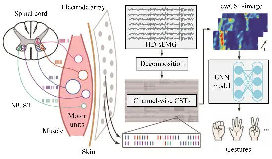
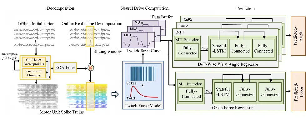
Continuous control imposes higher demands on sEMG-based motion decoding, making the extraction of MU discharge information from raw signals a research priority. Compared with traditional features, MU discharge information is closer to the essential nature of motoneuron pool output, enabling the relationship between muscle activity and motor output to be explained from a neural control perspective. Blind source separation (BSS) is the most widely adopted approach for sEMG decomposition, with convolution kernel compensation (CKC) [46] and independent component analysis (ICA) [47] as the most representative methods. The CKC-based method extends the observed signals in the spatiotemporal domain to identify strongly correlated waveform sequences, which are then identified as motor unit action potential trains (MUAPTs). In contrast, the ICA-based method seeks independent sources that maximize non-Gaussianity from the observed signals, which are then interpreted as MUAPTs, primarily through mutual information minimization or negentropy maximization. In recent years, researchers have further explored the application of sEMG decomposition in decoding movement intention. MU discharge information has gradually evolved from a neurophysiological analysis metric into an important feature for movement decoding. By establishing the mapping between MU discharge patterns and motor outputs, it has been successfully applied to various movement decoding tasks, including gesture recognition, continuous joint angle estimation, and grasp force prediction [48,49], as illustrated in Figure 2. Recent studies have further focused on improving the accuracy, long-term stability, and real-time performance of MU-driven decoders. Advances in MU decomposition algorithms, decoder training strategies, and deep learning-based frameworks have substantially improved decoding performance in complex movement scenarios, facilitating the translation of MU-driven neural interfaces toward practical HMI [5,50,51]. Nevertheless, the practical deployment of MU-driven decoding remains constrained by its reliance on HD-sEMG acquisition, robust MU decomposition under dynamic conditions, and the computational cost associated with real-time implementation.
To facilitate comparison, the major characteristics of representative neuromotor interface approaches are summarized in Table 1.
Table 1. Comparison of representative neuromotor interfaces.
|
Approach |
General Advantages |
Subtypes |
Advantages |
Limitations |
Clinical Readiness |
|---|---|---|---|---|---|
|
BCI |
Direct access to motor intention; broader applicability due to independence from residual muscle activity |
Invasive |
High spatial resolution and SNR |
Surgical risks and long-term signal instability |
Early clinical translation |
|
Semi-invasive (ECoG) |
Higher spatial resolution than non-invasive BCI; lower invasiveness than invasive BCI |
Craniotomy still required and long-term signal instability |
Early clinical translation |
||
|
Non-invasive (EEG) |
Safety; portability; low cost |
Low SNR and spatial resolution; frequent recalibration required |
Research and limited clinical applications |
||
|
Myoelectric interface |
Directly reflect neuromuscular activity and exhibit stronger motor relevance |
Invasive (iEMG) |
Higher SNR than non-invasive myoelectric interface |
Invasiveness; poor long-term stability; limited portability |
Early feasibility studies |
|
Non-invasive (sEMG) |
Intuitiveness; wearability; safety; low cost; portability |
Requires residual muscle activity; sensitive to electrode shift and fatigue |
Established clinical use |
3. Sensory Feedback Interface
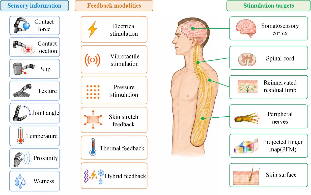
Sensory feedback interface aims to convert information acquired by prosthetic sensing systems into stimulation signals that can be perceived by the human nervous system through specific encoding strategies and stimulation modalities, as shown in Figure 3, thereby reconstructing the afferent pathway between the prosthesis and the user. Effective sensory feedback can reduce the user’s reliance on vision and improve grasp success, object recognition, and fine manipulation, while also enhancing prosthesis ownership and acceptance.
3.1. Sensory Feedback Modalities
3.1.1. Electrical Stimulation
Electrical stimulation feedback can be classified as either invasive or non-invasive. Invasive electrical stimulation connects the prosthetic sensing system with the nervous system through implanted electrodes, encoding information such as contact force, position, or texture into neural signals that the CNS can perceive. According to the stimulation target, invasive electrical stimulation can be further divided into peripheral nervous system (PNS) stimulation and CNS stimulation.
PNS stimulation typically involves implanting electrodes into or around peripheral nerves. By modulating stimulation amplitude, pulse width, frequency, and stimulation site, these interfaces can evoke somatotopically localized sensations perceived as originating from the phantom fingers or palm. Recent advances have further shifted implanted interfaces toward fully implantable, wirelessly connected, bidirectional systems. A first-in-human implementation of a high-channel-count implanted system was reported, in which intramuscular myoelectric recording and peripheral nerve stimulation were simultaneously supported, enabling three-degree-of-freedom prosthetic control and somatosensory feedback through wireless communication [52]. CNS stimulation targets the spinal cord or the somatosensory cortex and can evoke tactile or proprioceptive sensations perceived as arising from the distal region of the missing upper limb. Invasive electrical stimulation offers potential advantages such as high spatial selectivity, relatively intuitive percepts, and reduced learning burden. However, its clinical translation remains limited by surgical trauma, infection risk, and high cost.
Non-invasive electrical stimulation delivers electrical current to the body via surface electrodes; among these, transcutaneous electrical nerve stimulation (TENS) is the most widely used. TENS [53] can induce sensations not only at the local skin region beneath the electrodes, but also in the phantom hand by stimulating subcutaneous nerves or the projected finger map (PFM) of the residual limb, thereby eliciting somatotopically localized sensations referred to the missing hand. D’Anna et al. integrated TENS into a bidirectional prosthetic hand system and evoked phantom sensations associated with the median and ulnar nerve territories, thereby enabling somatotopic feedback coupled with prosthetic sensors [54]. Hao et al. further demonstrated that stimulation of the projected finger map in the residual limb can activate the corresponding finger representation in the primary somatosensory cortex, suggesting that this approach may transmit finger-specific information through a relatively natural afferent pathway. Beyond the restoration of cutaneous tactile sensations, transcutaneous electrostimulation has recently been explored for proprioceptive feedback. In a pilot study, stimulation of the residual finger flexor muscles was used to evoke an illusion of finger extension, demonstrating preliminary feasibility for object-size discrimination with a myoelectric prosthesis [55].
In addition, transcutaneous spinal cord stimulation (tSCS) has been proposed as an emerging non-invasive approach for sensory feedback [56]. By applying surface stimulation over the cervical spinal cord, tSCS can evoke touch, vibration, or movement sensations perceived as originating from the missing hand. This method may be particularly suitable for individuals with high-level amputation or impaired peripheral nerves, although its spatial selectivity and parameter stability still require further improvement.
3.1.2. Vibrotactile Stimulation
Vibrotactile feedback is delivered by placing vibration actuators on the user’s skin surface. By modulating parameters such as vibration frequency, amplitude, and duration, this approach can encode tactile information related to prosthetic hand contact state, grip force, and slippage. It is one of the most widely studied and relatively mature forms of mechanical feedback. Previous studies have shown that vibrotactile feedback can improve grasping success and prosthetic motion control accuracy, while also helping to reduce excessive grip force [57]. Its main advantages include fast response, lightweight structure, low cost, and ease of integration. However, the sensations it elicits are generally less natural and are more suitable for signaling discrete events, such as contact, slippage, or excessive force. Its ability to convey continuous force information and high-spatial-resolution tactile cues remains limited. In addition, prolonged or high-frequency stimulation may induce sensory adaptation, and stimulation thresholds are strongly affected by inter-individual variability and skin location. Therefore, vibrotactile feedback is gradually evolving from single-channel cueing toward multichannel encoding, spatiotemporal pattern encoding, and multimodal combined encoding.
Wei et al. proposed a feedback armband that conveys proprioceptive information through spatiotemporal vibrotactile patterns and provides tactile and proximity feedback using either combined electrotactile stimulation or vibrotactile-only stimulation [58]. Wu et al. also demonstrated that hybrid vibro-electrotactile feedback improved task success rates and reduced errors under different visual conditions [59]. In addition, vibration of tendons or muscles can induce proprioceptive illusions. Marasco et al. used vibration of residual muscles after targeted muscle reinnervation (TMR) to induce movement illusions, thereby improving prosthetic hand control accuracy and the subjective sense of agency in amputees [12]. Vibrotactile stimulation can also evoke illusory limb movements in individuals with congenital or acquired upper-limb differences [60]. Therefore, vibrotactile feedback can be used not only for sensory substitution but also as a strategy for proprioceptive restoration. However, its long-term stability, individual adaptability, and training effects require further validation.
3.1.3. Pressure Stimulation
Pressure feedback delivers normal forces to the skin through actuators, producing sensations that are relatively intuitive and natural. Studies have shown that pressure feedback, as a modality-matched strategy, outperforms vibrotactile feedback in multi-point discrimination tasks [61]. To improve wearability, pressure feedback devices have gradually evolved from rigid stimulation units toward pneumatic and socket-integrated designs. Huaroto et al. developed a soft pneumatic actuator as a haptic unit that can be incorporated into a silicone liner [62], and Barontini et al. further integrated soft pneumatic pressure feedback into a prosthetic socket for grip-force modulation [63]. Multichannel pneumatic armbands can also support complex grasping tasks when visual feedback is occluded [64]. Overall, pressure feedback provides intuitive, natural, and fatigue-resistant sensations; however, further optimization is still required in terms of miniaturization, power consumption, and response speed.
3.1.4. Other Stimulation Modalities
Other feedback modalities generally convey a narrower range of information and are therefore still insufficient to independently support closed-loop prosthetic control. However, they can serve as homologous feedback modalities to enrich sensory experience and improve perceptual naturalness. For example, skin-stretch feedback generates tangential displacement on the skin, making it suitable for encoding finger aperture or movement direction [65]. Thermal feedback [10,11] typically uses Peltier elements or thermal haptic displays to convey information about object temperature, thereby improving material discrimination and enhancing sensory naturalness. Nevertheless, its application is limited by response speed, power consumption, and thermal safety. Wetness feedback is commonly based on the mechanism by which cold–dry stimulation elicits an illusion of wetness. Studies have shown that amputees can discriminate among different moisture levels using thermal feedback devices [66].
3.2. Sensory Feedback Strategies
3.2.1. Sensory Substitution
Sensory substitution refers to the delivery of information acquired by prosthetic sensors to the human body through an alternative stimulation modality, thereby indirectly compensating for the missing sensory input. For example, contact force at the prosthetic fingertip can be encoded as vibrotactile stimulation delivered to the skin. When users no longer interpret the stimulus as an abstract cue but instead integrate it as part of prosthetic tactile or proprioceptive perception, effective sensory substitution can be considered to have been achieved [13].
Common approaches include vibrotactile feedback, TENS, and auditory feedback. Owing to its flexible implementation and relatively simple hardware integration, sensory substitution has become one of the most widely investigated strategies in prosthetic sensory feedback. However, this strategy usually requires users to learn the mapping between stimulation parameters and sensory information, such as associating vibration frequency with changes in joint angle, which inevitably increases cognitive burden.
3.2.2. Modality-Matched Feedback
Modality-matched feedback emphasizes the consistency between the perceived quality of feedback and the physical information detected by the prosthesis; that is, it aims to convey sensory variables through feedback channels that evoke percepts of the same or similar modality. Compared with sensory substitution, its main advantage lies in the greater intuitiveness of stimulation, which can reduce the need to learn artificial encoding rules and lower cognitive burden. Typical examples include using mechanical pressure or skin deformation to convey contact force and slip, thermal stimulation to convey object temperature, tendon vibration to evoke kinesthetic illusions, and skin stretch to convey proprioceptive cues.
In recent years, research has shifted from simple intensity encoding toward more natural reconstruction of sensory quality. For instance, peripheral nerve stimulation and TENS can evoke tactile sensations in the phantom limb; thermal feedback can enable amputees to perceive the temperature of objects touched by the prosthetic hand; and soft pneumatic or flexible haptic interfaces can improve the wearability of feedback devices while preserving pressure-based modality matching.
3.2.3. Somatotopic Matching
Amputation leads to the loss of sensory organs required for interaction with the environment. However, the sensory nerves in the residual limb and the CNS can still be accessed or engaged. Somatotopic matching aims to make sensory information from the prosthesis perceived as originating from the corresponding region of the missing limb, rather than from the actual stimulation site. This strategy is mainly achieved through neural mapping, targeted reinnervation, and direct neural stimulation.
Neural mapping typically exploits phantom hand maps on the residual-limb skin. By using mechanical stimulation or TENS to activate preserved afferent pathways, stimulation applied to specific skin regions can be perceived as originating from the phantom fingers or phantom palm. This approach can provide relatively natural feedback without electrode implantation. However, the completeness and stability of phantom limb maps vary markedly across individuals. Some amputees retain only partial maps, whereas others lack identifiable phantom finger areas [16,53,67].
Targeted reinnervation can establish biological interfaces for prosthetic control and sensory feedback by redirecting residual nerves to preserved muscles or skin regions. For example, vibration applied to reinnervated muscles after TMR can elicit phantom limb movement sensations associated with prosthetic hand motion, thereby improving prosthetic control [68,69].
Direct neural stimulation can be classified into PNS stimulation and CNS stimulation according to the stimulation target. The former stimulates residual-limb nerves through implanted electrodes to evoke tactile or proprioceptive sensations in the phantom fingers. D’Anna et al. implanted transverse intrafascicular multichannel electrodes into the ulnar and median nerves, enabling simultaneous delivery of tactile feedback and finger position information [70]. The latter reconstructs hand sensory representations by stimulating the somatosensory cortex [71], which may be suitable for individuals with severely impaired peripheral pathways. However, this approach is still limited by surgical invasiveness, long-term stability, and challenges in clinical translation.
4. Future Directions
The neural interaction system of intelligent upper-limb prostheses aims to decode motor intention, reconstruct sensory input, and achieve bidirectional closed-loop control. Therefore, future research should focus on three key levels: motor neural decoding, sensory neural feedback, and sensorimotor integration.
In terms of motor neural interfaces, the primary goal is to make prosthetic control more closely resemble the dynamic regulation process of the human upper limb.
First, continuous decoding. Traditional discrete commands are incapable of meeting the interaction demands of multi-degree-of-freedom systems for simultaneous proportional control (SPC). Neural interfaces that can continuously estimate kinematics have been a key research direction in the field of HMI. However, despite the achievements in decoding joint angles, movement velocities, and grip forces from signals such as sEMG and EEG, significant bottlenecks persist in real-time performance, accuracy, and cross-subject generalization. SPC directly determines the mechanical compliance and naturalness of prosthetic systems. In the future, it is necessary to promote this technology from laboratory to practice, enabling smoother and more natural HMI.
Second, interpretable neural decoding. Deep learning methods have significantly improved the performance of neural signal decoding. However, their decision-making processes lack physiological interpretability, limiting their applicability in scenarios requiring high interpretability, such as clinical diagnosis and neurorehabilitation. To address this limitation, research efforts are gradually shifting from end-to-end models toward interpretable neural decoding frameworks. On the one hand, explainable artificial intelligence (XAI) has been explored in neural decoding to reveal the intrinsic relationships between neural features and decoding outputs [72,73]. On the other hand, models incorporating physiological priors have been developed, such as sEMG decomposition techniques and musculoskeletal models [74,75]. These approaches collectively promote the alignment between internal model representations and known physiological mechanisms, thereby improving both physiological interpretability and generalization capability, and are emerging as a key direction for next-generation neural interfaces.
Third, multimodal fusion. Decoding models based on a single neural signal modality still face challenges in robustness under complex and dynamic environments. Integrating multimodal information has become an important direction for overcoming performance bottlenecks. Integrating heterogeneous sensor modalities, such as an inertial measurement unit (IMU), mechanomyography (MMG), force myography (FMG), sonomyography (SMG), and near infrared spectroscopy (NIRS), enables complementary measurement and joint modeling of movement intentions [76]. For upper-limb amputees, the integration of EEG and sEMG represents a novel and clinically valuable neural interface paradigm [77]. Accordingly, multimodal information fusion is poised to enhance system robustness, thereby facilitating more natural and reliable HMI.
In parallel with motor neural decoding, the core objective of sensory neural feedback is to restore the afferent information pathway between the prosthesis and the human body. At present, the reconstruction of sensory neural feedback still faces several major challenges.
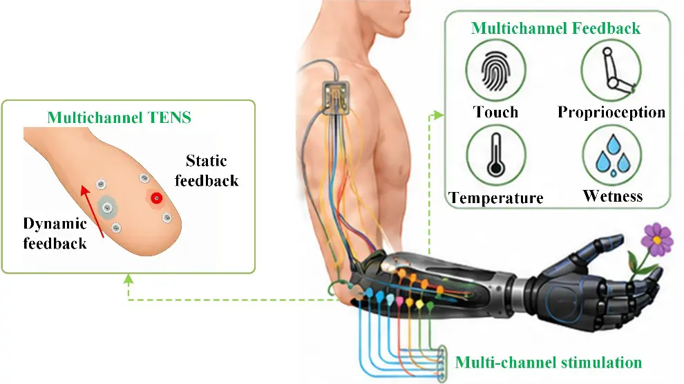
First, the development of multimodal feedback should address both multimodal physical information and multimodal feedback modalities. As shown in Figure 4, the former requires prosthetic sensing systems to extend beyond single fingertip force detection toward the acquisition of multisource information, including contact force, temperature, slippage, joint position, and kinematic state, in order to meet the sensory demands of different manipulation scenarios. The latter emphasizes the selection of modality-matched stimulation methods according to the attributes of the feedback variables. Mechanical stimulation, electrical stimulation, thermal stimulation, and their hybrid forms, as well as invasive and non-invasive approaches, should be used in a complementary manner to improve the discriminability and transmission efficiency of multiple sensory signals. Notably, multichannel TENS can enable higher-dimensional information encoding through electrode arrays. Static feedback can convey contact location and grip force magnitude by modulating stimulation site, amplitude, or pulse width, whereas dynamic feedback can encode prosthetic hand movement direction, movement speed, and changes in joint state through spatiotemporal activation sequences across electrodes. If the electrodes are further positioned over the phantom finger map of the residual limb, somatotopically matched feedback may be achieved.
Second, feedback strategies should be optimized to improve sensory naturalness. Most current closed-loop prosthetic systems still use linear mapping, in which sensor outputs are directly converted into stimulation intensity or frequency. Although this method is straightforward to implement, the evoked sensations are often insufficiently natural. Biomimetic encoding strategies proposed in recent years transmit tactile, pressure, and position information from prostheses by mimicking the neural coding mechanisms of the human sensory system. Such biomimetic encoding strategies, which simulate neural firing patterns, can improve sensory naturalness, tactile sensitivity, and dexterous hand manipulation ability [78,79,80,81]. On this basis, machine-learning-assisted adaptive encoding may become an important direction for next-generation sensory feedback. Its core concept is to use machine learning or adaptive control methods to dynamically optimize stimulation channels and parameter combinations according to individual perceptual thresholds, comfort levels, task performance, and electrode–skin interface conditions, thereby compensating for electrode displacement, skin impedance fluctuations, sensory adaptation, and other factors.
Third, standardized and objective evaluation metrics for sensory feedback should be established to enable reliable comparisons across studies and feedback modalities. Existing studies commonly use psychophysical experiments to evaluate the sensitivity and discriminability of evoked sensations. Although these methods can directly reflect subjective experience, they are susceptible to inter-individual variability, attentional state, and prior learning experience [82,83,84]. Moreover, quantitative comparison is difficult not only across different feedback modalities but also among studies using the same modality, because participant characteristics, stimulation sites, hardware configurations, encoding parameters, calibration procedures, experimental tasks, visual conditions, and outcome measures vary substantially. Even cross-modal congruency tasks cannot fully eliminate the influence of subjective reports and generally require validation in sufficiently large samples [85]. Future studies should therefore establish standardized experimental protocols, common functional tasks, and unified outcome measures, while combining subjective assessments with objective neural metrics. Neuroimaging and neurophysiological techniques, including functional magnetic resonance imaging, electroencephalography [86,87], and functional near-infrared spectroscopy, may help quantify stimulation-induced activity in afferent pathways and the somatosensory cortex. Such multimodal evaluation could support more reliable characterization of feedback localization, modality congruence, and spatiotemporal stability, and ultimately enable rigorous cross-study comparison and meta-analysis.
Once motor intention can be stably decoded and sensory information can be effectively fed back, the development of intelligent prostheses will further move toward the integration of motor control and sensory feedback. Future intelligent prostheses should no longer treat motor control and sensory feedback as two independent functional modules but instead establish truly closed-loop, bidirectional neural interaction systems. Several key issues need to be addressed.
First, the real-time performance and stability of the closed-loop pathway must be improved. Delays introduced by sensing, encoding, stimulation, and control computation should be minimized to prevent feedback latency from disrupting sensorimotor congruence, thereby ensuring natural control during continuous grasping, force modulation, and multi-degree-of-freedom manipulation.
Second, the compatibility between electrical stimulation feedback and neural signal acquisition remains a critical challenge. Because TENS and electromyographic electrodes share the same body-surface conductive environment, stimulation may degrade EMG signal quality or even cause signal saturation. Potential interference suppression strategies include optimizing electrode placement and structural design, as well as incorporating isolation circuits, to reduce crosstalk at the hardware level. In terms of stimulation waveform design, charge-balanced biphasic electrical stimulation can be used to minimize net charge accumulation and reduce polarization-related artifacts. At the algorithmic level, adaptive filtering, real-time artifact template updating based on prior stimulation information, and dynamic compensation methods can be combined to improve the usability of EMG signals during stimulation.
Third, long-term validation should be oriented toward functional performance. In addition to evaluating clinically relevant outcomes, such as cognitive burden, phantom limb pain, embodiment, and comfort, future studies should assess the long-term stability and reliability of the system during prolonged daily use. User training strategies should also be optimized to reduce calibration requirements and training burden while improving adaptation to bidirectional control. Furthermore, long-term evaluations should incorporate activities of daily living to systematically assess system usability, functional recovery, and clinical utility, thereby facilitating the translation of bidirectional neural prostheses into routine clinical practice.
5. Conclusions
Bidirectional sensorimotor neural interaction plays a decisive role in the performance of intelligent upper-limb prostheses. Neural motor control reconstructs the efferent pathway by decoding motor intentions through BCI and myoelectric interface, whereas sensory neural feedback restores the afferent pathway through electrical stimulation, mechanical stimulation, thermal feedback, and multimodal feedback. Existing studies have demonstrated that bidirectional interaction can improve control accuracy, reduce visual dependence, and enhance embodiment. However, current systems remain far from achieving natural, long-term, stable neural interaction. Future research should focus on continuous real-time control, multimodal integration, sensory naturalness, and real-time bidirectional integration, thereby promoting the development of upper-limb prostheses from functional restoration toward natural neural interaction systems.
Author Contributions
Conceptualization, Y.H., L.Z. and L.J.; Methodology, Y.H. and L.Z.; Investigation, Y.H. and L.Z.; Resources, L.J. and M.C.; Data Curation, Y.H. and L.Z.; Writing—Original Draft Preparation, Y.H. and L.Z.; Writing—Review & Editing, Y.H., L.Z. and L.J.; Visualization, Y.H. and L.Z.; Supervision, L.J.; Project Administration, L.J. and M.C. All authors have read and agreed to the published version of the manuscript.
Ethics Statement
Not applicable.
Informed Consent Statement
Not applicable.
Data Availability Statement
Data are contained within this article.
Funding
This work was supported in part by the National Natural Science Foundation of China (Grant No. 91948302); in part by the National Natural Science Foundation of China under the Basic Science Center Program for “Space Robot Intelligent Manipulation” (Grant No. T2388101).
Declaration of Competing Interest
The authors declare that they have no known competing financial interests or personal relationships that could have appeared to influence the work reported in this paper.
References
-
Wang H, Zhang Y, Karrenbach M, Ding Y, He B. Sensory-guided human-machine joint learning accelerates the acquisition of motor imagery brain computer interface control. Nat. Commun. 2026, 17, 6177. DOI:10.1038/s41467-026-75435-5 [Google Scholar]
-
CTRL-labs at Reality Labs, Sussillo D, Kaifosh P, Reardon T. A generic noninvasive neuromotor interface for human-computer interaction. Biorxiv 2024, 2024-02. DOI:10.1101/2024.02.23.581779 [Google Scholar]
-
Yu Y, Chen C, Zhao J, Sheng X, Zhu X. Surface Electromyography Image-Driven Torque Estimation of Multi-DoF Wrist Movements. IEEE Trans. Ind. Electron. 2022, 69, 795–804. DOI:10.1109/TIE.2021.3050367 [Google Scholar]
-
Hajian G, Morin E. Deep Multi-Scale Fusion of Convolutional Neural Networks for EMG-Based Movement Estimation. IEEE Trans. Neural Syst. Rehabil. Eng. 2022, 30, 486–495. DOI:10.1109/TNSRE.2022.3153252 [Google Scholar]
-
Grison A, Mendez Guerra I, Clarke AK, Muceli S, Ibáñez J, Farina D. Unlocking the full potential of high-density surface EMG: Novel non-invasive high-yield motor unit decomposition. J. Physiol. 2025, 603, 2281–2300. DOI:10.1113/JP287913 [Google Scholar]
-
Panchal M, Tanzarella S, Jung MK, Farina D. Mapping Intrinsic and Extrinsic Muscle Myoelectric Activity During Natural Dynamic Movements Into Finger and Wrist Kinematics Using Deep Learning Prediction Models. IEEE Trans. Hum.-Mach. Syst. 2023, 53, 924–934. DOI:10.1109/THMS.2023.3302613 [Google Scholar]
-
Lin C, Zhao C, Zhang J, Chen C, Jiang N, Farina D, et al. Continuous Estimation of Hand Kinematics From Electromyographic Signals Based on Power-and Time-Efficient Transformer Deep Learning Network. IEEE Trans. Neural Syst. Rehabil. Eng. 2025, 33, 58–67. DOI:10.1109/TNSRE.2024.3514938 [Google Scholar]
-
Deng H, Wei Z, Hu X, Zeng H, Song A, Zhang D, et al. Online Unsupervised Adaptation of Latent Representation for Myoelectric Control During User-Decoder Co-Adaptation. IEEE Trans. Neural Syst. Rehabil. Eng. 2025, 33, 1026–1037. DOI:10.1109/TNSRE.2025.3545818 [Google Scholar]
-
Gholinezhad S, Farina D, Dosen S, Dideriksen J. Encoding force modulation in two electrotactile feedback parameters strengthens sensory integration according to maximum likelihood estimation. Sci. Rep. 2023, 13, 12461. DOI:10.1038/s41598-023-38753-y [Google Scholar]
-
Iberite F, Muheim J, Akouissi O, Gallo S, Rognini G, Morosato F, et al. Restoration of natural thermal sensation in upper-limb amputees. Science 2023, 380, 731–735. DOI:10.1126/science.adf6121 [Google Scholar]
-
Osborn LE, Venkatasubramanian R, Himmtann M, Moran CW, Pierce JM, Gajendiran P, et al. Evoking natural thermal perceptions using a thin-film thermoelectric device with high cooling power density and speed. Nat. Biomed. Eng. 2023, 8, 1004–1017. DOI:10.1038/s41551-023-01070-w [Google Scholar]
-
Marasco PD, Hebert JS, Sensinger JW, Shell CE, Schofield JS, Thumser ZC, et al. Illusory movement perception improves motor control for prosthetic hands. Sci. Transl. Med. 2018, 10, eaao6990. DOI:10.1126/scitranslmed.aao6990 [Google Scholar]
-
Shokur S, Bassolino M, Kang N, Ackerley R, Micera S. Hand prostheses with somatosensory feedback: Functional and clinical benefits. Lancet Neurol. 2026, 25, 421–432. DOI:10.1016/S1474-4422(25)00493-4 [Google Scholar]
-
Lecompte O, Achiche S, Mohebbi A. A Review of Proprioceptive Feedback Strategies for Upper-Limb Myoelectric Prostheses. IEEE Trans. Med. Robot. Bionics 2024, 6, 930–939. DOI:10.1109/TMRB.2024.3407532 [Google Scholar]
-
Donati E, Valle G. Neuromorphic hardware for somatosensory neuroprostheses. Nat. Commun. 2024, 15, 556. DOI:10.1038/s41467-024-44723-3 [Google Scholar]
-
Zhang J, Chou C-H, Hao M, Liang W, Zhang Z, Xie A, et al. Somatotopically Evoked Tactile Sensation via Transcutaneous Electrical Nerve Stimulation Improves Prosthetic Sensorimotor Performance. IEEE Trans. Neural Syst. Rehabil. Eng. 2024, 32, 2815–2825. DOI:10.1109/TNSRE.2024.3435570 [Google Scholar]
-
Bockbrader M. Upper limb sensorimotor restoration through brain-computer interface technology in tetraparesis. Curr. Opin. Biomed. Eng. 2019, 11, 85–101. DOI:10.1016/j.cobme.2019.09.002 [Google Scholar]
-
Chandrasekaran S, Wandelt SK, Jangam A, Elias Z, Ibroci E, Maffei C, et al. A neuroprosthesis for restoring hand movement and sensation in a person with complete tetraplegia. Nat. Med. 2026, 32, 2591–2601. DOI:10.1038/s41591-026-04498-0 [Google Scholar]
-
Liu X-Y, Wang W-L, Liu M, Chen M-Y, Pereira T, Doda DY, et al. Recent applications of EEG-based brain-computer-interface in the medical field. Mil. Med. Res. 2025, 12, 14. DOI:10.1186/s40779-025-00598-z [Google Scholar]
-
Patrick-Krueger KM, Burkhart I, Contreras-Vidal JL. The state of clinical trials of implantable brain–computer interfaces. Nat. Rev. Bioeng. 2024, 3, 50–67. DOI:10.1038/s44222-024-00239-5 [Google Scholar]
-
Hameed I, Khan DM, Ahmed SM, Aftab SS, Fazal H. Enhancing motor imagery EEG signal decoding through machine learning: A systematic review of recent progress. Comput. Biol. Med. 2025, 185, 109534. DOI:10.1016/j.compbiomed.2024.109534 [Google Scholar]
-
Edelman BJ, Zhang S, Schalk G, Brunner P, Müller-Putz G, Guan C, et al. Non-Invasive Brain-Computer Interfaces: State of the Art and Trends. IEEE Rev. Biomed. Eng. 2025, 18, 26–49. DOI:10.1109/RBME.2024.3449790 [Google Scholar]
-
Huang Q, Zhang Z, Yu T, He S, Li Y. An EEG-/EOG-Based Hybrid Brain-Computer Interface: Application on Controlling an Integrated Wheelchair Robotic Arm System. Front. Neurosci. 2019, 13, 1243. DOI:10.3389/fnins.2019.01243 [Google Scholar]
-
Hong J, Wang W, Najafizadeh L. ChatBCI, a P300 speller BCI with context-driven word prediction leveraging large language models, from concept to evaluation. Sci. Rep. 2026, 16, 6379. DOI:10.1038/s41598-025-25660-7 [Google Scholar]
-
Kalra J, Mittal P, Mittal N, Arora A, Tewari U, Chharia A, et al. How Visual Stimuli Evoked P300 is Transforming the Brain–Computer Interface Landscape: A PRISMA Compliant Systematic Review. IEEE Trans. Neural Syst. Rehabil. Eng. 2023, 31, 1429–1439. DOI:10.1109/TNSRE.2023.3246588 [Google Scholar]
-
Tang J, Xu M, Han J, Liu M, Dai T, Chen S, et al. Optimizing SSVEP-Based BCI System towards Practical High-Speed Spelling. Sensors 2020, 20, 4186. DOI:10.3390/s20154186 [Google Scholar]
-
Muller-Putz GR, Pfurtscheller G. Control of an Electrical Prosthesis With an SSVEP-Based BCI. IEEE Trans. Biomed. Eng. 2008, 55, 361–364. DOI:10.1109/TBME.2007.897815 [Google Scholar]
-
Albán-Escobar M, Navarrete-Arroyo P, De La Cruz-Guevara DR, Tobar-Quevedo J. Assistance Device Based on SSVEP-BCI Online to Control a 6-DOF Robotic Arm. Sensors 2024, 24, 1922. DOI:10.3390/s24061922 [Google Scholar]
-
Cheng L, Li W, Liang L, Zhou J, Zhang Q. Hybrid BCI robotic arm control system based on MI and SSVEP. Biomed. Signal Process. Control 2025, 104, 107477. DOI:10.1016/j.bspc.2024.107477 [Google Scholar]
-
Yu Y, Liu Y, Jiang J, Yin E, Zhou Z, Hu D. An Asynchronous Control Paradigm Based on Sequential Motor Imagery and Its Application in Wheelchair Navigation. IEEE Trans. Neural Syst. Rehabil. Eng. 2018, 26, 2367–2375. DOI:10.1109/TNSRE.2018.2881215 [Google Scholar]
-
Tonin L, Bauer FC, Del R. Millan J. The Role of the Control Framework for Continuous Teleoperation of a Brain–Machine Interface-Driven Mobile Robot. IEEE Trans. Robot. 2020, 36, 78–91. DOI:10.1109/TRO.2019.2943072 [Google Scholar]
-
Al-Saegh A, Dawwd SA, Abdul-Jabbar JM. Deep learning for motor imagery EEG-based classification: A review. Biomed. Signal Process. Control 2021, 63, 102172. DOI:10.1016/j.bspc.2020.102172 [Google Scholar]
-
Zhang H, Zhou Q-Q, Chen H, Hu X-Q, Li W-G, Bai Y, et al. The applied principles of EEG analysis methods in neuroscience and clinical neurology. Mil. Med. Res. 2023, 10, 67. DOI:10.1186/s40779-023-00502-7 [Google Scholar]
-
Jiang X, Meng L, Chen X, Wu D. A CSP-based retraining framework for motor imagery based brain-computer interfaces. Sci. China Inf. Sci. 2024, 67, 189403. DOI:10.1007/s11432-024-4081-x [Google Scholar]
-
Lotte F, Bougrain L, Cichocki A, Clerc M, Congedo M, Rakotomamonjy A, et al. A review of classification algorithms for EEG-based brain–computer interfaces: A 10 year update. J. Neural Eng. 2018, 15, 031005. DOI:10.1088/1741-2552/aab2f2 [Google Scholar]
-
Song T, Zheng W, Song P, Cui Z. EEG Emotion Recognition Using Dynamical Graph Convolutional Neural Networks. IEEE Trans. Affect. Comput. 2020, 11, 532–541. DOI:10.1109/TAFFC.2018.2817622 [Google Scholar]
-
Yao Y, Wang H, Chen L, Peng Y, Luo J. Foundation models for EEG decoding: current progress and prospective research. J. Neural Eng. 2025, 22, 061002. DOI:10.1088/1741-2552/ae17e9 [Google Scholar]
-
Wang T, Yao S, Shao L-H, Zhu Y. Stretchable Ag/AgCl Nanowire Dry Electrodes for High-Quality Multimodal Bioelectronic Sensing. Sensors 2024, 24, 6670. DOI:10.3390/s24206670 [Google Scholar]
-
Rolandino G, Zangrandi C, Vieira T, Luigi Cerone G, Andrews B, FitzGerald JJ. HDE-Array: Development and Validation of a New Dry Electrode Array Design to Acquire HD-sEMG for Hand Position Estimation. IEEE Trans. Neural Syst. Rehabil. Eng. 2024, 32, 4004–4013. DOI:10.1109/TNSRE.2024.3490796 [Google Scholar]
-
Hudgins B, Parker P, Scott RN. A new strategy for multifunction myoelectric control. IEEE Trans. Biomed. Eng. 1993, 40, 82–94. DOI:10.1109/10.204774 [Google Scholar]
-
Tkach D, Huang H, Kuiken TA. Study of stability of time-domain features for electromyographic pattern recognition. J. NeuroEng. Rehabil. 2010, 7, 21. DOI:10.1186/1743-0003-7-21 [Google Scholar]
-
Phinyomark A, Phukpattaranont P, Limsakul C. Feature reduction and selection for EMG signal classification. Expert Syst. Appl. 2012, 39, 7420–7431. DOI:10.1016/j.eswa.2012.01.102 [Google Scholar]
-
Yang D, Liu H. An EMG-Based Deep Learning Approach for Multi-DOF Wrist Movement Decoding. IEEE Trans. Ind. Electron. 2022, 69, 7099–7108. DOI:10.1109/TIE.2021.3097666 [Google Scholar]
-
Guo W, Jiang N, Farina D, Su J, Wang Z, Lin C, et al. Multi-Attention Feature Fusion Network for Accurate Estimation of Finger Kinematics From Surface Electromyographic Signals. IEEE Trans. Hum.-Mach. Syst. 2023, 53, 512–519. DOI:10.1109/THMS.2023.3252817 [Google Scholar]
-
Shin Y, Kim J, Choi S-I. Palm sEMG-based user identification during doorknob rotation using a convolutional neural network. Sci. Rep. 2026, 16, 22244. DOI:10.1038/s41598-026-46294-3 [Google Scholar]
-
Holobar A, Zazula D. Multichannel Blind Source Separation Using Convolution Kernel Compensation. IEEE Trans. Signal Process. 2007, 55, 4487–4496. DOI:10.1109/TSP.2007.896108 [Google Scholar]
-
Negro F, Muceli S, Castronovo AM, Holobar A, Farina D. Multi-channel intramuscular and surface EMG decomposition by convolutive blind source separation. J. Neural Eng. 2016, 13, 026027. DOI:10.1088/1741-2560/13/2/026027 [Google Scholar]
-
Yu Y, Zhou Z, Xu Y, Chen C, Guo W, Sheng X. Toward Hand Gesture Recognition Using a Channel-Wise Cumulative Spike Train Image-Driven Model. Cyborg Bionic Syst. 2025, 6, 0219. DOI:10.34133/cbsystems.0219 [Google Scholar]
-
Li D, Chen C, Zhu K, Guo R, Shull PB. Integrating Motor Unit Activity With Deep Learning for Real-Time, Simultaneous and Proportional Wrist Angle and Grasp Force Estimation. IEEE Trans. Biomed. Eng. 2025, 72, 3682–3693. DOI:10.1109/TBME.2025.3575252 [Google Scholar]
-
Meng L, Hu X. Motor unit-driven decoders for long-term motion intention: Effects of training strategies. Biomed. Signal Process. Control 2026, 114, 109310. DOI:10.1016/j.bspc.2025.109310 [Google Scholar]
-
Li D, Liu Y, Chen X, Zhou P, Zhang X. Real-time decoding fine motor intentions from a set of motor unit activities using set transformer. Biomed. Signal Process. Control 2026, 117, 109597. DOI:10.1016/j.bspc.2026.109597 [Google Scholar]
-
Cady SR, Lambrecht JM, Dsouza KT, Dunning JL, Anderson JR, Malone KJ, et al. First-in-human implementation of a bidirectional somatosensory neuroprosthetic system with wireless communication. J. NeuroEng. Rehabil. 2025, 22, 90. DOI:10.1186/s12984-025-01613-z [Google Scholar]
-
Chai G, Sui X, Li S, He L, Lan N. Characterization of evoked tactile sensation in forearm amputees with transcutaneous electrical nerve stimulation. J. Neural Eng. 2015, 12, 066002. DOI:10.1088/1741-2560/12/6/066002 [Google Scholar]
-
D’Anna E, Petrini FM, Artoni F, Popovic I, Simanić I, Raspopovic S, et al. A somatotopic bidirectional hand prosthesis with transcutaneous electrical nerve stimulation based sensory feedback. Sci. Rep. 2017, 7, 10930. DOI:10.1038/s41598-017-11306-w [Google Scholar]
-
Fontana E, Natili G, Catalano MG, Grioli G, Bicchi A. Exploring transcutaneous electrostimulation for proprioceptive feedback in upper limb prostheses: A pilot study. J. NeuroEng. Rehabil. 2025, 22, 242. DOI:10.1186/s12984-025-01772-z [Google Scholar]
-
Kharboush R, Valdunciel AP, Boesendorfer A, Baumgartner B, Aszmann OC, Pereda JI, et al. Transcutaneous Spinal Cord Stimulation Provides Sensations to the Missing Hand of Individuals with Upper Limb Amputation. IEEE Trans. Neural Syst. Rehabil. Eng. 2026, 34, 1119–1130. DOI:10.1109/TNSRE.2026.3661849 [Google Scholar]
-
Pylatiuk C, Kargov A, Schulz S. Design and Evaluation of a Low-Cost Force Feedback System for Myoelectric Prosthetic Hands. JPO J. Prosthet. Orthot. 2006, 18, 57–61. DOI:10.1097/00008526-200604000-00007 [Google Scholar]
-
Wei Z, Song A, Guo F, Dosen S, Hu X, Zhao Z, et al. A Multimodal Stimulation System for Conveying Diverse Feedback in Hand Prosthetics: Preliminary Assessment. IEEE Trans. Neural Syst. Rehabil. Eng. 2026, 34, 57–68. DOI:10.1109/TNSRE.2025.3636435 [Google Scholar]
-
Wu G, Gu W, Luo Y, Zhang X, Li L, Hou J, et al. Study on Prosthetic Hand Proprioception Feedback Based on Hybrid Vibro-Electrotactile Stimulation. IEEE Trans. Neural Syst. Rehabil. Eng. 2025, 33, 2967–2976. DOI:10.1109/TNSRE.2025.3593354 [Google Scholar]
-
Leskovar R, Kyberd PJ, Moore JM, Exell TA, Ostler C. The proprioception illusion can simulate limb movement in persons with limb difference. Sci. Rep. 2026, 16, 3897. DOI:10.1038/s41598-025-30687-x [Google Scholar]
-
Antfolk C, D’Alonzo M, Controzzi M, Lundborg G, Rosen B, Sebelius F, et al. Artificial Redirection of Sensation from Prosthetic Fingers to the Phantom Hand Map on Transradial Amputees: Vibrotactile Versus Mechanotactile Sensory Feedback. IEEE Trans. Neural Syst. Rehabil. Eng. 2013, 21, 112–120. DOI:10.1109/TNSRE.2012.2217989 [Google Scholar]
-
Huaroto JJ, Suarez E, Krebs HI, Marasco PD, Vela EA. A Soft Pneumatic Actuator as a Haptic Wearable Device for Upper Limb Amputees: Toward a Soft Robotic Liner. IEEE Robot. Autom. Lett. 2019, 4, 17–24. DOI:10.1109/LRA.2018.2874379 [Google Scholar]
-
Barontini F, Catalano MG, Grioli G, Bianchi M, Bicchi A. Wearable Integrated Soft Haptics in a Prosthetic Socket. IEEE Robot. Autom. Lett. 2021, 6, 1785–1792. DOI:10.1109/LRA.2021.3060432 [Google Scholar]
-
Abd MA, Ingicco J, Hutchinson DT, Tognoli E, Engeberg ED. Multichannel haptic feedback unlocks prosthetic hand dexterity. Sci. Rep. 2022, 12, 2323. DOI:10.1038/s41598-022-04953-1 [Google Scholar]
-
Colella N, Bianchi M, Grioli G, Bicchi A, Catalano MG. A Novel Skin-Stretch Haptic Device for Intuitive Control of Robotic Prostheses and Avatars. IEEE Robot. Autom. Lett. 2019, 4, 1572–1579. DOI:10.1109/LRA.2019.2896484 [Google Scholar]
-
Ploumitsakou M, Muheim J, Felouzis A, Carbonell Muñoz N-I, Iberite F, Akouissi O, et al. Remapping Wetness Perception in Upper Limb Amputees. Adv. Intell. Syst. 2024, 6, 2300512. DOI:10.1002/aisy.202300512 [Google Scholar]
-
Zhang J, Hao M, Yang F, Liang W, Sun A, Chou C-H, et al. Evaluation of multiple perceptual qualities of transcutaneous electrical nerve stimulation for evoked tactile sensation in forearm amputees. J. Neural Eng. 2022, 19, 026041. DOI:10.1088/1741-2552/ac6062 [Google Scholar]
-
Dumanian GA, Potter BK, Mioton LM, Ko JH, Cheesborough JE, Souza JM, et al. Targeted Muscle Reinnervation Treats Neuroma and Phantom Pain in Major Limb Amputees A Randomized Clinical Trial. Ann. Surg. 2019, 270, 238–246. DOI:10.1097/SLA.0000000000003088 [Google Scholar]
-
Gardetto A, Müller-Putz GR, Eberlin KR, Bassetto F, Atkins DJ, Turri M, et al. Restoration of Genuine Sensation and Proprioception of Individual Fingers Following Transradial Amputation with Targeted Sensory Reinnervation as a Mechanoneural Interface. J. Clin. Med. 2025, 14, 417. DOI:10.3390/jcm14020417 [Google Scholar]
-
D’Anna E, Valle G, Mazzoni A, Strauss I, Iberite F, Patton J, et al. A closed-loop hand prosthesis with simultaneous intraneural tactile and position feedback. Sci. Robot. 2019, 4, eaau8892. DOI:10.1126/scirobotics.aau8892 [Google Scholar]
-
Flesher SN, Downey JE, Weiss JM, Hughes CL, Herrera AJ, Tyler-Kabara EC, et al. A brain-computer interface that evokes tactile sensations improves robotic arm control. Science 2021, 372, 831–836. DOI:10.1126/science.abd0380 [Google Scholar]
-
Sujatha Ravindran A, Contreras-Vidal J. An empirical comparison of deep learning explainability approaches for EEG using simulated ground truth. Sci. Rep. 2023, 13, 17709. DOI:10.1038/s41598-023-43871-8 [Google Scholar]
-
Rajpura P, Cecotti H, Kumar Meena Y. Explainable artificial intelligence approaches for brain–computer interfaces: A review and design space. J. Neural Eng. 2024, 21, 041003. DOI:10.1088/1741-2552/ad6593 [Google Scholar]
-
Zhao Y, Zhang Z, Li Z, Yang Z, Dehghani-Sanij AA, Xie S. An EMG-Driven Musculoskeletal Model for Estimating Continuous Wrist Motion. IEEE Trans. Neural Syst. Rehabil. Eng. 2020, 28, 3113–3120. DOI:10.1109/TNSRE.2020.3038051 [Google Scholar]
-
Han J, Ding Q, Xiong A, Zhao X. A State-Space EMG Model for the Estimation of Continuous Joint Movements. IEEE Trans. Ind. Electron. 2015, 62, 4267–4275. DOI:10.1109/TIE.2014.2387337 [Google Scholar]
-
Zhou H, Alici G. Non-Invasive Human-Machine Interface (HMI) Systems With Hybrid On-Body Sensors for Controlling Upper-Limb Prosthesis: A Review. IEEE Sens. J. 2022, 22, 10292–10307. DOI:10.1109/JSEN.2022.3169492 [Google Scholar]
-
Li X, Samuel OW, Zhang X, Wang H, Fang P, Li G. A motion-classification strategy based on sEMG-EEG signal combination for upper-limb amputees. J. NeuroEng. Rehabil. 2017, 14, 2. DOI:10.1186/s12984-016-0212-z [Google Scholar]
-
Valle G. Peripheral neurostimulation for encoding artificial somatosensations. Eur. J. Neurosci. 2022, 56, 5888–5901. DOI:10.1111/ejn.15822 [Google Scholar]
-
Valle G, Katic Secerovic N, Eggemann D, Gorskii O, Pavlova N, Petrini FM, et al. Biomimetic computer-to-brain communication enhancing naturalistic touch sensations via peripheral nerve stimulation. Nat. Commun. 2024, 15, 1151. DOI:10.1038/s41467-024-45190-6 [Google Scholar]
-
Wei Y, Marshall AG, McGlone FP, Makdani A, Zhu Y, Yan L, et al. Human tactile sensing and sensorimotor mechanism: From afferent tactile signals to efferent motor control. Nat. Commun. 2024, 15, 6857. DOI:10.1038/s41467-024-50616-2 [Google Scholar]
-
Chen L, Karilanova S, Chaki S, Wen C, Wang L, Winblad B, et al. Spike timing–based coding in neuromimetic tactile system enables dynamic object classification. Science 2024, 384, 660–665. DOI:10.1126/science.adf3708 [Google Scholar]
-
Valle G, Petrini FM, Strauss I, Iberite F, D’Anna E, Granata G, et al. Comparison of linear frequency and amplitude modulation for intraneural sensory feedback in bidirectional hand prostheses. Sci. Rep. 2018, 8, 16666. DOI:10.1038/s41598-018-34910-w [Google Scholar]
-
Geng B, Dong J, Jensen W, Dosen S, Farina D, Kamavuako EN. Psychophysical Evaluation of Subdermal Electrical Stimulation in Relation to Prosthesis Sensory Feedback. IEEE Trans. Neural Syst. Rehabil. Eng. 2018, 26, 709–715. DOI:10.1109/TNSRE.2018.2803844 [Google Scholar]
-
Guémann M, Halgand C, Bastier A, Lansade C, Borrini L, Lapeyre É, et al. Sensory substitution of elbow proprioception to improve myoelectric control of upper limb prosthesis: experiment on healthy subjects and amputees. J. NeuroEng. Rehabil. 2022, 19, 59. DOI:10.1186/s12984-022-01038-y [Google Scholar]
-
Blustein D, Wilson A, Sensinger J. Assessing the quality of supplementary sensory feedback using the crossmodal congruency task. Sci. Rep. 2018, 8, 6203. DOI:10.1038/s41598-018-24560-3 [Google Scholar]
-
Su S, Chai G, Meng J, Sheng X, Mouraux A, Zhu X. Towards optimizing the non-invasive sensory feedback interfaces in a neural prosthetic control. J. Neural Eng. 2022, 19, 016028. DOI:10.1088/1741-2552/ac4e1b [Google Scholar]
-
Wang Y, Fang P, Tang X, Jiang N, Tian L, Li X, et al. Effective Evaluation of Finger Sensation Evoking by Non-Invasive Stimulation for Sensory Function Recovery in Transradial Amputees. IEEE Trans. Neural Syst. Rehabil. Eng. 2022, 30, 519–528. DOI:10.1109/TNSRE.2022.3155756 [Google Scholar]