Found 6 results
Open Access
Review
20 April 2026Molecular Targets and Emerging Therapeutics in Cardiac Fibrosis
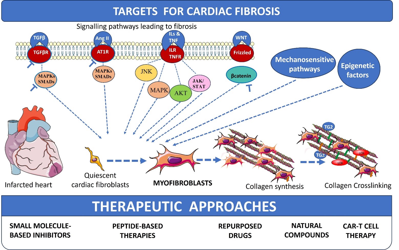
Cardiac fibrosis represents a global health crisis, observed in nearly all forms of heart disease, and contributes significantly to the progression of heart failure. Driven by diverse etiologies such as chronic hypertension, myocardial infarction, and metabolic disorders, cardiac fibrosis is characterized by the excessive deposition of extracellular matrix proteins. At the cellular level, the activation of cardiac fibroblasts into myofibroblasts serves as the primary mechanism for this structural remodelling. Excessive collagen deposition, crosslinking, and pathological scarring lead to increased ventricular stiffness, electrical arrhythmias, and a profound decline in cardiac function, affecting the quality of life for millions of patients worldwide. The review discusses the existing well-known profibrotic signals and molecular signalling pathways leading to cardiac fibroblast activation, collagen synthesis, and crosslinking. Mechanosensitive pathways, signalling mechanisms involved in collagen crosslinking, and epigenetic factors of cardiac fibrosis are also discussed along with their potential antifibrotic targets and therapeutic drugs. Further, small-molecule inhibitors, peptide-based therapies, natural compounds, and repurposed drugs for fibrosis are also discussed. This review concludes with recent approaches of chimeric antigen receptor (CAR)-T cell therapy for cardiac fibrosis.
Open Access
Review
04 September 2025Collagen Biosynthesis to Engineered Biomaterials: Molecular Design, Synthetic Strategy, and Biomedical Application
Collagen, a principal component of the extracellular matrix, provides mechanical strength and stability to tissues and organs through its structural organization. Its biocompatibility has established it as a crucial material in biomedical applications such as drug delivery systems, cell culture matrices, and tissue engineering scaffolds. However, the use of animal-derived collagen carries risks of pathogen transmission, which has driven research towards developing synthetic collagen alternatives. Advances in AI-assisted protein engineering are accelerating the design of synthetic collagens and their applications in biomaterials. This review examines collagen’s structural characteristics, biosynthesis strategies, biological activities as well as AI-assisting engineering.
Open Access
Review
27 June 2025Targeting Collagen Secretion as a Potential Therapeutic Strategy to Modulate Fibrosis
Fibrotic diseases are driven by the excessive accumulation of extracellular matrix (ECM), particularly collagens, leading to progressive tissue stiffness and organ dysfunction. While many factors contribute to fibrosis—including cytokine signaling, integrin-mediated mechanotransduction, and altered ECM degradation—the synthesis and secretion of collagen remain central bottlenecks. Collagen biosynthesis is a complex process involving extensive post-translational modification and intracellular trafficking. The export of procollagen from the endoplasmic reticulum (ER) requires Transport and Golgi Organisation 1 (TANGO1), a transmembrane organizer of ER exit sites that coordinates cargo selection, membrane remodeling, and connectivity between the ER and the ER-Golgi-Intermediate-Comaprtment (ERGIC). By assembling into ring-like structures at ER exit sites, TANGO1 builds a secretory route for bulky cargoes that bypasses conventional vesicle constraints. Loss of TANGO1 disrupts collagen secretion and causes developmental defects across various species. In fibrotic tissues, TANGO1 expression is upregulated, linking secretory machinery to pathological matrix deposition. Recent work has identified specific interfaces within the complex of TANGO1 with its vertebrate paralogue Cutaneous T-cell lymphoma-associated antigen 5 (cTAGE5) as targets for cell-permeant peptide inhibitors. Inhibitors that selectively and specifically block TANGO1 complex formation reduce collagen secretion in fibroblasts and scar formation in vivo, offering a new strategy to modulate fibrotic processes.

Open Access
Review
23 October 2024The Notch3 Pathway in Organ Fibrosis
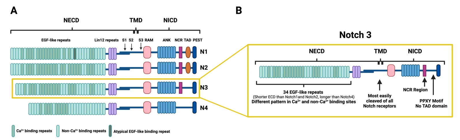
Fibrosis occurs in many organs, including the lung, heart, skin, liver or kidney, and is characterized by progressive tissue scarring in response to repetitive or chronic non-resolving injury, ultimately leading to organ failure and death. It is, in fact, a major cause of morbidity and mortality worldwide, being estimated to account for 45% of deaths in the world. Despite this fact, little progress has been made therapeutically, and fibrosis remains a major clinical and therapeutic challenge. Although significant advances in our understanding of cellular and molecular mechanisms driving tissue fibrosis have been made, the lack of an efficient treatment reflects the limited insight into the pathophysiological mechanisms underlying the initiation and progression of the fibrotic process. Thus, there is an urgent need for better understanding of tissue fibrosis and repair mechanisms that later lead to the development of new therapeutic approaches to fight fibrosis. The Notch pathway is a highly conserved signaling pathway that has been linked to tissue fibrosis in many organs and promises to open new therapeutic opportunities. This manuscript reviews the relevance of Notch signaling in the development and progression of tissue fibrosis in several organs with a special focus on the Notch3 pathway due to the unique features of this receptor.
Open Access
Review
21 December 2023TANGO1 Dances to Export of Procollagen from the Endoplasmic Reticulum
The endoplasmic reticulum (ER) to Golgi secretory pathway is an elegantly complex process whereby protein cargoes are manufactured, folded, and distributed from the ER to the cisternal layers of the Golgi stack before they are delivered to their final destinations. The export of large bulky cargoes such as procollagen and its trafficking to the Golgi is a sophisticated mechanism requiring TANGO1 (Transport ANd Golgi Organization protein 1. It is also called MIA3 (Melanoma Inhibitory Activity protein 3). TANGO1 has two prominent isoforms, TANGO1-Long and TANGO1-Short, and each isoform has specific functions. On the luminal side, TANGO1-Long has an HSP47 recruitment domain and uses this protein to collect collagen. It can also tether its paralog isoforms cTAGE5 and TALI and along with these proteins enlarges the vesicle to accommodate procollagen. Recent studies show that TANGO1-Long combines retrograde membrane flow with anterograde cargo transport. This complex mechanism is highly activated in fibrosis and promotes the excessive deposition of collagen in the tissues. The therapeutic targeting of TANGO1 may prove successful in the control of fibrotic disorders. This review focuses on TANGO1 and its complex interaction with other procollagen export factors that modulate increased vesicle size to accommodate the export of procollagen.

Open Access
Article
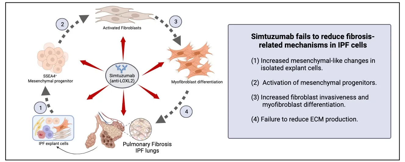
28 November 2023Translational Studies Reveal the Divergent Effects of Simtuzumab Targeting LOXL2 in Idiopathic Pulmonary Fibrosis
The composition of extracellular matrix (ECM) is altered during pathologic scarring in damaged organs including the lung. One major change in the ECM involves the cross-linking of collagen, which promotes fibroblast to myofibroblast differentiation. We examined the role of lysyl oxidase (LOX)-like 2 in lung progenitors and fibroblasts cultured from normal or IPF lung samples and in a humanized mouse model of IPF using a monoclonal antibody (Simtuzumab). Primary lung fibroblasts from normal donor lungs and IPF lung explants were examined for expression of LOXL2. Targeting LOXL2 with Simtuzumab on normal and IPF fibroblasts was examined both in vitro and in vivo for synthetic, functional, and profibrotic properties. LOXL2 was increased at transcript and protein level in IPF compared with normal lung samples. In a dose-dependent manner, Simtuzumab enhanced differentiation of fibroblasts into myofibroblasts. Inhibition of LOXL2 also enhanced fibroblast invasion and accelerated the outgrowth of fibroblasts from dissociated human lung cell preparations. Finally, preventative or delayed delivery of Simtuzumab enhanced lung fibrosis in a humanized mouse model of pulmonary fibrosis. Consistent with its failure in a Phase 2 clinical trial, Simtuzumab exhibited no therapeutic efficacy in translational in vitro and in vivo assays.