
The Four-Hit Framework of Aortic Aneurysm Progression: From Wall Injury to Imaging and Targeted Intervention
The Four-Hit Framework of Aortic Aneurysm Progression: From Wall Injury to Imaging and Targeted Intervention
Manhong Yi
1,†
Hongjiu Zhang
2,†
Keyi Fan
2
Jiang Han
2
Shirong Zhu
2
Yuwen Wang
2,*
Heng Wang
3,4,*
 Guoping Zheng
3,4,*
Guoping Zheng
3,4,*
Received: 19 January 2026 Revised: 28 April 2026 Accepted: 06 August 2026 Published: 14 August 2026

© 2026 The authors. This is an open access article under the Creative Commons Attribution 4.0 International License (https://creativecommons.org/licenses/by/4.0/).
Graphical Abstract
1. Introduction
Aortic aneurysm (AA) is a life-threatening vascular disorder characterized by segmental and progressive dilatation of the aorta, degenerative remodeling of the aortic wall, and an increased risk of rupture. Clinically, AA is commonly classified into thoracic aortic aneurysm (TAA) and abdominal aortic aneurysm (AAA), which differ in anatomical location, epidemiology, risk adrs, genetic contribution, and dominant pathological features. The increasing global burden of AA, together with the high mortality of rupture and the substantial cost of surgical or endovascular repair, highlights the need for improved approaches to disease assessment and intervention [1]. Current clinical management relies largely on diameter-based surveillance, but aortic diameter does not fully reflect the biological activity within the aneurysm wall, including inflammation, metabolic dysregulation, extracellular matrix remodeling, impaired repair, and cellular senescence [2]. At present, no pharmacological therapy has been established in routine clinical practice to effectively halt AA progression [3].
Over the past two decades, studies using animal models, human aneurysm tissues, genetic analyses, multi-omics approaches, single-cell sequencing, and vascular biology methods have identified multiple mechanisms involved in AA pathogenesis, including endothelial dysfunction, smooth muscle cell phenotypic switching, immune-cell infiltration, oxidative stress, proteolytic matrix degradation, maladaptive repair, and aging-associated vascular degeneration [4,5,6,7,8,9,10]. However, these processes are often discussed as separate pathological events, and their relative importance may differ between AAA and TAA, between genetic and non-genetic aneurysms, and across disease stages. In this review, we propose the Four-Hit Hypothesis as an integrative framework for understanding AA progression. The framework includes four broad biological domains: disruption of the aortic barrier environment, immune-inflammatory invasion, maladaptive proliferative repair, and aging-associated structural destruction. Rather than representing a new standalone mechanism or a universal linear pathway, the Four-Hit Hypothesis is used here as an organizing framework to summarize current evidence and discuss implications for biomarkers, imaging, and therapeutic development.
2. Clinical and Biological Heterogeneity of Aortic Aneurysm
An aortic aneurysm should be viewed as a spectrum of biologically distinct disorders rather than a single, uniform disease entity. Although thoracic aortic aneurysm (TAA) and abdominal aortic aneurysm (AAA) share the final phenotype of progressive aortic dilatation and wall weakening, they differ markedly in developmental origin, anatomical environment, cellular composition, genetic architecture, inflammatory burden, and clinical behavior [11]. In TAA, particularly heritable forms such as Marfan syndrome, Loeys-Dietz syndrome, vascular Ehlers-Danlos syndrome, and familial nonsyndromic TAA, disease initiation often stems from intrinsic medial vulnerability, including defects in extracellular matrix integrity, transforming growth factor-β signaling, smooth muscle cell contractile function, and mechanotransduction. However, recent studies indicate that TAA is not simply a passive consequence of structural weakness. Zhang et al. showed that aortic stress activates an adaptive smooth muscle cell program that preserves aortic strength and protects against aneurysm and dissection in mice [6]. Huang et al. demonstrated that CX3CR1-positive macrophages can promote aneurysm progression in a Marfan syndrome model [12]. These findings suggest that TAA progression reflects a dynamic balance among inherited wall vulnerability, stress-responsive smooth muscle cell adaptation, immune cell activity, and maladaptive repair (Table 1).
Table 1. Representative evidence illustrating mechanistic heterogeneity across aortic aneurysm subtypes.
|
Four-Hit Component |
Representative Evidence |
Aneurysm Context |
Heterogeneity Highlighted |
|---|---|---|---|
|
Hits 1/3: Barrier disruption/maladaptive proliferative repair |
Stress-responsive adaptive SMC program (Zhang et al.) |
TAA model |
Medial vulnerability and wall-stress response |
|
Hit 2: Immune-inflammatory invasion. |
CX3CR1 + macrophage-driven progression (Huang et al.) |
Marfan-associated TAA |
Immune modification of a genetic aneurysm |
|
Hit 2: Immune-inflammatory invasion. |
IFN-inducible monocyte/macrophage states (Le et al.) |
AAA single-cell analysis |
Immune-cell state heterogeneity |
|
Hit 2: Immune-inflammatory invasion. |
Microbiome-NET axis (Tian et al.) |
Experimental AAA |
Systemic inflammatory input |
|
Hit 4: Aging-associated structural destruction. |
miR-1204-MYLK aging axis (Liu et al.) |
AAD model |
Aging-driven disease amplification |
|
Hit 4: Aging-associated structural destruction. |
Age-associated aortic proteomic remodeling (Tyrrell et al.) |
Human aorta/TAA |
Age-related molecular remodeling |
|
Cross-cutting heterogeneity |
Shared genetic susceptibility with cardiometabolic traits (Zheng et al.) |
AAA genetic analysis |
Genetic-metabolic convergence |
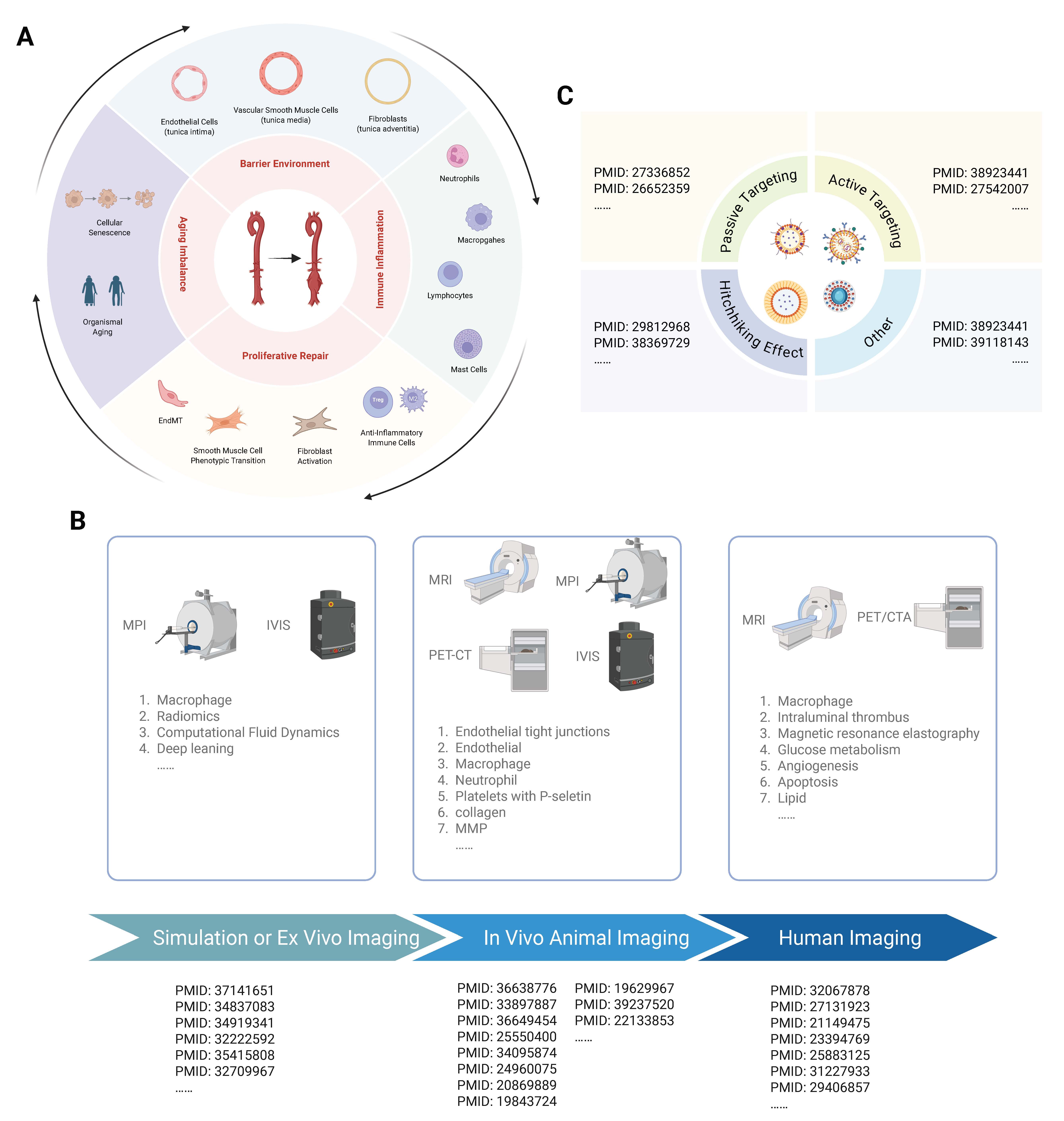
By contrast, degenerative AAA is more closely linked to aging, smoking, cardiometabolic risk factors, intraluminal thrombus, chronic inflammation, and proteolytic extracellular matrix destruction. Sex-related hormonal factors may further contribute to aneurysm heterogeneity. Androgen signaling exerts context-dependent effects on endothelial function, inflammation, vascular smooth muscle cell homeostasis, and extracellular matrix remodeling, with its vascular consequences varying with hormone levels, cellular context, and disease state [13]. This disease is increasingly recognized as an active thrombo-inflammatory and immune-remodeling process rather than a simple mechanical enlargement of the infrarenal aorta. Single-cell and mechanistic studies have strengthened this concept: Le et al. identified interferon-inducible monocytes/macrophages as candidate cellular drivers of AAA progression and rupture risk [14]; Tian et al. linked gut microbiome dysbiosis to neutrophil extracellular trap formation and aneurysm development [15]; and Wang et al. showed that macrophage ILF3 promotes AAA by inducing an inflammatory imbalance [16]. At the same time, the boundary between genetic and acquired disease is not absolute. Zheng et al. reported shared genetic susceptibility between AAA and cardiometabolic traits related to lipid metabolism and inflammation [17], while Liu et al. and Tyrrell et al. highlighted aging-associated molecular and proteomic remodeling as active modifiers of aneurysm biology [4,18]. Together, these findings support a model in which aneurysm subtype, etiology, disease stage, and dominant biological activity determine the relative contribution of barrier disruption, immune-inflammatory invasion, maladaptive repair, and aging-associated structural destruction (Figure 1).
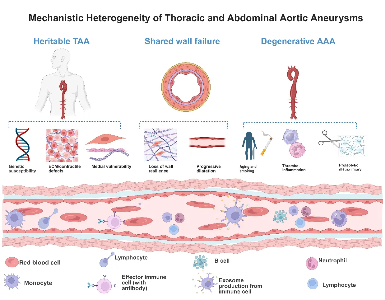
Figure 1. Mechanistic heterogeneity of thoracic and abdominal aortic aneurysms. Distinct mechanisms underlying TAA and AAA converge on a shared phenotype of aortic wall weakening and progressive dilatation. AAA, abdominal aortic aneurysm; ECM, extracellular matrix; TAA, thoracic aortic aneurysm.
Thus, the Four-Hit Hypothesis is best understood not as a universal linear pathway or a new standalone mechanism, but as a comparative framework or tool for synthesizing established and emerging evidence and organizing heterogeneous mechanisms across aneurysm subtypes.
3. The Four-Hit Hypothesis of Aortic Aneurysm Progression
Aortic aneurysm progression can be framed as a breakdown of coordinated aortic wall maintenance. The normal aortic wall depends on endothelial integrity, vascular smooth muscle cell homeostasis, extracellular matrix organization, mechanosensitive signaling, and controlled immune surveillance to preserve tensile strength under continuous pulsatile stress. Disruption of these systems has been implicated at multiple biological levels: endothelial tight-junction dysfunction has been linked to thoracic aortic aneurysm and dissection [19], improved endothelial barrier function has been shown to decelerate early abdominal aortic aneurysm progression [20], altered smooth muscle mechanosensation can promote aneurysm formation [21], and epigenetic control of vascular smooth muscle cell homeostasis has been shown to protect against abdominal aortic aneurysm formation [22]. Building on these observations, the Four-Hit Hypothesis organizes aneurysm progression into four partially overlapping biological domains: disruption of the aortic barrier environment, immune-inflammatory invasion, maladaptive proliferative repair, and aging-associated structural destruction. This framework provides a structured way to discuss how loss of wall integrity, inflammatory amplification, incomplete repair, and progressive tissue degeneration may jointly shape aneurysm evolution.
3.1. Hit 1: Disruption of the Aortic Barrier Environment
The first hit can be conceptualized as a failure of the aortic wall to preserve compartmental integrity and buffer mechanical stress. The aorta is not simply a passive conduit; it is a multilayered biological barrier in which endothelial junctions regulate luminal permeability, medial smooth muscle cells and elastic lamellae absorb pulsatile load, extracellular matrix networks maintain tensile strength, and the adventitia participates in immune surveillance and repair. When this barrier environment is disturbed, circulating inflammatory mediators, metabolic stress, thrombotic signals, and oxidative injury can more readily access or reshape the vessel wall, lowering the threshold for downstream inflammation and structural degeneration. Recent studies support this barrier-centered view at the luminal interface: Yang et al. showed that endothelial tight junction disruption contributes to thoracic aortic aneurysm and dissection [19], whereas another study by Yang et al. demonstrated that improving endothelial barrier function through ALDH2-LIN28B-ELK3 signaling slows early progression of abdominal aortic aneurysm. Luo et al. further linked endothelial dysfunction to aneurysm and dissection through an HDAC1-ZEB2-NuRD complex that regulates protein S-sulfhydration [23], and Stammer et al. reported VE-cadherin shedding in patients with aortic aneurysm and dissection [24]. These findings suggest that endothelial instability may contribute to the creation of a permissive interface for inflammatory entry and wall injury.
Barrier disruption also involves the medial compartment’s failure to sense and appropriately absorb mechanical stress. Qian et al. showed that microskeletal stiffness promotes aneurysm formation by sustaining pathological vascular smooth muscle cell mechanosensation through Piezo1 [21], while Zhao et al. reported that BAF60c protects against abdominal aortic aneurysm formation through epigenetic control of vascular smooth muscle cell homeostasis. These studies extend the concept of “barrier” beyond the endothelial monolayer to encompass medial stress buffering and maintenance of smooth muscle cell state. In heritable TAA, this first hit may be dominated by intrinsic medial vulnerability and abnormal mechanotransduction; in degenerative AAA, it may be more closely linked to endothelial dysfunction, metabolic injury, thrombotic signaling, adventitial inflammation, and proteolytic matrix damage. Thus, disruption of the aortic barrier environment creates the permissive biological ground on which immune-inflammatory invasion and maladaptive repair can subsequently develop.
3.2. Hit 2: Immune-Inflammatory Invasion
Following disruption of the aortic barrier environment, inflammation becomes a mechanism of biological amplification rather than a nonspecific bystander response. The injured aneurysmal wall provides multiple entry points and niches for immune activation, including endothelial dysfunction, medial injury, adventitial remodeling, intraluminal thrombus, and perivascular tissue. Within these compartments, macrophages, neutrophils, lymphocytes, mast cells, platelets, fibroblasts, and smooth muscle cells can form multicellular inflammatory circuits that sustain cytokine signaling, protease activity, oxidative stress, and regulated cell death. Recent studies have moved the field beyond the idea of diffuse inflammatory infiltration toward a more resolved view of cell-cell communication and immune niches. This reciprocal immune-vascular interaction is not unique to aneurysm disease. In pulmonary arterial hypertension, crosstalk among immune cells, endothelial cells, smooth muscle cells, and fibroblasts similarly sustains endothelial dysfunction, proliferative remodeling, and fibrosis. Although the vascular territories differ, these observations reinforce the concept that inflammation is maintained through bidirectional cellular communication rather than isolated immune-cell infiltration [25]. Wu et al. showed that NINJ1 facilitates abdominal aortic aneurysm formation by enhancing macrophage infiltration through the TLR4-ANXA2 axis [26]. Thayaparan et al. linked endothelial dysfunction to macrophage-dependent abdominal aortic aneurysm formation in an atherosclerotic plaque context [27]. Wagenhäuser et al. further demonstrated that platelet crosstalk with macrophages and fibroblasts aggravates inflammation, aortic wall stiffening, and osteopontin release in abdominal aortic aneurysm [28]. In parallel, neutrophil-related injury illustrates how innate immune activation can be converted into structural damage: Chen et al. reported that mesenchymal stem cell-derived extracellular vesicles protect against abdominal aortic aneurysm formation by inhibiting NET-induced ferroptosis [29], Qi et al. showed that NET-induced ferroptosis promotes aneurysm formation through SLC25A11-mediated mitochondrial glutathione depletion [30], and Xiong et al. demonstrated that PI3Kγ promotes NET formation through noncanonical pyroptosis in abdominal aortic aneurysm [31].
The inflammatory profile of the second hit is unlikely to be uniform across aneurysm subtypes. In a degenerative abdominal aortic aneurysm, immune-inflammatory invasion is often a dominant feature, supported by macrophage and neutrophil infiltration, intraluminal thrombus-associated inflammation, NET formation, oxidative stress, and proteolytic matrix remodeling. In contrast, in many forms of heritable thoracic aortic aneurysm, inflammation may act more as a disease modifier than as the initiating event, superimposed on intrinsic medial vulnerability, smooth muscle cell dysfunction, and extracellular matrix defects. Moreover, immune activation is not uniformly destructive. Depending on timing and cellular state, macrophage polarization, regulatory T cells, and reparative stromal responses may either restrain inflammation or contribute to maladaptive remodeling. Single-cell immune profiling has begun to capture this complexity, with Yuan et al. deconstructing immune-cell distributions in experimental abdominal aortic aneurysm [32]. Thus, the second hit should be interpreted as a context-dependent immune remodeling program rather than a simple accumulation of inflammatory cells. Its pathological consequence is to amplify matrix degradation, vascular cell dysfunction, and the transition toward maladaptive repair.
3.3. Hit 3: Maladaptive Proliferative Repair
The third hit captures a central paradox in aneurysm biology: the same reparative programs that may preserve wall integrity can, when dysregulated, accelerate wall failure. After barrier disruption and immune-mediated injury, the aortic wall activates smooth muscle cell plasticity, extracellular matrix remodeling, fibroblast and stromal-cell expansion, endothelial-to-mesenchymal transition, and growth factor signaling in an attempt to restore mechanical competence. These responses are not intrinsically harmful; in early or localized injury, they may reinforce the wall and compensate for matrix loss. However, when repair is prolonged, spatially disorganized, or uncoupled from the original injury, it can produce fibrosis, stiffness, cellular exhaustion, matrix disarray, and progressive loss of tensile strength. Recent studies support this shift from adaptive to maladaptive repair. Song et al. showed that SLC44A2 regulates vascular smooth muscle cell phenotypic switching and aortic aneurysm formation [33], whereas Zhang et al. reported that HINT1 aggravates aneurysm by targeting the ITGA6/FAK axis in vascular smooth muscle cells [34]. Guo et al. further linked LXRα to abdominal aortic aneurysm formation through UHRF1-mediated epigenetic modification of miR-26b-3p [35]. These findings place smooth muscle cell state transitions at the center of the repair response, where plasticity may be either compensatory or pathogenic depending on timing, context, and persistence of injury.
The form of maladaptive repair differs across aneurysm subtypes. In heritable thoracic aortic aneurysm, repair failure is often superimposed on intrinsic medial weakness, abnormal smooth muscle cell contractility, extracellular matrix instability, and altered mechanobiological signaling; in this setting, insufficient or misdirected repair may be unable to restore medial strength. In a degenerative abdominal aortic aneurysm, repair occurs within a more inflammatory and proteolytic environment shaped by thrombus-associated injury, adventitial activation, macrophage and neutrophil signaling, and matrix degradation. Non-smooth muscle cell compartments also contribute to this process: Pan et al. identified SM22α-lineage perivascular stromal cells as contributors to abdominal aortic aneurysm [36], and Millar et al. linked endothelial-to-mesenchymal transition to interleukin-1 pathway activity during aneurysm formation [37]. From a translational perspective, Wei et al. showed that extravascular administration of IGF1R agonists protects against aneurysm formation in rodent and porcine models [38], suggesting that repair-associated growth factor pathways may be therapeutically modifiable. Thus, the third hit is context-dependent in at least four respects: the aneurysm subtype in which repair occurs, the injury milieu that shapes the reparative response, the timing and persistence of repair activation, and the cellular source and matrix quality of the newly formed tissue. A response that is stabilizing in one setting may become pathogenic in another when chronic inflammation, thrombus-derived proteases, or persistent growth factor signaling drive disorganized fibrosis and wall stiffening. Therefore, maladaptive proliferative repair should be understood not simply as “excess repair” or “failed repair”, but as a remodeling state in which compensatory programs become insufficient, misdirected, or biologically costly.
3.4. Hit 4: Aging-Associated Structural Destruction
The fourth hit represents the point at which aneurysm biology shifts from potentially compensated remodeling to progressive structural failure. Aging should not be interpreted simply as a demographic risk factor; it changes the biological reserve of the aortic wall. With aging, vascular smooth muscle cells and stromal compartments become more vulnerable to senescence, mitochondrial dysfunction, oxidative stress, proteostatic failure, and regulated cell death. These processes reduce the capacity of the wall to restore matrix organization, resolve inflammation, and maintain mechanical resilience after repeated injury. Recent work has begun to connect these aging-related programs with aneurysm vulnerability. Sun et al. showed that pro-ferroptotic signaling promotes arterial aging through vascular smooth muscle cell senescence [39], whereas Zhang et al. reported that ganglioside GM3 protects against abdominal aortic aneurysm by suppressing ferroptosis [40]. Wang et al. further demonstrated that targeting the smooth muscle cell Keap1-Nrf2-GSDMD-pyroptosis axis prevents abdominal aortic aneurysm formation [41]. These studies suggest that late-stage wall destruction is not merely the passive result of matrix wear, but may reflect active coupling among senescence, oxidative injury, ferroptosis, pyroptosis, and failed repair.
The consequences of aging are likely to differ between aneurysm subtypes. In a degenerative abdominal aortic aneurysm, aging interacts with smoking, intraluminal thrombus, chronic inflammation, and metabolic injury, creating a milieu in which oxidative stress, immune activation, and proteolytic matrix degradation reinforce one another [42]. Evidence from peripheral artery disease further suggests that smoking-related vascular injury may be biologically embedded through accelerated epigenetic aging. Sheng et al. found that a DNA methylation-derived measure of aging partially mediated the association between early-life smoking and lower ankle-brachial index and increased peripheral artery disease risk. Although obtained in a different vascular disorder, these findings provide a mechanistic precedent for linking environmental exposure, epigenetic aging, and persistent vascular vulnerability [43]. In thoracic aortic aneurysm, aging may lower the threshold at which medial degeneration, abnormal mechanotransduction, extracellular matrix instability, or stress-response pathways become clinically expressed [44,45,46]. In both settings, however, aging reduces the compensatory reserve needed to buffer the earlier hits: barrier disruption becomes more difficult to repair, immune-inflammatory signals are less effectively resolved, and proliferative repair is more likely to become exhausted, fibrotic, or misdirected. Consistent with this model, GSDME-dependent pyroptosis has been linked to abdominal aortic aneurysm through promotion of vascular senescence [47], and smooth muscle cell oxidative-inflammatory signaling through STING has been shown to exacerbate aortic aneurysm and dissection [48] (Figure 2).
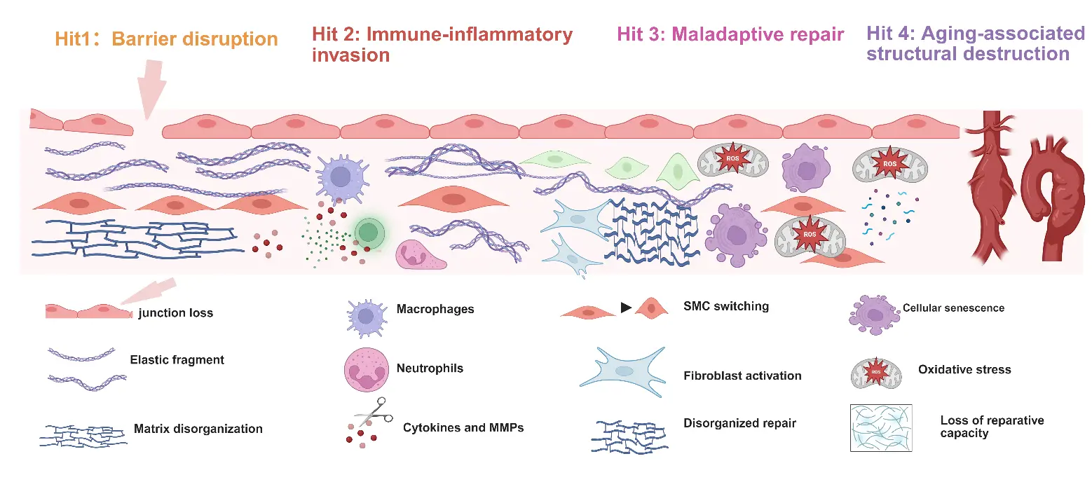
Figure 2. The Four-Hit Framework of Aortic Aneurysm Progression. Four interacting pathological processes promote aortic dilatation, wall degeneration, and rupture. The four domains overlap and interact dynamically and should not be interpreted as a fixed temporal sequence.
Thus, aging-associated structural destruction should be understood as a disease-amplifying state in which the aneurysmal wall progressively loses resilience, making rapid expansion and rupture more likely outcomes.
4. Biomarkers and Imaging Readouts of the Four-Hit Framework
Maximum aortic diameter remains the dominant criterion for aneurysm surveillance and intervention, yet it represents an anatomical consequence rather than a direct measure of disease activity. This distinction is important within the Four-Hit framework because barrier dysfunction, immune-inflammatory activation, maladaptive repair, and aging-associated structural destruction may emerge before overt enlargement and may continue despite apparently stable diameter. Increasing evidence indicates that aneurysm progression is influenced by biological and biomechanical features beyond size alone. Intraluminal thrombus has been associated with rapid abdominal aortic aneurysm growth [49], and proteolytic activity has been linked to aneurysm wall morphology and thrombus volume [50]. Biomechanical analyses further suggest that rupture-related parameters can evolve independently of vessel geometry during aneurysm growth [51], while finite element analysis has shown that predicted rupture risk may correlate with aortic wall histology in individual patients [52]. These findings support the view that diameter-based imaging is often downstream of the biological processes that determine wall instability. Accordingly, biomarkers and imaging readouts should be considered complementary tools for identifying active aneurysm biology, refining patient phenotypes, and supporting mechanism-based clinical trial design, rather than replacements for established anatomical thresholds (Table 2).
Table 2. Translational landscape of aortic aneurysm assessment tools.
|
Assessment Domain |
Representative Readouts |
Biological Information |
Potential Application |
Evidence Stage |
Key Limitation |
|---|---|---|---|---|---|
|
Circulating inflammation |
CRP, IL-6, chemokines |
Systemic and vascular inflammation |
Activity assessment and longitudinal monitoring |
Clinical investigation |
Limited aortic specificity |
|
Matrix turnover |
MMP-2, MMP-9, elastin- and collagen-derived fragments |
Extracellular matrix degradation |
Detection of active wall remodeling |
Clinical investigation |
Assay and cohort heterogeneity |
|
Thrombus activity |
D-dimer, fibrin degradation products |
Thrombus formation and fibrinolysis |
Assessment of thrombus-associated AAA activity |
Clinical investigation |
Confounding by systemic thrombosis |
|
Molecular signatures |
miRNAs, extracellular vesicles, proteomic profiles |
Cell communication and molecular remodeling |
Patient stratification and biomarker panels |
Exploratory |
Limited external validation |
|
Ultrasound and CTA |
Diameter, growth, volume, morphology, and intraluminal thrombus |
Structural disease burden |
Surveillance and procedural planning |
Clinical standard |
Limited biological information |
|
MRI and 4D-flow MRI |
Wall characteristics, flow patterns, and hemodynamics |
Tissue remodeling and altered flow |
Functional phenotyping |
Emerging clinical |
Cost and technical complexity |
|
PET imaging |
Tracer uptake in the aneurysm wall or thrombus |
Inflammation, metabolism, or matrix activity |
Identification of biologically active regions |
Clinical investigation |
Variable tracer specificity |
|
Molecular imaging |
Targeted probes for proteases, inflammation, or thrombus |
Specific molecular activity in vivo |
Early detection and treatment monitoring |
Predominantly preclinical |
Translation and safety barriers |
Circulating biomarkers provide a minimally invasive approach to interrogating aneurysm biology, but their clinical utility remains limited by insufficient disease specificity and biological overlap with systemic inflammation, thrombosis, atherosclerosis, renal dysfunction, and aging. Matrix remodeling and thrombus-associated markers remain among the most extensively investigated candidates. Circulating matrix metalloproteinase-9 has been associated with the presence of abdominal aortic aneurysm in a meta-analysis [53], whereas plasma fibrinogen and D-dimer have been linked to aneurysm presence, consistent with activation of coagulation and fibrinolysis, and intraluminal thrombus biology [54]. D-dimer has also been reported as a predictor of abdominal aortic aneurysm progression [55] and as a diagnostic biomarker in patients with peripheral artery disease [56]. More recent studies have moved toward multidimensional biomarker discovery. Circulating cardiovascular biomarkers have been associated with aneurysm volume [57], blood-based biomarker discovery has been explored for abdominal aortic aneurysm detection [58], and targeted proteomics and metabolomics have been applied to biomarker discovery in abdominal aortic aneurysm and post-EVAR sac volume assessment [59]. These data suggest that single biomarkers are unlikely to capture the complexity of aneurysm progression. Within the Four-Hit framework, biomarker signals should instead be interpreted as overlapping biological signatures, in which proteolysis, thrombus activity, inflammation, repair remodeling, and tissue degeneration converge.
Imaging provides a spatial and anatomical context that circulating biomarkers cannot offer. Ultrasound, computed tomography angiography, and magnetic resonance angiography remain indispensable for measuring diameter, defining anatomy, detecting thrombus, and planning open or endovascular repair. However, functional imaging may provide additional insight into active biological processes within the aneurysm wall and thrombo-inflammatory microenvironment. In abdominal aortic aneurysm, 18F-sodium fluoride positron emission tomography identifies focal microcalcification activity and was associated with aneurysm progression and clinical events in the SoFIA3 study [60]. 18F-fluorodeoxyglucose PET has been used to assess vascular inflammation; increased uptake has been associated with inflammatory features, wall instability, and acute symptoms in some studies [61], although systematic evaluation has highlighted heterogeneity in its relationship with aneurysm growth and rupture [62]. This inconsistency may reflect differences in patient selection, imaging protocols, background atherosclerosis, partial-volume correction, and the dynamic nature of inflammatory activity. Advanced magnetic resonance approaches offer a complementary perspective. Four-dimensional flow MRI can quantify abnormal flow patterns, flow stasis, wall shear stress, and thrombus-related hemodynamic changes in abdominal aortic aneurysm [63,64]. These methods may help connect local mechanics with barrier injury, thrombus-wall interaction, and regional remodeling, but require further standardization and prospective validation before routine clinical implementation.
Molecular and nanoparticle-based imaging approaches extend biological readouts beyond morphology and hemodynamics, particularly in preclinical and early translational studies. PET imaging of vessel-wall matrix metalloproteinase activity has demonstrated the feasibility of visualizing proteolytic remodeling within abdominal aortic aneurysm tissue [65]. Experimental imaging strategies have also targeted macrophage-rich inflammation, including computed tomography imaging of macrophage phagocytic activity in abdominal aortic aneurysm [66], and CXCR4-targeted magnetic particle imaging has been explored to detect inflammatory cell accumulation and assess aneurysm activity [67]. Conceptually, these approaches align with the Four-Hit framework: endothelial or thrombus-associated probes may report barrier disruption, macrophage- or neutrophil-targeted probes may reflect immune-inflammatory invasion, MMP- or collagen-turnover probes may indicate maladaptive repair and matrix remodeling, and future probes directed at senescence or regulated cell death may help capture late structural destruction. Nevertheless, most molecular probes and nanoparticle platforms remain investigational.
Their near-term translational value is more likely to lie in mechanistic studies, biological phenotyping, and the enrichment of clinical trials with patients who have active inflammatory, proteolytic, thrombotic, or repair-associated disease. A realistic goal is therefore not to replace anatomical imaging, but to integrate anatomical, biochemical, molecular, and biomechanical information into a more informative model of aneurysm risk (Figure 3).
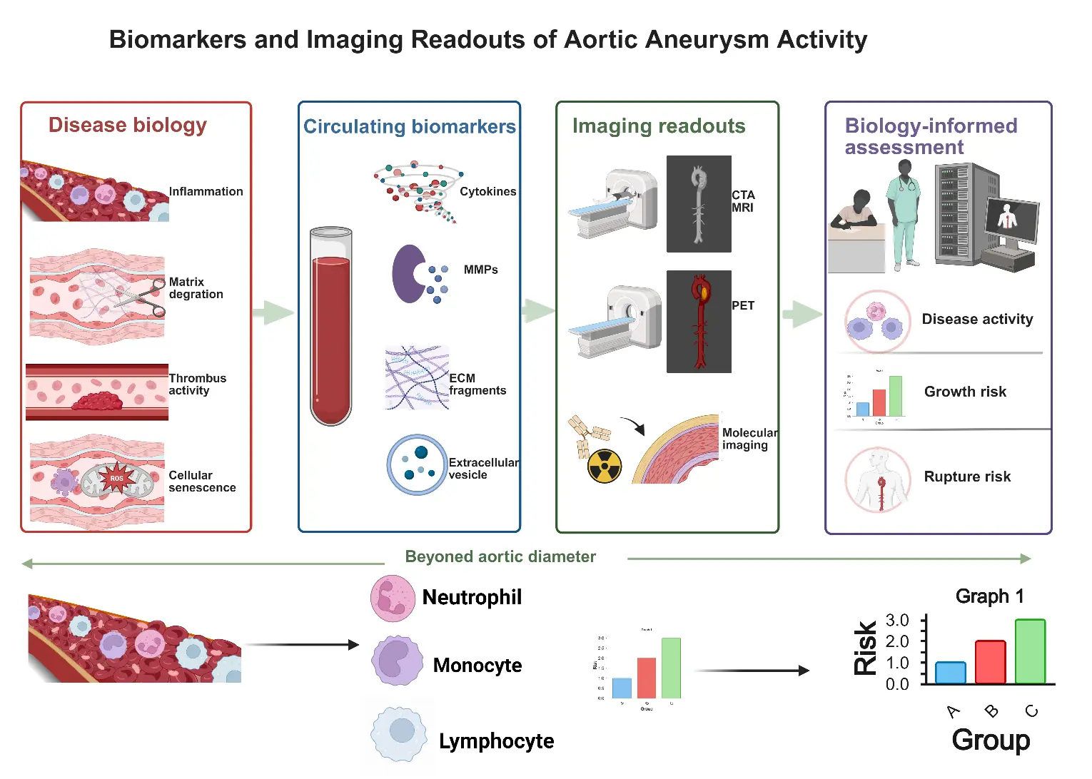
Figure 3. Biomarkers and Imaging Readouts of Aortic Aneurysm Activity. Circulating biomarkers and imaging modalities capture complementary aspects of aneurysm biology and may support biology-informed assessment of disease activity, growth, and rupture risk. Most circulating biomarkers and molecular imaging approaches remain investigational and are not established replacements for diameter-based surveillance.
5. Therapeutic Implications and Future Directions
The expanding mechanistic landscape of aortic aneurysm has yet to translate into an effective disease-modifying therapy. No pharmacological intervention has been conclusively shown to prevent expansion of abdominal aortic aneurysm (AAA) or to avert the need for surgical or endovascular repair, and aggregate clinical trial evidence remains largely neutral [68]. The failure of apparently rational therapies may reflect not only insufficient target engagement but also the biological heterogeneity of enrolled populations and the use of anatomical growth as a late therapeutic endpoint. Doxycycline exemplifies this disconnect: despite its matrix metalloproteinase–inhibitory activity, it did not reduce the growth of small infrarenal AAAs in a randomized trial [69]. By contrast, observational evidence has linked metformin use to slower AAA growth, although residual confounding precludes designating it as an aneurysm-specific therapy [69,70,71]. Therapeutic principles are also unlikely to be interchangeable across aneurysm subtypes. In Marfan syndrome, losartan may modestly attenuate aortic root dilatation, but its incremental benefit over β-blockade and its efficacy across genetic backgrounds remain uncertain [72,73]. These findings argue against a uniform pharmacological strategy for degenerative AAA and genetically mediated thoracic aortic aneurysm.
Preclinical evidence nevertheless identifies several therapeutically tractable processes, including endothelial dysfunction, immune-cell activation, neutrophil extracellular trap formation, smooth muscle cell instability, proteolytic matrix remodeling, senescence, and regulated cell death. Their relevance, however, is likely to depend on timing and pathological context. Inhibition of neutrophil extracellular traps attenuated experimental AAA progression in a thrombus-dependent manner, indicating that local disease architecture can modify therapeutic efficacy [74]. Pharmacological inhibition of gasdermin D also suppressed angiotensin II–induced aneurysm formation, implicating pyroptotic signaling as a potential target [75], whereas restoration of GPX4 activity limited smooth muscle cell ferroptosis and aneurysmal remodeling [76]. These observations should not be interpreted as support for indiscriminate suppression of inflammation or cellular plasticity. Immune responses and smooth muscle cell state transitions may be either destructive or protective depending on disease stage, cellular context, and the capacity for compensatory repair. The Four-Hit framework may therefore be most useful for distinguishing interventions that preserve wall homeostasis from those that restrain inflammatory amplification, redirect maladaptive remodeling, or limit irreversible structural loss, without imposing a fixed therapeutic sequence.
Targeted delivery systems may help address this biological complexity by increasing drug exposure within inflamed or proteolytically active aneurysm tissue while limiting systemic toxicity. Reactive oxygen species–responsive nanoparticles have demonstrated targeted antioxidant and anti-inflammatory effects in experimental AAA [77], and nanoparticle-mediated delivery of pitavastatin to monocytes and macrophages inhibited aneurysm formation in mice [78]. More recent platforms have combined inflammatory suppression with extracellular matrix stabilization or enabled targeted small-interfering RNA delivery [79,80]. Nevertheless, these technologies remain preclinical, and their translation will require rigorous assessment of biodistribution, clearance, immunogenicity, manufacturing consistency, long-term toxicity, and efficacy in large-animal models. Future trials should align therapeutic selection with aneurysm subtype, dominant biological activity, and disease stage, supported where appropriate by biomarkers and functional imaging. The principal translational value of the Four-Hit framework is therefore not the proposal of a universal treatment algorithm, but the organization of therapeutic hypotheses and the development of biologically enriched clinical trials (Figure 4).
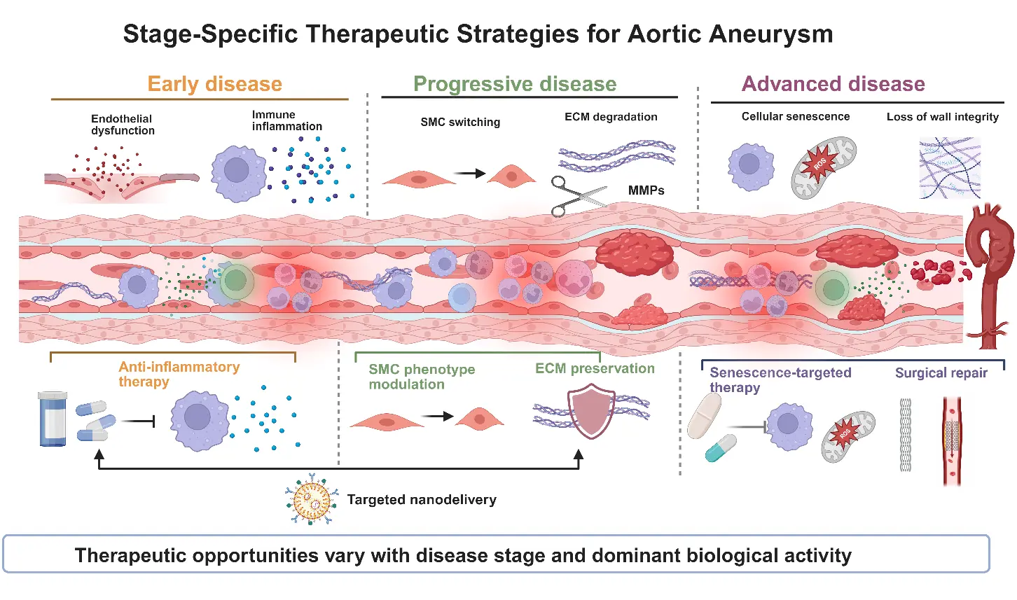
Figure 4. Stage-Specific Therapeutic Strategies for Aortic Aneurysm. Potential interventions targeting inflammation, vascular wall remodeling, matrix degradation, and cellular senescence are shown across conceptual disease phases. These phases do not represent a validated clinical staging system. Most pharmacological and targeted-delivery strategies remain investigational, whereas open or endovascular repair remains the established treatment for anatomically advanced disease. (The graphical elements and symbols used in Figure 4 are consistent with those defined and annotated in Figure 1, Figure 2 and Figure 3.)
6. Conclusions
Aortic aneurysm is a heterogeneous group of disorders in which genetic susceptibility, vascular wall injury, immune-inflammatory activation, maladaptive repair, and aging-related degeneration converge to promote progressive dilatation and rupture. The Four-Hit Hypothesis integrates these processes into four partially overlapping biological domains: disruption of the aortic barrier environment, immune-inflammatory invasion, maladaptive proliferative repair, and aging-associated structural destruction. This framework is not intended to define a universal linear pathway or propose a new standalone mechanism, but to organize established and emerging evidence across aneurysm subtypes and disease stages. Its potential value lies in connecting mechanistic heterogeneity with complementary biomarkers, functional imaging, and biologically informed therapeutic development. Future studies should validate these relationships in longitudinal human cohorts, distinguish clinically actionable signals from preclinical observations, and determine whether biological stratification can improve risk assessment and clinical trial design beyond anatomical measurements alone.
Author Contributions
Conceptualization, H.W. and G.Z.; Investigation, M.Y., H.Z., K.F., J.H., S.Z. and Y.W.; Writing—Original Draft Preparation, M.Y. and H.Z.; Writing—Review & Editing, H.W. and G.Z.; Visualization, M.Y., H.Z. and K.F.; Supervision, H.W. and G.Z. All authors have read and agreed to the published version of the manuscript.
Ethics Statement
Not applicable.
Informed Consent Statement
Not applicable.
Data Availability Statement
No new data were generated or analyzed in this review.
Funding
This research received no external funding.
Declaration of Competing Interest
The authors declare that they have no known competing financial interests or personal relationships that could have appeared to influence the work reported in this paper.
References
- Wang H, Li Y, Fan K, Zhao T, Xu K, Zahin M, et al. Global Epidemiology of Early-Onset Aortic Aneurysm: Temporal Trends, Risk Factors, and Future Burden Projections. J. Epidemiol. Glob. Health 2025, 15, 25. DOI:10.1007/s44197-025-00369-y [Google Scholar]
- Thorbjørnsen K, Svensjö S, Mani K, Wanhainen A. Morphological factors associated with progression of subaneurysmal aortas. Br. J. Surg. 2023, 110, 489–497. DOI:10.1093/bjs/znad030 [Google Scholar]
- Chen J, Hu L, Liu Z. Medical treatments for abdominal aortic aneurysm: An overview of clinical trials. Expert Opin. Investig. Drugs 2024, 33, 979–992. DOI:10.1080/13543784.2024.2377747 [Google Scholar]
- Tyrrell DJ, Chen J, Li BY, Wood SC, Rosebury-Smith W, Remmer HA, et al. Aging Alters the Aortic Proteome in Health and Thoracic Aortic Aneurysm. Arterioscler. Thromb. Vasc. Biol. 2022, 42, 1060–1076. DOI:10.1161/ATVBAHA.122.317643 [Google Scholar]
- Yang P, Liu H, Wang S, Xiao X, Jiang L, Le S, et al. PIEZO1 attenuates Marfan syndrome aneurysm development through TGF-β signaling pathway inhibition via TGFBR2. Eur. Heart J. 2025, 46, 958–974. DOI:10.1093/eurheartj/ehae786 [Google Scholar]
- Zhang C, Li Y, Chakraborty A, Li Y, Rebello KR, Ren P, et al. Aortic Stress Activates an Adaptive Program in Thoracic Aortic Smooth Muscle Cells That Maintains Aortic Strength and Protects Against Aneurysm and Dissection in Mice. Arterioscler. Thromb. Vasc. Biol. 2023, 43, 234–252. DOI:10.1161/ATVBAHA.122.318135 [Google Scholar]
- Chi Z, Chen S, Yang D, Cui W, Lu Y, Wang Z, et al. Gasdermin D-mediated metabolic crosstalk promotes tissue repair. Nature 2024, 634, 1168–1177. DOI:10.1038/s41586-024-08022-7 [Google Scholar]
- Chakraborty A, Li Y, Zhang C, Li Y, Rebello KR, Li S, et al. Epigenetic Induction of Smooth Muscle Cell Phenotypic Alterations in Aortic Aneurysms and Dissections. Circulation 2023, 148, 959–977. DOI:10.1161/CIRCULATIONAHA.123.063332 [Google Scholar]
- Li Y, Ren P, Dawson A, Vasquez HG, Ageedi W, Zhang C, et al. Single-Cell Transcriptome Analysis Reveals Dynamic Cell Populations and Differential Gene Expression Patterns in Control and Aneurysmal Human Aortic Tissue. Circulation 2020, 142, 1374–1388. DOI:10.1161/CIRCULATIONAHA.120.046528 [Google Scholar]
- Pedroza AJ, Dalal AR, Shad R, Yokoyama N, Nakamura K, Cheng P, et al. Embryologic Origin Influences Smooth Muscle Cell Phenotypic Modulation Signatures in Murine Marfan Syndrome Aortic Aneurysm. Arterioscler. Thromb. Vasc. Biol. 2022, 42, 1154–1168. DOI:10.1161/ATVBAHA.122.317381 [Google Scholar]
- Berger T, Dumfarth J, Kreibich M, Minatoya K, Ziganshin BA, Czerny M. Thoracic aortic aneurysm. Nat. Rev. Dis. Primers 2025, 11, 34. DOI:10.1038/s41572-025-00617-2 [Google Scholar]
- Huang J, Liu H, Liu Z, Wang Z, Xu H, Li Z, et al. Inhibition of aortic CX3CR1+ macrophages mitigates thoracic aortic aneurysm progression in Marfan syndrome in mice. J. Clin. Investig. 2025, 135, e178198. DOI:10.1172/JCI178198 [Google Scholar]
- Zhang H, Lun Y, Wei H, Yab H, Zhang J. Role of Androgen in Vascular Pathology: Protective or Harmful? Vasc. Health 2026, 1, 17–28. DOI:10.64187/vh.2026.v1.i1.002 [Google Scholar]
- Le S, Wu J, Liu H, Du Y, Wang D, Luo J, et al. Single-cell RNA sequencing identifies interferon-inducible monocytes/macrophages as a cellular target for mitigating the progression of abdominal aortic aneurysm and rupture risk. Cardiovasc. Res. 2024, 120, 1351–1364. DOI:10.1093/cvr/cvae117 [Google Scholar]
- Tian Z, Zhang Y, Zheng Z, Zhang M, Zhang T, Jin J, et al. Gut microbiome dysbiosis contributes to abdominal aortic aneurysm by promoting neutrophil extracellular trap formation. Cell Host Microbe 2022, 30, 1450–1463.e8. DOI:10.1016/j.chom.2022.09.004 [Google Scholar]
- Wang ZY, Cheng J, Wang Y, Yuan HT, Bi SJ, Wang SX, et al. Macrophage ILF3 promotes abdominal aortic aneurysm by inducing inflammatory imbalance in male mice. Nat. Commun. 2024, 15, 7249. DOI:10.1038/s41467-024-51030-4 [Google Scholar]
- Zheng S, Tsao PS, Pan C. Abdominal aortic aneurysm and cardiometabolic traits share strong genetic susceptibility to lipid metabolism and inflammation. Nat. Commun. 2024, 15, 5652. DOI:10.1038/s41467-024-49921-7 [Google Scholar]
- Liu ZL, Li Y, Lin YJ, Shi MM, Fu MX, Li ZQ, et al. Aging aggravates aortic aneurysm and dissection via miR-1204-MYLK signaling axis in mice. Nat. Commun. 2024, 15, 5985. DOI:10.1038/s41467-024-50036-2 [Google Scholar]
- Yang X, Xu C, Yao F, Ding Q, Liu H, Luo C, et al. Targeting endothelial tight junctions to predict and protect thoracic aortic aneurysm and dissection. Eur. Heart J. 2023, 44, 1248–1261. DOI:10.1093/eurheartj/ehac823 [Google Scholar]
- Yang K, Cui S, Wang J, Xu T, Du H, Yue H, et al. Early Progression of Abdominal Aortic Aneurysm is Decelerated by Improved Endothelial Barrier Function via ALDH2‐LIN28B‐ELK3 Signaling. Adv. Sci. 2023, 10, e2302231. DOI:10.1002/advs.202302231 [Google Scholar]
- Qian W, Hadi T, Silvestro M, Ma X, Rivera CF, Bajpai A, et al. Microskeletal stiffness promotes aortic aneurysm by sustaining pathological vascular smooth muscle cell mechanosensation via Piezo1. Nat. Commun. 2022, 13, 512. DOI:10.1038/s41467-021-27874-5 [Google Scholar]
- Zhao G, Zhao Y, Lu H, Chang Z, Liu H, Wang H, et al. BAF60c prevents abdominal aortic aneurysm formation through epigenetic control of vascular smooth muscle cell homeostasis. J. Clin. Investig. 2022, 132. DOI:10.1172/JCI158309 [Google Scholar]
- Luo S, Kong C, Zhao S, Tang X, Wang Y, Zhou X, et al. Endothelial HDAC1-ZEB2-NuRD Complex Drives Aortic Aneurysm and Dissection Through Regulation of Protein S-Sulfhydration. Circulation 2023, 147, 1382–1403. DOI:10.1161/CIRCULATIONAHA.122.062743 [Google Scholar]
- Stammer P, Terhorst I, Guo J, Ibrahim A, Oberhuber A, Eierhoff T. VE-cadherin shedding in vitro and in patients with aortic aneurysm and dissection. Sci. Rep. 2024, 14, 26743. DOI:10.1038/s41598-024-77940-3 [Google Scholar]
- Zhang Y, Fang Y, Zhang H, Yan Y. Immune-Vascular Crosstalk in Pulmonary Arterial Hypertension. Vasc. Health 2026, 1, 126–152. DOI:10.64187/vh.2026.v1.i1.010 [Google Scholar]
- Wu Z, Xu Z, Pu H, Ding A, Hu J, Lei J, et al. NINJ1 Facilitates Abdominal Aortic Aneurysm Formation via Blocking TLR4‐ANXA2 Interaction and Enhancing Macrophage Infiltration. Adv. Sci. 2024, 11, e2306237. DOI:10.1002/advs.202306237 [Google Scholar]
- Thayaparan D, Emoto T, Khan AB, Besla R, Hamidzada H, El-Maklizi M, et al. Endothelial dysfunction drives atherosclerotic plaque macrophage-dependent abdominal aortic aneurysm formation. Nat. Immunol. 2025, 26, 706–721. DOI:10.1038/s41590-025-02132-8 [Google Scholar]
- Wagenhäuser MU, Mulorz J, Krott KJ, Bosbach A, Feige T, Rhee YH, et al. Crosstalk of platelets with macrophages and fibroblasts aggravates inflammation, aortic wall stiffening, and osteopontin release in abdominal aortic aneurysm. Cardiovasc. Res. 2024, 120, 417–432. DOI:10.1093/cvr/cvad168 [Google Scholar]
- Chen L, Liu Y, Wang Z, Zhang L, Xu Y, Li Y, et al. Mesenchymal stem cell-derived extracellular vesicles protect against abdominal aortic aneurysm formation by inhibiting NET-induced ferroptosis. Exp. Mol. Med. 2023, 55, 939–951. DOI:10.1038/s12276-023-00986-2 [Google Scholar]
- Qi Y, Chen L, Ding S, Shen X, Wang Z, Qi H, et al. Neutrophil extracellular trap-induced ferroptosis promotes abdominal aortic aneurysm formation via SLC25A11-mediated depletion of mitochondrial glutathione. Free Radic. Biol. Med. 2024, 221, 215–224. DOI:10.1016/j.freeradbiomed.2024.05.036 [Google Scholar]
- Xiong Y, Liu S, Liu Y, Zhao J, Sun J, Li Y, et al. PI3Kγ promotes neutrophil extracellular trap formation by noncanonical pyroptosis in abdominal aortic aneurysm. JCI Insight 2024, 9, e183237. DOI:10.1172/jci.insight.183237 [Google Scholar]
- Yuan Z, Shu L, Fu J, Yang P, Wang Y, Sun J, et al. Single-Cell RNA Sequencing Deconstructs the Distribution of Immune Cells Within Abdominal Aortic Aneurysms in Mice. Arterioscler. Thromb. Vasc. Biol. 2024, 44, 1986–2003. DOI:10.1161/ATVBAHA.124.321129 [Google Scholar]
- Song T, Zhao S, Luo S, Chen C, Liu X, Wu X, et al. SLC44A2 regulates vascular smooth muscle cell phenotypic switching and aortic aneurysm. J. Clin. Investig. 2024, 134, e173690. DOI:10.1172/JCI173690 [Google Scholar]
- Zhang Y, Wu W, Yang X, Luo S, Wang X, Da Q, et al. HINT1 aggravates aortic aneurysm by targeting ITGA6/FAK axis in vascular smooth muscle cells. J. Clin. Investig. 2025, 135, e186628. DOI:10.1172/JCI186628 [Google Scholar]
- Guo X, Zhong J, Zhao Y, Fu Y, Sun LY, Yuan A, et al. LXRα Promotes Abdominal Aortic Aneurysm Formation Through UHRF1 Epigenetic Modification of miR-26b-3p. Circulation 2024, 150, 30–46. DOI:10.1161/CIRCULATIONAHA.123.065202 [Google Scholar]
- Pan X, Zhang R, Lu B, Chen S, Chen H, Li M, et al. SM22α-Lineage Perivascular Stromal Cells Contribute to Abdominal Aortic Aneurysm. Circ. Res. 2025, 137, 4–22. DOI:10.1161/CIRCRESAHA.124.325750 [Google Scholar]
- Millar JK, Salmon M, Nasser E, Malik S, Kolli P, Lu G, et al. Endothelial to mesenchymal transition in the interleukin-1 pathway during aortic aneurysm formation. J. Thorac. Cardiovasc. Surg. 2024, 167, e146–e158. DOI:10.1016/j.jtcvs.2023.11.010 [Google Scholar]
- Wei Y, Jiang H, Li F, Chai C, Xu Y, Xing M, et al. Extravascular administration of IGF1R antagonists protects against aortic aneurysm in rodent and porcine models. Sci. Transl. Med. 2024, 16, eadh1763. DOI:10.1126/scitranslmed.adh1763 [Google Scholar]
- Sun DY, Wu WB, Wu JJ, Shi Y, Xu JJ, Ouyang SX, et al. Pro-ferroptotic signaling promotes arterial aging via vascular smooth muscle cell senescence. Nat. Commun. 2024, 15, 1429. DOI:10.1038/s41467-024-45823-w [Google Scholar]
- Zhang F, Li K, Zhang W, Zhao Z, Chang F, Du J, et al. Ganglioside GM3 Protects Against Abdominal Aortic Aneurysm by Suppressing Ferroptosis. Circulation 2024, 149, 843–859. DOI:10.1161/CIRCULATIONAHA.123.066110 [Google Scholar]
- Wang J, Ye W, Zou J, Yang P, Jin M, Zheng Z, et al. Targeting the smooth muscle cell Keap1-Nrf2-GSDMD-pyroptosis axis by cryptotanshinone prevents abdominal aortic aneurysm formation. Theranostics 2024, 14, 6516–6542. DOI:10.7150/thno.98400 [Google Scholar]
- Cai D, Chen SY. Elastin in the Pathogenesis of Abdominal Aortic Aneurysm. Cells 2025, 14, 1597. DOI:10.3390/cells14201597 [Google Scholar]
- Sheng C, Zhang Z, Yang P, Wang W. Early-life Smoking, Ankle-Brachial Index, and Peripheral Artery Disease: A Mediation Analysis of DNA Methylation-Derived Aging Markers. Vasc. Health 2025, 1, 1–15. DOI:10.64187/vh.2026.v1.i1.001 [Google Scholar]
- Ganizada BH, Veltrop RJA, Akbulut AC, Koenen RR, Accord R, Lorusso R, et al. Unveiling cellular and molecular aspects of ascending thoracic aortic aneurysms and dissections. Basic. Res. Cardiol. 2024, 119, 371–395. DOI:10.1007/s00395-024-01053-1 [Google Scholar]
- Wang Z, Mu W, Xu R, Zhong J, Xiong W, Zhao X, et al. Perivascular adipose tissue dysfunction contributes to thoracic aortic aneurysm development. Cardiovasc. Diabetol. 2025, 24, 223. DOI:10.1186/s12933-025-02765-x [Google Scholar]
- Rochano-Ortiz A, San Sebastián-Jaraba I, Zamora C, Simó C, García-Cañas V, Martínez-Albaladejo S, et al. Excessive glycosylation drives thoracic aortic aneurysm formation through integrated stress response. Eur. Heart J. 2025, 46, 4988–5005. DOI:10.1093/eurheartj/ehaf556 [Google Scholar]
- Sun SJ, Zhang Z, Zhang GY, Wu JJ, Zhang YX, Wu WB, et al. GSDME-dependent pyroptosis drives abdominal aortic aneurysm via promoting vascular senescence. Nat. Commun. 2025, 16, 11248. DOI:10.1038/s41467-025-66103-1 [Google Scholar]
- Liu H, Yang P, Chen S, Wang S, Jiang L, Xiao X, et al. Ncf1 knockout in smooth muscle cells exacerbates angiotensin II–induced aortic aneurysm and dissection by activating the STING pathway. Cardiovasc. Res. 2024, 120, 1081–1096. DOI:10.1093/cvr/cvae081 [Google Scholar]
- Zhu C, Leach JR, Wang Y, Gasper W, Saloner D, Hope MD. Intraluminal Thrombus Predicts Rapid Growth of Abdominal Aortic Aneurysms. Radiology 2020, 294, 707–713. DOI:10.1148/radiol.2020191723 [Google Scholar]
- Sladojevic M, Koncar I, Zlatanovic P, Stanojevic Z, Matejevic D, Vidicevic Novakovic S, et al. Correlation Between Proteolytic Activity and Abdominal Aortic Aneurysm Wall Morphology with Intraluminal Thrombus Volume. Ann. Vasc. Surg. 2022, 87, 487–494. DOI:10.1016/j.avsg.2022.05.039 [Google Scholar]
- Zschäpitz D, Bohmann B, Lutz B, Eckstein HH, Reeps C, Maegdefessel L, et al. Rupture risk parameters upon biomechanical analysis independently change from vessel geometry during abdominal aortic aneurysm growth. JVS-Vasc. Sci. 2023, 4, 100093. DOI:10.1016/j.jvssci.2022.10.004 [Google Scholar]
- Erhart P, Grond-Ginsbach C, Hakimi M, Lasitschka F, Dihlmann S, Böckler D, et al. Finite Element Analysis of Abdominal Aortic Aneurysms: Predicted Rupture Risk Correlates With Aortic Wall Histology in Individual Patients. J. Endovasc. Ther. 2014, 21, 556–564. DOI:10.1583/14-4695.1 [Google Scholar]
- Takagi H, Manabe H, Kawai N, Goto SN, Umemoto T. Circulating matrix metalloproteinase-9 concentrations and abdominal aortic aneurysm presence: A meta-analysis. Interact. CardioVasc. Thorac. Surg. 2009, 9, 437–440. DOI:10.1510/icvts.2009.208835 [Google Scholar]
- Takagi H, Manabe H, Kawai N, Goto S, Umemoto T. Plasma Fibrinogen and D-dimer Concentrations are Associated with the Presence of Abdominal Aortic Aneurysm: A Systematic Review and Meta-analysis. Eur. J. Vasc. Endovasc. Surg. 2009, 38, 273–277. DOI:10.1016/j.ejvs.2009.05.013 [Google Scholar]
- Vele E, Kurtcehajic A, Zerem E, Maskovic J, Alibegovic E, Hujdurovic A. Plasma D‐dimer as a predictor of the progression of abdominal aortic aneurysm. J. Thromb. Haemost. 2016, 14, 2298–2303. DOI:10.1111/jth.13487 [Google Scholar]
- Cai H, Pan B, Xu J, Liu S, Wang L, Wu K, et al. D-Dimer Is a Diagnostic Biomarker of Abdominal Aortic Aneurysm in Patients With Peripheral Artery Disease. Front. Cardiovasc. Med. 2022, 9, 890228. DOI:10.3389/fcvm.2022.890228 [Google Scholar]
- Bouwens E, Vanmaele A, Hoeks SE, Verhagen HJ, Fioole B, Moelker A, et al. Circulating biomarkers of cardiovascular disease are related to aneurysm volume in abdominal aortic aneurysm. Vasc. Med. 2023, 28, 433–442. DOI:10.1177/1358863X231181159 [Google Scholar]
- Li B, Khan H, Shaikh F, Zamzam A, Abdin R, Qadura M. Identification and Evaluation of Blood-Based Biomarkers for Abdominal Aortic Aneurysm. J. Proteome Res. 2024, 23, 2279–2287. DOI:10.1021/acs.jproteome.4c00254 [Google Scholar]
- Vanmaele A, Bouwens E, Hoeks SE, Kindt A, Lamont L, Fioole B, et al. Targeted proteomics and metabolomics for biomarker discovery in abdominal aortic aneurysm and post-EVAR sac volume. Clin. Chim. Acta 2024, 554, 117786. DOI:10.1016/j.cca.2024.117786 [Google Scholar]
- Forsythe RO, Dweck MR, McBride OMB, Vesey AT, Semple SI, Shah ASV, et al. 18F–Sodium Fluoride Uptake in Abdominal Aortic Aneurysms. J. Am. Coll. Cardiol. 2018, 71, 513–523. DOI:10.1016/j.jacc.2017.11.053 [Google Scholar]
- Reeps C, Essler M, Pelisek J, Seidl S, Eckstein HH, Krause BJ. Increased 18F-fluorodeoxyglucose uptake in abdominal aortic aneurysms in positron emission/computed tomography is associated with inflammation, aortic wall instability, and acute symptoms. J. Vasc. Surg. 2008, 48, 417–423. DOI:10.1016/j.jvs.2008.03.059 [Google Scholar]
- Jalalzadeh H, Indrakusuma R, Planken RN, Legemate DA, Koelemay MJW, Balm R. Inflammation as a Predictor of Abdominal Aortic Aneurysm Growth and Rupture: A Systematic Review of Imaging Biomarkers. Eur. J. Vasc. Endovasc. Surg. 2016, 52, 333–342. DOI:10.1016/j.ejvs.2016.05.002 [Google Scholar]
- Ziegler M, Welander M, Lantz J, Lindenberger M, Bjarnegård N, Karlsson M, et al. Visualizing and quantifying flow stasis in abdominal aortic aneurysms in men using 4D flow MRI. Magn. Reson. Imaging 2019, 57, 103–110. DOI:10.1016/j.mri.2018.11.003 [Google Scholar]
- Aalbregt E, Indrakusuma R, Jalalzadeh H, Planken RN, van Schuppen J, Meijboom L, et al. Four‐Dimensional Flow MRI‐Derived Hemodynamics in Abdominal Aortic Aneurysms: Reproducibility and Associations With Diameter, Intraluminal Thrombus Volume, and Vorticity. Magn. Reson. Imaging 2024, 60, 878–888. DOI:10.1002/jmri.29138 [Google Scholar]
- Toczek J, Gona K, Liu Y, Ahmad A, Ghim M, Ojha D, et al. Positron Emission Tomography Imaging of Vessel Wall Matrix Metalloproteinase Activity in Abdominal Aortic Aneurysm. Circ Cardiovasc. Imaging 2023, 16, e014615. DOI:10.1161/CIRCIMAGING.122.014615 [Google Scholar]
- Toczek J, Boodagh P, Sanzida N, Ghim M, Salarian M, Gona K, et al. Computed tomography imaging of macrophage phagocytic activity in abdominal aortic aneurysm. Theranostics 2021, 11, 5876–5888. DOI:10.7150/thno.55106 [Google Scholar]
- Cao G, Zhang R, Jia X, Jiang B, Li Y, Xuan X, et al. CXCR4-targeted sensitive magnetic particle imaging for abdominal aortic aneurysm early detection and prognosis evaluation by recognizing total inflammatory cells. Cardiovasc. Res. 2025, 121, 324–338. DOI:10.1093/cvr/cvae255 [Google Scholar]
- Su Z, Guo J, Gu Y. Pharmacotherapy in Clinical Trials for Abdominal Aortic Aneurysms: A Systematic Review and Meta-Analysis. Clin. Appl. Thromb. Hemost. 2022, 28, 10760296221120423. DOI:10.1177/10760296221120423 [Google Scholar]
- Baxter BT, Matsumura J, Curci JA, McBride R, Larson L, Blackwelder W, et al. Effect of Doxycycline on Aneurysm Growth Among Patients With Small Infrarenal Abdominal Aortic Aneurysms. JAMA 2020, 323, 2029. DOI:10.1001/jama.2020.5230 [Google Scholar]
- Manenti A, Manco G, Silingardi R, Coppi F. Comment on: Influence of cardiometabolic medications on abdominal aortic aneurysm growth in the UK Aneurysm Growth Study: Metformin and angiotensin-converting enzyme inhibitors associated with slower aneurysm growth. Br. J. Surg. 2024, 111, znae055. DOI:10.1093/bjs/znae055 [Google Scholar]
- Dewangga R, Winston K, Ilhami LG, Indriani S, Siddiq T, Adiarto S. Association of metformin use with abdominal aortic aneurysm: A systematic review and meta-analysis. Asian Cardiovasc. Thorac. Ann. 2024, 32, 148–156. DOI:10.1177/02184923231225794 [Google Scholar]
- Groenink M, den Hartog AW, Franken R, Radonic T, de Waard V, Timmermans J, et al. Losartan reduces aortic dilatation rate in adults with Marfan syndrome: A randomized controlled trial. Eur. Heart J. 2013, 34, 3491–3500. DOI:10.1093/eurheartj/eht334 [Google Scholar]
- Elbadawi A, Omer MA, Elgendy IY, Abuzaid A, Mohamed AH, Rai D, et al. Losartan for Preventing Aortic Root Dilatation in Patients with Marfan Syndrome: A Meta-Analysis of Randomized Trials. Cardiol. Ther. 2019, 8, 365–372. DOI:10.1007/s40119-019-00149-3 [Google Scholar]
- Ibrahim N, Bleichert S, Klopf J, Kurzreiter G, Hayden H, Knöbl V, et al. Reducing Abdominal Aortic Aneurysm Progression by Blocking Neutrophil Extracellular Traps Depends on Thrombus Formation. JACC Basic Transl. Sci. 2024, 9, 342–360. DOI:10.1016/j.jacbts.2023.11.003 [Google Scholar]
- Guo J, Shi J, Qin M, Wang Y, Li Z, Shoji T, et al. Pharmacological Inhibition of Gasdermin D Suppresses Angiotensin II-Induced Experimental Abdominal Aortic Aneurysms. Biomolecules 2023, 13, 899. DOI:10.3390/biom13060899 [Google Scholar]
- Shi Y, Zhao Y, Sun SJ, Lan XT, Wu WB, Zhang Z, et al. Targeting GPX4 alleviates ferroptosis and retards abdominal aortic aneurysm formation. Biochem. Pharmacol. 2025, 234, 116800. DOI:10.1016/j.bcp.2025.116800 [Google Scholar]
- Cheng J, Zhang R, Li C, Tao H, Dou Y, Wang Y, et al. A Targeting Nanotherapy for Abdominal Aortic Aneurysms. J. Am. Coll. Cardiol. 2018, 72, 2591–2605. DOI:10.1016/j.jacc.2018.08.2188 [Google Scholar]
- Katsuki S, Koga JI, Matoba T, Umezu R, Nakashiro S, Nakano K, et al. Nanoparticle-Mediated Delivery of Pitavastatin to Monocytes/Macrophages Inhibits Angiotensin II-Induced Abdominal Aortic Aneurysm Formation in Apoe−/− Mice. J. Atheroscler. Thromb. 2022, 29, 111–125. DOI:10.5551/jat.54379 [Google Scholar]
- Zu HL, Zhuang PP, Peng Y, Peng C, Peng C, Zhu ZJ, et al. Dual‐Drug Nanomedicine Assembly with Synergistic Anti‐Aneurysmal Effects via Inflammation Suppression and Extracellular Matrix Stabilization. Small 2024, 20, e2402141. DOI:10.1002/smll.202402141 [Google Scholar]
- Wu Z, Zhang P, Yue J, Wang Q, Zhuang P, Jehan S, et al. Tea polyphenol nanoparticles enable targeted siRNA delivery and multi-bioactive therapy for abdominal aortic aneurysms. J. Nanobiotechnol. 2024, 22, 471. DOI:10.1186/s12951-024-02756-2 [Google Scholar]