
Sepsis-Induced Cardiomyopathy and Acute Respiratory Distress Syndrome
Sepsis-Induced Cardiomyopathy and Acute Respiratory Distress Syndrome
Bikram Otal
1,†
 Gabriel Escudero
1,†
Yuri Matusov
2,*
Gabriel Escudero
1,†
Yuri Matusov
2,*
Received: 31 December 2025 Revised: 14 April 2026 Accepted: 18 May 2026 Published: 23 July 2026

© 2026 The authors. This is an open access article under the Creative Commons Attribution 4.0 International License (https://creativecommons.org/licenses/by/4.0/).
1. Introduction
Sepsis is the leading global cause of acute respiratory distress syndrome (ARDS) [1,2,3], accounting for 35–45% of ARDS presentations and associated with more severe hypoxemia [3]. Septic cardiomyopathy (SCM) is highly prevalent but frequently under-recognized. Reversible myocardial depression occurs in approximately 40–60% of patients with septic shock [4,5]. Early descriptions by Parker et al. identified rapid onset, global ventricular dysfunction which resolved within 7–10 days [6]. Studies using modern echocardiography confirm this phenotype and highlight significant right ventricular (RV) dysfunction as a common subphenotype, especially in the presence of ARDS [7,8].
The overlap between ARDS and SCM is substantial. Patients with sepsis-associated ARDS have higher rates of RV dysfunction and myocardial strain due to increased pulmonary vascular resistance (PVR), mechanical ventilation effects, and systemic inflammation [7,8,9]. These epidemiologic patterns underscore the need to conceptualize SCM and ARDS as interconnected manifestations of a shared cardiopulmonary pathobiology rather than isolated organ failures.
Most reviews address ARDS and SCM separately, even though these conditions frequently coexist and arise from overlapping molecular and physiologic mechanisms [5,6,10,11,12,13,14]. Treating ARDS as a pulmonary disorder and SCM as a cardiac complication fails to account for the substantial heart-lung interaction seen in septic shock, where pulmonary vascular dysfunction, hypoxemia, mechanical ventilation, and inflammatory signaling directly influence myocardial performance [7,8].
Recent insights highlight shared pathways, such as cytokine-driven endothelial injury, glycocalyx shedding, mitochondrial dysfunction, microcirculatory collapse, and dysregulated autonomic signaling [5,6,10,11,12,13,14]. These mechanisms simultaneously impair oxygen delivery, cardiac output, alveolar stability, and ventilation-perfusion matching. Understanding these common pathways is essential for accurate diagnosis and optimization of therapy.
Diagnostic strategies for sepsis associated cardiopulmonary dysfunction are rapidly evolving. Echocardiography, lung ultrasound, computerized tomography (CT) phenotyping, and biomarker assays provide critical complementary information while emerging modalities, including cardiac magnetic resonance imaging (CMR), electrical impedance tomography, and machine learning-enhanced ultrasound, offer unprecedented insights into regional physiology [6,15,16,17,18,19]. However, no existing review integrates these diagnostic approaches into a unified framework.
This narrative review aims to use the existing literature to introduce a conceptual framework that (1) synthesizes the shared pathophysiology linking cardiomyopathy and ARDS in sepsis, and (2) provides a comprehensive, multimodal diagnostic and management strategy for evaluating these intertwined syndromes.
2. Definitions
SCM is defined in this review as an acute, reversible myocardial dysfunction occurring during sepsis or septic shock, typically presenting with global systolic impairment, reduced myocardial strain, and frequent right ventricular involvement [4,5].
Sepsis-associated ARDS is defined according to the Berlin criteria, which require the presence of bilateral pulmonary opacities within one week of an infectious insult and hypoxemia not fully explained by cardiac failure or fluid overload [1]. ARDS is further graded based on PaO2/FiO2 thresholds, reflecting the severity of gas exchange impairment [1,3]. In sepsis, ARDS typically reflects a diffuse alveolocapillary injury pattern with inflammatory exudation, surfactant dysfunction, and pulmonary vascular remodeling [2].
2.1. Scope of Review
This narrative review focuses on the shared pathobiology and diagnostic evaluation of cardiomyopathy and lung injury in the setting of sepsis-associated ARDS. The scope is intentionally integrative, emphasizing the mechanisms and diagnostic tools that unify these two organ dysfunctions rather than treating them as isolated syndromes.
Specifically, this review will address:
-
Overlapping mechanisms of injury, synthesizing data on cytokine-mediated myocardial depression, endothelial and glycocalyx injury, mitochondrial dysfunction, microcirculatory failure, and autonomic dysregulation. These mechanisms are implicated in both SCM and ARDS [5,6,10,11,12,13,14,16]. These pathways form the conceptual foundation for understanding the sepsis induced cardiopulmonary axis.
-
Heart-lung interactions in sepsis. Ventilatory mechanics, PVR, and RV function tightly interact in sepsis [7,8]. The review highlights the physiologic bidirectionality linking the heart and lungs, including the impact of positive pressure ventilation on hemodynamics and oxygen delivery.
-
Multimodal diagnostics. We describe the evidence supporting echocardiography, lung ultrasound (LUS), CT morphology, biomarkers, and hemodynamic monitoring as essential components of diagnostic evaluation [6,15,20,21,22,23]. Emerging modalities are reviewed for their potential to refine phenotyping [16,17,18,19].
-
Conceptual reframing of sepsis-induced cardiopulmonary failure. The review suggests that SCM and ARDS can be viewed as a unique unified pathophysiologic condition rather than discrete complications. Understanding this integrated framework may inform future research and therapeutic strategies that target shared injury pathways.
The review does not address the following: non-septic causes of ARDS (e.g., trauma, transfusion), chronic cardiomyopathies, heart failure due to acute coronary syndrome or primary structural heart disease, and detailed management strategies (e.g., fluid resuscitation, ventilator settings) except as specific to this phenotype, and pediatric-specific data.
2.2. Search Strategy
A comprehensive literature search was performed using the electronic databases PubMed and Embase. Keywords included “ARDS”, “acute respiratory distress syndrome”, “septic cardiomyopathy”, “septic shock”, “cardiogenic shock”, “cardiopulmonary disease” and “hemodynamic management” were used. Boolean operators (“AND”, “OR”) were used to refine the search. Selected articles were primarily published from January 2000–December 2025, with preference for articles published after January 2010.
3. Pathophysiology
SCM is pathophysiologically distinct from atherosclerotic ischemia. Rather than resulting from obstructive coronary disease, myocardial dysfunction in sepsis reflects cytokine-mediated depression, mitochondrial injury, and microcirculatory dysregulation, producing a reversible pattern of global rather than regional contractile impairment [5,6,10,16].
A full biochemical review of sepsis-associated ARDS and SCM is beyond the scope of this article. However, these conditions arise from intertwined inflammatory, endothelial, and metabolic disturbances that simultaneously affect the heart and lungs. Cytokine-mediated microvascular dysfunction and mitochondrial injury create a unified cardiopulmonary phenotype that evolves with the host response to sepsis [4,5,24,25]. Sepsis induces a hyperinflammatory response characterized by elevated TNF-α, IL-1β, IL-6, IFN-γ, and chemokines that directly impair cardiomyocyte contractility through impaired calcium cycling, reduced β-adrenergic signaling, and nitric oxide (NO) mediated myofilament desensitization [5,10]. These same cytokines drive ARDS by disrupting epithelial tight junctions, promoting neutrophil recruitment, and producing diffuse alveolar damage (DAD) [2]. Neutrophil extracellular traps, elastases, and reactive oxygen species further damage the alveolocapillary barrier [2,26].
Endothelial dysfunction is central to both cardiac and pulmonary injury. Glycocalyx degradation by way of syndecan-1 shedding increases vascular permeability, promoting myocardial interstitial edema and alveolar flooding [11,12]. Injury to the endothelial barrier promotes microthrombosis and loss of vascular regulation, which impairs both coronary and pulmonary perfusion [13,14].
Cardiomyocytes in sepsis exhibit reduced oxidative phosphorylation and impaired ATP generation, reflecting mitochondrial bioenergetic failure rather than ischemic necrosis [4]. These changes produce the reversible myocardial depression first described by Parker et al. [6]. In the lung, mitochondrial damage in type II pneumocytes reduces surfactant production and impairs alveolar fluid clearance, worsening alveolar collapse and lung compliance [27].
Sepsis produces profound microvascular heterogeneity due to endothelial swelling, leukocyte sequestration, microthrombosis, and impaired autoregulation [13,16]. These abnormalities worsen myocardial contractility and increase pulmonary dead space and pulmonary hypertension [7].
Sepsis is also associated with an adrenergic imbalance characterized by β-receptor desensitization, catecholamine hyporesponsiveness, and increased catecholamine demand [5]. Catecholamines (endogenous and exogenous) increase myocardial oxygen consumption and may worsen RV strain in ARDS [7]. Simultaneously, dysregulation of nitric oxide and endothelin-1 contributes to vasoplegia and impaired coronary perfusion [24].
Direct cellular injury represents the downstream expression of the inflammatory and endothelial mechanisms described earlier. In the myocardium, cytokine-mediated membrane permeability, oxidative stress, and microvascular dysregulation lead to troponin release and reversible myocyte dysfunction, without the necrosis typical of ischemic injury [20]. These inflammatory effects, together with mitochondrial impairment, produce the global, reversible depression characteristic of SCM [5,6,10].
In the lung, diffuse alveolocapillary injury arises from cytokine signaling, endothelial barrier disruption, and neutrophil-derived oxidants, resulting in alveolar epithelial injury, hyaline membrane formation, and impaired surfactant production [2,21,22,27]. Cellular injury in both the heart and lung reflects the final common pathway of systemic inflammation, endothelial dysfunction, and mitochondrial injury, linking the earlier mechanistic steps to the structural manifestations of SCM and ARDS.
Treatment strategies in sepsis-associated ARDS may exacerbate these underlying factors. In patients who require invasive mechanical ventilation (IMV), the use of a higher positive end-expiratory pressure (PEEP) strategy, often favored in ARDS, may contribute to increased RV afterload in and may produce a septal shift with reduced left ventricular (LV) filling and consequent reduction in stroke volume [7]. Lung hyperinflation elevates PVR and decreases venous return, contributing to the RV failure sometimes seen in ARDS [7,8]. Allowing for permissive hypercapnia and acidosis can directly impair right ventricular (RV) contractility and increase PVR, thereby increasing RV afterload [7,8]. Vasopressors with predominant α-adrenergic activity, such as phenylephrine, may increase PVR and further exacerbate RV afterload [28]. Although early fluid resuscitation is recommended by sepsis management guidelines [24], liberal fluid administration may worsen ventricular performance and tissue perfusion in patients with impaired myocardial contractility [4,7].
SCM and ARDS result from overlapping pathways. Cytokine mediated injury, endothelial disruption, mitochondrial dysfunction, microcirculatory collapse, and maladaptive heart-lung interactions result in a unified cardiopulmonary phenotype of sepsis. A summary of these pathways can be found in Figure 1.
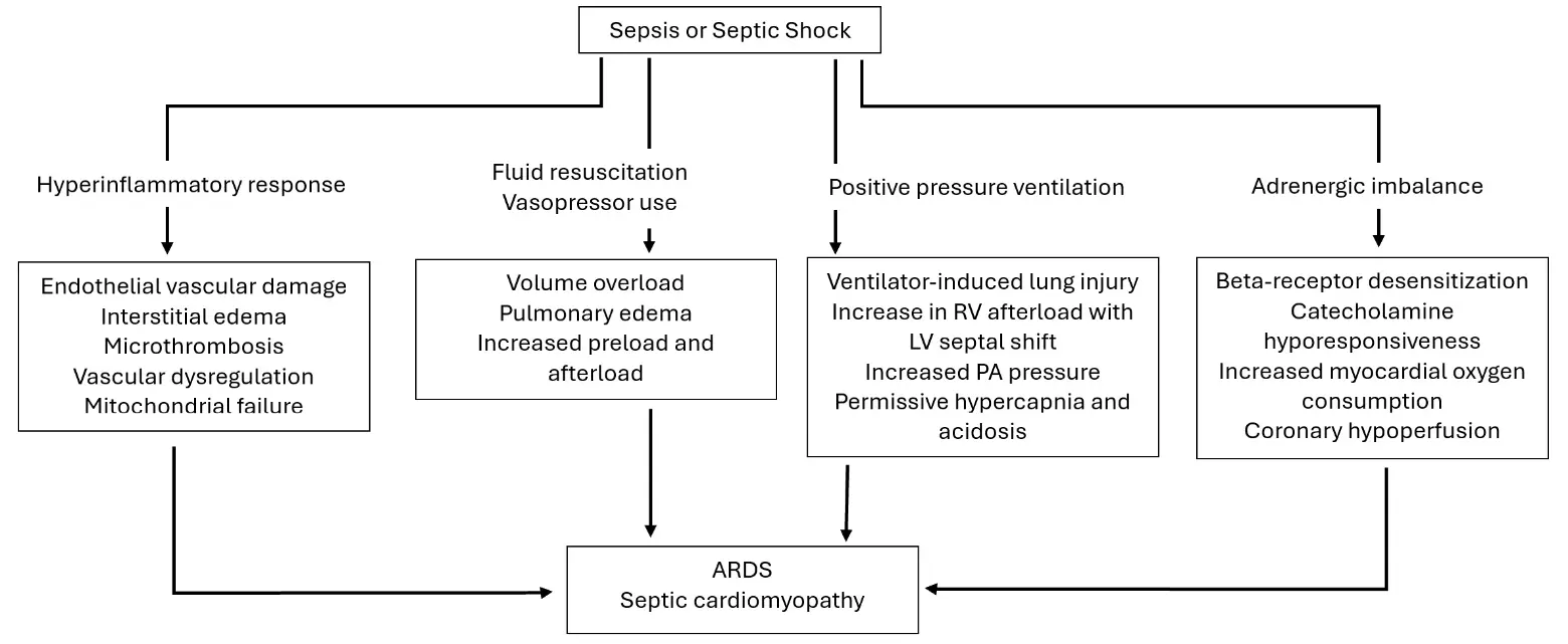
Figure 1. Broad overview of the common pathophysiologic drivers of ARDS and septic cardiomyopathy. RV, right ventricle; LV, left ventricle; ARDS, acute respiratory distress syndrome. Shared pathophysiologic causes leading to an inflammatory response affecting cardiac function, whereas therapeutic strategies for ARDS, sepsis, and shock can potentially impact cardiopulmonary interactions.
4. Detection and Diagnostics
The diagnostic approach to SCM and sepsis-associated ARDS requires multimodal integration: clinical evaluation, echocardiography, lung imaging, biomarkers, and hemodynamic monitoring. Because SCM and ARDS frequently coexist, careful evaluation is required to differentiate intrinsic myocardial dysfunction from pulmonary vascular strain and distributive physiology.
4.1. Clinical Features and Bedside Phenotyping
Septic shock is a distributive state characterized by low systemic vascular resistance (SVR) and a compensatory high cardiac output [4]. When SCM develops, this expected high-output state is lost, and patients exhibit signs of inadequate myocardial contractility in addition to vasodilation [5]. Thus, clinical features that suggest SCM reflect the development of a mixed septic and cardiogenic shock state. These patients have hypotension disproportionate to the degree of vasoplegia, a narrow pulse pressure, tachycardia with an inappropriately low stroke volume, and persistent shock despite adequate fluids and vasopressors [5]. In distributive shock, the expected cardiac response is hyperdynamic; therefore, the presence of apparently ‘normal’ cardiac function in septic shock may reflect an inappropriately blunted response and underlying myocardial insufficiency [4,5,6].
In contrast to classic distributive shock, which is typically characterized by low SVR and warm extremities, patients with a substantial cardiogenic component may present with cooler extremities, reflecting reduced cardiac output and relatively increased SVR [4,5,6]. Although no lab test is specific for SCM, patients may have an elevated B-type natriuretic peptide (BNP) and low mixed venous oxygen saturation (MvO2); in contrast, patients with isolated septic shock tend to have MvO2 > 70% [4,5,6,29,30].
In sepsis-associated ARDS, patients develop progressive hypoxemia despite supplemental oxygen, diffuse infiltrates, decreased lung compliance, and increasing ventilatory requirements [2]. These features overlap with those seen in cardiogenic pulmonary edema, and in patients with suspected myocardial dysfunction, it is therefore essential to determine whether pulmonary findings are a cause or consequence of cardiomyopathy [1,5,7]. Bedside tools to aid in this differentiation are described below. Anecdotally, patients with isolated cardiogenic pulmonary edema tend to have greater improvement in oxygenation with the institution of positive-pressure ventilation and diuresis, compared with patients with ARDS.
4.2. Echocardiography
Echocardiography is the cornerstone of diagnosing SCM and typically reveals a pattern of global, rather than regional, LV hypokinesis, distinguishing it from ischemic cardiomyopathy [6]. Although a reduced ejection fraction (EF) may be present, EF can appear deceptively normal in states of profound vasoplegia, making it an unreliable standalone measure of systolic function. Global longitudinal strain (GLS) offers greater sensitivity for detecting early myocardial impairment and often identifies dysfunction even when EF is preserved [17]. Diastolic abnormalities are frequently observed in septic cardiomyopathy and contribute to reduced cardiac output by impairing ventricular filling. Echocardiographic markers include elevated E/e′ ratios, abnormal transmitral inflow patterns, and tissue Doppler indices consistent with increased filling pressures and impaired relaxation [4]. These findings are particularly relevant in septic shock, where tachycardia and altered loading conditions amplify the hemodynamic consequences of diastolic dysfunction [4]. The pattern first described by Parker et al., characterized by reversible biventricular depression, remains consistent with findings from modern strain-based studies that confirm the dynamic and reversible nature of septic myocardial dysfunction [6].
The RV is particularly vulnerable in ARDS and septic shock due to elevated PVR, hypoxic pulmonary vasoconstriction, mechanical ventilation effects (PEEP), and pulmonary microvascular obstruction [7,16]. Key echocardiographic findings of RV dysfunction include RV dilation, reduced tricuspid annular plane systolic excursion (TAPSE) and S′, decreased fractional area change, and septal flattening (“D-sign”), reflecting pressure overload and impaired RV–pulmonary arterial (PA) coupling [8]. In addition to these cardiac findings, systemic venous congestion can be assessed at the bedside using venous Doppler ultrasound. Abnormal flow patterns in the hepatic, portal, and intrarenal veins, summarized by the venous excess ultrasound (VEXUS) score, reflect elevated right-sided filling pressures and impaired venous return and have been associated with organ congestion and adverse outcomes [31]. Collectively, these echocardiographic and venous ultrasound markers highlight the central role of RV dysfunction and venous congestion in ARDS, a relationship emphasized by Vieillard-Baron [32] and Petit [8], who identified RV failure as a major determinant of mortality.
4.3. Lung Ultrasound (LUS)
LUS provides characteristic findings in sepsis-associated ARDS, including heterogeneous B-line patterns, subpleural consolidations, dynamic air bronchograms, pleural line thickening or irregularity, and non-dependent spared areas, with progressive loss of aeration on serial examinations [9,33]. Importantly, this heterogeneous and regional distribution distinguishes ARDS from cardiogenic pulmonary edema, which typically shows bilateral, symmetric, gravity-dependent B-lines, a smooth pleural line, and minimal consolidations [33]. LUS has been established as a rapid, bedside modality for detecting acute lung injury and for differentiating ARDS from cardiogenic edema based on these sonographic patterns [33]. Additional tools have included LUS reaeration score, which quantifies aeration changes in response to PEEP adjustments and provides practical guidance for titrating ventilatory support [9]. More recently, Costamagna [15] showed that LUS can reliably distinguish focal versus non-focal ARDS phenotypes, an important observation, given that recruitability and optimal ventilator strategies differ significantly between these morphologic patterns.
4.4. Chest CT
CT remains the gold standard for structural lung assessment and ARDS phenotyping. In early (exudative) ARDS, CT typically demonstrates bilateral ground-glass opacities, often with dependent consolidations, impaired aeration with relative ventral sparing, air bronchograms, and regions of mosaic attenuation, reflecting alveolar flooding, inflammatory infiltrates, and surfactant dysfunction [2]. As ARDS evolves into the fibroproliferative and late phases, CT findings increasingly include traction bronchiectasis, architectural distortion, reduced lung compliance, and, in some cases, reticular changes consistent with fibrotic remodeling [2]. Differentiating early from late CT patterns is clinically relevant, as early disease may retain greater recruitability, whereas late-stage changes reflect irreversible structural injury with important implications for ventilatory strategy and prognosis. Focal and nonfocal ARDS can be distinguished on CT based on the distribution and morphology of lung injury, a distinction with important prognostic and management implications [15]. Focal ARDS is characterized by localized consolidations, often involving dependent or lobar regions with relatively preserved aeration elsewhere, whereas nonfocal ARDS demonstrates diffuse, bilateral loss of aeration and widespread ground-glass opacities [15]. These morphologic patterns are clinically relevant because nonfocal ARDS is generally associated with lower lung compliance, reduced recruitability, and worse outcomes, while focal ARDS may tolerate lower PEEP strategies and targeted recruitment approaches [15].
In contrast, cardiogenic pulmonary edema typically exhibits symmetric, gravity-dependent ground-glass opacities, interlobular septal thickening, pleural effusions, and perihilar predominance without the regional heterogeneity or spared areas characteristic of ARDS [2]. Differentiating these CT patterns is essential, as misclassification may lead to inappropriate ventilatory strategies or fluid management, particularly in patients with concomitant myocardial dysfunction. Equally important is consideration of other etiologies of parenchymal pulmonary disease, such as acute eosinophilic pneumonia, as well as underlying interstitial lung disease exacerbations.
RV strain can be identified on CT by RV dilation, interventricular septal flattening, and PA enlargement, as well as reflux of intravenous contrast into the inferior vena cava and hepatic veins, reflecting elevated right-sided pressures and pulmonary vascular load [3,7,8]. These findings correlate strongly with RV dysfunction on echocardiography and are associated with increased mortality [7,8].
4.5. Biomarkers
Cardiac biomarkers complement imaging for phenotyping and prognostication. Commonly used markers in sepsis-induced cardiomyopathy include troponin and BNP/NT-proBNP. Troponin is elevated in approximately 40–70% of patients with septic shock and typically reflects inflammatory membrane injury and microvascular dysfunction rather than plaque rupture. Importantly, troponin elevations in sepsis are generally modest compared with the marked elevations seen in acute coronary syndromes, and often lack a dynamic rise-and-fall pattern or regional wall motion abnormalities consistent with ischemia [20].
4.6. Hemodynamic Monitoring
Advanced hemodynamic monitoring may be helpful when shock is mixed or when echocardiographic findings are equivocal. PA catheterization (PAC) is useful for distinguishing cardiogenic vs. non-cardiogenic pulmonary edema and for quantifying PVR, RV afterload, and mixed venous oxygen saturation. Historically, PAC was frequently used for hemodynamic monitoring in septic shock, and PAC-derived parameters were incorporated into early goal-directed therapy protocols and used as clinical trial endpoints [34]. More recent large-scale randomized trials demonstrated that protocolized resuscitation strategies do not improve outcomes compared with usual care, leading to a shift away from rigid hemodynamic targets in septic shock management [35,36,37]. After the ESCAPE and PAC-MAN trials demonstrated no mortality benefit and potential harm associated with routine PAC use, including increased complications without improved outcomes, PAC-guided management fell out of favor in both cardiogenic and septic shock [38,39]. However, in selected patients and in experienced hands, PAC can provide a wealth of information. In patients with SCM, PAC measurements may demonstrate reduced cardiac output with elevated PA pressures, while patients with concomitant LV dysfunction often exhibit marked elevation of pulmonary capillary wedge pressure, reflecting impaired forward flow and elevated filling pressures [4,6,7]. Sampling of mixed-venous blood gases from a PA catheter can help characterize shock physiology. Patients with isolated distributive septic shock typically demonstrate preserved or elevated MvO2 (>70%), reflecting high cardiac output and impaired oxygen extraction, whereas patients with significant myocardial depression or inadequate compensatory response often exhibit reduced MvO2 (<70%) [4,6,34]. These data can be used to make decisions on vasopressors, inotropes, and fluid management.
Minimally invasive hemodynamic monitoring tools are increasingly used in critically ill patients. For example, the Pulse index Continuous Cardiac Output (PiCCO) system uses transpulmonary thermodilution to estimate cardiac output and derive additional parameters, including extravascular lung water (EVLW), which quantifies pulmonary edema, and global end-diastolic volume (GEDV), a volumetric surrogate of cardiac preload [23]. These measurements can aid in differentiating cardiogenic from non-cardiogenic pulmonary edema and in guiding fluid and ventilatory management. Other minimally invasive monitoring systems, such as FloTrac, NICOM, and bioreactance methods, have been evaluated in shock. However, a recent systematic review noted that most cardiac output monitors demonstrate poor agreement with reference methods and that their true clinical utility remains unclear, particularly regarding precision and trending under dynamic conditions [40].
4.7. Emerging Diagnostic Modalities
Emerging diagnostic modalities offer additional insight into the cardiopulmonary dysfunction seen in sepsis. Cardiac MRI (CMR) can identify non-ischemic inflammatory injury, including myocardial edema and elevated T1/T2 relaxation times, which help confirm myocarditis-like patterns characteristic of SCM [19]. Electrical impedance tomography (EIT) provides real-time, regional assessments of ventilation, allowing clinicians to evaluate PEEP responsiveness, detect overdistension, and assess recruitment heterogeneity at the bedside [17]. Advances in machine learning-enhanced LUS further improve diagnostic reproducibility by using deep-learning algorithms to classify ARDS phenotypes and detect pathologic LUS patterns [18]. Additionally, sublingual microcirculatory imaging can reveal reductions in perfused vessel density and increased flow heterogeneity, physiologic abnormalities that correlate with organ failure and mortality in sepsis [16]. Collectively, these modalities complement traditional imaging and hemodynamic assessment by providing higher-resolution physiologic data relevant to both SCM and sepsis-associated ARDS.
Several biomarkers of pulmonary injury have been investigated in sepsis-associated ARDS, including receptor for advanced glycation end products (RAGE), surfactant protein-D (SP-D), and angiopoietin-2 (Ang-2). RAGE reflects type I pneumocyte injury and is associated with ARDS severity and mortality [21]. SP-D is a type II pneumocyte injury marker predictive of ARDS development and outcomes [22], while Ang-2 reflects endothelial activation and permeability and correlates with mortality and ARDS severity [11]. These biomarkers are not routinely used in clinical practice, but provide important mechanistic insight and may inform future diagnostic or prognostic strategies.
4.8. Integrated Diagnostic Approach
A structured diagnostic strategy synthesizes the following:
-
Clinical phenotype—shock type, hypoxemia severity
-
Bedside ultrasonography—LV/RV function + LUS findings + markers of systemic venous congestion (e.g., by VEXUS)
-
Biomarkers—troponin, BNP
-
Hemodynamic monitoring—cardiac output, PA pressure, pulmonary capillary wedge pressure
-
CT phenotyping—distinguish focal versus nonfocal ARDS morphology, evaluation for non-ARDS causes of bilateral pulmonary infiltrates, and diagnostic differentiation from cardiogenic pulmonary edema
-
Assessment of heart-lung interaction—RV strain, TAPSE/PASP ratio
-
Serial re-evaluation
5. Interventions and Therapeutic Strategies
A framework for visualizing therapeutic strategies by ARDS phenotype with SCM can be found in Table 1. This framework uses the practical patient presentation of hypoxia and bilateral pulmonary infiltrates, along with clinical tools, to provide a phenotypic characterization of the patient. Fundamentally, it is key to distinguish ARDS from cardiogenic pulmonary edema and, thereafter, determine the ARDS phenotype in relation to SCM. As Table 1 suggests, this is not always clear, and diagnostic features of ARDS and pulmonary edema due to cardiomyopathy frequently overlap. It is also important, in this framework, to consider more rare causes of ARDS, non-ARDS causes of bilateral pulmonary infiltrates, coexisting valvular disease, and the possibility of coexisting thromboembolic disease.
Table 1. A basic framework for conceptualizing the treatment approach to the patient with bilateral pulmonary infiltrates and hypoxia, highlighting shared attributes and differences. ARDS, acute respiratory distress syndrome; SCM, septic cardiomyopathy; RV, right ventricle; LVEF, left ventricular ejection fraction; CT, computerized tomography; IVC, inferior vena cava; PA, pulmonary artery; PAWP, pulmonary artery wedge pressure; PRN, as‑needed; MvO2, mixed venous oxygen saturation; VEXUS, venous excess ultrasound; VAV, veno‑arterio‑venous; VV, veno‑venous; VA, veno‑arterial; ECMO, extracorporeal membrane oxygenation; IABP, intra‑aortic balloon pump.
|
Data |
Findings |
Sepsis-Related ARDS + SCM with Biventricular Failure |
Sepsis-Related ARDS + SCM with RV Failure |
Sepsis-Related ARDS with Appropriate Cardiac Response |
Pulmonary Edema Due to Primary Cardiomyopathy without ARDS |
|---|---|---|---|---|---|
|
Physical exam |
Cool extremities, bradycardia |
+ or − |
+ or − |
− |
+ |
|
Warm extremities, tachycardia |
+ or − |
+ or − |
+ |
− |
|
|
Echocardiogram |
LVEF reduction |
+ |
− |
− |
+ |
|
RV dilation/dysfunction |
+ |
+ |
− |
+ |
|
|
Wall motion abnormalities |
+ or − |
− |
− |
+ or − |
|
|
CT of the lungs |
Dependent consolidations |
+ |
+ |
+ |
− |
|
Diffuse groundglass opacities |
+ |
+ |
+ |
+ |
|
|
Interlobular septal thickening |
+ |
− |
− |
+ |
|
|
RV dilation/IVC reflux |
+ or − |
+ |
− |
+ or − |
|
|
Hemodynamics |
High PA pressure |
+ |
+ |
− |
+ |
|
Low cardiac output |
+ |
+ |
− |
+ |
|
|
High PAWP |
+ |
− |
− |
+ |
|
|
Mixed venous oxygen saturation |
≤70% |
+ or − |
+ or − |
− |
+ |
|
>70% |
+ or − |
+ or − |
+ |
− |
|
|
Norepinephrine + PRN vasopressin Early addition of dobutamine/milrinone or epinephrine, target MvO2 VEXUS $$\rightarrow$$ diurese if congested Lung protective ventilation Prone positioning Consider early steroids Treat primary cause of ARDS Consider VAV ECMO in refractory cases |
Norepinephrine + early vasopressin Early addition of dobutamine/milrinone or epinephrine, target MvO2 Inhaled nitric oxide/epoprostenol Lung protective ventilation but avoid permissive hypercapnia and acidosis Prone positioning Consider early steroids Treat primary cause of ARDS Consider VV/VAV ECMO in refractory cases |
Norepinephrine + PRN vasopressin VEXUS $$\rightarrow$$ diurese if congested Lung protective ventilation Prone positioning Consider early steroids Treat primary cause of ARDS Consider VV ECMO in refractory cases |
Norepinephrine Early addition of dobutamine/milrinone or epinephrine, target MvO2 VEXUS $$\rightarrow$$ diurese if congested Treat primary cause of cardiomyopathy Consider IABP, Impella, or VA ECMO in refractory cases |
5.1. Hemodynamic Management
5.1.1. Intravascular Volume Management
Early fluid resuscitation to maintain cardiac output and tissue perfusion remains a cornerstone of sepsis management [41,42]. However, in patients with SCM or ARDS, there is a delicate balance between restoring systemic perfusion and avoiding iatrogenic pulmonary edema. The amount of fluid infused during early periods of hospitalization may be associated with the development of lung injury and ARDS, with positive fluid balance a poor prognostic factor [43]. A retrospective cohort study of 598 patients with sepsis and acute decompensated heart failure found that, compared to the guideline-recommended 30 mL/kg crystalloid administration [42], resuscitation volumes of 10–15 mL/kg may have a lower risk of in-hospital mortality; however, that study focused on those with existing heart failure versus cardiomyopathy induced by sepsis [44].
There is also emerging evidence via the ANDROMEDA-SHOCK 2 trial, which utilized a personalized resuscitation algorithm based around capillary refill time (CRT)—highlighting the use of dynamic measures of fluid responsiveness to guide individualized fluid resuscitation, rather than a “one-size-fits-all” approach [45]. These approaches aim to prevent excessive RV preload, which can worsen right-heart failure, impair LV filling, and exacerbate oxygenation deficits in ARDS [23,46]. Here, echocardiography plays a key role in measuring parameters such as inferior vena cava (IVC) distensibility to predict fluid responsiveness, or in identifying significant RV failure, which may skew other measures of responsiveness, such as pulse-pressure variation (PPV) and stroke volume variation (SVV) [23,47]. Studies have shown that dynamic echocardiographic indices, such as respiratory variation in superior vena cava diameter or change in aortic velocity-time integral, have good predictive value for fluid responsiveness in ARDS [48]. Given the known risks of lung injury with over-administration of fluids, regular monitoring of these dynamic measurements should be done to accurately guide fluid therapy [49].
Finding the balance between fluid resuscitation and vascular decongestion in patients with mixed septic and cardiogenic shock, as often seen with SCM, can be a significant therapeutic challenge [50]. Currently, there is a need for more robust data on the use of resuscitation measures such as CRT in patients with SCM and SCM-ARDS overlap. American College of Cardiology guidelines emphasize that patients with signs of venous congestion on echocardiogram or invasive monitoring should be treated with loop diuretics, with the addition of thiazide diuretics or renal replacement therapy if needed [51]. The aforementioned VEXUS bedside ultrasound protocol uses IVC diameter and Doppler flow patterns of hepatic, portal, and renal veins to grade overall systemic venous congestion on a scale of zero to three. This has a good association with mean PA and right atrial pressures as measured by PAC, and a good association between weight change and congestion in patients with heart failure undergoing diuresis [52]. Recent studies show no significant association between VEXUS assessments and AKI occurrence [53,54], and this tool is currently being studied for its use in patients with septic shock [55].
5.1.2. Vasopressors
Norepinephrine. Norepinephrine (NE) remains the first-line hemodynamic agent for undifferentiated shock, with both vasopressor and inotropic properties [42,49]. However, increasing SVR may lead to increased LV afterload, which should be considered in patients with SCM or ARDS. Small cohort studies of patients with septic shock found that patients with normal LV function prior to administration of NE developed global LV hypokinesia within 24 to 48 h, thought to be related to an increase in afterload unmasking underlying myocardial dysfunction [56]. Increasing afterload may impose additional LV stress and worsen RV dysfunction, which can be compounded by hypoxic pulmonary vasoconstriction seen with ARDS [57]. There are, however, studies that show NE may augment RV myocardial oxygen delivery without meaningfully altering PA pressures or PVR [58,59]. With this in mind, NE should still be used as the first-choice vasopressor, but additional vasopressor/inotrope combinations can be considered based on clinical exam, echocardiography, and other parameters of perfusion [42]. Recent studies also underscore NE’s potential to modulate microcirculatory flow, which may influence myocardial and pulmonary perfusion simultaneously, although this has mainly been studied in the setting of anesthesia-related hypotension or endotoxic shock [60].
Epinephrine. Epinephrine is a vasopressor with more potent inotropic properties, with lower doses functioning primarily on beta-1 adrenergic receptors [42]. In experimental rat models of septic shock with severe myocardial dysfunction, both epinephrine and NE improved mean arterial pressure and cardiac output, and preload-recruitable stroke work. Notably, epinephrine was associated with increased heart rate, myocardial oxygen consumption, and arrhythmia incidence [61]. A separate randomized controlled trial (RCT) and network meta-analysis comparing either NE plus dobutamine versus epinephrine alone in patients with persistent hypoperfusion despite adequate volume status showed no significant difference in mortality between the two approaches [62,63].
Vasopressin. Vasopressin is a secondary option that may have an additional potential benefit of reducing PA pressure. Recent studies in neonates show improvements in pulmonary hemodynamics with vasopressin. One study of newborns with refractory acute pulmonary hypertension showed decreased oxygen requirements and improved oxygenation index after administration of vasopressin [64]. A different observational study of mechanically ventilated infants found that vasopressin significantly decreased echocardiographic indices of pulmonary hypertension, such as tricuspid regurgitation velocities, and improved biventricular output [65]. However, these studies primarily focused on infant patients; there is less data on vasopressin-induced reductions in PA pressures in adult patients, and thus we use caution when extrapolating from neonatal studies. Recent adult data from a single-center randomized crossover trial with 153 cardiac surgery patients ultimately showed no significant difference in the ratio of mean PA pressure to systemic arterial pressure when comparing vasopressin to norepinephrine [66]. At higher doses, however, vasopressin may increase PA and coronary artery vasoconstriction [67].
Phenylephrine. Phenylephrine is generally not a favorable choice in SCM with ARDS, given its primarily alpha-1 adrenergic vasoconstrictor properties, which elevate SVR and could increase myocardial workload and decrease LV output [68]. Additionally, phenylephrine has been shown to cause unopposed pulmonary vasoconstriction and increased PVR [69,70,71], which could make this unsuitable for ARDS patients who have RV dysfunction. However, phenylephrine may be a useful adjunct in patients who have arrhythmias, particularly atrial fibrillation with rapid ventricular response, related to either epinephrine or NE, as it does not stimulate beta-1 receptors and thus will not accelerate arrhythmias. This is relevant as patients with SCM are highly dependent on adequate atrial filling and emptying to provide ventricular preload.
Angiotensin II. While not yet studied in SCM specifically, angiotensin II has been shown to be effective in raising blood pressure in those with vasodilatory shock refractory to other conventional vasopressors [72,73]. Animal models of septic shock found that angiotensin II was associated with less myocardial oxygen consumption and expression of myocardial inflammatory markers compared to norepinephrine [74], which could suggest benefits with SCM. Additionally, a post-hoc analysis of ARDS patients suggested that angiotensin II improved P/F ratios and oxygenation indices compared to placebo [75].
5.1.3. Inotropes
Dobutamine. For patients with impaired contractility secondary to sepsis, inotropes are used to improve cardiac output, with the beta-1 receptor agonist dobutamine one of the more well-studied options. Both experimental models [76,77] and clinical studies have shown improvement in cardiac output, splanchnic perfusion, and tissue oxygenation in patients with septic shock [78]. However, it is important to note that dobutamine is also a peripheral vasodilator, and Surviving Sepsis guidelines explicitly warn that dobutamine infusions may cause vasodilation in distributive shock and lower MAP further [42]. Dobutamine in conjunction with NE can mitigate these issues, and there is data showing that the addition of dobutamine with reduced doses of NE is associated with improved LV hypokinesia and hemodynamics [56]. Despite promising results regarding its physiological effects, the effects of dobutamine on actual patient outcomes remain unclear. In fact, some studies suggest an association with increased mortality, or at least its use as an independent predictor of 90-day mortality [79]. The efficacy of dobutamine in SCM specifically is even less clear; there is currently an ongoing RCT assessing its use in patients with sepsis-induced LV failure [80].
Milrinone. Milrinone is a phosphodiesterase III inhibitor that prevents the breakdown of cAMP and cGMP in myocardial and vascular musculature, respectively, giving it both inotropic and vasodilatory properties [81]. An RCT compared milrinone to placebo in the treatment of adults with non-hypotensive septic shock with adequate fluid resuscitation, but with signs of poor tissue perfusion (defined as elevated lactate or decreased urine output) or echocardiogram evidence of LV dysfunction, and showed that milrinone had a significantly improved cardiac output and greater percentage change in cardiac index. However, 28-day mortality was identical between the two groups [82]. Another recent propensity-matched retrospective study of patients with sepsis-induced myocardial injury showed that milrinone treatment significantly improved 365-day survival of 66.7% from 59.7% [83]. Compared with dobutamine, recent evidence shows that, although there is no mortality difference between the two, patients treated with milrinone required longer inotropic support, longer hospital stays, and more antiarrhythmic agents [84].
Levosimendan. Levosimendan is a myofilament calcium-sensitizer with inotropic properties, as well as anti-inflammatory and antioxidant characteristics [85]. This mechanism of action is largely separate from beta adrenergic activity and theoretically results in a more favorable inotropic effect by increasing the sensitivity of myocytes to already existing stores of calcium, avoiding the risk of arrhythmia seen with adrenergic modulators [86,87,88]. However, the LeoPARDS large-scale RCT found no significant reduction in organ failure or mortality in patients with septic shock treated with levosimendan when compared to placebo in addition to standard care [89], and further subgroup analysis showed no association with lower mortality in patients specified to have evidence of cardiac dysfunction via cardiac biomarkers [90]. Despite the lack of evidence supporting mortality benefits, levosimendan has been associated with lowering of serum lactate, increased cardiac contractility, and augmented cardiac index and LV EF [91,92]. Some studies comparing levosimendan to dobutamine suggested a benefit to the former in patients with myocardial dysfunction, with improvement in cardiac index, lactate levels, and biomarkers of myocardial injury, although there were no significant differences in mortality or ICU length of stay between the two interventions [93,94]. The role of levosimendan in sepsis complicated by ARDS remains uncertain, and it is currently not approved by the Food and Drug Administration (FDA) for use in the United States.
5.1.4. Beta-Adrenergic Blockers
Modulation of heart rate with short-acting beta-blockers has gained increased interest as a strategy to prevent tachyarrhythmias and reduce the harmful effects of catecholamines in sepsis and ARDS. These agents may improve ventricular efficiency and reduce oxygen consumption without compromising perfusion when carefully titrated.
Esmolol. In one RCT assessing esmolol use in SCM with elevated heart rates (HR), all patients achieved their target HR of less than 100 beats/minute without a significant reduction in LV contractility, while also improving short-term mortality [95]. Two meta-analyses also concluded that esmolol had a significant decrease in 28-day mortality and improved levels of cardiac troponin. However, the effects of esmolol on oxygen metabolism are less clear. One analysis showed a significant increase in central venous oxygen saturation (ScvO2), with shortened duration of mechanical ventilation and ICU length of stay, whereas another overall found no difference in ICU length of stay, ScvO2, or PO2/FiO2 ratio [96,97].
Landiolol. Landiolol is a short-acting beta-blocker that, while currently FDA-approved only for the treatment of supraventricular tachycardia in the U.S., has also been studied for use in sepsis with mixed results. One study showed effectiveness in HR reduction without increasing pressor requirements in patients with septic shock and ongoing tachycardia, but there were no significant differences in adverse effects and 28-day mortality compared to standard of care [98]. Infusions of landiolol have not been shown to reduce organ failure within septic shock patients, giving evidence that landiolol should not be used for the management of tachyarrhythmias in septic shock. Furthermore, it is speculated that patients with SCM may require elevated heart rates to maintain adequate cardiac output, thus rendering beta-blocker usage a controversial topic in the realm of sepsis management [99].
Ivabradine. Ivabradine, a selective sinoatrial node inhibitor, is an alternative to beta-blockers that theoretically offers rate control and reduced myocardial oxygen demand without negative inotropic effects, making it a potentially safer option in patients with impaired myocardial function [100]. The MODIFY trial looked at 70 patients with multiple organ dysfunction syndrome due to cardiac or septic shock, and treatment with ivabradine for 96 h showed improvements of cardiac performance markers such as cardiac index [101]. Another RCT supported the findings of improved cardiac function, although, as with studies of beta-blockers, there was no difference in 30-day mortality [102]. Overall, ivabradine shows promise as a rate-stabilizing agent in patients with septic shock, although it is important to note that its use in patients specifically with cardiomyopathy or ARDS is not as well-documented. Overall, inotropes’ combined impact on both cardiac function and pulmonary physiology remains an area of active inquiry.
5.1.5. Pulmonary Vasodilators
Inhaled nitric oxide (iNO) can selectively reduce PVR and improve RV afterload, which may be beneficial for ARDS patients with RV failure. It has been well established that iNO reduces PA pressures and PVR [103]. While past trials have not shown consistent mortality benefit with iNO [104,105], a more recent study of patients with severe ARDS with RV dysfunction showed that iNO may improve mortality and reduce the need for renal replacement therapy, although it did not affect the length of stay in the ICU or hospital [106]. Despite these promising results in ARDS, the use of iNO in patients with SCM is less understood and may even be contraindicated, as the synthesis of nitric oxide via the IL-1 inflammatory pathway is implicated in the reduction of myocardial contractility in sepsis [86], and theoretically may lead to pulmonary edema due to volume equilibration in the PA and pulmonary venous compartments as pressure between them approximate [107]. Inhaled prostacyclins, such as epoprostenol, have shown similar benefits in reducing PA pressure and possible improvements in RV function, although, like iNO, their effects on SCM are not well-studied [108,109,110].
5.2. Ventilation Strategies
5.2.1. Cardiopulmonary Effects with Mechanical Ventilation
Mechanical ventilation (MV) is a mainstay of the management of ARDS. However, it can substantially affect cardiac preload and afterload—an important consideration in patients with SCM, particularly in those who may already experience RV strain due to hypoxic pulmonary vasoconstriction [57]. Elevated intrathoracic pressures can reduce venous return and further impair RV output, although this effect can be mitigated with lung-protective tidal volumes, which are now commonly used in ARDS protocols [111,112].
Positive end-respiratory pressure (PEEP) settings can affect cardiac output as well. Higher PEEP increases intrathoracic pressures and inversely affects venous return, which can exacerbate RV strain [113]. Higher PEEP may simultaneously decrease LV preload and afterload due to baroreceptor response to aortic compression, which may actually benefit patients with left-sided heart failure, although overall effects of PEEP on hemodynamics will vary based on each patients’ preload dependence, LV function, and severity of RV failure [51]. Excessively low PEEP, however, can worsen hypoxemia and result in microvascular injury, further exacerbating underlying heart and lung injury [114,115]. Thus, it is crucial to understand the patient-specific balance of PEEP tolerance, especially in patients with combined ARDS and SCM. There are emerging strategies tailored to protect right heart function, including more careful titration of PEEP, lowering plateau pressures, and avoiding hypercapnia [116,117].
5.2.2. Prone Positioning
Proning is a cornerstone in the management of ARDS, shown in the PROSEVA trial to improve oxygenation and mortality in ARDS [118]. Prone positioning may also reduce RV afterload. A study using transesophageal echocardiography before and after proning sessions found an association between reductions in plateau pressures and PaO2, and improvements in septal dyskinesia and RV end-diastolic area/LV end-diastolic area ratios [119]. The mechanisms of this effect include improvement of lung compliance, leading to decreased plateau and driving pressures. Additionally, homogenization of atelectatic lung allows expansion of pulmonary vessels and improved oxygenation, thereby decreasing pulmonary vasoconstriction that may contribute to RV failure. Lastly, proning can increase intra-abdominal pressure, which may augment venous return and cardiac preload [119,120,121]. Unfortunately, there has been little exploration regarding actual cardiovascular outcomes from early prone positioning, although the PROSEVA trial showed fewer cardiac arrests and more cardiac failure-free days at 28 days in its prone ventilation arm [118,122].
5.3. Extracorporeal Membrane Oxygenation (ECMO)
ECMO Modalities
Venoarterial (VA)-ECMO is a form of extracorporeal circulation support that allows oxygenated blood to cycle throughout the body while bypassing the heart completely [123]. Studies have shown that, for cardiogenic shock due to SCM, VA-ECMO can be used to provide full cardiac support. In one study of fourteen patients with refractory septic shock—defined as LVEF < 25%, cardiac index < 2.2 L/min/m2, persistent hypotension, extensive skin mottling, or elevated lactate levels despite high-dose catecholamines—the median LVEF had significantly improved to 60% after discontinuation of ECMO. It is speculated that this apparent reversal of cardiomyopathy is due to ECMO giving the failing heart more time to recover [124]. A separate retrospective study of 82 patients with refractory shock found the use of VA-ECMO led to improved 90-day survival, rapid reduction in pressor requirements, and enhanced lactate clearance. These results were achieved even with VA-ECMO patients having worse cardiac index, LVEF, and organ failure in comparison to control groups [125].
Venovenous (VV)-ECMO has been used in severe cases of ARDS and may benefit by reducing cardiac dysfunction without the need for VA-ECMO. VV-ECMO allows for control of oxygenation and carbon dioxide levels, which are two drivers for increased PVR, thereby reducing RV afterload [126]. VV-ECMO can reduce plateau pressures and driving pressures during mechanical ventilation, which could benefit right heart function [8]. A large randomized clinical trial of VV-ECMO did not meet its statistically significant primary endpoint of 60-day mortality but demonstrated a substantial benefit in patients with severe ARDS as compared with usual care [127].
Alternatively, veno-arteriovenous (VAV)-ECMO offers hybrid support for combined respiratory and cardiac failure, often used in patients who are already on VA or VV-ECMO [128]. One retrospective study assessed outcomes of severe ARDS patients on VV-ECMO who needed additional VAV ECMO support. These patients had an ICU survival rate of 52%, with long-term outcomes of survival and organ functional status comparable to patients who underwent more classical VA or VV-ECMO [129]. The use of VAV-ECMO is an area of further study with potential benefit in combined heart and lung injury, although overall the choice between ECMO modalities largely depends on the predominant physiologic derangements.
Percutaneous LV assist devices (LVAD) such as microaxial flow pumps or “Impella” devices have been approved for use in patients with cardiogenic shock secondary to acute myocardial infarction, postcardiotomy care, or acute left heart failure; however, SCM is not among these indications [130]. The recent DanGer Shock trial showed lower all-cause 180-day mortality among patients treated with Impella versus standard care, but looked at patients with post-STEMI cardiogenic shock [131]. Ultimately, the literature supports the use of VA-ECMO as the preferred device for temporary circulatory support in patients with SCM [132,133].
Intra-aortic balloon pumps (IABP) have also historically been used for cardiogenic shock secondary to myocardial infarction, although the IABP-SHOCK II trial showed no significant reduction in 30-day mortality, and a more recent meta-analysis of 12 RCTs confirmed the lack of improvement [134,135].
5.4. Anti-Inflammatory Therapies
5.4.1. Corticosteroids and Mineralocorticoids
Steroids are an important consideration in the management of septic shock and ARDS, given their ability to modulate systemic inflammation, stabilize endothelial barriers, and improve shock reversal [136]. Recent guidelines underscore steroids’ role in reducing mortality, ICU and hospital length-of-stay, and mechanical ventilation days [136]. A 2018 RCT showed that a combination of hydrocortisone and fludrocortisone demonstrated accelerated weaning from vasopressor therapy and a reduction in 90-day mortality for patients in septic shock [137]. More recent trials also suggest that combinations of hydrocortisone and fludrocortisone are superior to hydrocortisone alone [138]. However, while broader sepsis literature demonstrates clear benefits to steroid use, there is a lack of trials focusing on patients with SCM specifically. One prospective study investigating steroids’ effects on echocardiographic data in early septic shock showed significant improvements in arterial pressure, heart rate, LV afterload, and multiple parameters of LV contractility, including GLS and the valve peak systolic wave, providing insights into how they can benefit cardiac function in patients with SCM [139].
5.4.2. Immunomodulators
Agents targeting cytokine pathways, such as IL-6 inhibitors and TNF-alpha blockers, and JAK inhibitors, are being investigated for their use in acutely severe inflammatory states. Their effects on both cardiac contractility and pulmonary endothelial activity remain under active study. Increased levels of IL-6, TNF-alpha, and IL-1-beta have been recorded in patients with SCM, which may indicate these cytokines as crucial targets for diagnostics and treatment for SCM [140]. While not specifically studied for use in SCM, the IL-6 receptor antagonist tocilizumab has been shown to decrease systemic inflammation and myocardial injury in patients surviving out-of-hospital cardiac arrest, indicating potential for treatment in other cardiac-related dysfunctions [141]. Lastly, a study of mice with induced SCM investigated the use of 1-deoxynojirimycin (DNJ), a modulator of the JAK2/STAT6 pathway, and results suggest attenuation of myocardial injury, decreased oxidative damage, and inflammation [142].
6. Knowledge Gaps and Future Directions
6.1. Integrated Cardio-Pulmonary Phenotyping
There is growing recognition that current diagnostic and therapeutic strategies inadequately capture the intertwined nature of myocardial and pulmonary injury in sepsis. By stratifying broad groups of SCM or ARDS patients into more specific phenotypes, one can possibly guide more personalized therapies and identify high-risk groups. One study used transesophageal echocardiography (TEE) to split septic shock patients into five distinct clusters based on cardiac function, which included groups with LV dysfunction, hyperkinetic function, RV failure, and persistent hypovolemia. Each cluster exhibited significant variance in mortality outcomes, with those with LV failure and RV failure having the highest mortality rates [143]. A different study used transthoracic echocardiography (TTE) and stratified patients into five groups based on LV EF and found that the extremes of LVEF (<25% and >70%) were linked with significantly higher in-hospital mortality [144]. These studies show distinct outcome differences among clusters of patients with septic shock, and it is possible that similar findings may be seen in SCM and ARDS.
Phenotyping is a highly variable field of study, with multiple studies using different methods to stratify their patients. A meta-analysis revealed many clustering variables that appeared to predict treatment response, including echocardiographic measurements, inflammatory and coagulation markers, and various transcriptomic and genomic expression profiles [145]. While this shows potential in the utility of phenotyping, it also illustrates that current approaches are extremely heterogeneous and require further validation, with the hope of developing a more authenticated method of clustering that would allow for individualized therapies.
6.2. Therapies Targeting Organ Cross-Talk
Interventions that interrupt harmful signaling between the heart and lungs are an emerging area of interest. Neutrophil elastase inhibitors, such as sivelestat, have shown promise in mitigating alveolar damage and may also protect cardiac tissue from inflammatory proteolytic injury. Multiple animal and molecular studies have detailed sivelestat’s ability to reduce apoptosis, inhibit the release of inflammatory factors, and downregulate inflammation-related protein expression in damaged myocardium [146,147]. A randomized trial of 97 patients with both ARDS and SCM compared treatment with sivelestat against controls, and showed significantly lower levels of IL-6, IL-8, and INF-alpha levels. More importantly, stroke volume, E/A values, and TAPSE measurements were significantly higher in the sivelestat group, implying that sivelestat could be efficacious for treating SCM [148]. Another retrospective study investigated sivelestat in patients with ARDS, showing a significant reduction in 30-day mortality and cytokine levels without a significant increase in side effects [149]. Whether these therapies improve outcomes in combined cardiopulmonary dysfunction remains to be tested, but these findings suggest neutrophil elastase inhibitors could be compelling future therapies for those with SCM and ARDS.
6.3. Artificial Intelligence and Multi-Parametric Monitoring
The use of artificial intelligence (AI) in the management of ARDS and SCM is rapidly evolving. Machine learning applied to hemodynamics, ventilator waveforms, echocardiographic data, and biomarkers may enable earlier identification of patients developing dual organ injury. Machine learning models have been used to rapidly cluster sepsis-associated ARDS patients, determine clinical outcomes and treatment responses within those clusters, such as mortality rate, lactic acid levels, and ICU length-of-stay, and identify risk factors. They have also been used to explore mortality rates among different PEEP levels within these specific clusters, showcasing the potential utility of real-time AI tools to support individualized titration of ventilation parameters [150,151,152]. Machine learning has also been used to generate models for predicting fluid responsiveness in septic shock by analyzing TTE parameters, with similar predictive value to more established methods such as passive leg raising, again showing promise in AI as a tool for more personalized titration of fluids [153]. Lastly, studies have used machine learning models to phenotype and predict mortality in patients requiring VA-ECMO, with good accuracy and reliability. While these studies were not tailored towards patients with ARDS or SCM specifically, they illustrate the importance of the evolving field of AI as a future tool for targeted ECMO intervention [154,155].
There is a lack of published research into the use of AI in the diagnosis, treatment, and clinical outcomes of SCM. Machine learning has been used to identify biomarkers, regulatory molecules, and genes associated with SCM, which may provide insights into future therapies [156,157,158], but AI models assessing clinical parameters require further research. Machine learning models may become a key tool in the diagnosis of sepsis-related sequelae; however, the dependability and practicality of AI in the clinical setting is an area of ongoing study [159].
6.4. Underrepresentation of RV Dysfunction
Right-heart failure is underrecognized in ARDS trials and registries. One scoping review of 51 studies on ARDS interventions found that only 27% of studies aimed to modify RV function, and only 5.9% were randomized controlled trials [160]. Further reviews have emphasized that, despite RV injury being a critical consideration in ARDS, there are limited large-scale trials to properly evaluate effective treatment strategies [57,161].
Notably, VA-ECMO has not been systematically studied for isolated RV failure in ARDS, leaving a significant evidence gap. VA-ECMO has generally been reserved for cases with concomitant LV failure or shock, and there are no large-scale trials evaluating its use in isolated RV failure, with most data from small, retrospective studies [162,163]. RV-protective strategies require more rigorous evaluation in both clinical trials and translational models.
6.5. Temporal Evolution of Dual-Organ Injury
The trajectories of SCM and ARDS differ markedly, yet their time courses overlap in clinically important ways. Understanding the temporal sequence of inflammation, microvascular injury, and functional decline could inform optimal timing of therapies such as prone positioning, β-blockade, or ECMO. The dynamic interplay and timing of these processes remain poorly understood. For example, although emerging evidence indicates that inflammation, microvascular injury, and myocardial edema are key players in SCM, the temporal relationships among them are not fully characterized [164]. Furthermore, although advances in molecular and imaging techniques have occurred, the sequence of events from the initial inflammatory insult to microvascular injury to functional decline remains unclear [165,166]. The literature underscores a critical gap in understanding the temporal evolution of inflammation and injury within SCM and ARDS, and how this may be a barrier to the development of effective, early interventions.
7. Conclusions
ARDS due to sepsis remains a heterogeneous condition; when SCM complicates ARDS, it can pose additional diagnostic and treatment challenges. Characterizing the specific cardiopulmonary interaction of SCM and ARDS allows for nuanced phenotyping and patient-oriented treatment strategies. A significant amount of research remains in the field of ARDS subphenotyping and treatment options.
Author Contributions
B.O. and G.E. conducted the literature review and wrote the initial manuscript and provided revisions. Y.M. conceived of the review, oversaw manuscript writing and contributed to revisions.
Ethics Statement
Not applicable.
Informed Consent Statement
Not applicable.
Data Availability Statement
Not applicable.
Funding
This research received no external funding.
Declaration of Competing Interest
B.O. and G.E. declare no conflicts of interest. Y.M. has received research funding from Mallickrodt and BioAegis.
References
- Ranieri VM, Rubenfeld GD, Thompson BT, Ferguson ND, Caldwell E, Fan E, et al. Acute respiratory distress syndrome: The Berlin Definition. JAMA 2012, 307, 2526–2533. DOI:10.1001/jama.2012.5669 [Google Scholar]
- Huppert LA, Matthay MA, Ware LB. Pathogenesis of acute respiratory distress syndrome. In Seminars in Respiratory and Critical Care Medicine; Thieme Medical Publishers: New York, NY, USA, 2019. [Google Scholar]
- Bellani G, Laffey JG, Pham T, Fan E, Brochard L, Esteban A, et al. Epidemiology, Patterns of Care, and Mortality for Patients With Acute Respiratory Distress Syndrome in Intensive Care Units in 50 Countries. JAMA 2016, 315, 788–800. DOI:10.1001/jama.2016.0291 [Google Scholar]
- Beesley SJ, Weber G, Sarge T, Nikravan S, Grissom CK, Shahul S, et al. Septic cardiomyopathy. Crit. Care Med. 2018, 46, 625–634. DOI:10.1097/CCM.0000000000002851 [Google Scholar]
- L’Heureux M, Sternberg M, Brath L, Turlington J, Kashiouris MG. Sepsis-induced cardiomyopathy: A comprehensive review. Curr. Cardiol. Rep. 2020, 22, 35. DOI:10.1007/s11886-020-01277-2 [Google Scholar]
- Parker MM, Shelhamer JH, Bacharach SL, Green MV, Natanson C, Frederick TM, et al. Profound but reversible myocardial depression in septic shock. Ann. Intern. Med. 1984, 100, 483–490. DOI:10.7326/0003-4819-100-4-483 [Google Scholar]
- Kakoullis L, Giannopoulou E, Papachristodoulou E, Pantzaris ND, Karamouzos V, Kounis NG, et al. The utility of brain natriuretic peptides in septic shock as markers for mortality and cardiac dysfunction: A systematic review. Int. J. Clin. Pract. 2019, 73, e13374. DOI:10.1111/ijcp.13374 [Google Scholar]
- Petit M, Jullien E, Vieillard-Baron A. Right Ventricular Function in Acute Respiratory Distress Syndrome: Impact on Outcome, Respiratory Strategy and Use of Veno-Venous Extracorporeal Membrane Oxygenation. Front. Physiol. 2022, 12, 797252. DOI:10.3389/fphys.2021.797252 [Google Scholar]
- Bouhemad B, Brisson H, Le-Guen M, Arbelot C, Lu Q, Rouby JJ, et al. Bedside Ultrasound Assessment of Positive End-Expiratory Pressure–induced Lung Recruitment. Am. J. Respir. Crit. Care Med. 2011, 183, 341–347. DOI:10.1164/rccm.201003-0369OC [Google Scholar]
- Sato R, Nasu M. A review of sepsis-induced cardiomyopathy. J. Intensive Care 2015, 3, 48. DOI:10.1186/s40560-015-0112-5 [Google Scholar]
- Calfee CS, Gallagher D, Abbott J, Thompson BT, Matthay MA, Eisner MD, et al. Plasma angiopoietin-2 in clinical acute lung injury. Crit. Care Med. 2012, 40, 1731–1737. DOI:10.1097/CCM.0b013e3182451c87 [Google Scholar]
- Murphy LS, Wickersham N, McNeil JB, Shaver CM, May AK, Bastarache JA, et al. Endothelial glycocalyx degradation is more severe in patients with non-pulmonary sepsis compared to pulmonary sepsis and associates with risk of ARDS and other organ dysfunction. Ann Intensive Care. 2017, 7, 102. DOI:10.1186/s13613-017-0325-y [Google Scholar]
- Shvilkina T, Shapiro N. Sepsis-Induced myocardial dysfunction: heterogeneity of functional effects and clinical significance. Front Cardiovasc Med. 2023, 10, 1200441. DOI:10.3389/fcvm.2023.1200441 [Google Scholar]
- Guarino M, Luppi F, Maroncelli G, Baldin P, Costanzini A, Maritati M, et al. From cardiac injury to omics signatures: a narrative review on biomarkers in septic cardiomyopathy. Clin. Exp. Med. 2025, 25, 298. DOI:10.1007/s10238-025-01842-5 [Google Scholar]
- Costamagna A, Pivetta E, Goffi A, Steinberg I, Arina P, Mazzeo AT, et al. Clinical performance of lung ultrasound in predicting ARDS morphology. Ann. Intensive Care 2021, 11, 51. DOI:10.1186/s13613-021-00837-1 [Google Scholar]
- De Backer D, Donadello K, Sakr Y, Ospina-Tascón G, Salgado D, Scolletta S, et al. Microcirculatory alterations in patients with severe sepsis: impact of time of assessment and relationship with outcome. Crit. Care Med. 2013, 41, 791–799. DOI:10.1097/CCM.0b013e3182742e8b [Google Scholar]
- Costa EL, Lima RG, Amato MB. Electrical impedance tomography. Curr. Opin. Crit. Care 2009, 15, 18–24. DOI:10.1097/mcc.0b013e3283220e8c [Google Scholar]
- Roy S, Menapace W, Oei S, Luijten B, Fini E, Saltori C, et al. Deep Learning for Classification and Localization of COVID-19 Markers in Point-of-Care Lung Ultrasound. IEEE Trans Med Imaging. 2020, 39, 2676–2687. DOI:10.1109/TMI.2020.2994459 [Google Scholar]
- Om SY, Yoo SY, Cho GY, et al. Diagnostic and Prognostic Value of Ergonovine Echocardiography for Noninvasive Diagnosis of Coronary Vasospasm. JACC Cardiovasc. Imaging 2020, 13, 1875–1887. DOI:10.1016/j.jcmg.2020.03.008 [Google Scholar]
- Seymour CW, Liu VX, Iwashyna TJ, Brunkhorst FM, Rea TD, Scherag A, et al. Assessment of Clinical Criteria for Sepsis: For the Third International Consensus Definitions for Sepsis and Septic Shock (Sepsis-3). JAMA 2016, 315, 762–774. DOI:10.1001/jama.2016.0288 [Google Scholar]
- Calfee CS, Ware LB, Eisner MD, Parsons PE, Thompson BT, Wickersham N, et al. Plasma receptor for advanced glycation end products and clinical outcomes in acute lung injury. Thorax 2008, 63, 1083–1089. DOI:10.1136/thx.2008.095588 [Google Scholar]
- Ware LB, Koyama T, Zhao Z, Janz DR, Wickersham N, Bernard GR, et al. Biomarkers of lung epithelial injury and inflammation distinguish severe sepsis patients with acute respiratory distress syndrome. Crit Care 2013, 17, R253. DOI:10.1186/cc13080 [Google Scholar]
- Zakynthinos GE, Giamouzis G, Xanthopoulos A, et al. Septic Cardiomyopathy: Difficult Definition, Challenging Diagnosis, Unclear Treatment. J. Clin. Med. 2025, 14, 986. DOI:10.3390/jcm14030986 [Google Scholar]
- Singer M, Deutschman CS, Seymour CW, Shankar-Hari M, Annane D, Bauer M, et al. The Third International Consensus Definitions for Sepsis and Septic Shock (Sepsis-3). JAMA 2016, 315, 801–810. DOI:10.1001/jama.2016.0287 [Google Scholar]
- Thompson BT, Chambers RC, Liu KD. Acute respiratory distress syndrome. N. Engl. J. Med. 2017, 377, 562–572. DOI:10.1056/NEJMra1608077 [Google Scholar]
- Yang SC, Tsai YF, Pan YL, Hwang TL. Understanding the role of neutrophils in acute respiratory distress syndrome. Biomed J. 2021, 44, 439–446. DOI:10.1016/j.bj.2020.09.001 [Google Scholar]
- Wang Y, Wang L, Ma S, Cheng L, Yu G. Repair and Regeneration of the Alveolar Epithelium in Lung Injury. FASEB J. 2024, 38, e23612. DOI:10.1096/fj.202400088R [Google Scholar]
- Price LC, Wort SJ, Finney SJ, Marino PS, Brett SJ. Pulmonary vascular and right ventricular dysfunction in adult critical care: Current and emerging options for management: A systematic literature review. Crit. Care 2010, 14, R169. DOI:10.1186/cc9264 [Google Scholar]
- Charpentier J, Luyt CE, Fulla Y, Vinsonneau C, Cariou A, Grabar S, et al. Brain natriuretic peptide: A marker of myocardial dysfunction and prognosis during severe sepsis. Crit. Care Med. 2004, 32, 660–665. DOI:10.1097/01.CCM.0000114827.93410.D8 [Google Scholar]
- Varpula M, Tallgren M, Saukkonen K, Voipio-Pulkki LM, Pettilä V. Hemodynamic variables related to outcome in septic shock. Intensive Care Med. 2005, 31, 1066–1071. DOI:10.1007/s00134-005-2688-z [Google Scholar]
- Beaubien-Souligny W, Rola P, Haycock K, Bouchard J, Lamarche Y, et al. Quantifying systemic congestion with point-of-care ultrasound: Development of the venous excess ultrasound grading system. Ultrasound J. 2020, 12, 16. DOI:10.1186/s13089-020-00163-w [Google Scholar]
- Vieillard-Baron A, Schmitt JM, Augarde R, Fellahi J, Prin S, Page B, et al. Acute cor pulmonale in acute respiratory distress syndrome submitted to protective ventilation: Incidence, clinical implications, and prognosis. Crit. Care Med. 2001, 29, 1551–1555. Available online: https://journals.lww.com/ccmjournal/abstract/00003246-200108000-00009~acute-cor-pulmonale-in-acute-respiratory-distress-syndrome?redirectionsource=fulltextview (accessed on 26 November 2025).
- Lichtenstein D, Mezière G. Relevance of lung ultrasound in the diagnosis of acute respiratory failure: The BLUE protocol. Chest 2008, 134, 117–125. DOI:10.1378/chest.07-2800. [Google Scholar]
- Rivers E, Nguyen B, Havstad S, Ressler J, Muzzin A, Knoblich B, et al. Early goal-directed therapy in the treatment of severe sepsis and septic shock. N. Engl. J. Med. 2001, 345, 1368–1377. DOI:10.1056/NEJMoa010307 [Google Scholar]
- ProCESS Investigators. A randomized trial of protocol-based care for early septic shock. N. Engl. J. Med. 2014, 370, 1683–1693. DOI:10.1056/NEJMoa1401602 [Google Scholar]
- Mouncey PR, Osborn TM, Power GS, Harrison DA, Sadique MZ, Grieve RD, et al. Trial of early, goal-directed resuscitation for septic shock. N. Engl. J. Med. 2015, 372, 1301–1311. DOI:10.1056/NEJMoa1500896 [Google Scholar]
- The ARISE Investigators, The ANZICS Clinical Trials Group. Goal-directed resuscitation for patients with early septic shock. N. Engl. J. Med. 2014, 371, 1496–1506. DOI:10.1056/NEJMoa1404380 [Google Scholar]
- The ESCAPE Investigators, ESCAPE Study Coordinators. Evaluation study of congestive heart failure and pulmonary artery catheterization effectiveness: The ESCAPE trial. JAMA 2005, 294, 1625–1633. DOI:10.1001/jama.294.13.1625 [Google Scholar]
- Harvey S, Harrison DA, Singer M, Ashcroft J, Jones CM, Elbourne D, et al. Assessment of the clinical effectiveness of pulmonary artery catheters in management of patients in intensive care (PAC-Man): A randomized controlled trial. Lancet 2005, 366, 472–477. DOI:10.1016/S0140-6736(05)67061-4 [Google Scholar]
- Lamarche-Fontaneto R, Oud L, Howell KD, Ganeriwal SA, Manek GU, Schleicher MC, et al. Cardiac output monitors in septic shock: Do they deliver what matters? A systematic review and meta-analysis. Crit. Care 2025, 29, 299. DOI:10.1186/s13054-025-05547-9 [Google Scholar]
- Zampieri FG, Bagshaw SM, Semler MW. Fluid therapy for critically ill adults with sepsis: A review. JAMA 2023, 329, 1967–1980. DOI:10.1001/jama.2023.7560 [Google Scholar]
- Evans L, Rhodes A, Alhazzani W, Antonelli M, Coopersmith CM, French C, et al. Surviving Sepsis Campaign: International guidelines for management of sepsis and septic shock 2021. Crit. Care Med. 2021, 49, e1063–e1143. DOI:10.1097/CCM.0000000000005337 [Google Scholar]
- Seethala RR, Hou PC, Aisiku IP, Frendl G, Park PK, Mikkelsen ME, et al. Early risk factors and the role of fluid administration in developing acute respiratory distress syndrome in septic patients. Ann. Intensive Care 2017, 7, 11. DOI:10.1186/s13613-017-0233-1 [Google Scholar]
- Weng J, Xu Z, Song J, Liu C, Jin H, Cheng Q, et al. Optimal fluid resuscitation targets in septic patients with acutely decompensated heart failure. BMC Med. 2024, 22, 492. DOI:10.1186/s12916-024-03715-2 [Google Scholar]
- Pálizas F, Lattanzio B, Durandal N, Alonso ME, Duque J, Falcon N, et al. Personalized hemodynamic resuscitation targeting capillary refill time in early septic shock: The ANDROMEDA-SHOCK-2 randomized clinical trial. JAMA 2025, 334, 1988–1999. DOI:10.1001/jama.2025.20402 [Google Scholar]
- Piccioni A, Rozzi G, Spaziani G, Novelli M, Fuorlo M, Candelli M, et al. From fluid responsiveness to prognosis: The emerging role of point-of-care echocardiography in sepsis. Diagnostics 2025, 15, 2612. DOI:10.3390/diagnostics15202612 [Google Scholar]
- Guerin L, Monnet X, Teboul JL. Monitoring volume and fluid responsiveness: From static to dynamic indicators. Best. Pract. Res. Clin. Anaesthesiol. 2013, 27, 177–185. DOI:10.1016/j.bpa.2013.06.002 [Google Scholar]
- Joseph A, Evrard B, Petit M, Goudelin M, Prat G, Slama M, et al. Fluid responsiveness in acute respiratory distress syndrome patients: A post hoc analysis of the HEMOPRED study. Intensive Care Med. 2024, 50, 1850–1860. DOI:10.1007/s00134-024-07639-6 [Google Scholar]
- Coopersmith CM, De Backer D, Deutschman CS, Ferrer R, Lat I, Machado FR, et al. Surviving sepsis campaign: Research priorities for sepsis and septic shock. Intensive Care Med. 2018, 44, 1400–1426. DOI:10.1007/s00134-018-5175-z [Google Scholar]
- Urina Jassir D, Chaanine AH, Desai S, Rajapreyar I, Le Jemtel TH. Therapeutic dilemmas in mixed septic-cardiogenic shock. Am. J. Med. 2023, 136, 27–32. DOI:10.1016/j.amjmed.2022.09.022 [Google Scholar]
- Sinha SS, Morrow DA, Kapur NK, Kataria R, Roswell RO. 2025 concise clinical guidance: An ACC expert consensus statement on the evaluation and management of cardiogenic shock. J. Am. Coll. Cardiol. 2025, 85, 1618–1641. DOI:10.1016/j.jacc.2025.02.018 [Google Scholar]
- Longino A, Martin K, Leyba K, Siegel G, Thai TN, Riscinti M, et al. Prospective evaluation of venous excess ultrasound for estimation of venous congestion. Chest 2024, 165, 590–600. DOI:10.1016/j.chest.2023.09.029 [Google Scholar]
- Song J, Chen G, Lai D, Zhong L, Fan H, Hu W, et al. Association between the venous excess ultrasound (VExUS) score and acute kidney injury in critically ill patients with sepsis: A multicenter prospective observational study. Ann. Intensive Care 2025, 15, 105. DOI:10.1186/s13613-025-01529-w [Google Scholar]
- Andrei S, Bahr PA, Nguyen M, Bouhemad B, Guinot PG. Prevalence of systemic venous congestion assessed by Venous Excess Ultrasound Grading System (VExUS) and association with acute kidney injury in a general ICU cohort: A prospective multicentric study. Crit. Care 2023, 27, 224. DOI:10.1186/s13054-023-04524-4 [Google Scholar]
- Prager R, Argaiz E, Pratte M, Rola P, Arntfield R, Beaubien-Souligny W, et al. Doppler identified venous congestion in septic shock: Protocol for an international, multi-centre prospective cohort study (Andromeda-VEXUS). BMJ Open 2023, 13, e074843. DOI:10.1136/bmjopen-2023-074843 [Google Scholar]
- Vieillard-Baron A, Caille V, Charron C, Belliard G, Page B, Jardin F. Actual incidence of global left ventricular hypokinesia in adult septic shock. Crit. Care Med. 2008, 36, 1701–1706. DOI:10.1097/CCM.0b013e318174db05 [Google Scholar]
- Zochios V, Parhar K, Tunnicliffe W, Roscoe A, Gao F, et al. The right ventricle in ARDS. Chest 2017, 152, 181–193. DOI:10.1016/j.chest.2017.02.019 [Google Scholar]
- Schreuder WO, Schneider AJ, Groeneveld ABJ, Thijs LG. Effect of dopamine versus norepinephrine on hemodynamics in septic shock: Emphasis on right ventricular performance. Chest 1989, 95, 1282–1288. DOI:10.1378/chest.95.6.1282 [Google Scholar]
- Kerbaul F, Rondelet B, Motte S, Fesler P, Hubloue I, Ewalenko P, et al. Effects of norepinephrine and dobutamine on pressure load-induced right ventricular failure. Crit. Care Med. 2004, 32, 1035–1040. DOI:10.1097/01.CCM.0000120052.77953.07 [Google Scholar]
- Ospina-Tascón GA, Aldana JL, García Marín AF, Calderón-Tapia LE, Marulanda A, Escobar EP, et al. Immediate norepinephrine in endotoxic shock: Effects on regional and microcirculatory flow. Crit. Care Med. 2023, 51, e157–e168. DOI:10.1097/CCM.0000000000005885 [Google Scholar]
- Ducrocq N, Kimmoun A, Furmaniuk A, Hekalo Z, Maskali F, Poussier S, et al. Comparison of equipressor doses of norepinephrine, epinephrine, and phenylephrine on septic myocardial dysfunction. Anesthesiology 2012, 116, 1083–1091. DOI:10.1097/ALN.0b013e31824f9669 [Google Scholar]
- Jia L, Wang P, Li C, Xie J. The efficacy and safety of vasopressors for septic shock patients: A systematic review and network meta-analysis. Shock 2023, 60, 746–752. DOI:10.1097/SHK.0000000000002193 [Google Scholar]
- Annane D, Vignon P, Renault A, Bollaert PE, Charpentier C, Martin C, et al. Norepinephrine plus dobutamine versus epinephrine alone for management of septic shock. Lancet 2007, 370, 676–684. DOI:10.1016/S0140-6736(07)61344-0 [Google Scholar]
- Ouellet S, Drolet C, Morissette G, Pellerin A, Hébert A. Vasopressin in newborns with refractory acute pulmonary hypertension. Pediatr. Res. 2024, 95, 1572–1577. DOI:10.1038/s41390-023-02995-3 [Google Scholar]
- Santelices F, Masoli D, Kattan J, Toso A, Luco M. Vasopressin as adjunctive therapy in pulmonary hypertension associated with refractory systemic hypotension in term newborns. J. Perinatol. 2024, 44, 1448–1453. DOI:10.1038/s41372-024-02015-0 [Google Scholar]
- Geube M, Abraham A, Kelava M, Bustamante SE, Alfirevic A, Assaad S, et al. Cardiopulmonary effects of vasopressin versus norepinephrine in patients undergoing cardiac surgery: A single-center, cluster-randomized crossover trial. J. Thorac. Cardiovasc. Surg. 2026, 171, 1265–1274.e2. DOI:10.1016/j.jtcvs.2025.11.009 [Google Scholar]
- Leather HA, Segers P, Berends N, Vandermeersch E, Wouters PF. Effects of vasopressin on right ventricular function in an experimental model of acute pulmonary hypertension. Crit. Care Med. 2002, 30, 2548–2552. DOI:10.1097/00003246-200211000-00024 [Google Scholar]
- Thiele RH, Nemergut EC, Lynch CI. The physiologic implications of isolated alpha1 adrenergic stimulation. Anesth. Analg. 2011, 113, 284–296. DOI:10.1213/ANE.0b013e3182124c0e [Google Scholar]
- Rajagopal S, Ruetzler K, Ghadimi K, Horn EM, Kelava M, Kudelko KT, et al. Evaluation and Management of Pulmonary Hypertension in Noncardiac Surgery: A Scientific Statement from the American Heart Association. Circulation 2023, 147, 1317–1343. DOI:10.1161/CIR.0000000000001136 [Google Scholar]
- Jiang C, Qian H, Luo S, Lin J, Yu J, Li Y, et al. Vasopressors induce passive pulmonary hypertension by blood redistribution from systemic to pulmonary circulation. Basic. Res. Cardiol. 2017, 112, 21. DOI:10.1007/s00395-017-0611-8 [Google Scholar]
- Juhl-Olsen P, Berg-Hansen K, Nørskov J, Enevoldsen J, Hermansen JL. The haemodynamic effects of phenylephrine after cardiac surgery. Acta Anaesthesiol. Scand. 2023, 67, 869–876. DOI:10.1111/aas.14256 [Google Scholar]
- Wieruszewski PM, Wittwer ED, Kashani KB, Brown DR, Butler SO, Clark AM, et al. Angiotensin II infusion for shock: A multicenter study of postmarketing use. Chest 2021, 159, 596–605. DOI:10.1016/j.chest.2020.08.2074 [Google Scholar]
- Khanna A, English SW, Wang XS, Ham K, Tumlin J, Szerlip H, et al. Angiotensin II for the treatment of vasodilatory shock. N. Engl. J. Med. 2017, 377, 419–430. DOI:10.1056/NEJMoa1704154 [Google Scholar]
- Garcia B, Su F, Dewachter L, Favory R, Khaldi A, Moiroux-Sahraoui A, et al. Myocardial effects of angiotensin II compared to norepinephrine in an animal model of septic shock. Crit. Care 2022, 26, 281. DOI:10.1186/s13054-022-04161-3 [Google Scholar]
- Leisman DE, Handisides DR, Chawla LS, Albertson TE, Busse LW, Boldt DW, et al. Angiotensin II treatment is associated with improved oxygenation in ARDS patients with refractory vasodilatory shock. Ann. Intensive Care 2023, 13, 128. DOI:10.1186/s13613-023-01227-5 [Google Scholar]
- Kamijo T, Tomaru T, Miwa AY, Nakamura F, Kido H, Sugimoto T, et al. The effects of dobutamine, propranolol and nitroglycerin on an experimental canine model of congestive heart failure. Jpn. J. Pharmacol. 1994, 65, 223–231. DOI:10.1016/S0021-5198(19)35753-1 [Google Scholar]
- How OJ, Røsner A, Kildal AB, Stenberg TA, Gjessing PF, Hermansen SE, et al. Dobutamine-norepinephrine, but not vasopressin, restores the ventriculoarterial matching in experimental cardiogenic shock. Transl. Res. 2010, 156, 273–281. DOI:10.1016/j.trsl.2010.07.011 [Google Scholar]
- Dubin A, Mugno M. The effects of dobutamine in septic shock: An updated narrative review of clinical and experimental studies. Medicina 2024, 60, 751. DOI:10.3390/medicina60050751 [Google Scholar]
- Wilkman E, Kaukonen KM, Pettilä V, Kuitunen A, Varpula M. Association between inotrope treatment and 90-day mortality in patients with septic shock. Acta Anaesthesiol. Scand. 2013, 57, 431–442. DOI:10.1111/aas.12056 [Google Scholar]
- Vignon P, Lger J, Evrard B, Goudelin M, Vaidie J, Brit S, et al. Adjunctive dobutamine in patients with septic cardiomyopathy and tissue hypoperfusion: A blinded randomised controlled multicentre trial study protocol of the ADAPT-dobutamine trial. BMJ Open 2025, 15, e101200. DOI:10.1136/bmjopen-2025-101200 [Google Scholar]
- Ayres JK, Maani CV. Milrinone. In StatPearls; StatPearls Publishing: Treasure Island, FL, USA, 2025. Available online: https://www.ncbi.nlm.nih.gov/books/NBK532943/ (accessed on 26 November 2025).
- Tongyoo S, Chobngam S, Yolsiriwat N, Jiranakorn C. Effects of adjunctive milrinone versus placebo on hemodynamics in patients with septic shock: A randomized controlled trial. Ann. Med. 2025, 57, 2484464. DOI:10.1080/07853890.2025.2484464 [Google Scholar]
- Yuan Z, She T, Wu Z, Li Q. The effect of milrinone on short- and mid-term outcomes in patients with sepsis-related myocardial injury: A propensity score matched retrospective study. Heart Lung 2026, 76, 74–80. DOI:10.1016/j.hrtlng.2025.11.014 [Google Scholar]
- Li B, Gershengorn HB, Vail EA, Wunsch H, Walkey AJ, Law AC, et al. Inotrope selection in mixed cardiogenic shock with sepsis: A comparative analysis between milrinone and dobutamine. Ann. Am. Thorac. Soc. 2026, 23, 252–260. DOI:10.1513/AnnalsATS.202503-339OC [Google Scholar]
- Papp Z, Édes I, Fruhwald S, De Hert SG, Salmenperä M, Leppikangas H, et al. Levosimendan: Molecular mechanisms and clinical implications: Consensus of experts on the mechanisms of action of levosimendan. Int. J. Cardiol. 2012, 159, 82–87. DOI:10.1016/j.ijcard.2011.07.022 [Google Scholar]
- Lukić I, Mihić D, Varžić SC, Relatić KS, Zibar L, Loinjak D, et al. Septic cardiomyopathy. Rev. Cardiovasc. Med. 2024, 25, 23. DOI:10.31083/j.rcm2501023 [Google Scholar]
- Wieruszewski ED, Jones GM, Samarin MJ, Kimmons LA. Predictors of dysrhythmias with norepinephrine use in septic shock. J. Crit. Care 2021, 61, 133–137. DOI:10.1016/j.jcrc.2020.10.023 [Google Scholar]
- Nasra AW. How to induce arrhythmias with dobutamine. In Arrhythmia Induction in the EP Lab; Cismaru G, Ed.; Springer: Cham, Switzerland, 2019; pp. 71–79. DOI:10.1007/978-3-319-92729-9_8 [Google Scholar]
- Gordon AC, Santhakumaran S, Al-Beidh F, Orme RM, Perkins GD, Singer M, et al. Levosimendan to prevent acute organ dysfunction in sepsis: The LeoPARDS RCT. Effic. Mech. Eval. 2018, 5, 1–94. DOI:10.3310/eme05060 [Google Scholar]
- Antcliffe DB, Santhakumaran S, Orme RML, Ward JK, Al-Beidh F, O’Dea K, et al. Levosimendan in septic shock in patients with biochemical evidence of cardiac dysfunction: A subgroup analysis of the LeoPARDS randomised trial. Intensive Care Med. 2019, 45, 1392–1400. DOI:10.1007/s00134-019-05731-w [Google Scholar]
- Chang W, Xie JF, Xu JY, Yang Y. Effect of levosimendan on mortality in severe sepsis and septic shock: A meta-analysis of randomised trials. BMJ Open 2018, 8, e019338. DOI:10.1136/bmjopen-2017-019338 [Google Scholar]
- Hiraiwa H, Kasugai D, Okumura T, Murohara T. Clinical implications of septic cardiomyopathy: A narrative review. Medicine 2024, 103, e37940. DOI:10.1097/MD.0000000000037940 [Google Scholar]
- Liu DH, Ning YL, Lei YY, Chen J, Liu YY, Lin XF, et al. Levosimendan versus dobutamine for sepsis-induced cardiac dysfunction: A systematic review and meta-analysis. Sci. Rep. 2021, 11, 20333. DOI:10.1038/s41598-021-99716-9 [Google Scholar]
- Meng JI, Hu MH, Lai ZZ, Ji CL, Xu XJ, Zhang G, et al. Levosimendan Versus Dobutamine in Myocardial Injury Patients with Septic Shock: A Randomized Controlled Trial. Med. Sci. Monit. Int. Med. J. Exp. Clin. Res. 2016, 22, 1486–1496. DOI:10.12659/MSM.898457 [Google Scholar]
- Wang J, Gao X, He Z, Wang J, Xu G, Li T. Evaluating the effects of Esmolol on cardiac function in patients with Septic cardiomyopathy by Speck-tracking echocardiography—A randomized controlled trial. BMC Anesth. 2023, 23, 51. DOI:10.1186/s12871-023-01983-8 [Google Scholar]
- Zhang J, Chen C, Liu Y, Yang Y, Yang X, Yang J. Benefits of esmolol in adults with sepsis and septic shock: An updated meta-analysis of randomized controlled trials. Medicine 2022, 101, e29820. DOI:10.1097/MD.0000000000029820 [Google Scholar]
- Wei Y, Bo F, Wang J, Fu J, Qiu Y, Bi H, et al. The role of esmolol in sepsis: A meta-analysis based on randomized controlled trials. BMC Anesthesiol. 2024, 24, 326. DOI:10.1186/s12871-024-02714-3 [Google Scholar]
- Rehberg S, Frank S, Černý V, Cihlář R, Borgstedt R, Biancofiore G, et al. Landiolol for heart rate control in patients with septic shock and persistent tachycardia. A multicenter randomized clinical trial (Landi-SEP). Intensive Care Med. 2024, 50, 1622–1634. DOI:10.1007/s00134-024-07587-1 [Google Scholar]
- Whitehouse T, Hossain A, Perkins GD, Gordon AC, Bion J, Young D, et al. Landiolol and Organ Failure in Patients with Septic Shock: The STRESS-L Randomized Clinical Trial. JAMA 2023, 330, 1641–1652. DOI:10.1001/jama.2023.20134 [Google Scholar]
- Pasetto M, Calabrò LA, Annoni F, Scolletta S, Labbé V, Donadello K, et al. Ivabradine in Septic Shock: A Narrative Review. J. Clin. Med. 2024, 13, 2338. DOI:10.3390/jcm13082338 [Google Scholar]
- Nuding S, Schröder J, Presek P, Wienke A, Müller-Werdan U, Ebelt H, et al. Reducing Elevated Heart Rates in Patients with Multiple Organ Dysfunction Syndrome with The If (Funny Channel Current) Inhibitor Ivabradine. Shock 2018, 49, 402–411. DOI:10.1097/SHK.0000000000000992 [Google Scholar]
- Datta PK, Rewari V, Ramachandran R, Singh PM, Ray BR, Aravindan A, et al. Effectiveness of enteral ivabradine for heart rate control in septic shock: A randomised controlled trial. Anaesth. Intensive Care 2021, 49, 366–378. DOI:10.1177/0310057X211009913 [Google Scholar]
- Krafft P, Fridrich P, Fitzgerald RD, Koc D, Steltzer H. Effectiveness of Nitric Oxide Inhalation in Septic ARDS. Chest 1996, 109, 486–493. DOI:10.1378/chest.109.2.486 [Google Scholar]
- Adhikari NKJ, Dellinger RP, Lundin S, Payen D, Vallet B, Gerlach H, et al. Inhaled Nitric Oxide Does Not Reduce Mortality in Patients With Acute Respiratory Distress Syndrome Regardless of Severity: Systematic Review and Meta-Analysis. Crit. Care Med. 2014, 42, 404–412. DOI:10.1097/CCM.0b013e3182a27909 [Google Scholar]
- Adhikari NKJ, Burns KEA, Friedrich JO, Granton JT, Cook DJ, Meade MO. Effect of nitric oxide on oxygenation and mortality in acute lung injury: Systematic review and meta-analysis. BMJ 2007, 334, 779. DOI:10.1136/bmj.39139.716794.55 [Google Scholar]
- Fritzmann EA, Yuen AD, Caudill A, Lewis MI, Matusov Y. Impact of inhaled nitric oxide on clinical outcomes in severe acute respiratory distress syndrome with associated right ventricular dysfunction. Respir. Med. 2025, 250, 108507. DOI:10.1016/j.rmed.2025.108507 [Google Scholar]
- Dickstein ML, Burkhoff D. A theoretic analysis of the effect of pulmonary vasodilation on pulmonary venous pressure: Implications for inhaled nitric oxide therapy. J. Heart Lung Transpl. 1996, 15, 715–721. Available online: https://europepmc.org/article/med/8820788 (accessed on 26 November 2025).
- Radermacher P, Santak B, Wüst HJ, Tarnow J, Falke KJ. Prostacyclin and right ventricular function in patients with pulmonary hypertension associated with ARDS. Intensive Care Med. 1990, 16, 227–232. DOI:10.1007/BF01705156 [Google Scholar]
- Searcy RJ, Morales JR, Ferreira JA, Johnson DW. The role of inhaled prostacyclin in treating acute respiratory distress syndrome. Ther. Adv. Respir. Dis. 2015, 9, 302–312. DOI:10.1177/1753465815599345 [Google Scholar]
- Kallet RH, Burns G, Zhuo H, Ho K, Phillips JS, Pangilinan LP, et al. Severity of Hypoxemia and Other Factors That Influence the Response to Aerosolized Prostacyclin in ARDS. Respir. Care 2017, 62, 1014–1022. DOI:10.4187/respcare.05268 [Google Scholar]
- Grübler MR, Wigger O, Berger D, Bloechlinger S. Basic concepts of heart-lung interactions during mechanical ventilation. Swiss Med. Wkly. 2017, 147, w14491. DOI:10.4414/smw.2017.14491 [Google Scholar]
- Mahmood SS, Pinsky MR. Heart-lung interactions during mechanical ventilation: The basics. Ann. Transl. Med. 2018, 6, 349. DOI:10.21037/atm.2018.04.29 [Google Scholar]
- Luecke T, Pelosi P. Clinical review: Positive end-expiratory pressure and cardiac output. Crit. Care 2005, 9, 607. DOI:10.1186/cc3877 [Google Scholar]
- Yi H, Li X, Mao Z, Liu C, Hu X, Song R, et al. Higher PEEP versus lower PEEP strategies for patients in ICU without acute respiratory distress syndrome: A systematic review and meta-analysis. J. Crit. Care 2022, 67, 72–78. DOI:10.1016/j.jcrc.2021.09.026 [Google Scholar]
- Guo L, Xie J, Huang Y, Pan C, Yang Y, Qiu H, et al. Higher PEEP improves outcomes in ARDS patients with clinically objective positive oxygenation response to PEEP: A systematic review and meta-analysis. BMC Anesthesiol. 2018, 18, 172. DOI:10.1186/s12871-018-0631-4 [Google Scholar]
- Gordo-Vidal F, Enciso-Calderón V. Acute respiratory distress syndrome, mechanical ventilation and right ventricular function. Med. Intensiv. Engl. Ed. 2012, 36, 138–142. DOI:10.1016/j.medine.2012.03.003 [Google Scholar]
- Zakynthinos GE, Tsolaki V, Mantzarlis K, Xanthopoulos A, Oikonomou E, Kalogeras K, et al. Navigating Heart–Lung Interactions in Mechanical Ventilation: Pathophysiology, Diagnosis, and Advanced Management Strategies in Acute Respiratory Distress Syndrome and Beyond. J. Clin. Med. 2024, 13, 7788. DOI:10.3390/jcm13247788 [Google Scholar]
- Guérin C, Reignier J, Richard JC, Beuret P, Gacouin A, Boulain T, et al. Prone Positioning in Severe Acute Respiratory Distress Syndrome. N. Engl. J. Med. 2013, 368, 2159–2168. DOI:10.1056/NEJMoa1214103 [Google Scholar]
- Vieillard-Baron A, Charron C, Caille V, Belliard G, Page B, Jardin F. Prone positioning unloads the right ventricle in severe ARDS. Chest 2007, 132, 1440–1446. DOI:10.1378/chest.07-1013 [Google Scholar]
- Vieillard-Baron A, Boissier F, Pesenti A. Hemodynamic impact of prone position. Let’s protect the lung and its circulation to improve prognosis. Intensive Care Med. 2023, 49, 692–694. DOI:10.1007/s00134-023-07001-2 [Google Scholar]
- Du X, Guo T, Pei S, Yang Y, Xie K. Changes in right heart load in patients with ARDS and the benefits of prone ventilation. Am. J. Med. Sci. 2025, 371, 2–8. DOI:10.1016/j.amjms.2025.08.007 [Google Scholar]
- Zochios V, Parhar K, Vieillard-Baron A. Protecting the Right Ventricle in ARDS: The Role of Prone Ventilation. J. Cardiothorac. Vasc. Anesth. 2018, 32, 2248–2251. DOI:10.1053/j.jvca.2018.01.007 [Google Scholar]
- ELSO. Types of ECMO | Extracorporeal Membrane Oxygenation | ECLS. 2025. Available online: https://www.elso.org/ecmo-resources/types-of-ecmo.aspx (accessed on 28 Novermber 2025).
- Bréchot N, Luyt CE, Schmidt M, Leprince P, Trouillet JL, Léger P, et al. Venoarterial Extracorporeal Membrane Oxygenation Support for Refractory Cardiovascular Dysfunction During Severe Bacterial Septic Shock. Crit. Care Med. 2013, 41, 1616–1626. DOI:10.1097/CCM.0b013e31828a2370 [Google Scholar]
- Bréchot N, Hajage D, Kimmoun A, Demiselle J, Agerstrand C, Montero S, et al. Venoarterial extracorporeal membrane oxygenation to rescue sepsis-induced cardiogenic shock: A retrospective, multicentre, international cohort study. Lancet 2020, 396, 545–552. DOI:10.1016/S0140-6736(20)30733-9 [Google Scholar]
- Miranda DR, van Thiel R, Brodie D, Bakker J. Right Ventricular Unloading after Initiation of Venovenous Extracorporeal Membrane Oxygenation. Am. J. Respir. Crit. Care Med. 2015, 191, 346–348. DOI:10.1164/rccm.201408-1404LE [Google Scholar]
- Combes A, Hajage D, Capellier G, Demoule A, Lavoué S, Guervilly C, et al. Extracorporeal Membrane Oxygenation for Severe Acute Respiratory Distress Syndrome. N. Engl. J. Med. 2018, 378, 1965–1975. DOI:10.1056/NEJMoa1800385 [Google Scholar]
- Saxena A, Curran J, Ahmad D, Nasher N, Miyamoto T, Brailovsky E, et al. Utilization and outcomes of V-AV ECMO: A systematic review and meta-analysis. Artif. Organs 2023, 47, 1559–1566. DOI:10.1111/aor.14610 [Google Scholar]
- Erlebach R, Wild LC, Seeliger B, Rath AK, Andermatt R, Hofmaenner DA, et al. Outcomes of patients with acute respiratory failure on veno-venous extracorporeal membrane oxygenation requiring additional circulatory support by veno-venoarterial extracorporeal membrane oxygenation. Front. Med. 2022, 9, 1000084. DOI:10.3389/fmed.2022.1000084 [Google Scholar]
- Bernhardt AM, Copeland H, Deswal A, Gluck J, Givertz MM, Garan AR, et al. The International Society for Heart and Lung Transplantation/Heart Failure Society of America Guideline on Acute Mechanical Circulatory Support. J. Card. Fail. 2023, 29, 304–374. DOI:10.1016/j.cardfail.2022.11.003 [Google Scholar]
- Møller JE, Engstrøm T, Jensen LO, Eiskjær H, Mangner N, Polzin A, et al. Microaxial Flow Pump or Standard Care in Infarct-Related Cardiogenic Shock. N. Engl. J. Med. 2024, 390, 1382–1393. DOI:10.1056/NEJMoa2312572 [Google Scholar]
- Pineton de Chambrun M, Bréchot N, Combes A. Mechanical circulatory devices in acute heart failure. Curr. Opin. Crit. Care 2018, 24, 286–291. DOI:10.1097/MCC.0000000000000520 [Google Scholar]
- Combes A, Price S, Slutsky AS, Brodie D. Temporary circulatory support for cardiogenic shock. Lancet 2020, 396, 199–212. DOI:10.1016/S0140-6736(20)31047-3 [Google Scholar]
- Thiele H, Zeymer U, Neumann FJ, Ferenc M, Olbrich HG, Hausleiter J, et al. Intraaortic Balloon Support for Myocardial Infarction with Cardiogenic Shock. N. Engl. J. Med. 2012, 367, 1287–1296. DOI:10.1056/NEJMoa1208410 [Google Scholar]
- Ahmad Y, Sen S, Shun-Shin MJ, Ouyang J, Finegold JA, Al-Lamee RK, et al. Intra-aortic Balloon Pump Therapy for Acute Myocardial Infarction: A Meta-analysis. JAMA Intern. Med. 2015, 175, 931–939. DOI:10.1001/jamainternmed.2015.0569 [Google Scholar]
- Chaudhuri D, Nei AM, Rochwerg B, Balk RA, Asehnoune K, Cadena R, et al. 2024 Focused Update: Guidelines on Use of Corticosteroids in Sepsis, Acute Respiratory Distress Syndrome, and Community-Acquired Pneumonia. Crit. Care Med 2024, 52, e219–e233. DOI:10.1097/CCM.0000000000006172 [Google Scholar]
- Annane D, Renault A, Brun-Buisson C, Megarbane B, Quenot JP, Siami S, et al. Hydrocortisone plus Fludrocortisone for Adults with Septic Shock. N. Engl. J. Med. 2018, 378, 809–818. DOI:10.1056/NEJMoa1705716 [Google Scholar]
- Bosch NA, Teja B, Law AC, Pang B, Jafarzadeh SR, Walkey AJ. Comparative Effectiveness of Fludrocortisone and Hydrocortisone vs Hydrocortisone Alone Among Patients with Septic Shock. JAMA Intern. Med. 2023, 183, 451–459. DOI:10.1001/jamainternmed.2023.0258 [Google Scholar]
- Bagate F, Coppens A, Masi P, de Prost N, Carteaux G, Razazi K, et al. Cardiac and vascular effects of low-dose steroids during the early phase of septic shock: An echocardiographic study. Front. Cardiovasc. Med. 2022, 9, 948231. DOI:10.3389/fcvm.2022.948231 [Google Scholar]
- Deng P, Tang N, Li L, Zou G, Xu Y, Liu Z. Diagnostic value of combined detection of IL-1β, IL-6, and TNF-α for sepsis-induced cardiomyopathy. Med. Clínica Engl. Ed. 2022, 158, 413–417. DOI:10.1016/j.medcle.2021.04.032 [Google Scholar]
- Meyer MAS, Wiberg S, Grand J, Meyer ASP, Obling LER, Frydland M, et al. Treatment Effects of Interleukin-6 Receptor Antibodies for Modulating the Systemic Inflammatory Response After Out-of-Hospital Cardiac Arrest (The IMICA Trial). Circulation 2020, 143, 1841–1851. DOI:10.1161/CIRCULATIONAHA.120.053318 [Google Scholar]
- Jiang L, Zhang L, Yang J, Shi H, Zhu H, Zhai M, et al. 1-Deoxynojirimycin attenuates septic cardiomyopathy by regulating oxidative stress, apoptosis, and inflammation via the JAK2/STAT6 signaling pathway. Biomed. Pharmacother. 2022, 155, 113648. DOI:10.1016/j.biopha.2022.113648 [Google Scholar]
- Geri G, Vignon P, Aubry A, Fedou AL, Charron C, Silva S, et al. Cardiovascular clusters in septic shock combining clinical and echocardiographic parameters: A post hoc analysis. Intensive Care Med. 2019, 45, 657–667. DOI:10.1007/s00134-019-05596-z [Google Scholar]
- Dugar S, Sato R, Chawla S, You JY, Wang X, Grimm R, et al. Is Left Ventricular Systolic Dysfunction Associated With Increased Mortality Among Patients With Sepsis and Septic Shock? Chest 2023, 163, 1437–1447. DOI:10.1016/j.chest.2023.01.010 [Google Scholar]
- Li H, Markal A, Balch JA, Loftus TJ, Efron PA, Ozrazgat-Baslanti T, et al. Methods for Phenotyping Adult Patients in Sepsis and Septic Shock: A Scoping Review. Crit. Care Explor. 2022, 4, e0672. DOI:10.1097/CCE.0000000000000672 [Google Scholar]
- Zhang R, Gao X, Hu F, Chen Q, Lei Z, Yang Y, et al. Myocardial protective effect of sivelestat sodium in rat models with sepsis-induced myocarditis. J. Thorac. Dis. 2022, 14, 4003–4011. DOI:10.21037/jtd-22-1309 [Google Scholar]
- Geng H, Zhang H, Cheng L, Dong S. Sivelestat ameliorates sepsis-induced myocardial dysfunction by activating the PI3K/AKT/mTOR signaling pathway. Int. Immunopharmacol. 2024, 128, 111466. DOI:10.1016/j.intimp.2023.111466 [Google Scholar]
- Lv H, Huang L, Yang X, Zhang C, Yu H, Shang X. The clinical effectiveness of sivelestat in treating sepsis patients with both acute respiratory distress syndrome and septic cardiomyopathy. J. Cardiothorac. Surg. 2024, 19, 399. DOI:10.1186/s13019-024-02835-3 [Google Scholar]
- Wang R, Yin J, Li J, Bai X, Liu H, Cheng M, et al. Clinical utility of the neutrophil elastase inhibitor sivelestat for the treatment of ALI/ARDS patients with COVID-19. Heliyon 2024, 10, e36337. DOI:10.1016/j.heliyon.2024.e36337 [Google Scholar]
- Bai Y, Xia J, Huang X, Chen S, Zhan Q. Using machine learning for the early prediction of sepsis-associated ARDS in the ICU and identification of clinical phenotypes with differential responses to treatment. Front. Physiol. 2022, 13, 1050849. DOI:10.3389/fphys.2022.1050849 [Google Scholar]
- Xu Z, Zhang K, Liu D, Fang X. Predicting mortality and risk factors of sepsis related ARDS using machine learning models. Sci. Rep. 2025, 15, 13509. DOI:10.1038/s41598-025-96501-w [Google Scholar]
- Lin J, Gu C, Sun Z, Zhang S, Nie S. Machine learning-based model for predicting the occurrence and mortality of nonpulmonary sepsis-associated ARDS. Sci. Rep. 2024, 14, 28240. DOI:10.1038/s41598-024-79899-7 [Google Scholar]
- Bataille B, de Selle J, Moussot P-E, Marty P, Silva S, Cocquet P. Machine learning methods to improve bedside fluid responsiveness prediction in severe sepsis or septic shock: An observational study. Br. J. Anaesth. 2021, 126, 826–834. DOI:10.1016/j.bja.2020.11.039 [Google Scholar]
- Wang S, Wang L, Du Z, Yang F, Hao X, Wang X, et al. Phenotyping Refractory Cardiogenic Shock Patients Receiving Venous–Arterial Extracorporeal Membrane Oxygenation Using Machine Learning Algorithms. Rev. Cardiovasc. Med. 2024, 25, 303. DOI:10.31083/j.rcm2508303 [Google Scholar]
- Wang S, Tao S, Zhu Y, Gu Q, Ni P, Zhang W, et al. AI-powered model for predicting mortality risk in VA-ECMO patients: A multicenter cohort study. Sci. Rep. 2025, 15, 10362. DOI:10.1038/s41598-025-94734-3 [Google Scholar]
- Li Y, Yu J, Li R, Zhou H, Chang X. New insights into the role of mitochondrial metabolic dysregulation and immune infiltration in septic cardiomyopathy by integrated bioinformatics analysis and experimental validation. Cell. Mol. Biol. Lett. 2024, 29, 21. DOI:10.1186/s11658-024-00536-2 [Google Scholar]
- Chen M, Kong C, Zheng Z, Li Y. Identification of Biomarkers Associated with Septic Cardiomyopathy Based on Bioinformatics Analyses. J. Comput. Biol. 2020, 27, 69–80. DOI:10.1089/cmb.2019.0181 [Google Scholar]
- Li X, Jiang S, Wang B, He S, Guo X, Lin J, et al. Integrated multi-omics analysis and machine learning developed diagnostic markers and prognostic model based on Efferocytosis-associated signatures for septic cardiomyopathy. Clin. Immunol. 2024, 265, 110301. DOI:10.1016/j.clim.2024.110301 [Google Scholar]
- Agnello L, Vidali M, Padoan A, Lucis R, Mancini A, Guerranti R, et al. Machine learning algorithms in sepsis. Clin. Chim. Acta 2024, 553, 117738. DOI:10.1016/j.cca.2023.117738 [Google Scholar]
- Ganeriwal S, Alves dos Anjos G, Schleicher M, Hockstein MA, Tonelli AR, Duggal A, et al. Right ventricle-specific therapies in acute respiratory distress syndrome: A scoping review. Crit. Care 2023, 27, 104. DOI:10.1186/s13054-023-04395-9 [Google Scholar]
- Yogeswaran A, Kremer NC, Janetzko P, Schäfer S, Rako ZA, Vadász I, et al. Right-sided heart failure in acute respiratory distress syndrome. Eur. Respir. Rev. 2025, 34, 250060. DOI:10.1183/16000617.0060-2025 [Google Scholar]
- Grant C, Jr., Richards JB, Frakes M, Cohen J, Wilcox SR. ECMO and Right Ventricular Failure: Review of the Literature. J. Intensive Care Med. 2021, 36, 352–360. DOI:10.1177/0885066619900503 [Google Scholar]
- Hockstein MA, Fan E. The Roles of Venopulmonary Arterial Extracorporeal Membrane Oxygenation. Crit. Care Med. 2024, 52, 297. DOI:10.1097/CCM.0000000000006094 [Google Scholar]
- Fan D, Wu R. Mechanisms of the septic heart: From inflammatory response to myocardial edema. J. Mol. Cell. Cardiol. 2024, 195, 73–82. DOI:10.1016/j.yjmcc.2024.08.003 [Google Scholar]
- Martin L, Derwall M, Al Zoubi S, Zechendorf E, Reuter DA, Thiemermann C, et al. The Septic Heart: Current Understanding of Molecular Mechanisms and Clinical Implications. CHEST 2019, 155, 427–437. DOI:10.1016/j.chest.2018.08.1037 [Google Scholar]
- Hollenberg SM, Singer M. Pathophysiology of sepsis-induced cardiomyopathy. Nat. Rev. Cardiol. 2021, 18, 424–434. DOI:10.1038/s41569-020-00492-2 [Google Scholar]